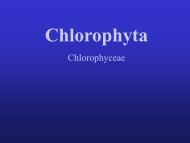
Trebouxiophyceae, Chlorophyta - (S)FTP hesla na Botany
Trebouxiophyceae, Chlorophyta - (S)FTP hesla na Botany
Trebouxiophyceae, Chlorophyta - (S)FTP hesla na Botany

Create successful ePaper yourself
Turn your PDF publications into a flip-book with our unique Google optimized e-Paper software.
Paper 2. Chloroplast morphology in Asterochloris<br />
Comparative study of chloroplast morphology and ontogeny<br />
in Asterochloris (<strong>Trebouxiophyceae</strong>, <strong>Chlorophyta</strong>)<br />
Pavel ŠKALOUD 1 1, 2<br />
& Ondřej PEKSA<br />
1 Charles University in Prague, Faculty of Science, Department of <strong>Botany</strong>, Benátská 2, Praha<br />
2, CZ-12801, Czech Republic; e-mail: skaloud@<strong>na</strong>tur.cuni.cz<br />
2 The Westbohemian Museum in Pilsen, Kopeckého sady 2, CZ-30100 Plzeň, Czech Republic;<br />
e-mail: opeksa@zcm.cz<br />
Abstract: Confocal laser scanning microscopy was utilized to compare the chloroplast morphology<br />
and ontogeny among five strains of the green alga Asterochloris. Parsimony a<strong>na</strong>lysis<br />
inferred from the rDNA ITS sequences confirmed their placement in three distinct lineages:<br />
Asterochloris phycobiontica, Trebouxia pyriformis and Asterochloris sp. Exami<strong>na</strong>tion by confocal<br />
microscopy revealed the existence of interspecific differences in the chloroplast ontogeny<br />
of Asterochloris; this was based upon either specific chloroplast structures observed in a<br />
single species, or on the differential timing of particular ontogenetic sequences. The occurrence<br />
of flat parietal chloroplasts prior to cell division, considered as a basic morphological<br />
discrimi<strong>na</strong>tive character of Asterochloris, was clearly associated with the process of aplanosporogenesis.<br />
By contrast, chloroplast transformation prior to the formation of autospores<br />
proceeded simply by the multiple fission of the chloroplast matrix in the cell lumen.<br />
Key words: Asterochloris, chloroplast morphology, confocal microscopy, ITS, molecular<br />
phylogeny, Trebouxia.<br />
Introduction<br />
The Swiss botanist Schwendener (1867) was the first to demonstrate that the microscopic<br />
green bodies in lichen thalli, the so-called gonidia, are in fact green or blue-green algae. Prior<br />
to that, lichenologists thought that the green bodies origi<strong>na</strong>ted from the tips of colorless hyphae,<br />
even though their resemblance to algae was noticed. At present, an estimated 100 species<br />
in 40 genera of algae are reported as photobionts of various lichen species (Tschermak-<br />
Woess 1988; Friedl & Büdel 1996). In the majority of the associations, the phycobiont belongs<br />
to one of three genera, <strong>na</strong>mely: Trebouxia Puymaly sensu lato, Trentepohlia Martius<br />
and Nostoc Vaucher ex Bornet et Flahault.<br />
Among the leading researchers in lichen symbiosis was Elisabeth Tschermak-Woess<br />
(1917 – 2001) who greatly increased our knowledge of the morphology and systematics of<br />
many photobionts. Her extensive scientific work includes descriptions and morphological<br />
observations of some novel or rare photobiont species, e.g. those of the genera Dictyochloropsis<br />
Geitler, Myrmecia Printz, Trebouxia Puymaly and Elliptochloris Tschermak-Woess. In<br />
particular, she was recognized as an exceptio<strong>na</strong>l cytologist, sometimes working at the limits<br />
of the laws of optics (for more information see Hesse 2001). In 1980, she described a new<br />
algal genus and species, Asterochloris phycobiontica Tschermak-Woess, based on her observations<br />
of the phycobiont of lichen Anzi<strong>na</strong> carneonivea (Anzi) Scheidegger (Tschermak-<br />
Woess 1980). She delimited the genus as having a mainly parietal, radially lobed cup-shaped<br />
30