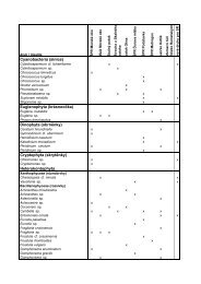
Trebouxiophyceae, Chlorophyta - (S)FTP hesla na Botany
Trebouxiophyceae, Chlorophyta - (S)FTP hesla na Botany
Trebouxiophyceae, Chlorophyta - (S)FTP hesla na Botany

Create successful ePaper yourself
Turn your PDF publications into a flip-book with our unique Google optimized e-Paper software.
Paper 3. Revision of Asterochloris<br />
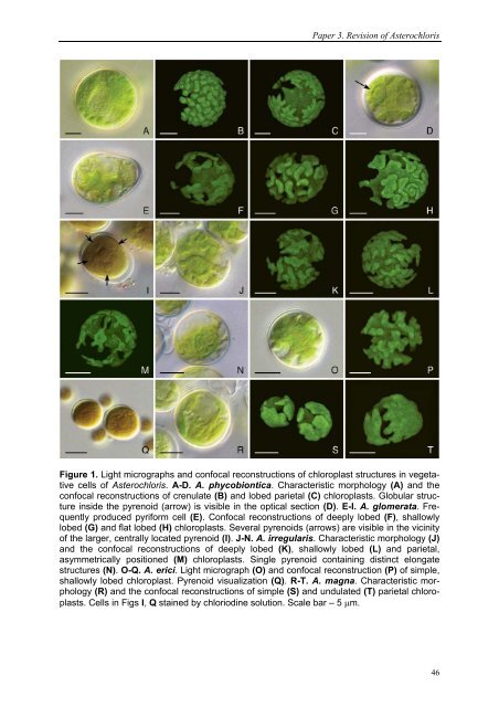
Figure 1. Light micrographs and confocal reconstructions of chloroplast structures in vegetative<br />
cells of Asterochloris. A-D. A. phycobiontica. Characteristic morphology (A) and the<br />
confocal reconstructions of crenulate (B) and lobed parietal (C) chloroplasts. Globular structure<br />
inside the pyrenoid (arrow) is visible in the optical section (D). E-I. A. glomerata. Frequently<br />
produced pyriform cell (E). Confocal reconstructions of deeply lobed (F), shallowly<br />
lobed (G) and flat lobed (H) chloroplasts. Several pyrenoids (arrows) are visible in the vicinity<br />
of the larger, centrally located pyrenoid (I). J-N. A. irregularis. Characteristic morphology (J)<br />
and the confocal reconstructions of deeply lobed (K), shallowly lobed (L) and parietal,<br />
asymmetrically positioned (M) chloroplasts. Single pyrenoid containing distinct elongate<br />
structures (N). O-Q. A. erici. Light micrograph (O) and confocal reconstruction (P) of simple,<br />
shallowly lobed chloroplast. Pyrenoid visualization (Q). R-T. A. mag<strong>na</strong>. Characteristic morphology<br />
(R) and the confocal reconstructions of simple (S) and undulated (T) parietal chloroplasts.<br />
Cells in Figs I, Q stained by chloriodine solution. Scale bar – 5 μm.<br />
46