Trebouxiophyceae, Chlorophyta - (S)FTP hesla na Botany
Trebouxiophyceae, Chlorophyta - (S)FTP hesla na Botany
Trebouxiophyceae, Chlorophyta - (S)FTP hesla na Botany

You also want an ePaper? Increase the reach of your titles
YUMPU automatically turns print PDFs into web optimized ePapers that Google loves.
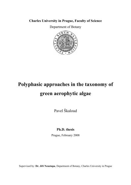
Charles University in Prague, Faculty of Science<br />
Department of <strong>Botany</strong><br />
Polyphasic approaches in the taxonomy of<br />
green aerophytic algae<br />
Pavel Škaloud<br />
Ph.D. thesis<br />
Prague, February 2008<br />
Supervised by: Dr. Jiří Neustupa, Department of <strong>Botany</strong>, Charles University in Prague
Contents<br />
LIST OF PAPERS ..................................................................................................................................................i<br />
ACKNOWLEDGEMENT.................................................................................................................................... ii<br />
1 GENERAL INTRODUCTION .................................................................................................................... 1<br />
1.1 TRADITIONAL MORPHOLOGY ................................................................................................................... 2<br />
1.2 CONFOCAL MICROSCOPY - AN EXAMPLE OF MODERN MORPHOLOGICAL APPROACHES .................................. 3<br />
1.3 MOLECULAR DATA....................................................................................................................................<br />
5<br />
1.4 SPECIES CONCEPTS....................................................................................................................................<br />
6<br />
2 AIMS OF THE THESIS...............................................................................................................................<br />
8<br />
3 OUTLINE OF THE THESIS.......................................................................................................................<br />
8<br />
3.1 SPECIES CONCEPT IN KLEBSORMIDIUM........................................................................................................<br />
8<br />
3.2 TAXONOMY OF ASTEROCHLORIS.................................................................................................................<br />
8<br />
TAXONOMY OF DICTYOCHLOROPSIS<br />
3.3 ........................................................................................................... 9<br />
PAPERS............................................................................................................................................................... 10<br />
PAPER 1. VARIATION AND TAXONOMIC SIGNIFICANCE OF SOME MORPHOLOGICAL FEATURES IN EUROPEAN<br />
STRAINS OF KLEBSORMIDIUM (KLEBSORMIDIOPHYCEAE, STREPTOPHYTA)................................................... 10<br />
PAPER 2. COMPARATIVE STUDY OF CHLOROPLAST MORPHOLOGY AND ONTOGENY IN ASTEROCHLORIS<br />
(TREBOUXIOPHYCEAE, CHLOROPHYTA)........................................................................................................ 29<br />
PAPER 3. PHYLOGENY, MORPHOLOGY, REPRODUCTION AND TAXONOMIC REVISION OF SYMBIOTIC ALGA<br />
ASTEROCHLORIS (TREBOUXIOPHYCEAE, CHLOROPHYTA) .............................................................................. 40<br />
PAPER 4. CONFOCAL MICROSCOPY OF CHLOROPLAST MORPHOLOGY AND ONTOGENY IN THREE STRAINS OF<br />
DICTYOCHLOROPSIS (TREBOUXIOPHYCEAE, CHLOROPHYTA)........................................................................ 75<br />
PAPER 5. MORPHOLOGY, MOLECULAR PHYLOGENY AND TAXONOMY OF GREEN ALGAL GENERA<br />
AEROSPHAERA AND DICTYOCHLOROPSIS (TREBOUXIOPHYCEAE, CHLOROPHYTA) WITH DESCRIPTION OF<br />
FOUR NEW SPECIES ........................................................................................................................................ 85<br />
4 CONCLUSIONS ....................................................................................................................................... 107<br />
5 REFERENCES..........................................................................................................................................<br />
108<br />
6 CURRICULUM VITAE – PAVEL ŠKALOUD.......................................................................................<br />
112
This thesis is based on the following five papers, referred to in the text as Papers 1-5:<br />
1. Variation and taxonomic significance of some morphological features in European<br />
strains of Klebsormidium (Klebsormidiophyceae, Streptophyta).<br />
Pavel Škaloud<br />
Nova Hedwigia (2006), 83(3-4): 533-550.<br />
2. Comparative study of chloroplast morphology and ontogeny in Asterochloris (<strong>Trebouxiophyceae</strong>,<br />
<strong>Chlorophyta</strong>).<br />
Pavel Škaloud & Ondřej Peksa<br />
Biologia (2008), in press.<br />
3. Phylogeny, morphology, reproduction and taxonomic revision of symbiotic alga Asterochloris<br />
(<strong>Trebouxiophyceae</strong>, <strong>Chlorophyta</strong>)<br />
Pavel Škaloud & Ondřej Peksa<br />
Submitted manuscript.<br />
4. Confocal microscopy of chloroplast morphology and ontogeny in three strains of Dictyochloropsis<br />
(<strong>Trebouxiophyceae</strong>, <strong>Chlorophyta</strong>).<br />
Pavel Škaloud, Jiří Neustupa, Barbora Radochová & Lucie Kubínová<br />
Phycologia (2005), 44: 261-269.<br />
5. Morphology, molecular phylogeny and taxonomy of green algal genera Aerosphaera<br />
and Dictyochloropsis (<strong>Trebouxiophyceae</strong>, <strong>Chlorophyta</strong>) with description of four new<br />
species.<br />
Pavel Škaloud, Thomas Friedl & Jiří Neustupa<br />
Manuscript.<br />
I hereby declare that I made this thesis independently, using the listed references, or in<br />
the co-operation with other authors of the papers. I did submit neither the thesis nor its any<br />
part to acquire any other academic title.<br />
I was in general responsible for morphological investigations, confocal microscopy and<br />
writing papers. I was also responsible for molecular a<strong>na</strong>lyses in papers 2, 3. O. Peksa collected<br />
lichen samples, isolated and cultivated algal strains of Asterochloris and helped with<br />
DNA isolation and following PCR reactions (papers 2, 3). Jiří Neustupa helped with morphological<br />
investigation of Dictyochloropsis strains (papers 4, 5). In paper 5, he also helped with<br />
forming the leading idea and fi<strong>na</strong>l writing of the manuscript. Barbora Radochová and Lucie<br />
Kubínová helped with the techniques of confocal microscopy (paper 4). Thomas Friedl performed<br />
molecular a<strong>na</strong>lyses of Dictyochloropsis strains (paper 5).<br />
All co-authors helped to polish the manuscript text.<br />
On behalf of all co-authors, we declare the keynote participation of Pavel Škaloud in<br />
acquiring the results and writing the papers, as described above.<br />
i
Acknowledgement<br />
I am indebted to my Thesis adviser, Jiří Neustupa, for his expert advice, help, inspiration,<br />
and the freedom to work independently.<br />
I kindly thank Ondřej Peksa (Charles University, Prague) for the wonderful cooperation<br />
in Asterochloris research, many interesting ideas and great supply of lichen literature. I would<br />
like to thank Thomas Friedl (University of Göttingen, Germany), Frederik Leliaert (Ghent<br />
University, Belgium) and Matthew P. Nelsen (Michigan Technological University) for valuable<br />
suggestions and consultations concerning the molecular phylogenetic a<strong>na</strong>lyses. For the<br />
assistance with DNA isolation and sequencing techniques I greatly thank to Veronika<br />
Machalová (Charles University, Prague), Eva Rejzková (Charles University, Prague), Opayi<br />
Mudimu (University of Göttingen, Germany) and Kathrin Mohr (University of Göttingen,<br />
Germany). For the assistance with the confocal microscope I thank to Barbora Radochová<br />
(Academy of Sciences, Prague), Lucie Kubínová (Academy of Sciences, Prague) and Ondřej<br />
Šebesta (Charles University, Prague). I thank Maike Lorenz (SAG Culture Collection, Germany),<br />
Thomas Pröschold (CCAP Culture Collection, Great Britain) and Toni Henning<br />
(UTEX Culture Collection, USA) for supplying algal strains and friendly relations. For improving<br />
the English style, I am very grateful to Mari<strong>na</strong> Julian and Michael Taylor. I would<br />
like to express my deep thanks to Magda Škaloudová for reading the manuscripts, valuable<br />
comments and various supports.<br />
The Thesis was supported by the grant 136/2006/B-BIO/PrF of the Grant Agency of the<br />
Charles University in Prague, by the grant of the Czech Ministry of Environment (No. VaV-<br />
SM/2/90/05, by the research project of the Czech Ministry of Education (No. 0021620828)<br />
and by FRVŠ grant No. 2826/2003.<br />
ii
1 General introduction<br />
General introduction<br />
At the generic and species levels, green microalgae are traditio<strong>na</strong>lly classified according<br />
to morphological characters of vegetative stages in their life cycle (Ettl & Gärtner 1995).<br />
However, since the 1990s, the application of phylogenetic a<strong>na</strong>lyses of molecular markers has<br />
demonstrated that this morphological concept is artificial for most of the green algal genera<br />
and needs to be revised (Chapman & Buchheim 1991; Huss et al. 1999; Krienitz et al. 2001;<br />
Pröschold et al. 2001). For example, the species of morphologically defined aero-terrestrial<br />
genus Chlorella were revealed to be dispersed over two classes of chlorophytes, the Chlorophyceae<br />
and the <strong>Trebouxiophyceae</strong> (Huss et al. 1999). It was demonstrated that only four species<br />
could be considered as true Chlorella taxa, whereas all other Chlorella species should be<br />
transferred into separate genera (Fig. 1). According to the present knowledge, the traditio<strong>na</strong>lly<br />
conceived genus Chlorella forms 9 particular lineages corresponding to different genera<br />
(Chlorella, Mycho<strong>na</strong>stes, “Heterochlorella”, “Glaphyrella”, Parachlorella, Auxenochlorella,<br />
“C. ellipsoidea clade”, Scenedesmus and Desmodesmus; An et al. 1999; Huss et al.<br />
1999; Kali<strong>na</strong> unpubl.; Krienitz et al. 2004). Similarly, the rDNA data suggest the splitting of<br />
the well-known, widely-distributed freshwater genus Pediastrum into five particular genera:<br />
Pediastrum, Mo<strong>na</strong>ctinus, Parapediastrum, Pseudopediastrum and Stauridium (Buchheim et<br />
al. 2005). The same results were also reported for marine green algae. Yamamoto et al. (2003)<br />
investigated the genus Nannochloris using 18S rDNA and actin genes and determined nonmonophyly<br />
of these small planktonic,<br />
coccoid forms.<br />
As demonstrated above, most of<br />
the green algal genera are polyphyletic<br />
and their status as well as<br />
species number needs a revision. Differentiation<br />
of genera and species<br />
based on single characters (e.g. morphology<br />
of vegetative cells, ultrastructural<br />
features) becomes to be insufficient<br />
and often leads to ambiguous<br />
classification (Pröschold & Leliaert<br />
2007). Therefore, the usage of polyphasic<br />
approaches (i.e. combining<br />
morphological, ultrastructural, molecular,<br />
ecological or biochemical<br />
data) could reveal the real biodiversity<br />
and taxa relationships among the<br />
green algae. Polyphasic approaches<br />
can clearly distinguish and delimit<br />
species and genera, which predomi<strong>na</strong>ntly<br />
lead to revealing more biological<br />
species (e.g. Fabry et al. 1999;<br />
Neustupa et al. 2007), but can also<br />
result to a reduction of described species<br />
(e.g. Pröschold et al. 2001). Fol-<br />
lowing chapters briefly introduce the<br />
application, benefits and disadvantages<br />
of particular approaches to<br />
study green microalgal species.<br />
Fig. 1. Phylogenetic tree inferred from 18S rRNA gene sequences<br />
showing the polyphyly of the genus Chlorella within the <strong>Trebouxiophyceae</strong>/Chlorophyceae.<br />
Taxa that were traditio<strong>na</strong>lly assigned to<br />
Chlorella are circled (Huss et al. 1999).<br />
1
1.1 Traditio<strong>na</strong>l morphology<br />
General introduction<br />
Traditio<strong>na</strong>lly, green algae were classified according to the morphological species concept<br />
based on the organisation level of the vegetative state (Brunnthaler 1915; Ettl & Gärtner<br />
1988; Hindák 1977, 1980, 1984; Komárek & Fott 1983). The main morphological and structural<br />
characteristics used as taxonomic criteria in green microalgae were: form and dimensions<br />
of cells, cell wall characteristics, number of nuclei, chloroplast morphology, presence or<br />
absence of pyrenoid, number and size of autospores, and possibility to form zoospores (Ettl &<br />
Gärtner 1995; Komárek & Fott 1983). Morphological observations and species descriptions<br />
made by a number of phycologists considerably increased our knowledge of the biodiversity,<br />
morphological variability and systematics of green algae. Morphological studies of several<br />
prominent cytologists are still much accounted for their quality of observations, being sometimes<br />
at the limits of the laws of optics (Fott & Nováková 1969 1 ; Gärtner 1985; Tschermak-<br />
Woess 1989).<br />
However, recent molecular and physiological<br />
studies have demonstrated high plasticity of several<br />
morphological characters used for species delimitation<br />
and they evoked need for critical evaluation of<br />
observed morphological differences among the species.<br />
For example, presence of pyrenoid has been<br />
widely used to distinguish particular green algal<br />
species and genera, e.g. to differentiate flagellate<br />
species Chlamydomo<strong>na</strong>s and Chloromo<strong>na</strong>s (Ettl<br />
1983, Fig. 2). However, phylogenetic a<strong>na</strong>lyses revealed<br />
that strains of both traditio<strong>na</strong>l genera could<br />
belong to the same clade and, in the case of<br />
Chloromo<strong>na</strong>s reticulata, even to the same species<br />
(Buchheim et al. 1997, Pröschold et al. 2001). Nozaki et al. (1998) demonstrated that presence<br />
or absence of pyrenoids in Chlorogonium depends primarily on culture conditions (autotrophics<br />
vs. heterotrophics), instead on evolutio<strong>na</strong>ry differentiation. Similarly, production of<br />
mucilaginous envelopes around the cells, considered as discrimi<strong>na</strong>tive character of green algal<br />
family Radiococcaceae (Komárek & Fott 1983), occurs in various unrelated algal lineages<br />
(Wolf et al. 2003). Moreover, it now appears that mucilaginous production often depends<br />
rather on the environmental<br />
conditions<br />
(Buzzelli et al. 1997,<br />
Reynolds 2007) than on<br />
phylogenetic position.<br />
Another example includes<br />
the genera Chlorella<br />
and Micractinium<br />
(Fig. 3), traditio<strong>na</strong>lly<br />
classified into different<br />
families of Chlorococcales<br />
(Chlorellaceae and<br />
Micractiniaceae). The<br />
18S rDNA and ITS data<br />
revealed close relation-<br />
Fig. 2. Morphological distinction between genera<br />
Chlamydomo<strong>na</strong>s (a) and Chloromo<strong>na</strong>s (b) (modified<br />
after Ettl 1983).<br />
Fig. 3. Morphology of Chlorella vulgaris (a) and Micractinium pusillum (b) (modified<br />
after Komárek & Fott 1983).<br />
ship of these genera (Krienitz et al. 2004). Moreover, formation of colonies and cell wall bris-<br />
1 cited paper has presently 114 citations in WOS (ISI Web of Science, http://portal.isiknowledge.com/portal.cgi)<br />
2
General introduction<br />
tles that characterize the genus Monoraphidium was proven to represent phenotypic adaptation<br />
against grazer pressure of Brachionus calciflorus (Luo et al. 2006).<br />
Fi<strong>na</strong>lly, morphology<br />
can be considerably<br />
influenced by the bacterial<br />
or fungal contami<strong>na</strong>tion.<br />
Common marine<br />
green alga Ulva is characterized<br />
by foliose morphology.<br />
However, by<br />
adding specific marine<br />
bacteria, strains of this<br />
genus are able to develop<br />
Fig. 4. Enteromorpha-like morphology of cultivated Ulva lactuca, induced by<br />
specific marine bacteria (after Provasoli & Pintner 1980).<br />
the Enteromorpha-like tubular thallus (Fig. 4, Provasoli & Pintner 1980).<br />
The phylogenetic a<strong>na</strong>lyses of ITS rDNA sequences later proved that green seaweeds<br />
Ulva and Enteromorpha are not distinct genera, and therefore, Enteromorpha was included<br />
into the genus Ulva (Hayden et al. 2003). The<br />
same effect of considerable variation in morphology<br />
occurs in green microalgae. Morphology<br />
of “Interfilum massjukiae” seems to<br />
vary from filamentous to packet-like in dependence<br />
to degree of bacterial contami<strong>na</strong>tion<br />
(own observations, Mikhailyuk et al.<br />
2007). Similarly, massive contami<strong>na</strong>tion of<br />
Pseudococcomyxa simplex by unidentified<br />
ascomycote fungus probably causes significant<br />
changes of cell width (Fig. 5, Nemjová<br />
2007).<br />
Fig. 5. Cell width variation in three Pseudococcomyxa<br />
simplex strains. Shaded box-plot indicates strain contamined<br />
by ascomycote fungus (modified after Nemjová 2007).<br />
1.2 Confocal microscopy - an example of modern morphological approaches<br />
In late 1980s, advent of a new procedure<br />
for optical sectioning of plant tissues<br />
using the confocal laser scanning microscope<br />
(CLSM) provided an exciting new tool for<br />
the morphological observation of living and<br />
intact cells. Although the principle of CLSM<br />
has been patented in 1957, the commercial<br />
application of this method in general started<br />
three decades later. The main advantages of<br />
CLSM consist in the increasing of<br />
micrograph contrast and the possibility to<br />
reconstruct three-dimensio<strong>na</strong>l images of investigated<br />
structures. The special confocal<br />
pinhole elimi<strong>na</strong>tes out-of-focus information,<br />
so that just the light within the focal plane<br />
can be detected (Fig. 6, Pawley 2006). Resulting<br />
image quality is much better than that<br />
of wide-field images obtained by conven-<br />
Fig. 6. The mechanics of confocal microscopy (from<br />
http://www.microscopyu.com/).<br />
3
General introduction<br />
tio<strong>na</strong>l light or fluorescent microscopes. Moreover, three-dimensio<strong>na</strong>l objects displayed by<br />
CLSM can be virtually cut into several optical slices, which can be later used for computer<br />
a<strong>na</strong>lyses and shape reconstructions.<br />
Above mentioned characteristics thus facilitate and greatly improve studies of plant<br />
chloroplasts, e<strong>na</strong>bling their exami<strong>na</strong>tion directly inside living cells using autofluorescence of<br />
the chlorophyll. Van Spronsen et al. (1989) presented some of the first observations of plant<br />
tissue using CLSM. To achieve their excellent images, they had simply placed whole pieces<br />
of leaf tissue between coverslips and then performed the confocal observations. Recently,<br />
CLSM has been repeatedly applied for the exami<strong>na</strong>tion of chloroplast morphology and structural<br />
dy<strong>na</strong>mics in higher plants (see Hepler & Gunning 1998; Wildman et al. 2004). However,<br />
it has only rarely been used in the investigations of algal chloroplasts, so far. The essential<br />
paper dealing with the algal chloroplast autofluorescence was published by Gunning &<br />
Schwartz (1999). These authors examined heterogeneity of chlorophyll fluorescence in<br />
chloroplasts of selected green algae and revealed differences in chloroplast ultrastructure between<br />
<strong>Chlorophyta</strong> and Streptophyta. Afterwards, CLSM was further used to investigate<br />
chloroplast morphology in Eugle<strong>na</strong> geniculata (Zakrys et al. 2002), changes in the distribution<br />
of the fluorescence intensity within plastids of Eugle<strong>na</strong> gracilis exposed to manganese<br />
excess (Ferroni et al. 2004) and plastid division in several Mallomo<strong>na</strong>s species (Weatherill et<br />
al. 2007).<br />
Despite its contemporary sporadic use, confocal microscopy and subsequent threedimensio<strong>na</strong>l<br />
reconstructions can add useful information in studies of the phenotypic plasticity<br />
of algal chloroplasts and for detailed investigation of chloroplast ontogeny during cell cycle<br />
(Fig. 7). Therefore, CLSM represents an important tool to facilitate the morphological delimitation<br />
of particular species in taxonomic studies. Especially in green microalgae, the morphological<br />
investigation of often structurally complicated chloroplasts can be essential for the<br />
identification of even small morphological differences among particular species (Škaloud &<br />
Radochová 2004).<br />
Fig. 7. Three-dimensio<strong>na</strong>l reconstructions of chloroplast morphology in several algae, as observed by confocal laser<br />
scanning microscope (from left to right: Zygnema, Mougeotia, Cosmarium, Spirogyra (after Hepler & Gunning 1998).<br />
4
1.3 Molecular data<br />
General introduction<br />
In the 1990s, application of phylogenetic a<strong>na</strong>lyses based on specific molecular markers<br />
was introduced into the systematics and taxonomy of algae. Molecular data now allow to<br />
formulate specific phylogenetic hypotheses and to trace the phylogenetic relationships among<br />
the taxa. Recent technological advantages allow rapid and accurate determi<strong>na</strong>tion of the sequence<br />
of nucleotides in a nucleic acid molecule. Typical genetic markers sequenced in phycological<br />
studies are e.g. the nuclear ribosomal operon (SSU, 5.8S and LSU, including ITS1<br />
and ITS2 regions), actin gene, several chloroplast genes (rbcL, atpB) and mitochondrial genes<br />
(coxI). Phylogenetic a<strong>na</strong>lyses of slowly evolving genes for small and large subunit ribosomal<br />
RNA (SSU and LSU rDNA) are often exploited in investigation of evolutio<strong>na</strong>ry divergences<br />
that led to the separation of major algal phyla and classes. However, SSU rDNA also contains<br />
some relatively variable regions, so that this marker can also be used at lower taxonomic levels,<br />
including microevolutio<strong>na</strong>ry investigations. Phylogenetic a<strong>na</strong>lyses of SSU rDNA provided<br />
solid support for existence of two main lineages among the green plants - Streptophyta<br />
and <strong>Chlorophyta</strong> (Friedl 1997), they revealed <strong>Trebouxiophyceae</strong> as a new green algal class<br />
(Friedl 1995), and demonstrated the polyphyly of many genera (e.g. Buchheim et al. 1997,<br />
2005; Huss et al. 1999; Krienitz et al. 2004; Senousy et al. 2004).<br />
For investigation of recent speciation<br />
events, the rapidly evolving sequences are<br />
preferred. The regions of the nuclearencoded<br />
ribosomal RNA genes known as<br />
the inter<strong>na</strong>l transcribed spacers (ITS) have<br />
proved to be very useful in such studies<br />
(Fig. 8). In fact, ITS has now become the<br />
single most frequently utilized DNA region<br />
in plant studies (Hershkovitz et al. 1999). In<br />
taxonomy of green microalgae, ITS sequences<br />
were recently used e.g. in discovery<br />
of genetic variability among strains of<br />
Chlorella and Micractinium (Luo et al 2006), in investigation of relationships between Paramecium<br />
symbionts (Hoshi<strong>na</strong> et al. 2005; Summerer et al. 2007) or in investigation of specificity<br />
and genetic variation in the genus Asterochloris (Nelsen & Gargas 2008; Yahr et al. 2006).<br />
Within the ITS region, the 5.8S gene sequence is highly conserved and it is useful for verifying<br />
identity of sequences. The ITS1 and ITS2 regions are<br />
much more variable in its primary sequence. Nevertheless,<br />
they exhibit a common secondary structure within all Viridiplantae<br />
(Mai & Coleman 1997). Although ITS represents<br />
the untranslated DNA region, the initial transcript of<br />
a nuclear ribosomal cistron is a long RNA that includes all<br />
three RNA genes plus the ITS. It promptly folds, and only<br />
then the ITS regions degraded (Venema & Tollervey<br />
1999). Once secondary structure has been established for a<br />
group of related organisms, it can serve not only as a guide<br />
to alignment of all the nucleotide positions, but it may also<br />
contain information potentially useful for comparisons at<br />
family, order and even higher levels (Fig. 9, Coleman<br />
2003). Nowadays, more than 550 ITS2 sequences along<br />
with the secondary structures are available for <strong>Chlorophyta</strong><br />
(Schultz et al. 2006).<br />
Fig. 8. Diagram illustrating the organization of the nuclear<br />
ribosomal cistrons (gray boxes) of a typical eukaryote and<br />
the position of ITS region in their primary RNA transcript<br />
(after Coleman 2003).<br />
Fig. 9. ITS2 secondary structure of Volvocaceae.<br />
Relatively conserved nucleotide<br />
positions are in bold (after Coleman<br />
2000).<br />
5
General introduction<br />
More variable markers, such as the gene introns, provide high resolution in phylogenetic<br />
studies and they are useful in identifying and delimiting cryptic and phylogenetic species<br />
and for use in population genetic studies. One such variable marker, the actin type I intron,<br />
was reported by Liss et al. (1997), who observed high levels of its variation in Chlamydomo<strong>na</strong>s<br />
and Volvox (Fig. 10). Afterwards, actin introns were used to investigate the cryptic<br />
diversity in lichen photobionts Trebouxia s. str. (Kroken & Taylor 2000) and Asterochloris<br />
(Nelsen & Gargas 2006, 2008).<br />
Fig. 10. Diagram illustrating the organisation of the actin genes of Chlamydomo<strong>na</strong>s reinhardtii with respect to exons<br />
(shaded boxes) and introns (open boxes) (After Liss et al. 1997).<br />
1.4 Species concepts<br />
All the above-mentioned morphological and molecular markers are frequently applied<br />
for description and delimitation of particular species of coccal green algae. However, given<br />
two organisms, how can we distinguish, whether they belong to the same species or not? The<br />
answer may be straightforward for two divergent organisms, but it can be extremely difficult<br />
and laborious in closely related ones. That is why many different concepts of species have<br />
been and are held by biologists.<br />
According to the morphological<br />
species concept, species are the<br />
smallest groups that can be repeatedly<br />
defined by structural characteristics<br />
that are relatively easy to distinguish<br />
(Fig. 11). However, some morphological<br />
species have been observed to<br />
undergo seaso<strong>na</strong>l succession or distinct<br />
morphological variability depending<br />
on environmental conditions<br />
(Luo et al. 2006; Stoyneva et al. 2007;<br />
van Holthoon et al. 2003; Verschoor<br />
et al. 2004). Therefore, frequently<br />
observed intraspecific morphological<br />
variation avoids the descriptions of<br />
species based solely on the morpho-<br />
logical differences. Moreover, traditio<strong>na</strong>l morphological species concept alone is not sufficient<br />
for species descriptions, since it does not recognize cryptic or sibling species (Behnke et<br />
al. 2004). However, due to its long tradition, previously published morphological investigations<br />
represent valuable information sources about morphological variability, uniqueness and<br />
distribution of particular green algal species.<br />
Fig. 11. Morphological species concept. Distinguishing Chlamydomo<strong>na</strong>s<br />
species according to the chloroplast morphology (after Ettl<br />
1983).<br />
The biological species concept, the most widely accepted concept among the biologists,<br />
defines species in terms of interbreeding. According to this concept, species are groups<br />
of interbreeding <strong>na</strong>tural populations that are reproductively isolated from other such groups<br />
(Mayr 1948). This is generally useful formulation for scientists working with living examples<br />
of the higher taxa like mammals, fish, and birds, but meaningless for organisms that do not<br />
reproduce sexually. In the majority of green microalgal genera, sexual reproduction has never<br />
been observed (Ettl & Gärtner 1995) and thus wide application of the biological concept is<br />
hard to imagine. However, indirect evidences on the interbreeding processes within the bio-<br />
6
General introduction<br />
logical species could be obtained, e.g. by the exami<strong>na</strong>tion of the recombi<strong>na</strong>tion events between<br />
two gene alleles (Kroken & Taylor 2000).<br />
Phycologists interested in algal evolution<br />
have advocated use of the phylogenetic species<br />
concept, in which a species is a smallest group of<br />
organisms that shares unique combi<strong>na</strong>tion of character<br />
states (nucleotide states as well) as separate<br />
species (Nixon & Wheeler 1990). Ideally, a phylogenetic<br />
species is also monophyletic, i.e. includes<br />
an ancestor and all its descendants. In contrast to<br />
monophyletic groups, paraphyletic groups do not<br />
include all of the descendants of a common ancestor.<br />
Polyphyletic groups include some members<br />
that are more closely related to taxa outside the<br />
group. The phylogenetic species concept can be<br />
applied practically for all organisms. However,<br />
strict application of this species concept, regarding<br />
all evolutio<strong>na</strong>ry end-products (even a number of<br />
clo<strong>na</strong>l asexual organisms) as unique species<br />
(Wheeler & Platnick 2000), could result in overestimating<br />
of real species number.<br />
According to the previously-mentioned<br />
characteristics, no single species concept seems to<br />
be the best for all groups of organisms. For species<br />
delimitation, the polyphasic approaches were<br />
suggested as the “gold standard” using a combi<strong>na</strong>tion<br />
of morphology, sequence data, physiological<br />
characteristics and ecological data. This way, a<br />
combi<strong>na</strong>tion of several species concepts applied for<br />
organisms studied will result in better delimitation<br />
of particular algal species. For example, the polyphasic<br />
approach was applied to recover cryptic<br />
species of Gonium pectorale by comparison of ITS<br />
Fig. 12. Phylogenetic species concept. Recognizing<br />
cryptic species within Trebouxia jamesii species<br />
complex, based on actin I intron phylogeny (after<br />
Kroken & Taylor 2000).<br />
sequences with a breeding data (Fabry et al. 1999; Coleman 2000) or to characterize the genera<br />
Oogamochlamys and Lobochlamys by means of combi<strong>na</strong>tion of SSU rDNA data, morphological<br />
characters and sexual reproduction processes (Pröschold et al. 2001). In this thesis, I<br />
present several examples of polyphasic approach in investigation of taxonomy and phylogeny<br />
of some genera of green aerophytic algae.<br />
7
2 Aims of the thesis<br />
Aims and outline of the thesis<br />
The general objective of the thesis was to taxonomically revise several genera of the<br />
green aerophytic algae, using both traditio<strong>na</strong>l morphological and modern molecular phylogenetic<br />
approaches. For the revision, I chose two coccal genera characterized by the complicated<br />
chloroplast structure (Asterochloris and Dictyochloropsis) and the ubiquitous filamentous<br />
genus Klebsormidium.<br />
In particular, two principal aims can be summarized as follows:<br />
1) To test the stability and suitability of current and newly suggested morphological<br />
discrimi<strong>na</strong>ting features for the distinction of the particular species<br />
(Papers 1, 2, 4).<br />
2) To revise the species concept of the genera, using the combi<strong>na</strong>tion of selected<br />
morphological features and molecular markers (Papers 3, 5).<br />
3 Outline of the thesis<br />
The thesis comprehends the taxonomical investigation of three aerophytic green algal<br />
genera – Klebsormidium, Asterochloris and Dictyochloropsis. In Klebsormidium, the species<br />
concept differentiating in morphological view two closely related species was investigated by<br />
the comparison of several morphological characters in 40 isolates. In Asterochloris and Dictyochloropsis,<br />
the combi<strong>na</strong>tion of detailed morphological study of chloroplast structure and<br />
the molecular phylogenetic a<strong>na</strong>lyses was performed to delimit particular species of the genera.<br />
3.1 Species concept in Klebsormidium<br />
The stability and suitability of several morphological characters was tested for the distinction<br />
between two <strong>na</strong>rrowly related species Klebsormidium flaccidum and K. nitens. In<br />
forty isolated strains, the variability in cell dimensions, filament length, character of zoosporangia<br />
and zoospore germi<strong>na</strong>tion was studied. Moreover, the habit of cell morphology in relation<br />
to the culture age was studied in six randomly chosen strains. In two selected strains, the<br />
effect of physico-chemical parameters (temperature, humidity, illumi<strong>na</strong>tion and pH) on cell<br />
width was further studied. The study showed variability in taxonomically relevant morphological<br />
features during growing of the species in cultures and impossibility to clearly define<br />
the species by the combi<strong>na</strong>tion of current morphological markers (Paper 1).<br />
The comparison of ascertained morphological features with the molecular phylogenetic<br />
data was not performed, since the broad phylogenetic study of Klebsormidium is presently in<br />
progress, based on the comparison of rbcL sequences of forty isolated strains (Fabio Rindi,<br />
pers. comm.).<br />
3.2 Taxonomy of Asterochloris<br />
First, the confocal microscopy was utilized to compare the chloroplast morphology and<br />
ontogeny among five strains of the green alga Asterochloris. The exami<strong>na</strong>tion revealed the<br />
existence of interspecific differences in the chloroplast ontogeny of Asterochloris, based upon<br />
the specific chloroplast structures observed in a single species (Paper 2).<br />
Next, the broad phylogenetic study was performed on the 34 cultured strains, using nuclear-encoded<br />
ITS rDNA and actin intron sequences as the phylogenetic markers. The combi<strong>na</strong>tion<br />
of molecular phylogenetic a<strong>na</strong>lyses and morphological exami<strong>na</strong>tion resulted in the revision<br />
of the genus. 13 species were newly delimited, including the description of 7 species<br />
8
Aims and outline of the thesis<br />
new for science (A. echi<strong>na</strong>ta, A. friedlii, A. gaertneri, A. leprariae, A. lobophora and A. woessiae).<br />
All species were defined by both unique actin sequences and combi<strong>na</strong>tion of selected<br />
morphological characters (chloroplast morphology, cell shape, etc.). Moreover, the isogamous<br />
sexual reproduction was observed in the genus, for the first time (Paper 3).<br />
3.3 Taxonomy of Dictyochloropsis<br />
Chloroplast morphology and ontogeny in three species of the genus Dictyochloropsis<br />
were investigated by using light and confocal microscopy. Four distinct morphological stages<br />
during the chloroplast ontogeny were revealed in all investigated strains. The stages were distinguished<br />
primarily by the number of differently structured chloroplast layers and by the inner<br />
structure of chloroplast lobes. The study detected significant differences in chloroplast<br />
morphology among the studied strains (Paper 4).<br />
These differences were further supported by the 18S rDNA sequence a<strong>na</strong>lyses. The phylogenetic<br />
a<strong>na</strong>lyses revealed that various Dictyochloropsis strains form two distinct lineages<br />
within the <strong>Trebouxiophyceae</strong>. Based on detailed morphological investigation and comparing<br />
with literature data, the lineages were assigned two different genera, Dictyochloropsis and<br />
Aerosphaera. Dictyochloropsis comprises algae with a reticulate chloroplast, forming distinct<br />
parallelly-arranged lobes at some ontogenetic stages, and which reproduce only by means of<br />
autospores. Aerosphaera encompasses algae with evenly perforated chloroplast that can reproduce<br />
also by the formation of zoospores with typical separate insertion of the flagella.<br />
Based on congruencies found between morphological features and rDNA sequence<br />
a<strong>na</strong>lyses, four new combi<strong>na</strong>tions were proposed and four new species were described (D. asterochloroides,<br />
A. handae, A. tropica and A. tschermakiae) (Paper 5).<br />
9
Paper 1. Significance of morphological features in Klebsormidium<br />
Paper 1<br />
Variation and taxonomic significance of some morphological<br />
features in European strains of Klebsormidium<br />
(Klebsormidiophyceae, Streptophyta)<br />
Pavel Škaloud<br />
Nova Hedwigia (2006), 83(3-4): 533-550<br />
Empty zoosporangia of Klebsormidium nitens with distinct apertures<br />
10
Paper 1. Significance of morphological features in Klebsormidium<br />
11
Paper 1. Significance of morphological features in Klebsormidium<br />
12
Paper 1. Significance of morphological features in Klebsormidium<br />
13
Paper 1. Significance of morphological features in Klebsormidium<br />
14
Paper 1. Significance of morphological features in Klebsormidium<br />
15
Paper 1. Significance of morphological features in Klebsormidium<br />
16
Paper 1. Significance of morphological features in Klebsormidium<br />
17
Paper 1. Significance of morphological features in Klebsormidium<br />
18
Paper 1. Significance of morphological features in Klebsormidium<br />
19
Paper 1. Significance of morphological features in Klebsormidium<br />
20
Paper 1. Significance of morphological features in Klebsormidium<br />
21
Paper 1. Significance of morphological features in Klebsormidium<br />
22
Paper 1. Significance of morphological features in Klebsormidium<br />
23
Paper 1. Significance of morphological features in Klebsormidium<br />
24
Paper 1. Significance of morphological features in Klebsormidium<br />
25
Paper 1. Significance of morphological features in Klebsormidium<br />
26
Paper 1. Significance of morphological features in Klebsormidium<br />
27
Paper 1. Significance of morphological features in Klebsormidium<br />
28
Paper 2<br />
Paper 2. Chloroplast morphology in Asterochloris<br />
Comparative study of chloroplast morphology and ontogeny in<br />
Asterochloris (<strong>Trebouxiophyceae</strong>, <strong>Chlorophyta</strong>)<br />
Pavel Škaloud & Ondřej Peksa<br />
Biologia (2008), in press<br />
Confocal reconstruction of deeply lobed Asterochloris chloroplast<br />
29
Paper 2. Chloroplast morphology in Asterochloris<br />
Comparative study of chloroplast morphology and ontogeny<br />
in Asterochloris (<strong>Trebouxiophyceae</strong>, <strong>Chlorophyta</strong>)<br />
Pavel ŠKALOUD 1 1, 2<br />
& Ondřej PEKSA<br />
1 Charles University in Prague, Faculty of Science, Department of <strong>Botany</strong>, Benátská 2, Praha<br />
2, CZ-12801, Czech Republic; e-mail: skaloud@<strong>na</strong>tur.cuni.cz<br />
2 The Westbohemian Museum in Pilsen, Kopeckého sady 2, CZ-30100 Plzeň, Czech Republic;<br />
e-mail: opeksa@zcm.cz<br />
Abstract: Confocal laser scanning microscopy was utilized to compare the chloroplast morphology<br />
and ontogeny among five strains of the green alga Asterochloris. Parsimony a<strong>na</strong>lysis<br />
inferred from the rDNA ITS sequences confirmed their placement in three distinct lineages:<br />
Asterochloris phycobiontica, Trebouxia pyriformis and Asterochloris sp. Exami<strong>na</strong>tion by confocal<br />
microscopy revealed the existence of interspecific differences in the chloroplast ontogeny<br />
of Asterochloris; this was based upon either specific chloroplast structures observed in a<br />
single species, or on the differential timing of particular ontogenetic sequences. The occurrence<br />
of flat parietal chloroplasts prior to cell division, considered as a basic morphological<br />
discrimi<strong>na</strong>tive character of Asterochloris, was clearly associated with the process of aplanosporogenesis.<br />
By contrast, chloroplast transformation prior to the formation of autospores<br />
proceeded simply by the multiple fission of the chloroplast matrix in the cell lumen.<br />
Key words: Asterochloris, chloroplast morphology, confocal microscopy, ITS, molecular<br />
phylogeny, Trebouxia.<br />
Introduction<br />
The Swiss botanist Schwendener (1867) was the first to demonstrate that the microscopic<br />
green bodies in lichen thalli, the so-called gonidia, are in fact green or blue-green algae. Prior<br />
to that, lichenologists thought that the green bodies origi<strong>na</strong>ted from the tips of colorless hyphae,<br />
even though their resemblance to algae was noticed. At present, an estimated 100 species<br />
in 40 genera of algae are reported as photobionts of various lichen species (Tschermak-<br />
Woess 1988; Friedl & Büdel 1996). In the majority of the associations, the phycobiont belongs<br />
to one of three genera, <strong>na</strong>mely: Trebouxia Puymaly sensu lato, Trentepohlia Martius<br />
and Nostoc Vaucher ex Bornet et Flahault.<br />
Among the leading researchers in lichen symbiosis was Elisabeth Tschermak-Woess<br />
(1917 – 2001) who greatly increased our knowledge of the morphology and systematics of<br />
many photobionts. Her extensive scientific work includes descriptions and morphological<br />
observations of some novel or rare photobiont species, e.g. those of the genera Dictyochloropsis<br />
Geitler, Myrmecia Printz, Trebouxia Puymaly and Elliptochloris Tschermak-Woess. In<br />
particular, she was recognized as an exceptio<strong>na</strong>l cytologist, sometimes working at the limits<br />
of the laws of optics (for more information see Hesse 2001). In 1980, she described a new<br />
algal genus and species, Asterochloris phycobiontica Tschermak-Woess, based on her observations<br />
of the phycobiont of lichen Anzi<strong>na</strong> carneonivea (Anzi) Scheidegger (Tschermak-<br />
Woess 1980). She delimited the genus as having a mainly parietal, radially lobed cup-shaped<br />
30
Paper 2. Chloroplast morphology in Asterochloris<br />
chloroplast (“sternförmig gegliederten Bechers”) with a single large, or up to seven additio<strong>na</strong>l<br />
pyrenoids. Later however, she recognized the close relationship of A. phycobiontica with<br />
those species of Trebouxia that reproduce only by means of aplanospores. In accordance with<br />
these observations, she transferred A. phycobiontica into the genus Trebouxia subg.<br />
Eleutherococcus (Warén) Tschermak-Woess under the desig<strong>na</strong>tion Trebouxia phycobiontica<br />
(Tschermak-Woess) Tschermak-Woess (Tschermak-Woess 1989). Additio<strong>na</strong>lly, Tschermak-<br />
Woess did not except the possible future elevation of the subgenera Trebouxia and Eleutherococcus<br />
as two separate genera; in that case, she suggested using the generic <strong>na</strong>me Asterochloris<br />
for those species producing no autospores (Tschermak-Woess 1989).<br />
Soon afterwards, ensuing molecular investigations revealed the polyphyly of the genus<br />
Trebouxia (DePriest 2004). Initially, Friedl & Zeltner (1994), Friedl (1995) and Friedl &<br />
Rokitta (1997) inferred from nrSSU and nrLSU rDNA sequence data that Trebouxia mag<strong>na</strong><br />
Archibald was more closely related to Myrmecia biatorellae Tschermak-Woess & Plessl than<br />
to Trebouxia s. str. In the light of this fact, Friedl (unpubl.) proposed a split of the genus Trebouxia<br />
into two genera, Asterochloris and Trebouxia, on the basis of congruencies found between<br />
morphology and DNA sequence a<strong>na</strong>lyses. In parallel, Rambold et al. (1998) referred to<br />
the lichen selectivity towards these two genera, assuming that all Asterochloris species would<br />
be the only compatible photobionts for the majority of the Cladoniaceae. Validity of Asterochloris<br />
was later supported by Piercey-Normore & DePriest (2001), who compared the nuclear<br />
inter<strong>na</strong>l transcribed spacer (ITS) sequences of many lichen photobionts and algal cultures.<br />
They revealed pairwise ITS sequence similarities among the Asterochloris taxa greater<br />
than 93%. Moreover, these sequences could not be aligned with those of Trebouxia s. str.<br />
Therefore, it appears that the Asterochloris algal symbionts are distinct from those of Trebouxia<br />
s. str. as proposed by Friedl (unpubl.).<br />
Eight species are presently considered to be affiliated with the genus Asterochloris,<br />
based on ITS sequences and morphological characteristics (Piercey-Normore & DePriest<br />
2001; Friedl & Gärtner 1988), including Asterochloris phycobiontica Tschermak-Woess, Trebouxia<br />
erici Ahmadjian, T. excentrica Archibald, T. glomerata (Warén) Ahmadjian, T. italia<strong>na</strong><br />
Archibald, T. irregularis Hildreth et Ahmadjian, T. mag<strong>na</strong> and T. pyriformis Archibald.<br />
In addition to considerably different ITS sequences, Asterochloris species can be recognized<br />
by their distinctive chloroplast ontogeny, as compared to Trebouxia. The chloroplasts of Asterochloris<br />
may flatten and assume a parietal position prior to cell division, while chloroplasts<br />
of Trebouxia species remain lobed and at a more central position during division (Ahmadjian<br />
1960; Hildreth & Ahmadjian 1981; Friedl & Gärtner 1988).<br />
In the present study, we observed the chloroplast morphology and ontogeny of selected Asterochloris<br />
species, using both type cultures and our own isolates from the lichen Lepraria<br />
Acharius. The main goals of this study were to investigate the process and function of chloroplast<br />
flattening prior to cell division, and to describe some additio<strong>na</strong>l patterns in chloroplast<br />
morphology that are typical for genus Asterochloris. A combi<strong>na</strong>tion of conventio<strong>na</strong>l light microscopy<br />
and confocal microscopy was utilized to better observe the morphological variations<br />
of chloroplasts during cell ontogeny in detail.<br />
Material and methods<br />
Species sampling and algal cultures<br />
Thallus fragments of three lichenized fungi, Lepraria borealis, Lepraria neglecta and<br />
Lepraria sp., were collected at various localities in Central Europe (Table 1). The algal symbionts<br />
were isolated into axenic culture according to the thallus fragmentation method of<br />
Ahmadjian (1993). Cultured strains of the isolated photobionts are maintained in the private<br />
culture collection of O. Peksa at the Department of <strong>Botany</strong>, Charles University in Prague. In<br />
addition, the type strains of Asterochloris phycobiontica and Trebouxia pyriformis were ob-<br />
31
Paper 2. Chloroplast morphology in Asterochloris<br />
tained from the Culture Collection of Algae at the University of Göttingen (SAG) and the<br />
Culture Collection of Algae at the University of Texas at Austin (UTEX), respectively (Table<br />
1). Observations of the algal isolates were made on cultures grown on 2% agar slants of<br />
Bold´s Basal Medium (BBM) as modified by Bischoff & Bold (1963). All cultures were<br />
grown under standard conditions: at a temperature of 15 °C, under an illumi<strong>na</strong>tion of 5-15<br />
μmol m -2 s -1 in a Helkama C5G cool box.<br />
Light and confocal microscopy<br />
Observations using a conventio<strong>na</strong>l light microscope and a confocal microscope were made<br />
regularly at 7 day intervals on 2-11 week old cultures. The pure algal samples were examined<br />
by a Leica TCS SP2 confocal laser scanning microscope, equipped with an Argon-Krypton<br />
laser, using a 488 nm excitation line and an AOBS filter free system collecting emitted light<br />
between 498 and 700 nm. A Leica 63x/1.4 N.A. oil immersion objective fitted on a Leica DM<br />
IRE2 inverted microscope was used. A series of optical sections through chloroplasts were<br />
captured and used for 3-dimensio<strong>na</strong>l reconstruction of their morphology. The autofluorescence<br />
of the chlorophyll was exploited for the visualization of the chloroplast structure. For<br />
the fi<strong>na</strong>l image processing we used Leica Confocal Software, version 2.61 (Leica Microsystems<br />
Heidelberg GmbH) and the Image J 1.34p program (Abramoff et al. 2004).<br />
DNA extraction, PCR and DNA sequencing<br />
Total genomic DNA was extracted from lyophilized algal cultures following the standard<br />
CTAB protocol (Doyle & Doyle 1987), with minor modifications. Algal DNA was resuspended<br />
in sterile dH2O and amplified by the polymerase chain reaction (PCR). The ITS1,<br />
ITS2, and 5.8S rDNA regions were amplified using the algal-specific primer nr-SSU-1780-5´<br />
(5´-CTG CGG AAG GAT CAT TGA TTC-3´; Piercey-Normore & DePriest 2001) and a universal<br />
primer ITS4-3´ (5´-TCC TCC GCT TAT TGA TAT GC-3´; White et al. 1990). All<br />
PCR were performed in 20 μl reaction volumes (15.1 μl sterile Milli-Q Water, 2 μl 10´ PCR<br />
buffer (Sigma), 0.4 μl dNTP (10 μM), 0.25 μl of primers (25 pmol/ml), 0.5 μl Red Taq DNA<br />
Polymerase (Sigma) (1U/ml), 0.5 μl of MgCl2, 1 μl of DNA (5 ng/ml)). After an initial de<strong>na</strong>turing<br />
step at 95 °C for 5 min, 35 cycles of de<strong>na</strong>turing at 95 °C for 1 min, annealing at 60 °C<br />
for 1 min and elongation at 72 °C for 1 min were performed, followed by a fi<strong>na</strong>l extension at<br />
72 °C for 7 min. The PCR products were quantified on 1% agarose gel stained with ethidium<br />
bromide and cleaned with GENOMED Jetquick kit. The purified amplification products were<br />
sequenced with a set of sequencing primers described above (nr-SSU-1780-5´ and ITS4-3´)<br />
using the protocol for the DNA sequencing kit (ABI Prism Big-Dye termi<strong>na</strong>tor cycle sequencing<br />
ready reaction, Applied BioSystems). Purified sequencing reactions were run on 3100-<br />
Table 1. Species and strains of Asterochloris used in this study.<br />
Phycobiont Strain number a<br />
Isolated from lichen Locality Collector Year<br />
Asterochloris<br />
phycobiontica<br />
SAG 26.81 Anzi<strong>na</strong> carneonivea<br />
Italy, Trento,<br />
Madon<strong>na</strong> di Campiglio.<br />
Tschermak-Woess E. 1976<br />
Asterochloris<br />
phycobiontica<br />
LEP 9 Lepraria neglecta<br />
Ukraine, East Carpathians,<br />
Breskul Mt.<br />
Slavíková Š. 2004<br />
Asterochloris sp. LEP 10 Lepraria borealis<br />
Bulgaria, Stara plani<strong>na</strong> Mts,<br />
Central Balkan NP.<br />
Slavíková Š. & Slavík M. 2004<br />
Asterochloris sp. LEP 36 Lepraria sp.<br />
Czech Republic,<br />
Máslovická stráň NR.<br />
Peksa O. & Jindráková Z. 2006<br />
Trebouxia<br />
pyriformis<br />
UTEX 1712 Cladonia squamosa<br />
USA, Massachusetts,<br />
Leverett<br />
Hutchinson W.A. 1969<br />
a<br />
SAG - culture collection of algae at the University of Göttingen (http://www.epsag.uni-goettingen.de/html/sag.html);<br />
UTEX - culture collection at the University of Austin, Texas (http://www.bio.utexas.edu/research/utex/); LEP – authors’<br />
strain desig<strong>na</strong>tion.<br />
32
Paper 2. Chloroplast morphology in Asterochloris<br />
Avant Genetic A<strong>na</strong>lyzer (Applied BioSystems). Sequencing reads were assembled and edited<br />
using SeqAssem (SequentiX Software). Newly obtained sequences were deposited in the<br />
EMBL Nucleotide Sequence Database with following accession numbers: AM900490 (Asterochloris<br />
phycobiontica, SAG 26.81), AM900491 (Asterochloris phycobiontica, LEP 9),<br />
AM900492 (Asterochloris sp., LEP 10), AM900493 (Asterochloris sp., LEP 36).<br />
Sequence alignment and phylogenetic a<strong>na</strong>lyses<br />
After initial automatic alignment using ClustalX 1.83 (Thompson et al. 1997), the 18S rDNA<br />
sequences were manually aligned using MEGA 3.1 (Kumar et al. 2004) with the following<br />
reference sequences taken from GenBank: AF345382 (Trebouxia glomerata UTEX 895),<br />
AF345404 (Trebouxia glomerata UTEX 896), AF345405 (Trebouxia glomerata UTEX 897),<br />
AF345406 (Trebouxia pyriformis UTEX 1712), AF345407 (Trebouxia pyriformis UTEX<br />
1713), AF345411 (Trebouxia irregularis UTEX 2236), AF345423 (Trebouxia mag<strong>na</strong> UTEX<br />
67), AF345433 (Trebouxia excentrica UTEX 1714), AF345439 (Trebouxia erici UTEX 910),<br />
AF345440 (Trebouxia erici UTEX 911), AF345441 (Trebouxia erici UTEX 912). Positions<br />
with deletions in most sequences were removed from the alignment, resulting in an alignment<br />
comprising 533 base positions. Alignment is available from EMBL-EBI (Accession No.<br />
ALIGN_001226). The phylogenetic tree was inferred from the aligned sequence data by the<br />
maximum parsimony (MP) method using the PAUP* 4.0b10 (Swofford 2003). Reliability of<br />
the resulting topology was tested using bootstrap a<strong>na</strong>lysis (10,000 replications). MP phylogenies<br />
were constructed using the branch-and-bound search option, with the simple addition of<br />
sequences and gap characters treated as a fifth base.<br />
Results<br />
The morphology of the isolated photobionts was compared with that of the type strains of<br />
Asterochloris phycobiontica and Trebouxia pyriformis. Comparisons under light microscopy<br />
revealed many shared morphological features, such as: the pyriform cell shape, chloroplast<br />
flattening prior to cell division, and frequent aplanosporogenesis. Further, the high similarity<br />
of photobiont ITS sequences with all available sequences from cultured strains of Asterochloris<br />
corroborated the assignment of the studied Lepraria photobionts to the genus Asterochloris,<br />
and revealed the close relationship among all studied strains. Parsimony a<strong>na</strong>lysis of<br />
the ITS data set recovered 14 most-parsimonious trees with a length of 33 steps. The resulting<br />
unrooted phylogeny of one of the most parsimonious trees is shown in Fig. 1. The tree topol-<br />
Fig. 1. Unrooted phylogeny of Asterochloris ITS rDNA sequences using the maximum parsimony method and a<br />
branch-and-bound search. Values at the nodes represent statistical support estimated by maximum parsimony bootstrapping.<br />
The scale indicates the distance due to two evolutio<strong>na</strong>ry steps. Investigated strains are indicated in bold.<br />
33
Paper 2. Chloroplast morphology in Asterochloris<br />
ogy corresponded with the results of Piercey-Normore & DePriest (2001), distinguishing the<br />
species Trebouxia glomerata, T. pyriformis and T. irregularis (Clade I sensu Piercey-<br />
Normore & DePriest) from all other species (bootstrap support 99 %). The sequence of strain<br />
LEP 9 was identical with the type species of Asterochloris phycobiontica SAG 26.81. The<br />
ITS sequences of LEP 10 and LEP 36 were identical, thus, indicating that they formed a distinct<br />
branch separate from lineages representing other species (bootstrap value 99 %). To investigate<br />
chloroplast ontogeny in Asterochloris, algal strains from three different evolutio<strong>na</strong>ry<br />
lineages were chosen: A. phycobiontica (strains SAG 26.81 and LEP 9), T. pyriformis (strain<br />
UTEX 1712) and Asterochloris sp. (strains LEP 10 and LEP 36).<br />
Figs 2-17. Confocal sections (CS) and maximum projections (MP) of chloroplast. 2-14: Asterochloris phycobiontica.<br />
2 – simple chloroplast of young cell (CS); 3 – crenulate chloroplast (CS); 4 – crenulate chloroplast (MP);<br />
5 – axial chloroplast with deep lobes (CS); 6 – parietal position of chloroplast (CS); 7 – chloroplast surface with<br />
many simple lobes (MP); 8 – parietal chloroplast with finger-like lobes (MP); 9 – smooth chloroplast surface<br />
with divided margi<strong>na</strong>l lobes (MP); 10 – pyrenoid multiplication (CS); 11 – dividing of smooth parietal chloroplast<br />
(MP); 12 – chloroplast division into two parts (CS); 13 – lobed surface of divided chloroplast parts (MP);<br />
14 – aplanospore production (MP). 15-17: Trebouxia pyriformis. 15 – simple chloroplast of young cells (MP,<br />
CS); 16 – deeply lobed axial chloroplast (CS); 17 – chloroplast with branched lobes (MP). Scale bar: 5 μm.<br />
34
Asterochloris phycobiontica (SAG 26.81, LEP 9)<br />
Paper 2. Chloroplast morphology in Asterochloris<br />
Young cells had a central crenulate chloroplast with many simple lobes and a central pyrenoid<br />
(Fig. 2). During cell growth, the chloroplasts either retained a crenulate form with a central<br />
mass of chloroplast matrix (Figs 3, 4), or had several deep incisions that cut the outer chloroplast<br />
layer into several separate lobes (Fig. 5). Very early in the cell ontogeny, the central asteroid<br />
chloroplast assumed a parietal position (Fig. 6). However, despite the eccentric chloroplast<br />
position, the simple crenulate chloroplast lobes were evenly distributed under the cell<br />
wall (Fig. 7). In the fully parietal stage, the chloroplast margin extended into simple, fingerlike<br />
lobes, that were frequently divided (Fig. 8). Simultaneous to the formation of these lobes,<br />
the chloroplast surface simplified, as the superficial lobes decreased in size. Fi<strong>na</strong>lly, the<br />
chloroplast assumed a parietal position, with the margins extended into the finger-like lobes<br />
(Fig. 9).<br />
In conjunction with the above-mentioned processes, the chloroplast structure underwent<br />
distinct changes prior to aplanosporogenesis. Initially, the single pyrenoid divided equally<br />
(Fig. 10) giving rise to 2 – 4 pyrenoids within the chloroplast. These pyrenoids assumed opposite<br />
positions in the cell and became the centres of the new daughter chloroplasts. The<br />
chloroplast matrix usually occupied the area around the pyrenoids leading to the division of<br />
the chloroplast into several parts. The new chloroplasts had a smooth surface and simple undulated<br />
margins (Figs 11, 12). Further chloroplast multiplication was sig<strong>na</strong>lled by further<br />
pyrenoid divisions, and by increased complexity of the chloroplast surface. The chloroplasts<br />
migrated towards the cell centre and their surface was divided into the characteristic elongated<br />
lobes (Fig. 13). Fi<strong>na</strong>lly, at the end of aplanosporogenesis, the chloroplast was separated into<br />
more than one hundred simple parts, entirely filling the cell lumen (Fig. 14).<br />
Trebouxia pyriformis (UTEX 1712)<br />
In young cells, the chloroplast assumed a central position with several lobes radiating towards<br />
the cell’s periphery. The lobes were clearly extended longitudi<strong>na</strong>lly at their ends, leading to<br />
an elongate appearance in surface view. Termi<strong>na</strong>l expansion was characterized by a T-shaped<br />
profile of the lobes as viewed in confocal optical sections (Fig. 15). Mature cells exhibited<br />
central chloroplasts with an asteroid or crenulate shape. Asteroid chloroplasts were characterized<br />
by deep lobes, emerging directly from the thin chloroplast layer spreading around the<br />
pyrenoid (Fig. 16). During cell growth, the chloroplast lobes branched both inside the cell and<br />
at the cell periphery (Fig. 17). The lobes then started to appear flattened over their entire<br />
length, with flat termi<strong>na</strong>l portions of variable shape (Fig. 18). Concurrently with the abovementioned<br />
increase of chloroplast complexity, the pyrenoids multiplied within the chloroplast<br />
matrix (Fig. 19). Before aplanosporogenesis, the chloroplast assumed a parietal position and<br />
began to divide (Fig. 20). The resulting smooth chloroplasts assumed an extremely flat shape,<br />
with no pyrenoids observed inside (Fig. 21). Fi<strong>na</strong>lly, further chloroplast multiplication led to<br />
the formation of many simple chloroplast parts, entirely filling the cell lumen (Fig. 22).<br />
Asterochloris sp. (LEP 10, LEP 36)<br />
As in T. pyriformis, the chloroplasts of young cells assumed a central position with several<br />
lobes spreading to the cell periphery. Elongate ends of the lobes were characterized by a Tshaped<br />
profile, as viewed in confocal optical sections (Fig. 23). Rarely, a centrally positioned<br />
crenulate chloroplast with many simple lobes was present (Fig. 24). Larger cells displayed a<br />
typical asteroid chloroplast with deep lobes that emerged directly from the thin chloroplast<br />
layer spreading around the pyrenoid (Figs 25, 26). Mature cell chloroplasts exhibited several<br />
ontogenetic stages, alter<strong>na</strong>ting during the cell’s ontogeny. Ordi<strong>na</strong>ry chloroplast lobes could<br />
35
Paper 2. Chloroplast morphology in Asterochloris<br />
change into the flattened ones, with flat peripheral endings of variable shapes (Fig. 27). Alter<strong>na</strong>tively,<br />
the chloroplast’s surface was sometimes cut into many tubular branched lobes (Fig.<br />
28). Fi<strong>na</strong>lly, some cells were characterized by having a sun-like shaped chloroplast, formed<br />
by many thin radial lobes that emerged from the deep chloroplast layer (Fig. 29).<br />
All the above-mentioned chloroplast stages could change as a consequence of the processes<br />
associated with asexual reproduction. Initial stages of aplanosporogenesis were sig<strong>na</strong>lled<br />
by multiplication of the pyrenoid, followed by the displacement of the chloroplast to a<br />
parietal position (Fig. 30). Then, the chloroplast matrix occupied the area around the pyrenoids,<br />
leading to the division of the chloroplast. The resulting smooth surfaced chloroplasts<br />
Figs 18-33. Confocal sections (CS) and maximum projections (MP) of chloroplast. 18-22: Trebouxia pyriformis.<br />
18 – flattened chloroplast lobes with flat termi<strong>na</strong>l parts (MP); 19 –pyrenoid multiplication (CS); 20 – parietal<br />
position of chloroplast (CS); 21 – smooth parietal chloroplast during its division (MP, CS); 22 – aplanospore<br />
production (MP). 23-33: Asterochloris sp. 23 – simple chloroplast of young cells (MP, CS); 24 – crenulate<br />
chloroplast (CS); 25 – deeply lobed chloroplast (CS); 26 – deeply lobed chloroplast (MP); 27 – flattened chloroplast<br />
lobes with flat termi<strong>na</strong>l parts (MP); 28 – chloroplast surface consisted of tubular lobes (MP); 29 – sun-like<br />
chloroplast (MP, CS); 30 – flattened parietal chloroplasts (CS); 31 – aplanospore production (MP); 32, 33 –<br />
chloroplast division during the autosporogenesis (CS). Scale bar: 5 μm.<br />
36
Paper 2. Chloroplast morphology in Asterochloris<br />
further broke up into a large number of simple parts, filling up the cell lumen (Fig. 31). By<br />
contrast, autospore production was characterized by the direct fission of a central asteroid<br />
chloroplast into several parts, without any migration to a parietal position (Figs 32, 33). During<br />
the subsequent separation the resulting chloroplast parts filled up the whole cell lumen.<br />
Discussion<br />
Molecular a<strong>na</strong>lysis of the ITS rDNA sequences clearly placed all investigated photobionts in<br />
three distinct lineages within Asterochloris. Strains LEP10 and LEP 36 formed a lineage that<br />
was separate from all described species as well as from published Asterochloris sequences.<br />
Therefore, they very probably represent a new species of Asterochloris (Fig. 1). Trebouxia<br />
italia<strong>na</strong>, the last described species of Asterochloris, with no published sequence, has a very<br />
different chloroplast morphology and cell dimensions compared to both investigated strains<br />
(Gärtner 1985). These results indicate the presence of obvious cryptic species diversity in<br />
Asterochloris, as has been recently shown in other trebouxiophycean clades (Kroken &<br />
Taylor 2000; Neustupa et al. 2007).<br />
Although it is undeniable that molecular characteristics play a leading role in the taxonomy<br />
of Trebouxia s.l., chloroplast morphology is still regarded as an important criterion in<br />
species delimitation (Beck et al. 1998; Friedl & Rokitta 1997). Despite the existance of different<br />
taxonomic concepts in Trebouxia s.l., the heterogeneity of Trebouxia was often demonstrated<br />
by the conspicuous differences in chloroplast structure. One of the main disparities,<br />
also applied recently for Trebouxia and Asterochloris separation, was the occurrence of flat<br />
parietal chloroplasts prior to cell division (Hildreth & Ahmadjian 1981; Friedl & Gärtner<br />
1988). Our observations confirm the validity of this distinction, as the ontogenetic stage with<br />
flat parietal chloroplasts was noticed in all studied strains of Asterochloris (Figs 12, 21, 30).<br />
Moreover, further exami<strong>na</strong>tion of mature cells revealed the specific occurrence of parietal<br />
chloroplasts only in the initial stages of aplano- or zoosporogenesis. On the other hand,<br />
chloroplast transformation prior to the formation of autospores occurred without chloroplast<br />
flattening, and simply involved the multiple fission of the chloroplast matrix in the cell lumen<br />
(Figs 32, 33). These observations appear to suggest the existence of two distinct ontogenetic<br />
pathways leading to chloroplast splitting prior to cell division. In the case of aplano- and zoosporogenesis,<br />
a large number (up to 128) of daughter cells is created compared to the production<br />
of autospores (Fig. 31). The requirement for a chloroplast to split into a large number of<br />
equal parts can lead to the necessity of chloroplast simplification prior to this process. The<br />
importance of this simplification can be demonstrated by A. phycobiontica: although the divided<br />
chloroplasts have a complicated structure with a lobed surface (Figs 13, 14), the splitting<br />
of the chloroplast precedes the formation of flat parietal chloroplasts with undulate margins<br />
(Figs 11, 12). Interestingly, these parietal chloroplasts have also been observed in T.<br />
erici, T. glomerata, T. irregularis and T. pyriformis (Friedl & Gärtner 1988), thus, in the majority<br />
of Asterochloris species. However, the specific stage of cell ontogeny demonstrating<br />
parietal chloroplasts has never been observed in Trebouxia, despite the evident prevalence of<br />
zoospores in this genus (Gärtner 1985). It would be interesting to compare the chloroplast<br />
ontogeny in Asterochloris and Trebouxia in greater detail which could clarify whether the<br />
parietal stage occurs in Trebouxia, or whether morphological transformation of chloroplasts<br />
during the process of aplano- and zoosporogenesis proceeds via a different ontogenetic pathway.<br />
In addition to the above-mentioned stage characterized by flat parietal chloroplasts,<br />
there are some further morphologically identical stages between either all three species studied<br />
or at least two of them. These include a central axial chloroplast with elongate lobes that<br />
are T-shaped in profile (Figs 15, 23), a massive crenulate chloroplast with many simple lobes<br />
(Figs 3, 4, 24), a central chloroplast with deep long lobes (Figs 5, 16, 25, 26), and an axial<br />
37
Paper 2. Chloroplast morphology in Asterochloris<br />
chloroplast with flattened lobes termi<strong>na</strong>ted by flat peripheral endings of variable shape (Figs<br />
18, 27). Although these stages are shared amongst the species of Asterochloris, specific differences<br />
primarily concern the different timing of the particular stages in chloroplast ontogeny.<br />
However, since we did not study synchronized cultures, we were not able to precisely<br />
time the occurrence and duration of particular stage within cell ontogeny. The results presented<br />
here correspond with the observations of Škaloud et al. (2005), who identified several<br />
distinct ontogenetic stages shared by species of coccal green alga Dictyochloropsis Geitler.<br />
However, we found that certain specific chloroplast developmental stages occurred in one<br />
species only, for example, the simple lobed chloroplast margin of mature cells and the parietal<br />
position of chloroplasts in A. phycobiontica.<br />
Although the results of molecular investigations demonstrated the polyphyly of Trebouxia<br />
and clearly segregated the genus Asterochloris (Piercey-Normore & DePriest 2001;<br />
DePriest 2004), the morphological diagnostic criteria of individual species remain vague. We<br />
hope that the distinctive differences in chloroplast ontogeny as demonstrated in this study will<br />
form a useful contribution towards future combined structural/molecular taxonomic investigations<br />
that are aimed at developing a clear species and genus concept in Trebouxia and Asterochloris.<br />
Acknowledgements<br />
We wish to thank Veronika Machalová, Eva Rejzková and Ondřej Šebesta for their valuable<br />
technical assistance. We also thank Štěpánka Slavíková for kindly providing specimens of<br />
Lepraria borealis. We are very grateful to Mari<strong>na</strong> Julian and Hans Sluiman for improving the<br />
English style and valuable comments. The study was supported by grant 136/2006/B-BIO/PrF<br />
of the Grant Agency of the Charles University in Prague and by a grant of the Czech Ministry<br />
of the Environment (No. VaV-SM/2/90/05).<br />
References<br />
Abramoff M.D., Magelhaes P.J. & Ram S.J. 2004. Image processing with ImageJ. Biophoton.<br />
Int. 11: 36-42.<br />
Ahmadjian V. 1960. Some new and interresting species of Trebouxia, a genus of lichenized<br />
algae. Am. J. Bot. 47: 677-683.<br />
Ahmadjian V. 1993. The lichen symbiosis. John Wiley & Sons, New York, NY, USA, 250<br />
pp.<br />
Beck A., Friedl T. & Rambold G. 1998. Selectivity of photobiont choice in a defined lichen<br />
community: inferences from cultural and molecular studies. New Phytol. 139: 709-720.<br />
Bischoff H.W. & Bold H.C. 1963. Phycological Studies. IV. Some soil algae from enchanted<br />
rock and related algal species. Univ. Texas Publ. 6318: 1-95.<br />
DePriest P.T. 2004. Early molecular investigations of lichen-forming symbionts: 1986-2001.<br />
Annu. Rev. Microbiol. 58: 273-301.<br />
Doyle J.J. & Doyle J.L. 1987. A rapid DNA isolation procedure for small quantities of fresh<br />
leaf tissue. Phytochem. Bull. 19: 11-15.<br />
Friedl T. 1995. Inferring taxonomic positions and testing genus level assignments in coccoid<br />
green lichen algae: A phylogenetic a<strong>na</strong>lysis of 18S ribosomal RNA sequences from Dictyochloropsis<br />
reticulata and from members of the genus Myrmecia (<strong>Chlorophyta</strong>, <strong>Trebouxiophyceae</strong><br />
Cl. Nov.). J. Phycol. 31: 632-639.<br />
Friedl T. & Büdel B. 1996. Photobionts, pp. 8-23. In: Nash T.H. (ed.), Lichen biology. Cambridge<br />
University Press, Cambridge, UK.<br />
Friedl T. & Gärtner G. 1988. Trebouxia (Pleurastrales, <strong>Chlorophyta</strong>) as a phycobiont in the<br />
lichen genus Diploschistes. Arch. Protistenkd. 135: 147-158.<br />
38
Paper 2. Chloroplast morphology in Asterochloris<br />
Friedl T. & Rokitta C. 1997. Species relationships in the lichen alga Trebouxia (<strong>Chlorophyta</strong>,<br />
<strong>Trebouxiophyceae</strong>): Molecular phylogenetic a<strong>na</strong>lyses of nuclear-encoded large subunit<br />
rRNA gene sequences. Symbiosis 23: 125-148.<br />
Friedl T. & Zeltner C.T. 1994. Assessing the relationships of some coccoid green lichen algae<br />
and the Microthamniales (<strong>Chlorophyta</strong>) with 18S ribosomal RNA gene sequence comparisons.<br />
J. Phycol. 30: 500-506.<br />
Gärtner G. 1985. Die Gattung Trebouxia Puymaly (Chlorellales, Chlorophyceae). Arch.<br />
Hydrobiol. Suppl., Algol. Stud. 41: 495-548.<br />
Hesse M. 2001. Zum Gedenken an Frau emer. O. Prof. Dr. Elisabeth Woess. Verh. Zool.-Bot.<br />
Ges. Österreich 138: 275-278.<br />
Hildreth K.C. & Ahmadjian V. 1981. A study of Trebouxia and Pseudotrebouxia isolates<br />
from different lichens. Lichenologist 13: 65-86.<br />
Kroken S. & Taylor J.W. 2000. Phylogenetic species, reproductive mode, and specifity of the<br />
green alga Trebouxia forming lichens with the fungal genus Letharia. Bryologist 103: 645-<br />
660.<br />
Kumar S., Tamura K. & Nei M. 2004. MEGA3: Integrated software for Molecular Evolutio<strong>na</strong>ry<br />
Genetics A<strong>na</strong>lysis and sequence alignment. Brief. Bioinform. 5: 150-163.<br />
Neustupa J., Eliáš M. & Šejnohová L. 2007. A taxonomic study of two Stichococcus species<br />
(<strong>Trebouxiophyceae</strong>, <strong>Chlorophyta</strong>) with a starch-enveloped pyrenoid. Nova Hedwigia 84:<br />
51-63.<br />
Piercey-Normore M.D. & DePriest P.T. 2001. Algal switching among lichen symbioses. Am.<br />
J. Bot. 88: 1490-1498.<br />
Rambold G., Friedl T. & Beck A. 1998. Photobionts in lichens: Possible indicators of phylogenetic<br />
relationships? Bryologist 101: 392-397.<br />
Schwendener S. 1867. Über die wahre Natur der Flechtengonidien. Verh. Schweiz.<br />
Naturforsch. Ges. 9-11.<br />
Škaloud P., Neustupa J., Radochová B. & Kubínová L. 2005. Confocal microscopy of chloroplast<br />
morphology and ontogeny in three strains of Dictyochloropsis (<strong>Trebouxiophyceae</strong>,<br />
<strong>Chlorophyta</strong>). Phycologia 44: 261-269.<br />
Swofford D.L. 2003. PAUP*. Phylogenetic a<strong>na</strong>lysis using parsimony (and other methods).<br />
Version 4. Si<strong>na</strong>uer Associates, Sunderland, Massachusetts.<br />
Thompson J.D., Gibson T.J., Plewniak F., Jeanmougin F. & Higgins D.G. 1997. The ClustalX<br />
Windows interface: flexible strategies for multiple sequence alignment aided by quality<br />
a<strong>na</strong>lysis tools. Nucl. Acid. Res. 25: 4876-4882.<br />
Tschermak-Woess E. 1980. Asterochloris phycobiontica, gen. at spec., nov., der Phycobiont<br />
der Flechte Varicellaria carneonivea. Pl. Syst. Evol. 135: 279-294.<br />
Tschermak-Woess E. 1988. The algal partner, pp. 39-92. In: Galun M. (ed.), CRC Handbook<br />
of Lichenology, Vol. 1., CRC-Press, Boca Raton, Florida.<br />
Tschermak-Woess E. 1989. Developmental studies in trebouxioid algae and taxonomical consequences.<br />
Pl. Syst. Evol. 164: 161-195.<br />
White T.J., Bruns T., Lee S. & Taylor J. 1990. Amplification and direct sequencing of fungal<br />
ribosomal RNA genes for phylogenetics, pp. 345-322. In: Innis M.A., Gelfand D.H., Sninsky<br />
J.J. & White T.J. (eds), PCR Protocols: A Guide to Methods and Applications, Academic<br />
Press, San Diego.<br />
39
Paper 3<br />
Paper 3. Revision of Asterochloris<br />
Phylogeny, morphology, reproduction and taxonomic revision of<br />
symbiotic alga Asterochloris (<strong>Trebouxiophyceae</strong>, <strong>Chlorophyta</strong>)<br />
Pavel Škaloud & Ondřej Peksa<br />
Submitted manuscript<br />
Specific flattened parietal chloroplast, appearing in all Asterochloris species prior to<br />
zoospore production<br />
40
Paper 3. Revision of Asterochloris<br />
Phylogeny, morphology, reproduction and<br />
taxonomic revision of the symbiotic alga As-<br />
terochloris (<strong>Trebouxiophyceae</strong>, <strong>Chlorophyta</strong>)<br />
Pavel Škaloud a a, b<br />
, Ondřej Peksa<br />
a<br />
Department of <strong>Botany</strong>, Faculty of Science, Charles University in Prague, Benátská 2, Praha<br />
2, CZ-12801, Czech Republic<br />
b<br />
The Westbohemian Museum in Pilsen, Kopeckého sady 2, CZ-30100 Plzeň, Czech Repub-<br />
lic<br />
Corresponding author:<br />
Name: Pavel Škaloud<br />
Fax: +420 221 951 645<br />
e-mail: skaloud@<strong>na</strong>tur.cuni.cz<br />
Abstract<br />
The genus Asterochloris is one of the most numerous lichen photobionts. We present a<br />
revision of the genus, based on the comparative morphological and molecular investigation<br />
of 34 cultured strains. For all strains, we determined nuclear-encoded ITS rDNA and partial<br />
actin I sequences. In addition, we studied morphology and reproduction by conventio<strong>na</strong>l light<br />
and confocal microscopy. Morphological comparisons reveal seven distinct chloroplast types<br />
appearing during cell ontogeny. We recognize and desig<strong>na</strong>te 13 monophyletic lineages, 7 of<br />
which represent currently described species (Asterochloris phycobiontica, Trebouxia erici, T.<br />
excentrica, T. glomerata, T. irregularis, T. italia<strong>na</strong> and T. mag<strong>na</strong>). Concurrently with proposing<br />
new combi<strong>na</strong>tions for former Trebouxia species, we describe 6 species new to science<br />
(A. echi<strong>na</strong>ta, A. friedlii, A. gaertneri, A. leprariae, A. lobophora and A. woessiae); furthermore,<br />
T. pyriformis is reduced to a synonym of A. glomerata. All species are defined by both<br />
unique actin sequences and a combi<strong>na</strong>tion of selected morphological characters (chloroplast<br />
morphology, cell shape, etc.). Variation in some of these characters correlates with genetic<br />
distances, and therefore, could be used for definition of particular clades. Asexual reproduction<br />
takes place by autospores, aplanospores and zoospores, which often bear special posterior<br />
extensions. Isogamous sexual reproduction was scarcely observed.<br />
Keywords<br />
Asterochloris, lichen photobionts, molecular phylogeny, morphology, reproduction,<br />
taxonomy<br />
41
Introduction<br />
Paper 3. Revision of Asterochloris<br />
Lichens are prime examples of symbiotic associations composed of a fungal (mycobiont)<br />
and a photosynthetic (photobiont) partner that may be either a green alga or cyanobacterium.<br />
The Swiss botanist Schwendener (1867) was the first to demonstrate that the microscopic<br />
green bodies in lichen thalli, the so-called gonidia, were in fact green or blue-green<br />
algae. Prior to that, lichenologists thought that the green bodies origi<strong>na</strong>ted from the tips of<br />
colorless hyphae, even though their resemblance to algae was noticed. At present, an estimated<br />
100 species in 40 genera of algae are reported as photobionts of various lichen taxa<br />
(Friedl and Büdel 1996; Tschermak-Woess 1988). The most common phycobiont genus,<br />
Trebouxia de Puymaly, is present in approximately 20% of all lichen species (DePriest 2004;<br />
Tschermak-Woess 1988).<br />
Since the beginning of investigations of Trebouxia algae, some researchers found a<br />
disunity in this genus, and recognized the existence of two species groups. Initially, Warén<br />
(1920) established two subgenera based on differences in asexual reproduction. Those algae<br />
which divided vegetatively he desig<strong>na</strong>ted as Eucystococcus, and those not exhibiting<br />
vegetative division but producing large numbers of autospores, he assigned to Eleuterococcus.<br />
Ahmadjian (1959b, 1960) divided Trebouxia into two main groups depending on the<br />
position of the chloroplast prior to sporogenesis as well as on cell shape. Group I was characterized<br />
by a parietal position of the chloroplast prior to cell division and rather oval cells,<br />
while group II was defined by a central position of dividing chloroplasts and rather spherical<br />
cells. Moreover, chloroplasts of group I algae were deeply lobed, with lobes reaching the cell<br />
periphery, as compared to group II algae containing a rather massive central chloroplast with<br />
a smoother surface. This differentiation was further confirmed by Jacobs and Ahmadjian<br />
(1968) and Peveling (1968) based on ultrastructural comparison of pyrenoid structure. The<br />
differences consisted of rather arcuate (group I) or swollen (group II) thylakoids penetrating<br />
the pyrenoid matrix and on a highly vesiculate pyrenoid in group II. Later, Archibald (1975)<br />
distinguished the genera Pseudotrebouxia Archibald and Trebouxia de Puymaly emend.<br />
Archibald, based on the occurrences of desmoschisis (Trebouxia) and eleutheroschisis<br />
(Pseudotrebouxia) in the process of asexual reproduction (Groover and Bold 1969). However,<br />
Gärtner (1985a, b) rejected establishment of Pseudotrebouxia because he exclusively<br />
observed protoplast division in all Trebouxia strains.<br />
In 1980, Tschermak-Woess described a new genus and species, Asterochloris phycobiontica<br />
Tschermak-Woess, based on her observations of the phycobiont of a lichen Anzi<strong>na</strong><br />
carneonivea (Anzi) Scheidegger (Tschermak-Woess 1980, as Varicellaria carneonivea). Although<br />
some morphological features were similar to Trebouxia species, she delimited the<br />
genus as having a mainly parietal, radially lobed, cup-shaped chloroplast with a single large,<br />
or up to seven additio<strong>na</strong>l, pyrenoids. Later, when revising the taxonomy of Trebouxia, she<br />
split the genus into two subgenera, Trebouxia Tschermak-Woess and Eleutherococcus<br />
(Warén) Tschermak-Woess (Tschermak-Woess 1989), following the genus division given by<br />
Warén (1920). In her opinion, Eleutherococcus was defined by the strict absence of autospores,<br />
which occurred in subg. Trebouxia. Moreover, she recognized the close relationship<br />
of A. phycobiontica with those species producing no autospores. In accordance with these<br />
observations, she transferred A. phycobiontica into the genus Trebouxia subg. Eleutherococcus<br />
under the desig<strong>na</strong>tion Trebouxia phycobiontica (Tschermak-Woess) Tschermak-Woess.<br />
Additio<strong>na</strong>lly, Tschermak-Woess did not exclude the possible of future reclassification of Trebouxia<br />
subgenera (Trebouxia and Eleutherococcus) into two separate genera; in that case,<br />
she suggested using the generic <strong>na</strong>me Asterochloris for those species producing no autospores<br />
(Tschermak-Woess 1989).<br />
Soon afterwards, molecular investigations revealed the polyphyly of genus Trebouxia.<br />
Initially, Friedl and Zeltner (1994), Friedl (1995) and Friedl and Rokitta (1997) inferred from<br />
nrSSU and nrLSU sequence data that Trebouxia mag<strong>na</strong> Archibald and Trebouxia erici<br />
Ahmadjian were more closely related to Myrmecia biatorellae Tschermak-Woess and Plessl<br />
than to Trebouxia s. str. In light of this, the genus Trebouxia was split into two genera, Asterochloris<br />
and Trebouxia, was proposed (Friedl unpublished observations, in Rambold et al.<br />
42
Paper 3. Revision of Asterochloris<br />
1998; Helms et al. 2001) based on the suggestion made by Tschermak-Woess (1989). Rambold<br />
et al. (1998) discussed the lichen selectivity towards these two genera and assumed<br />
that Asterochloris species would be the only compatible photobionts for the majority of the<br />
Cladoniaceae. Validity of Asterochloris was later supported by Piercey-Normore and De-<br />
Priest (2001), who compared the nuclear inter<strong>na</strong>l transcribed spacer (ITS) sequences of<br />
many lichen photobionts and algal cultures. They revealed pairwise ITS sequence similarities<br />
among the Asterochloris taxa greater than 93%. Moreover, these sequences could not be<br />
aligned with those of Trebouxia s. str.<br />
Comparing the ITS sequences of all species that cluster with Asterochloris phycobiontica,<br />
Piercey-Normore and DePriest (2001) reveal many discer<strong>na</strong>ble features to define the<br />
genus, as has been proposed by several authors in the past. They are as follows: parietal<br />
position of chloroplast prior to cell division (Ahmadjian 1960; Friedl and Gärtner 1988; Hildreth<br />
and Ahmadjian 1981), typical ultrastructure of pyrenoid matrix (Friedl 1989a; Jacobs<br />
and Ahmadjian 1968; Peveling 1968), deeply lobed chloroplast (Ahmadjian 1959b, 1960),<br />
rather oviform, elliptical and pyriform cell shape (Ahmadjian 1960; Hildreth and Ahmadjian<br />
1981), high proportion or strict presence of aplanospores (Friedl 1993; Tschermak-Woess<br />
1989; Warén 1920) and photobiont selection toward the Cladoniaceae (Ahmadjian and Jacobs<br />
1981; Rambold et al. 1998). The above-mentioned morphological and molecular features<br />
clearly differentiate Trebouxia and Asterochloris. Additio<strong>na</strong>lly, several recent studies<br />
have illustrated that a wide range of Asterochloris taxa occur as photobionts of various lichen<br />
species (e.g. Beiggi and Piercey-Normore 2007; Cordeiro et al. 2005; Nelsen and Gargas<br />
2008; Piercey-Normore 2004; Piercey-Normore and DePriest 2001; Yahr et al. 2004, 2006).<br />
Despite this, formal delimitation of Asterochloris as well as assignment of the species to that<br />
genus is still pending.<br />
In the present study, we propose the revision of genus Asterochloris based on the<br />
comparative investigation of nine authentic strains in addition to twenty-five our own isolates<br />
from various lichen species. The polyphasic taxonomical approach was adopted to clearly<br />
define the species concept within the genus. Hence, a combi<strong>na</strong>tion of conventio<strong>na</strong>l light and<br />
confocal microscopy was utilized in conjunction with the DNA sequencing of ITS rDNA and<br />
an intron-containing portion of the actin type I gene.<br />
Results<br />
Taxonomic revisions<br />
Asterochloris Tschermak-Woess 1980; Pl. Syst. Evol. 135, pp. 291, 292 emend. Škaloud et<br />
Peksa<br />
Type species: Asterochloris phycobiontica Tschermak-Woess emend. Škaloud et<br />
Peksa.<br />
Emended diagnosis: Single asteroid chloroplast of lobed, crenulate or echi<strong>na</strong>te form.<br />
Prior to aplano- and zoosporogenesis, the chloroplast flattens and assumes a parietal position.<br />
Asexual reproduction by (16-32-)64-128(-256) aplanospores and zoospores, occasio<strong>na</strong>lly<br />
by 2-4-8 autospores. Zoospores <strong>na</strong>ked, dorsiventrally flattened, 4-10 μm long x 1.5-4 μm<br />
wide, with two apical flagella and one posterior extension; stigma present or absent. Sexual<br />
reproduction by biflagellated isogamous gametes. Zygotes motile, quadriflagellate. Photobionts<br />
of many lichens (genera Anzi<strong>na</strong>, Cladia, Cladonia, Diploschistes, Lepraria, Pilophorus,<br />
Pycnothelia, Stereocaulon, etc.). Widely distributed, cosmopolitan.<br />
43
Paper 3. Revision of Asterochloris<br />
Key to the species<br />
1a. Chloroplast without pyrenoid A. mag<strong>na</strong><br />
1b. Chloroplast with single or several pyrenoids 2<br />
2a. Cells predomi<strong>na</strong>ntly contain structurally complex, deeply lobed chloroplast 3<br />
2b. Deeply lobed chloroplast occurs only occasio<strong>na</strong>lly or it is never produced 7<br />
3a. Young cells often pyriform 4<br />
3b. Young cells spherical 5<br />
4a. Several smaller pyrenoids occur around the large central pyrenoid, zoospore length 4-4.5<br />
μm A. glomerata<br />
4b. Pyrenoids of equal size, zoospore length 5-7 μm A. irregularis<br />
5a. Cells up to 17(-18) μm in diameter, never with a crenulate chloroplast A. excentrica<br />
5b. Cells up to 20(-24) μm in diameter, crenulate chloroplast can be produced 6<br />
6a. Single pyrenoid per chloroplast A. friedlii<br />
6b. Several pyrenoids per chloroplast A. woessiae<br />
7a. Cells commonly exhibit parietal lobed chloroplast 8<br />
7b. Parietal lobed chloroplast rare or absent 9<br />
8a. Single pyrenoid per cell, elongated lobe termi<strong>na</strong>tions produced A. lobophora<br />
8b. Several pyrenoids per cell, elongated lobe termi<strong>na</strong>tions absent A. phycobiontica<br />
9a. Mature cells often exhibit globular chloroplasts A. echi<strong>na</strong>ta<br />
9b. Globular chloroplast never produced 10<br />
10a. Cells up to 20 μm in diameter 11<br />
10b. Cells larger 12<br />
11a. Indistinct pyrenoid, spherical or slightly oval cells A. erici<br />
11b. Distinct, delimited pyrenoid, cells occasio<strong>na</strong>lly pyriform or cylindrical A. italia<strong>na</strong><br />
12a. Large cells, up to 27(-33) μm in diameter, several smaller pyrenoids occur around the<br />
large central pyrenoid A. leprariae<br />
12b. Smaller cells, up to 23(-25) μm in diameter, pyrenoids of equal size A. gaertneri<br />
Asterochloris phycobiontica Tschermak-Woess 1980; Pl. Syst. Evol. 135, p. 292 emend.<br />
Škaloud et Peksa<br />
Authentic strain: SAG 26.81<br />
Emended diagnosis: Vegetative cells usually spherical, occasio<strong>na</strong>lly oviform to pyriform,<br />
up to 24(-25) μm in diameter (Fig. 1A). Cell wall thin, mature cells occasio<strong>na</strong>lly with<br />
expressive local thickenings (up to 5 μm wide) or rarely with wholly thickened cell wall. In<br />
young cells chloroplast is crenulate with many simple lobes and central pyrenoid. In mature<br />
cells, the chloroplasts either retain a crenulate form (Fig. 1B), or, more commonly, assume a<br />
parietal position with the margin extended into divided finger-like lobes (Fig. 1C). The chloroplast<br />
lobes are either simply termi<strong>na</strong>ted, or termi<strong>na</strong>ted by finger-like extensions. The chloroplast<br />
contains from one to many distinct pyrenoids having well-defined globular structures or<br />
indistinct striations (Fig. 1D). Besides the typical, centrally located pyrenoid, up to seven<br />
smaller ones may be present in its vicinity. Starch grains are embedded in a layer around the<br />
pyrenoid. Asexual reproduction by 64-128 aplanospores, or zoospores, produced in large<br />
spherical sporangia. Zoospores 4-4.5 μm long x 2.5 μm wide with posterior extensions.<br />
Asterochloris glomerata (Warén) Škaloud et Peksa comb. nov.<br />
Basionym: Cystococcus glomeratus Warén 1920; Reinkulturen von Flechtengonidien,<br />
pp. 56-60, Taf. I., Fig. 6.<br />
Synonyms: Trebouxia glomerata (Warén) Ahmadjian 1960; Am. J. Bot. 47(8), p. 679,<br />
Figs 9, 10, 15. Trebouxia pyriformis Archibald 1975; Phycologia 14(3), pp. 130, 131, Fig. 11.<br />
Emended diagnosis: Young cells usually spherical, mature vegetative cells pyriform,<br />
oviform, ellipsoidal, spherical or irregular, up to 20(-21) μm long x 15 μm wide (Fig. 1E). Cell<br />
wall thin, mature cells sometimes with local polar thickening, or rarely, with irregularly thickened<br />
cell wall. Chloroplast in young cells assumes the central position with several lobes<br />
spreading towards the cell’s periphery. Mature cells exhibit central chloroplasts of either<br />
deeply lobed (Fig. 1F) or rarely shallowly lobed form (Fig. 1G). The chloroplast lobes are ex-<br />
44
Paper 3. Revision of Asterochloris<br />
tended either longitudi<strong>na</strong>lly or finger-like at their ends. Occasio<strong>na</strong>lly, the chloroplast lobes<br />
start to appear flattened over their entire length exhibiting flat termi<strong>na</strong>l portions of variable<br />
shape (Fig. 1H). Especially in older cells, the chloroplast can be asymmetrically positioned.<br />
The chloroplast contains from one to many distinct pyrenoids. Besides the typical, centrally<br />
located pyrenoid, up to four smaller ones are present in its vicinity (Fig. 1I). At times, indistinct<br />
striations or granulation can be visible inside pyrenoids. Starch grains are embedded in<br />
a layer around the pyrenoid. Asexual reproduction by 32-64-128 aplanospores or 64 zoospores<br />
that are produced in large ellipsoidal or spherical sporangia. Zoospores dorsiventrally<br />
flattened, 4-4.5 μm long x 3 μm wide.<br />
Asterochloris irregularis (Hildreth et Ahmadjian) Škaloud et Peksa comb. nov.<br />
Basionym: Trebouxia irregularis Hildreth et Ahmadjian 1981; Lichenologist 13(1), pp.<br />
82, 83, Fig. 2C.<br />
Emended diagnosis: Young cells usually spherical, mature vegetative cells pyriform,<br />
oviform, ellipsoidal, irregular or spherical, up to 20(-21) μm long x 11(-14) μm wide (Fig. 1J).<br />
Cell wall thin, in maturity often thickened, either unilaterally or along the entire surface.<br />
Chloroplast in young cells assumes the central position, with several lobes spreading towards<br />
the cell’s periphery. Mature cells exhibit central chloroplasts of either deeply lobed<br />
(Fig. 1K) or shallowly lobed form (Fig. 1L). The chloroplast lobes generally are either extended<br />
longitudi<strong>na</strong>lly at their ends or termi<strong>na</strong>ted by finger-like extensions. Especially in older<br />
cells, the chloroplast is apparently asymmetrically positioned. It does not occupy the whole<br />
cell lumen, but forms a parietal mass with long lobes spreading towards the cell’s periphery<br />
(Fig. 1M). In this case, the anterior part of the cell includes no chloroplast, containing a nucleus<br />
and cytoplasm. The chloroplast generally contains one conspicuous pyrenoid that can<br />
be indistinctly or distinctly striated (Fig. 1N). Sometimes, the pyrenoid is divided by invading<br />
thylakoids into several neighbouring parts. Asexual reproduction by 16-32-64-128 aplanospores<br />
or 16-32 zoospores produced in sporangia of variable, mainly pyriform shape. Zoospores<br />
5-7 μm long x 1.5-2.5 μm wide with posterior extensions.<br />
Asterochloris erici (Ahmadjian) Škaloud et Peksa comb. nov.<br />
Basionym: Trebouxia erici Ahmadjian 1960; Am. J. Bot. 47(8), pp. 680, 681, Figs 6,<br />
10, 15.<br />
Authentic strain: UTEX 912<br />
Emended diagnosis: Vegetative cells usually spherical, occasio<strong>na</strong>lly ellipsoidal, up to<br />
16 μm in diameter (Fig. 1O). Cell wall thin, mature cells occasio<strong>na</strong>lly with local thickenings.<br />
Chloroplast in young cells simply asteroid or band-shaped, assuming the central position<br />
within the cell. Mature cells exhibit central chloroplasts of shallowly lobed form (Fig. 1P). The<br />
chloroplast lobes are distinctively or slightly extended longitudi<strong>na</strong>lly at their ends. The chloroplast<br />
contains one indistinct pyrenoid without delimited margin (Fig. 1Q). Sometimes, invading<br />
thylakoids are visible under confocal microscopy. Starch grains are embedded evenly<br />
throughout the chloroplast matrix. Asexual reproduction by 16 aplanospores or zoospores<br />
produced in spherical or ellipsoidal sporangia with locally thickened cell wall. Zoospores dorsiventrally<br />
flattened, 4.5-7.5 μm long x 2.5-3 μm wide with posterior extensions.<br />
45
Paper 3. Revision of Asterochloris<br />
Figure 1. Light micrographs and confocal reconstructions of chloroplast structures in vegetative<br />
cells of Asterochloris. A-D. A. phycobiontica. Characteristic morphology (A) and the<br />
confocal reconstructions of crenulate (B) and lobed parietal (C) chloroplasts. Globular structure<br />
inside the pyrenoid (arrow) is visible in the optical section (D). E-I. A. glomerata. Frequently<br />
produced pyriform cell (E). Confocal reconstructions of deeply lobed (F), shallowly<br />
lobed (G) and flat lobed (H) chloroplasts. Several pyrenoids (arrows) are visible in the vicinity<br />
of the larger, centrally located pyrenoid (I). J-N. A. irregularis. Characteristic morphology (J)<br />
and the confocal reconstructions of deeply lobed (K), shallowly lobed (L) and parietal,<br />
asymmetrically positioned (M) chloroplasts. Single pyrenoid containing distinct elongate<br />
structures (N). O-Q. A. erici. Light micrograph (O) and confocal reconstruction (P) of simple,<br />
shallowly lobed chloroplast. Pyrenoid visualization (Q). R-T. A. mag<strong>na</strong>. Characteristic morphology<br />
(R) and the confocal reconstructions of simple (S) and undulated (T) parietal chloroplasts.<br />
Cells in Figs I, Q stained by chloriodine solution. Scale bar – 5 μm.<br />
46
Paper 3. Revision of Asterochloris<br />
Asterochloris mag<strong>na</strong> (Archibald) Škaloud et Peksa comb. nov.<br />
Basionym: Trebouxia mag<strong>na</strong> Archibald 1975; Phycologia 14(3), p. 130, Fig. 10.<br />
Synonym: Trebouxia lambii nomen nudum Ahmadjian 1959a: 55-57.<br />
Lectotype: Ahmadjian 1959a; The taxonomy and physiology of lichen algae and problems<br />
of lichen synthesis. Ph.D. Dissertation, Harvard University. Illustration “Pilophorus<br />
acicularis” A-H inserted between pages 58 and 59 (as T. lambii sp. nov.). <strong>Botany</strong> Farlow Library,<br />
22 Divinity Avenue, Cambridge, MA 02138, USA (hic desig<strong>na</strong>tus).<br />
Authentic strain: UTEX 902<br />
Emended diagnosis: Vegetative cells spherical or ellipsoidal, occasio<strong>na</strong>lly oval or pyriform,<br />
up to 19(-21) μm in diameter (Fig. 1R). Cell wall thin, mature cells sometimes with local<br />
thickenings. Chloroplast in young cells simply parietal. Mature cells exhibit smooth parietal<br />
chloroplasts that very quickly divide into several parts, equally distributed at the cell periphery<br />
(Fig. 1S). Larger cells can occasio<strong>na</strong>lly contain one parietal chloroplast with simply undulated<br />
margins (Fig. 1T). Generally, the chloroplast has a smooth surface and does not produce any<br />
extensions towards the cell’s periphery. The chloroplast does not contain any pyrenoids.<br />
Asexual reproduction by 64-128 aplanospores produced in large spherical sporangia. Occasio<strong>na</strong>lly<br />
autosporangia are formed, containing 4-8 spherical autospores. Zoospores dorsiventrally<br />
flattened, 4.5-6.5 μm long x 2-2.5 μm wide with posterior extensions.<br />
Asterochloris excentrica (Archibald) Škaloud et Peksa comb. nov.<br />
Basionym: Trebouxia excentrica Archibald 1975; Phycologia 14(3), pp. 128, 130, Fig. 7.<br />
Authentic strain: UTEX 1714<br />
Emended diagnosis: Young cells usually spherical, mature vegetative cells spherical,<br />
occasio<strong>na</strong>lly ellipsoidal or oviform, up to 17(-18) μm in diameter (Fig. 2A). Cell wall thin, mature<br />
cells sometimes with local polar thickenings. Chloroplast in young cells assumes the<br />
central position with several lobes spreading towards the cell’s periphery. Mature cells predomi<strong>na</strong>ntly<br />
exhibit central deeply lobed chloroplasts (Fig. 2B). Occasio<strong>na</strong>lly shallowly lobed<br />
chloroplasts are formed (Fig. 2C). The chloroplast can be slightly asymmetrically positioned<br />
in mature cells. The chloroplast lobes are generally termi<strong>na</strong>ted by flat extensions (Fig. 2D).<br />
Infrequently, they also can be extended longitudi<strong>na</strong>lly at their ends or termi<strong>na</strong>ted by fingerlike<br />
extensions. The chloroplast contains one distinct pyrenoid. Indistinct granulation or parallel<br />
striation can be visible inside the pyrenoid (Fig. 2E). Starch grains are embedded in a<br />
layer around the pyrenoid in the form of large granules. Asexual reproduction by 4-8 autospores,<br />
16-32-64 aplanospores or 16 zoospores. Zoospores dorsiventrally flattened, 4-7 μm<br />
long x 2-3.8 μm wide; with posterior extensions. Autospores are produced relatively frequently,<br />
as compared to another species of the genus.<br />
Asterochloris italia<strong>na</strong> (Archibald) Škaloud et Peksa comb. nov.<br />
Basionym: Trebouxia italia<strong>na</strong> Archibald 1975; Phycologia 14(3), p. 130, Fig. 9.<br />
Authentic strain: CCAP 219/5B<br />
Emended diagnosis: Vegetative cells usually ellipsoidal, occasio<strong>na</strong>lly spherical, cylindrical<br />
and pyriform, up to 20 μm in length and 13 μm in width (Fig. 2F). Cell wall thin, seldom<br />
a flat local thickening of the cell wall can be made out. Chloroplast in young cells simply asteroid,<br />
assuming the central position within the cell. Mature cells exhibit central chloroplasts<br />
of either shallowly lobed (Fig. 2G) or, less commonly, crenulate form (Fig. 2H). Sometimes,<br />
the termi<strong>na</strong>l lobes have broad bases and tapered ends, so the chloroplast itself looks like a<br />
many-pointed star. The chloroplast lobes are generally extended longitudi<strong>na</strong>lly at their ends,<br />
but occasio<strong>na</strong>lly they are only simply termi<strong>na</strong>ted. Pyrenoid generally one and distinct (Fig.<br />
2I), occasio<strong>na</strong>lly up to four smaller ones occur in its vicinity. Sometimes, an indistinct granulation<br />
can be visible inside the pyrenoid. Starch grains are embedded centrally around the<br />
pyrenoid. Asexual reproduction by 32-64 aplanospores or zoospores produced in spherical,<br />
ellipsoidal or long cylindrical sporangia. Occasio<strong>na</strong>lly, 4-8 autospores are also produced.<br />
Zoospores dorsiventrally flattened, 3- μm long x 2-2.5 μm wide with posterior extensions.<br />
47
Paper 3. Revision of Asterochloris<br />
Asterochloris woessiae Škaloud et Peksa sp. nov.<br />
Latin Diagnosis: Cellulae vegetativae sphericae, interdum ellipticae et piriformes, ad<br />
21(-24) μm mag<strong>na</strong>e. Parietes cellularum raro localiter incrassati. Chloroplastus unicus, saepe<br />
lobularis, interdum echi<strong>na</strong>tus, cre<strong>na</strong>tus aut parietalis, pyrenoide unica vel aliquibus<br />
praeditus. Pyrenoides granis amylaceis dispersis circumcinctae. Nucleus unus, parietalis.<br />
Propagatio asexualis per zoosporas, aplanosporas et autosporas. Aplanosporae et zoosporae<br />
128 per sporangio. Zoosporae compla<strong>na</strong>tae, 4.5-7.5 μm longae et 2.5-4.5 μm latae, cum<br />
caudae posteriorum. Autosporae 2 vel 4 per sporangio. Propagatio sexualis per coniunctionem<br />
isogametarum, forma zoosporis similum. Specimi<strong>na</strong> typica phycobionte Leprariae<br />
borealis Loht. & Tønsberg.<br />
Holotype: Freeze-dried material obtained from strain LEP 10 (culture collection of O.<br />
Peksa) isolated from Lepraria borealis and deposited at the CAUP (Culture Collection of algae<br />
of the Charles University in Prague, Department of <strong>Botany</strong>, Benátská 2, 12801, Praha 2,<br />
Czech Republic) under desig<strong>na</strong>tion LYO-H 1009 (hic desig<strong>na</strong>tus). The living cultures (exholotypes)<br />
have been deposited as CAUP H 1009 ibidem.<br />
Type locality: Bulgaria, Stara plani<strong>na</strong> Mts, Central Balkan Natio<strong>na</strong>l Park, on sunlit<br />
slate rock and over moss in fissures of rock, alt. 618 m – sample of Lepraria borealis,<br />
1.7.2004 collected Š. Bayerová & M. Slavík, deposited in PRA No. 3401.<br />
Etymology: The species epithet is in honour of the work of Dr. Elisabeth Tschermak-<br />
Woess, who described the genus Asterochloris.<br />
Diagnosis: Vegetative cells usually spherical, occasio<strong>na</strong>lly oviform and pyriform, up to<br />
21(-24) μm in diameter (Fig. 2J). Cell wall thin, seldom a flat local thickening of the cell wall<br />
can be made out. Very rarely the cell wall is slightly thickened along its entire surface.<br />
Chloroplast in young cells assumes the central position with several lobes spreading towards<br />
the cell’s periphery. Mature cells display a structurally complicated, central, deeply lobed<br />
chloroplast with branched lobes that emerge directly from the thin layer spreading around the<br />
pyrenoid (Fig. 2K). Mature cell chloroplasts can further exhibit several other ontogenetic<br />
stages, alter<strong>na</strong>ting during the cell’s ontogeny. The chloroplast lobes can appear flattened<br />
over their entire length (Fig. 2L), or the chloroplast can be formed by many thin radial lobes<br />
giving it a bristly appearance (Fig. 2M). Rarely, the crenulate and parietal chloroplast can be<br />
formed as well. Generally, the chloroplast lobes are termi<strong>na</strong>ted in three modes: they can be<br />
extended longitudi<strong>na</strong>lly at their ends, flat (Fig. 2N), or simply termi<strong>na</strong>ted. The chloroplast contains<br />
1-3 distinctively delimited pyrenoids (Fig. 2O). Sometimes, an indistinct granulation can<br />
be visible inside the pyrenoids. Starch grains are embedded in a layer around the pyrenoid.<br />
Asexual reproduction by 128 aplanospores or zoospores produced in large spherical or ellipsoidal<br />
sporangia. Occasio<strong>na</strong>lly, 2-4 autospores are also produced. Zoospores dorsiventrally<br />
flattened, 4.5-7.5 μm long x 2.5-4.5 μm wide with posterior extensions. Sexual reproduction<br />
by biflagellated isogamous gametes.<br />
Asterochloris leprariae Škaloud et Peksa sp. nov.<br />
Latin Diagnosis: Cellulae vegetativae sphericae, interdum ellipticae, oviformes et piriformes,<br />
ad 27(-33) μm mag<strong>na</strong>e. Parietes cellularum raro localiter incrassati. Chloroplastus<br />
unicus, crenulatus aut lobularis, interdum parietalis, pyrenoide unica vel aliquibus praeditus.<br />
Pyrenoides granis amylaceis dispersis circumcinctae. Nucleus unus, parietalis. Propagatio<br />
asexualis per 64-128 aplanosporas et 64 zoosporas, interdum per 2-4 autosporas. Zoosporae<br />
compla<strong>na</strong>tae et recurvatae, 6-10 μm longae et 2.8-4 μm latae, cum caudae posteriorum.<br />
Specimi<strong>na</strong> typica phycobionte Leprariae neglectae (Nyl.) Erichsen.<br />
48
Paper 3. Revision of Asterochloris<br />
Figure 2. Light micrographs and confocal reconstructions of chloroplast structures in<br />
vegetative cells of Asterochloris. A-E. A. excentrica. Characteristic morphology (A) and the<br />
confocal reconstructions of deeply lobed (B) and shallowly lobed (C) chloroplasts. Chloroplast<br />
lobes often termi<strong>na</strong>ted by flat extensions (D). Parallelly striated pyrenoid (E). F-I. A.<br />
italia<strong>na</strong>. Characteristic morphology (F) and confocal reconstructions of shallowly lobed (G)<br />
and crenulate (H) chloroplasts. Single pyrenoid (I). J-O. A. woessiae. Characteristic morphology<br />
(J) and the confocal reconstructions of deeply lobed (K) and flat lobed (L) chloroplasts.<br />
Echi<strong>na</strong>te chloroplast (M). Flat termi<strong>na</strong>tions of chloroplast lobes (N). Several pyrenoids<br />
(arrows) are formed within the chloroplast (O). P-T. A. leprariae. Light micrograph (P) and<br />
confocal reconstruction (Q) of crenulate chloroplast. Confocal reconstructions of shallowly<br />
lobed (R) and deeply lobed (S) chloroplasts. Several pyrenoids occur around the large central<br />
pyrenoid (T). Cells in Figs E, I, T stained by chloriodine solution. Scale bar – 5 μm.<br />
49
Paper 3. Revision of Asterochloris<br />
Holotype: Freeze-dried material obtained from strain LEP 16 (culture collection of O.<br />
Peksa) isolated from Lepraria neglecta and deposited at the CAUP (Culture Collection of<br />
algae of the Charles University in Prague, Department of <strong>Botany</strong>, Benátská 2, 12801, Praha<br />
2, Czech Republic) under desig<strong>na</strong>tion LYO-H 1010 (hic desig<strong>na</strong>tus). The living cultures (exholotypes)<br />
have been deposited as CAUP H 1010 ibidem.<br />
Type locality: Czech Republic, Šumava Mts, Modrava, Rybár<strong>na</strong>, on siliceous rock, alt.<br />
1000 m – sample of Lepraria neglecta, 23.5.2005 collected O. Peksa et al., deposited in herbarium<br />
of O. Peksa in PL, No. 183.<br />
Etymology: The species is <strong>na</strong>med in reference to the mycobiont genus Lepraria Ach.<br />
Diagnosis: Vegetative cells usually spherical, occasio<strong>na</strong>lly ellipsoidal, oviform and<br />
pyriform, up to 27(-33) μm in diameter (Fig. 2P). Cell wall thin, seldom a flat local thickening<br />
of the cell wall can be distinguished . Very rarely, the cell wall is slightly thickened along its<br />
entire surface. Chloroplast in young cells assumes the central position with several lobes<br />
spreading towards the cell’s periphery. Mature cells exhibit central chloroplasts of either<br />
crenulate (Fig. 2Q) or shallowly lobed form (Fig. 2R). Rarely, the deeply lobed (Fig. 2S) and<br />
the lobed parietal chloroplast is observed as well. Generally, the chloroplast lobes are simply<br />
termi<strong>na</strong>ted or extended longitudi<strong>na</strong>lly at their ends. Occasio<strong>na</strong>lly, the lobe ends are flat. The<br />
chloroplast contains from one to many distinct or indistinct pyrenoids. Besides the typical,<br />
centrally located pyrenoid, up to seven smaller ones may be present in its vicinity (Fig. 2T).<br />
Sometimes, an indistinct granulation or striation can be visible inside pyrenoids. Starch<br />
grains are embedded either in a layer around the pyrenoid or distributed evenly throughout<br />
the chloroplast. Asexual reproduction by 64-128 aplanospores or 64 zoospores produced in<br />
large spherical or ellipsoidal sporangia. Occasio<strong>na</strong>lly, 2-4 autospores are also produced.<br />
Zoospores dorsiventrally flattened, drop-shaped, arcuate in lateral view, 6-10 μm long x 2.8-4<br />
μm wide with posterior extensions.<br />
Asterochloris friedlii Škaloud et Peksa sp. nov.<br />
Latin Diagnosis: Cellulae vegetativae sphericae et aliquantum ellipticae, ad 20(-22)<br />
μm mag<strong>na</strong>e. Parietes cellularum raro localiter incrassati. Chloroplastus unicus, plerumque<br />
lobularis, interdum cre<strong>na</strong>tus aut parietalis, pyrenoide unica praeditus. Pyrenoides granis<br />
amylaceis dispersis circumcinctae. Nucleus unus, parietalis. Propagatio asexualis per<br />
aplanosporas et zoosporas, 64-128 pro sporangio. Zoosporae compla<strong>na</strong>tae, 4.5-7 μm longae<br />
et 3-3.5 μm latae, cum caudae posteriorum. Specimi<strong>na</strong> typica phycobionte Leprariae caesioalbae<br />
(de Lesd.) J. R. Laundon.<br />
Holotype: Freeze-dried material obtained from strain LEP 5 (culture collection of O.<br />
Peksa) isolated from Lepraria caesioalba and deposited at the CAUP (Culture Collection of<br />
algae of the Charles University in Prague, Department of <strong>Botany</strong>, Benátská 2, 12801, Praha<br />
2, Czech Republic) under desig<strong>na</strong>tion LYO-H 1011 (hic desig<strong>na</strong>tus). The living cultures (exholotypes)<br />
have been deposited as CAUP H 1011 ibidem.<br />
Type locality: Slovakia, Slovenské Rudohorie Mts, Klenovský Vepor Mt, on bryophytes<br />
on siliceous rock, alt. 1330 m – sample of Lepraria caesioalba, 12.7.2004 collected O. Peksa<br />
& Z.Jindráková, deposited in herbarium of O. Peksa in PL, No. 235.<br />
Etymology: The species epithet is in honour of the work of Dr. Thomas Friedl, who<br />
published several reports on Trebouxia s.l., and first proposed the polyphyly of the genus<br />
based on molecular data.<br />
Diagnosis: Vegetative cells spherical or slightly ellipsoidal, up to 20(-22) μm in diameter<br />
(Fig. 3A). Cell wall thin, seldom a flat local thickening of the cell wall can be detected.<br />
Chloroplast in young cells assumes the central position with several lobes spreading towards<br />
the cell’s periphery. Mature cells generally display a structurally complicated, central, deeply<br />
lobed chloroplast with branched lobes that emerge directly from the thin chloroplast layer<br />
spreading around the pyrenoid (Fig. 3B). Sometimes, the crenulate (Fig. 3C) or lobed parietal<br />
chloroplast (Fig. 3D) can be formed as well. In mature cells, the chloroplast can be slightly<br />
asymmetrically positioned. The chloroplast lobes are extended longitudi<strong>na</strong>lly at their ends,<br />
termi<strong>na</strong>ted by finger-like extensions, or simply termi<strong>na</strong>ted. The chloroplast generally contains<br />
a single distinct, granulated pyrenoid. Especially in older cells, the pyrenoid buds at its sur-<br />
50
Paper 3. Revision of Asterochloris<br />
face and give rise to several smaller ones in its vicinity (Fig. 3E). Starch grains are embedded<br />
in a layer around the pyrenoid in the form of large granules. Asexual reproduction by 64-<br />
128 aplanospores or zoospores produced in large spherical, ellipsoidal or irregular sporangia.<br />
Zoospores dorsiventrally flattened, 4.5-7 μm long x 3-3.5 μm wide with posterior extensions.<br />
Asterochloris echi<strong>na</strong>ta Škaloud et Peksa sp. nov.<br />
Latin Diagnosis: Cellulae vegetativae plerumque sphericae, interdum ellipticae, ad<br />
22(-24) μm mag<strong>na</strong>e. Parietes cellularum vix incrassati. Chloroplastus unicus, lobularis et<br />
echi<strong>na</strong>tus, interdum sphaericus, pyrenoide unica vel aliquibus praeditus. Pyrenoides granis<br />
amylaceis dispersis circumcinctae. Nucleus unus, parietalis. Propagatio asexualis per<br />
aplanosporas et zoosporas, 64-128 pro sporangio. Zoosporae raro formatae, compla<strong>na</strong>tae, 6<br />
μm longae et 4 μm latae. Specimi<strong>na</strong> typica phycobionte Leprariae sp.<br />
Holotype: Freeze-dried material obtained from strain LEP 31 (culture collection of O.<br />
Peksa) isolated from Lepraria sp. and deposited at the CAUP (Culture Collection of algae of<br />
the Charles University in Prague, Department of <strong>Botany</strong>, Benátská 2, 12801, Praha 2, Czech<br />
Republic) under desig<strong>na</strong>tion LYO-H 1012 (hic desig<strong>na</strong>tus). The living cultures (ex-holotypes)<br />
have been deposited as CAUP H 1012 ibidem.<br />
Type locality: Czech Republic, Lužické hory Mts, Klíč Mt., on bryophytes on siliceous<br />
rock, alt. 710 m – sample of Lepraria sp., 18.9.2004 collected O. Peksa & J. P. Halda, deposited<br />
in herbarium of O. Peksa in PL, No.186.<br />
Etymology: The species epithet is <strong>na</strong>med in reference to the echi<strong>na</strong>te shape of<br />
chloroplast, which appeared in certain ontogenetic stages.<br />
Diagnosis: Vegetative cells usually spherical, occasio<strong>na</strong>lly ellipsoidal, up to 22(-24) μm<br />
in diameter (Fig. 3F). Cell wall thin, without any local thickenings. Young and mature cells<br />
predomi<strong>na</strong>ntly exhibit central crenulate chloroplasts (Fig. 3G). At times, the chloroplast can<br />
transform into the echi<strong>na</strong>te form characterized by many thin radial lobes giving it a bristly<br />
appearance (Fig. 3H). The crenulate chloroplast of old cells frequently transform into a highly<br />
specific form that is characterized by a simple globular shape without any lobes (Fig. 3I). This<br />
form is typified by distinctive chloroplast ultrastructure in its central and margi<strong>na</strong>l regions. In<br />
the centre, the starch accumulation causes the decrease of thylakoid numbers, and subsequent<br />
modification of the chloroplast’s texture. The chloroplast lobes are either simply termi<strong>na</strong>ted,<br />
or very slightly extended longitudi<strong>na</strong>lly at their ends. The chloroplast contains from<br />
one to many distinct or indistinct pyrenoids. In the later case, up to eight smaller pyrenoids<br />
are present in the vicinity of the central one (Fig. 3J). Sometimes, an indistinct granulation<br />
can be visible inside pyrenoids. Starch grains are embedded evenly throughout the chloroplast.<br />
Asexual reproduction by 64-128 aplanospores produced in large spherical or ellipsoidal<br />
sporangia. Zoospores very rare, dorsiventrally flattened, 6 μm long x 4 μm wide.<br />
Asterochloris gaertneri Škaloud et Peksa sp. nov.<br />
Latin Diagnosis: Cellulae vegetativae plerumque sphericae et aliquantum ellipticae,<br />
interdum oviformes et piriformes, ad 23(-25) μm mag<strong>na</strong>e. Parietes cellularum raro localiter<br />
incrassati. Chloroplastus unicus, plerumque lobularis, interdum cre<strong>na</strong>tus, echi<strong>na</strong>tus aut parietalis,<br />
pyrenoide unica vel aliquibus praeditus. Pyrenoides granis amylaceis dispersis circumcinctae.<br />
Nucleus unus, parietalis. Propagatio asexualis per 64-128-256 aplanosporas et<br />
128 zoosporas, interdum per 4-8 autosporas. Zoosporae compla<strong>na</strong>tae, 6-7.5 μm longae et<br />
2.5-4 μm latae. Specimi<strong>na</strong> typica phycobionte Leprariae rigidulae (de Lesd.) Tønsberg.<br />
Holotype: Freeze-dried material obtained from strain LEP 6 (culture collection of O.<br />
Peksa) isolated from Lepraria rigidula and deposited at the CAUP (Culture Collection of algae<br />
of the Charles University in Prague, Department of <strong>Botany</strong>, Benátská 2, 12801, Praha 2,<br />
Czech Republic) under desig<strong>na</strong>tion LYO-H 1013 (hic desig<strong>na</strong>tus). The living cultures (exholotypes)<br />
have been deposited as CAUP H 1013 ibidem.<br />
51
Paper 3. Revision of Asterochloris<br />
Figure 3. Light micrographs and confocal reconstructions of chloroplast structures in vegetative<br />
cells of Asterochloris. A-E. A. friedlii. Characteristic morphology (A) and the confocal<br />
reconstructions of deeply lobed (B), shallowly lobed (C) and lobed parietal (D) chloroplasts.<br />
Single budding pyrenoid (E). F-J. A. echi<strong>na</strong>ta. Light micrograph (F) and confocal reconstruction<br />
(G) of crenulate chloroplast. Confocal reconstruction of echi<strong>na</strong>te chloroplast (H). Globular<br />
chloroplast with single distinct pyrenoid (I). Several pyrenoids occur around the large central<br />
pyrenoid (J). K-O. A. gaertneri. Characteristic morphology (K) and confocal reconstructions<br />
of shallowly lobed (L), crenulate (M) and echi<strong>na</strong>te (N) chloroplasts. Several pyrenoids<br />
of equal size (arrows) occur in the chloroplast’s centre (O). P-T. A. lobophora. Characteristic<br />
morphology (P) and confocal reconstructions of crenulate (Q), shallowly lobed (R) and parietal<br />
lobed (S) chloroplasts. A single pyrenoid is situated in the chloroplast lumen (T). Cells in<br />
Figs E, J, T stained by chloriodine solution. Scale bar – 5 μm.<br />
52
Paper 3. Revision of Asterochloris<br />
Type locality: Czech Republic, Králický Sněžník Mts, under Stříbrnická Mt., on bark of<br />
Acer pseudoplatanus, alt. 1030 m – sample of Lepraria rigidula, 10.2.2005 collected O.<br />
Peksa, deposited in herbarium of O. Peksa in PL, No. 236.<br />
Etymology: The species epithet is in honour of the work of Dr. Georg Gärtner, who<br />
published several reports on Trebouxia s.l. and revised the genus according to the morphological<br />
characters.<br />
Diagnosis: Vegetative cells spherical or slightly ellipsoidal, occasio<strong>na</strong>lly oviform to<br />
pyriform, up to 23(-25) μm in diameter (Fig. 3K). Cell wall thin, seldom a flat local thickening<br />
of the cell wall can be observed. Very rarely, the cell wall is slightly thickened along its entire<br />
surface. Chloroplast in young cells assumes the central position with several lobes spreading<br />
towards the cell’s periphery. Mature cells predomi<strong>na</strong>ntly exhibit shallowly lobed axial chloroplasts<br />
(Fig. 3L) characterized by a central irregular mass of chloroplast layer bearing simple<br />
or branched lobes termi<strong>na</strong>ted along the entire cell periphery. The chloroplasts can be further<br />
transformed into several other ontogenetic stages that alter<strong>na</strong>te during the cell’s ontogeny.<br />
The chloroplast can exhibit a crenulate shape (Fig. 3M), or it can be transformed into the<br />
echi<strong>na</strong>te form with many thin radial lobes (Fig. 3N). Rarely, deeply lobed or lobed parietal<br />
chloroplasts occur as well. The chloroplast lobes are simply termi<strong>na</strong>ted, extended longitudi<strong>na</strong>lly<br />
at their ends, or termi<strong>na</strong>ted by finger-like extensions. The chloroplast contains from one<br />
to many distinct pyrenoids. If many, they usually jointly occupy the chloroplast’s centre (Fig.<br />
3O). Sometimes, an indistinct striation can be visible inside pyrenoids. Starch grains are embedded<br />
in a layer around the pyrenoid. Asexual reproduction by 64-128-256 aplanospores or<br />
128 zoospores produced in large spherical or ellipsoidal sporangia. Occasio<strong>na</strong>lly, 4-8 autospores<br />
are also produced. Zoospores dorsiventrally flattened, 6-7.5 μm long x 2.5-4 μm wide.<br />
Asterochloris lobophora Škaloud et Peksa sp. nov.<br />
Latin Diagnosis: Cellulae vegetativae sphericae et ellipticae, ad 22(-25) μm mag<strong>na</strong>e.<br />
Parietes cellularum raro localiter incrassati. Chloroplastus unicus, plerumque cre<strong>na</strong>tus et<br />
lobularis, interdum parietalis, pyrenoide unica praeditus. Pyrenoides granis amylaceis dispersis<br />
circumcinctae. Nucleus unus, parietalis. Propagatio asexualis per 64-128 aplanosporas<br />
et 128-256 zoosporas. Zoosporae compla<strong>na</strong>tae, 4-7 μm longae et 2.5-3.5 μm latae, cum<br />
caudae posteriorum. Specimi<strong>na</strong> typica phycobionte Leprariae caesioalbae (de Lesd.) J. R.<br />
Laundon.<br />
Holotype: Freeze-dried material obtained from strain LEP 13 (culture collection of O.<br />
Peksa) isolated from Lepraria caesioalba and deposited at the CAUP (Culture Collection of<br />
algae of the Charles University in Prague, Department of <strong>Botany</strong>, Benátská 2, 12801, Praha<br />
2, Czech Republic) under desig<strong>na</strong>tion LYO-H 1014 (hic desig<strong>na</strong>tus). The living cultures (exholotypes)<br />
have been deposited as CAUP H 1014 ibidem.<br />
Type locality: Czech Republic, Šumava Mts, Kašperk Mt. (castle), on siliceous rock,<br />
alt. 900 m – sample of Lepraria caesioalba, 31.3.2005 collected O. Peksa, deposited in herbarium<br />
of O. Peksa in PL, No. 166.<br />
Etymology: The species epithet is <strong>na</strong>med in reference to the lobed chloroplast shape.<br />
Diagnosis: Vegetative cells spherical or ellipsoidal, up to 22(-25) μm in diameter (Fig.<br />
3P). Cell wall thin, seldom a flat local thickening of the cell wall can be distinguished. Very<br />
rarely, the cell wall is slightly thickened along its entire surface. Chloroplast in young cells<br />
assumes the central position with several lobes spreading towards the cell’s periphery. Mature<br />
cells exhibit central crenulate (Fig. 3Q), or shallowly lobed chloroplasts (Fig. 3R) frequently<br />
transformed into the parietal lobed form (Fig. 3S). Even though the chloroplast can<br />
sometimes appears to be deeply lobed, the lobes never emerge directly from the pyrenoid<br />
surroundings. The chloroplast lobes are simply termi<strong>na</strong>ted, extended longitudi<strong>na</strong>lly at their<br />
ends, or termi<strong>na</strong>ted by finger-like extensions. The chloroplast contains single distinct granulated<br />
pyrenoid (Fig. 3T). Particularly prior to cell division, the pyrenoid sometimes divides into<br />
two parts.<br />
53
Paper 3. Revision of Asterochloris<br />
Table 1. Asterochloris strains included in this study. Newly obtained sequences are in bold face.<br />
Algal species Strain Mycobiont species<br />
Accession numbers<br />
ITS Actin<br />
Asterochloris echi<strong>na</strong>ta CAUP H1012 Lepraria sp. AM905992 AM906017<br />
Asterochloris erici UTEX 911 Cladonia cristatella AF345440 AM906018<br />
Asterochloris excentrica UTEX 1714 Stereocaulon dactylophyllum AM905993 AM906019<br />
Asterochloris friedlii LEP 4 Lepraria cf. caesioalba AM905994 AM906020<br />
Asterochloris friedlii CAUP H1011 Lepraria caesioalba AM905995 AM906021<br />
Asterochloris friedlii LEP 33 Lepraria caesioalba AM905996 AM906022<br />
Asterochloris friedlii Nelsen 3974, 154 Lepraria lobificans DQ229877 DQ229898<br />
Asterochloris friedlii Nelsen 3966, L36 Lepraria caesioalba EU008664 EU008697<br />
Asterochloris friedlii Nelsen 3960, L12 Lepraria lobificans EU008675 EU008704<br />
Asterochloris friedlii Nelsen 3973, 153 Lepraria lobificans EU008678 EU008707<br />
Asterochloris friedlii Nelsen 2453, L59 Lepraria sp. EU008691 EU008716<br />
Asterochloris gaertneri CAUP H1013 Lepraria rigidula AM905997 AM906023<br />
Asterochloris glomerata UTEX 895 Stereocaulon evolutoides AF345382 AM906024<br />
Asterochloris glomerata UTEX 1712 Cladonia squamosa AF345406 AM906025<br />
Asterochloris glomerata DIP 2 Diploschistes muscorum AM905998 AM906026<br />
Asterochloris irregularis UTEX 2236 Stereocaulon sp. AF345411 AM906027<br />
Asterochloris irregularis STER 1 Stereocaulon pileatum AM905999 AM906028<br />
Asterochloris irregularis B13 Cladonia mitis AM906000 AM906029<br />
Asterochloris irregularis Talbot 153 Stereocaulon botyosum DQ229880 DQ229889<br />
Asterochloris irregularis Talbot 167 Stereocaulon subcoralloides DQ229881 DQ229890<br />
Asterochloris italia<strong>na</strong> CCAP 519/5B Xanthoria parieti<strong>na</strong> AM906001 AM906030<br />
Asterochloris italia<strong>na</strong> UTEX 67 Cladonia sp. AF345423 DQ229894<br />
Asterochloris leprariae CAUP H1010 Lepraria neglecta AM906002 AM906031<br />
Asterochloris leprariae LEP 23 Lepraria caesioalba chemotype 1 AM906003 AM906032<br />
Asterochloris leprariae LEP 25 Lepraria caesioalba chemotype 1 AM906004 AM906033<br />
Asterochloris leprariae LEP 30 Lepraria neglecta AM906005 AM906034<br />
Asterochloris lobophora LEP 1 Lepraria caesioalba chemotype 1 AM906006 AM906035<br />
Asterochloris lobophora LEP 2 Lepraria cf. caesioalba AM906007 AM906036<br />
Asterochloris lobophora CAUP H1014 Lepraria caesioalba chemotype 1 AM906008 AM906037<br />
Asterochloris lobophora LEP 27 Lepraria caesioalba chemotype 1 AM906009 AM906038<br />
Asterochloris lobophora LEP 28 Lepraria alpi<strong>na</strong> AM906010 AM906039<br />
Asterochloris lobophora DIP 1 Diploschistes muscorum AM906011 AM906040<br />
Asterochloris lobophora Nelsen 3950 Cladonia cf. bacillaris DQ229878 DQ229892<br />
Asterochloris mag<strong>na</strong> UTEX 902 Pilophorus acicularis AM906012 AM906041<br />
Asterochloris phycobiontica SAG 26.81 Anzi<strong>na</strong> carneonivea AM900490 AM906042<br />
Asterochloris phycobiontica LEP 9 Lepraria neglecta AM900491 AM906043<br />
Asterochloris phycobiontica LEP 7 Lepraria neglecta AM906013 AM906044<br />
Asterochloris sp. Nelsen 2211a, L54 Lepraria sp. EU008684 EU008711<br />
Asterochloris sp. Nelsen 2585, L60 Lepraria sp. EU008690 EU008715<br />
Asterochloris sp. Nelsen 2233f Pilophorns cf. cereolus DQ229883 DQ229895<br />
Asterochloris sp. Talbot 101 Stereocaulon paschale DQ229887 DQ229891<br />
Asterochloris sp. Talbot 400 Stereocaulon tomentosum DQ229882 DQ229893<br />
Asterochloris sp. Talbot 281 Stereocaulon vesuvianum DQ229885 DQ229888<br />
Asterochloris sp. Nelsen 2181b Stereocaulon sp. DQ229884 DQ229896<br />
Asterochloris woessiae CAUP H1009 Lepraria borealis AM900492 AM906045<br />
Asterochloris woessiae LEP 36 Lepraria nylanderia<strong>na</strong> AM900493 AM906046<br />
Asterochloris woessiae LEP 15 Lepraria caesioalba chemotype 1 AM906014 AM906047<br />
Asterochloris woessiae LEP 34 Lepraria borealis AM906015 AM906048<br />
Asterochloris woessiae CLAD 1 Cladonia foliacea AM906016 AM906049<br />
Asterochloris woessiae Nelsen 3637b, L55 Lepraria nigrocincta EU008681 EU008710<br />
Asterochloris woessiae Nelsen 2166a, L18 Lepraria sp. EU008687 EU008714<br />
Asterochloris woessiae Talbot KIS 187 Stereocaulon saxatile DQ229886 DQ229897<br />
54
Paper 3. Revision of Asterochloris<br />
Starch grains are embedded either in a layer around the pyrenoid or evenly throughout the<br />
chloroplast. Asexual reproduction by 64-128 aplanospores or 128-256 zoospores produced<br />
in large spherical or slightly ellipsoidal sporangia. Zoospores dorsiventrally flattened, 4-7 μm<br />
long x 2.5-3.5 μm wide with posterior extensions.<br />
Phylogenetic a<strong>na</strong>lyses<br />
Sequences of the ITS region and the actin type I gene (2 introns and 1 complete exon)<br />
were determined for all strains. Information on the accession numbers of the sequences obtained<br />
is given in Table 1.<br />
Specifications of the ITS, actin and concate<strong>na</strong>ted datasets, as well as node resolution<br />
statistics obtained from bootstrap a<strong>na</strong>lyses and posterior probabilities are given in Table 2.<br />
Although both loci consisted of similar numbers of a<strong>na</strong>lyzed nucleotide pairs, they differ considerably<br />
in the amount of phylogenetic sig<strong>na</strong>l. The actin dataset contains a significantly<br />
higher amount of both variable and parsimony informative sites than ITS region that leads to<br />
higher average sequence divergence and better resolution of taxa relationships.<br />
Table 2. Specification of data sets, model parameters obtained, and details on the ML, MP and BI a<strong>na</strong>lyses.<br />
ITS actin concate<strong>na</strong>ted<br />
Alignment length/a<strong>na</strong>lyzed 531/531 799/532 1330/1063<br />
Variable sites/parsimony informative sites (in %) 70/41 (13.2/7.7) 319/238 (60.0/44.8) 389/279 (36.6/26.2)<br />
Pairwise sequence divergence (max/average) 0.041/0.017 0.124/0.058 0.052/0.029<br />
Empirical base frequencies (A/C/G/T) 0.21/0.30/0.26/0.24 0.19/0.26/0.32/0.23 0.20/0.28/0.29/0.23<br />
Model estimated a<br />
GTR + I + Γ GTR + Γ GTR + Γ<br />
Node resolution b<br />
BI (GTR + I + Γ) 16/22/61/42.6 21/7/21/62.8 22/8/19/63.8<br />
BI (GTR + Γ) 16/18/65/42.4 22/7/20/62.8 22/8/19/63.6<br />
BI (GTR + I + Γ for ITS, GTR + Γ for Actin partition) - - 21/9/19/63.3<br />
ML (GTR + I + Γ) 12/14/74/28.6 21/4/24/51.5 20/6/23/53.0<br />
ML (GTR + Γ) 12/14/74/27.9 21/3/25/52.1 20/8/21/53.4<br />
wMP 12/16/71/31.7 22/8/19/59.7 22/11/16/61.1<br />
average resolution of all a<strong>na</strong>lyses 14/17/69/34.6 21/6/22/57.8 21/8/20/59.0<br />
a Estimated by the Akaike Information Criterion (AIC) with PAUP/MrModeltest 1.0b<br />
b Percentage of all nodes receiving high (more than 94 % for BI; more than 79 % for ML and MP)/moderate (50-94 %<br />
for BI; 50-79 % for ML and MP)/low (less than 50 % for BI, ML and MP) bootstrap support/average support of all nodes<br />
Phylograms inferred from ITS and actin sequences were constructed by the maximumlikelihood<br />
method. ITS phylogram (Fig. 4) contains a few well supported lineages corresponding<br />
to: species A. friedlii, A. italia<strong>na</strong> and A. woessiae; a pair of species A. glomerata<br />
and A. irregularis; and an undescribed lineage of Asterochloris. None of the other Asterochloris<br />
species, nor any inter<strong>na</strong>l branches, receive high bootstrap support. Distinct loss of<br />
phylogenetic sig<strong>na</strong>l occurs in a clade containing A. phycobiontica, with highly unresolved<br />
positions of four Asterochloris species and monophyly of A. lobophora. The actin phylogeny<br />
(Fig. 5) reveals support of all Asterochloris species represented by at least two a<strong>na</strong>lyzed representatives<br />
(i.e. A. friedlii, A. glomerata, A. italia<strong>na</strong>, A. irregularis, A. leprariae, A. lobophora,<br />
A. phycobiontica and A. woessiae). Results also indicate a sister-group relationship of A.<br />
glomerata to A. irregularis, A. mag<strong>na</strong> to A. erici and A. lobophora to A. phycobiontica. Generally,<br />
Asterochloris is composed of three major, well-supported clades: (1) a lineage composed<br />
of A. glomerata, A. irregularis, A. erici and A. mag<strong>na</strong>; (2) a lineage consisting of A.<br />
excentrica, A. gaertneri and A. leprariae; and (3) a large clade containing the remaining representatives<br />
of the genus, including a thus far undescribed, well-resolved lineage (clade 3A)<br />
consisting of Lepraria sp., Stereocaulon sp. and Pilophorus cf. cereolus photobionts isolated<br />
from lichens collected in North and Central America (Nelsen and Gargas 2006, 2008).<br />
55
Paper 3. Revision of Asterochloris<br />
Figure 4. Unrooted maximum likelihood tree based on ITS sequences. Evolutio<strong>na</strong>ry model:<br />
GTR + G. Values at the nodes indicate statistical support estimated by three methods -<br />
maximum likelihood bootstrap (top left), maximum parsimony bootstrap (top right) and<br />
MrBayes posterior node probability (lower). Thick branches represent nodes receiving high<br />
statistical support in at least two bootstrap/posterior probability a<strong>na</strong>lyses. ITS sequences determined<br />
in this study are given in bold face. Scale bar – substitutions per site.<br />
According to the partition homogeneity test (ILD test), the ITS and actin data sets were significantly<br />
incongruent (p = 0.001). Even if the ILD test was performed on parsimony<br />
informative sites only, the incogruence was still highly statistically significant (p = 0.001).<br />
Moreover, both Templeton (TT) and Kishino-Hasegawa (KHT) tests revealed significant incongruence<br />
between algal ITS and actin topologies (ITS data with actin topology: p = 0.0049<br />
(TT), 0.0046 (KHT); actin data with ITS topology: p = 0.0001 (both TT and KHT)). The a<strong>na</strong>lyses<br />
of the concate<strong>na</strong>ted data set led to slight increasing of average node resolutions. However,<br />
the proportion of nodes receiving high bootstrap support and posterior probabilities did<br />
not increase (Table 2). Regarding all above-mentioned results, we do not infer concate<strong>na</strong>ted<br />
ITS+actin phylogeny; rather, we use actin sequences alone to define individual Asterochloris<br />
species and to reveal the relationships among taxa.<br />
56
Paper 3. Revision of Asterochloris<br />
Figure 5. Unrooted maximum likelihood tree based on actin I locus sequences. Evolutio<strong>na</strong>ry<br />
model: GTR + I + G. Values at the nodes indicate statistical support estimated by three<br />
methods - maximum likelihood bootstrap (top left), maximum parsimony bootstrap (top right)<br />
and MrBayes posterior node probability (lower). Thick branches represent nodes receiving<br />
high statistical support in at least two bootstrap/posterior probability a<strong>na</strong>lyses. Actin sequences<br />
determined in this study are given in bold face. Strain affiliation to three particular<br />
clades is indicated. Scale bar – substitutions per site.<br />
Comparative morphology<br />
The major morphological characteristics used for species identification in Asterochloris are<br />
as follows: cell form, cell dimensions, cell wall, chloroplast morphology, pyrenoid number and<br />
structure, and the characteristics associated with asexual reproduction (Table 3). The cells’<br />
form and dimensions show sufficient variability, both among species and within strains belonging<br />
to a single species. The average dimensions of mature vegetative cells vary in the<br />
range of 13-24 μm. Cells are generally spherical, elliptical, or ovoid, and occasio<strong>na</strong>lly pyriform<br />
to irregular. Although the cell’s form varies during cell ontogeny, differences in the prevailing<br />
shape of a species is maintained. For example, pyriform cells are frequently produced<br />
in A. glomerata and A. irregularis, although rare in A. woessiae and A. phycobiontica, and<br />
never observed in A. erici and A. excentrica. The cell wall is thin, and occasio<strong>na</strong>lly a flat local<br />
thickening of the wall can be detected in mature cells (except A. echi<strong>na</strong>ta). In old cultures the<br />
cell wall of some cells can be slightly thickened along their entire surface. A single nucleus<br />
with distinct nucleolus is situated parietally in the broad chloroplast infolding.<br />
57
Paper 3. Revision of Asterochloris<br />
Table 3. Comparative table of morphological characters of Asterochloris species. Relative frequency<br />
of particular characters: *** prevailing, ** common, * occasio<strong>na</strong>l.<br />
species<br />
cell shape chloroplast types lobe termi<strong>na</strong>tions<br />
pyrifrem<br />
spherical<br />
oval/ovoid<br />
maximum cell size<br />
deeply-lobed<br />
shallowly-lobed<br />
crenulate<br />
parietal lobed<br />
flat-lobed<br />
echi<strong>na</strong>te<br />
globular<br />
excentrical<br />
phycobiontica * *** ** 24(-25) ** *** *** * many 4-4.5 x 2.5<br />
glomerata *** * ** 20(-21) *** ** * * *** * ** many *** 4-4.5 x 3<br />
irregularis *** * ** 20(-21) *** ** ** *** ** many 5-7 x 1.5-2.5<br />
erici *** ** 16 *** *** 1 4.5-7.5 x 2.5-3<br />
mag<strong>na</strong> * *** ** 19(-21) *** 0 4.5-6.5 x 2-2.5<br />
excentrica *** ** 17(-18) *** * * ** *** ** 1 4-7 x 2-3.8<br />
italia<strong>na</strong> * ** *** 20 *** * *** * 1 * 3-5 x 2-2.5<br />
woessiae * *** ** 21(-24) *** * * ** ** *** * ** 1-3 4.5-7.5 x 2.5-4.5<br />
leprariae * *** ** 27(-33) * ** *** * ** *** * many *** 6-10 x 2.8-4<br />
friedlii *** ** 20(-22) *** ** * * *** ** ** 1 ** 4.5-7 x 3-3.5<br />
echi<strong>na</strong>ta *** * 22(-24) *** ** ** * *** many *** 6 x 4<br />
gaertneri * *** ** 23(-25) * *** ** * ** ** *** ** many 6-7.5 x 2.5-4<br />
lobophora *** ** 22(-25) ** *** ** ** *** ** 1 4-7 x 2.5-3.5<br />
The chloroplast morphology in Asterochloris deserves special consideration, because<br />
its remarkable variability can be very useful in delimiting individual species. The chloroplast<br />
of young cells is not axial, but rather parietal or ribbon-shaped. Soon, it shifts to a central<br />
position and begins to develop into a lobed form. Thus, mature vegetative cells contain a<br />
central axial chloroplast with variously arranged lobes reaching the cell periphery. In the late<br />
ontogenetic stages, specifically prior to zoo- or aplanosporogenesis, the chloroplast transforms<br />
into the parietal type with smooth, never lobed, margins. After a short time, it begins to<br />
divide into numerous parts in preparation for asexual reproduction. The axial chloroplast of<br />
mature vegetative cells is characterized by a large morphological variability, e<strong>na</strong>bling the<br />
distinction of seven specific chloroplast types:<br />
Deeply lobed (Fig. 6A): axial chloroplast characterized by long, branched or unbranched<br />
lobes. These emerge directly from the thin chloroplast layer spreading around the<br />
pyrenoid (“Tieflappig Typ” sensu Gärtner 1985a).<br />
Shallowly lobed (Fig. 6B): similar to the deeply lobed type, but chloroplast lobes<br />
emerge from the central mass of chloroplast layer encircling the pyrenoid. The lobes never<br />
emerge near the pyrenoid (“Normaltyp” sensu Gärtner 1985a).<br />
Crenulate (Fig. 6C): central massive chloroplast with regularly nodulated surface<br />
(“Crenulater Typ” sensu Gärtner 1985b).<br />
Parietal lobed (Fig. 6D): characterized by parietally positioned nodulated chloroplast<br />
with the margins extended into divided finger-like lobes.<br />
Flat lobed (Fig. 6E): axial chloroplast with long lobes that appear flattened over their<br />
entire length.<br />
Echi<strong>na</strong>te (Fig. 6F): axial chloroplast with many thin radial lobes emerging uniformly<br />
from the central mass of chloroplast layer.<br />
Globular (Fig. 6G): simple spherical chloroplast without, or with very shallow, lobes.<br />
Type observed only in A. echi<strong>na</strong>ta.<br />
elongated<br />
simple<br />
tabular<br />
fingerlike<br />
number of pyrenoids<br />
satelite pyrenoids<br />
zoospore dimensions<br />
58
Paper 3. Revision of Asterochloris<br />
Figure 6. Schematic drawings of particular chloroplast and lobe termi<strong>na</strong>tion types in Asterochloris<br />
algae. A-G. Chloroplast types (left – surface view, right – view in optical section). A<br />
deeply lobed, B shallowly lobed, C crenulate, D parietal lobed, E flat lobed, F echi<strong>na</strong>te, G<br />
globular. H-K. Lobe termi<strong>na</strong>tion types. H elongated, I simple, J flat, K finger-like.<br />
In addition to these seven morphological chloroplast types, four types of lobe termi<strong>na</strong>tions<br />
can be distinguished:<br />
Elongated termi<strong>na</strong>tion (Fig. 6H): each chloroplast lobe is extended longitudi<strong>na</strong>lly at its<br />
end. Thus, giving the chloroplast a finny appearance in surface view (“Rippenförmig Typ”<br />
sensu Gärtner 1985a).<br />
Simple termi<strong>na</strong>tion (Fig. 6I): chloroplast lobes are simply termi<strong>na</strong>ted at their ends. This<br />
is specific to the crenulate, echi<strong>na</strong>te, and generally also parietal lobed types of chloroplast.<br />
Flat termi<strong>na</strong>tion (Fig. 6J): chloroplast lobes are termi<strong>na</strong>ted by irregular plates, perpendicularly<br />
oriented with respect to the lobe axis.<br />
Finger-like termi<strong>na</strong>tion (Fig. 6K): chloroplast lobes branch at their ends to form several<br />
finger-like projections. These are perpendicularly oriented with respect to the lobe axis,<br />
spreading below the plasma membrane.<br />
The pyrenoid is present in all Asterochloris species except A. mag<strong>na</strong>. Beside A. erici<br />
and A. lobophora having single pyrenoid, the cells can contain from one to several pyrenoids,<br />
lying in the chloroplast centre. A characteristic arrangement of pyrenoids occurs in many<br />
species; one large centrally located pyrenoid is surrounded by several smaller satellite ones,<br />
created by budding. The pyrenoids are generally distinct, only in A. erici does the pyrenoid<br />
gradually change over to a chloroplast matrix without a distinct pyrenoid margin. Various<br />
structures can be sometimes seen in the pyrenoid matrix, both under conventio<strong>na</strong>l light and<br />
confocal microscopy. The pyrenoids can be granulated, striated or perforated. In A. phycobiontica<br />
the pyrenoid contains distinct rings of puzzling origin. The frequency and markedness<br />
59
Paper 3. Revision of Asterochloris<br />
of subpyrenoidal structures significantly increase with cell age. Pyrenoids are usually surrounded<br />
by a conspicuous starch sheet (visualized by chloriodine solution in Figs 2I, 2T, 3J;<br />
dark area around pyrenoids).<br />
Reproduction<br />
Life cycle and the reproduction processes are schematically delineated in Fig. 7.<br />
Asexual reproduction occurs by the formation of aplanospores, zoospores and autospores<br />
(sensu Tschermak-Woess 1989). Autospore production is relatively rare, and observed<br />
only in some species. It initiates by slight cell enlargement and subsequent chloroplast<br />
division. The chloroplast does not markedly flatten in the course of division, and fills up<br />
the entire lumen of the cell (Fig. 7B). In general, autospores are formed in relatively small<br />
numbers (mostly 4 or 8), as compared to aplano- and zoospores (Fig. 8A). In young autosporangia<br />
the daughter cells are tightly enclosed within the mother cell wall and are pressed<br />
against each other, which causes the slightly angular shape of autospores. During the growth<br />
of the autospores, the mother cell wall expands and becomes thin. The cells are liberated by<br />
either decomposition or rupturing of the mother cell wall, without producing any special openings<br />
(Fig. 7D).<br />
Figure 7. Schematic representation of the life cycle and reproduction processes in Asterochloris.<br />
A vegetative cell. B-E. Autosporogenesis. B chloroplast division, C young autosporangium,<br />
D release of autospores, E mature autospore. F-N. Aplano-, zoosporogenesis. F<br />
chloroplast flattening, note thickening of the cell wall, G young aplano/zoosporangium, H<br />
mature aplanosporangium, I release of aplanospores, J young aplanospore, K release of<br />
zoospores, note evagi<strong>na</strong>tion of gelatinous vesicle, L zoospore packet, M releasing of zoospores<br />
from the packet, N single zoospore with posterior extension. O-P. Sexual reproduction.<br />
O gametes, P planozygote.<br />
60
Paper 3. Revision of Asterochloris<br />
When compared to autospores, the formation of aplanospores and zoospores is frequent.<br />
Initial development of aplanospores and zoospores is identical, leading to distinct<br />
enlarging of the young sporangium and formation of a special thickening of the cell wall<br />
(Tschermak-Woess 2000). Prior to the first cleavage, the chloroplast flattens, assumes the<br />
parietal position, and starts to divide (Figs 7F, 8B). Fi<strong>na</strong>lly, a high number of daughter cells<br />
(usually 64 or 128) is formed within the sporangium (Fig. 7H). Once all daughter cells are<br />
formed the distinction between aplanosporangia and zoosporangia is observable. In the case<br />
of aplanospores, the daughter cells round off and produce a cell wall of their own. Due to<br />
large sporangium size, pressure for space is not as drastic as in autosporangia, so they generally<br />
acquire a perfectly spherical form (Fig. 8C). Mature aplanospores are liberated by rupturing<br />
of the mother cell wall. During their liberation, they are often enclosed in a thin, gelatinous<br />
vesicle (Fig. 8D). Mature zoosporangia are discernible by the irregular shape of daughter<br />
cells (Fig. 8E). The zoospores are all liberated simultaneously with the rupturing of the<br />
mother cell wall. Similar to aplanospores, they are enclosed in a gelatinous vesicle that can<br />
slightly evagi<strong>na</strong>te from the mother cell wall during the liberation process (Fig. 7K). The zoospores<br />
are dorsiventrally flattened with two equal anterior flagella, posterior chloroplast, median<br />
to posterior nucleus and indistinct stigma (Figs 8F, G). Following liberation from the sporangium<br />
they swim in a packet, remaining together by their posterior extensions (Figs 7L,<br />
8H). Shortly afterwards, single zoospores start to detach from the packet and swim separately.<br />
Despite this, the majority of zoospores still bear posterior extensions even if observed<br />
several minutes after separation (Figs 7N, 8I).<br />
Sexual reproduction is very scarce, and was observed only twice in A. woessiae (strain<br />
CAUP H1009). Biflagellated isogamous gametes are morphologically indistinguishable from<br />
zoospores. After their fusion, they give rise to large quadriflagellate planozygotes (Figs 7P,<br />
8J).<br />
Figure 8. Light micrographs of reproductive structures in Asterochloris. A mature autosporangium,<br />
B remodelling of chloroplast prior to aplano-, zoospore production, C mature<br />
aplanosporangium, D release of autospores, note gelatinous vesicle containing liberated<br />
spores, E mature zoosporangium, F, G morphology of zoospores, H zoospore packet (arrow<br />
indicates association of posterior extensions), I single zoospore bearing the posterior extension<br />
(flagella are indicated by arrowheads, extension is marked by arrow), J planozygote<br />
(four flagella are indicated by arrowheads). Scale bar – 5 μm.<br />
61
A<strong>na</strong>lyses of morphological characters<br />
Mantel tests were performed to determine<br />
which morphological characters<br />
congruently change with increasing genetic<br />
distances; i.e. which characters can<br />
be used to morphologically delimit the<br />
species clusters, and to trace the microevolution<br />
processes that led to the differentiation<br />
of the species. Differences in cell<br />
shape, overall chloroplast structure and<br />
the pattern of lobe termi<strong>na</strong>tion significantly<br />
correlate with increasing genetic distance<br />
(Table 4). Specifically, the pyriform cell<br />
shape, the crenulate chloroplast type and<br />
the simple type of lobe termi<strong>na</strong>tions fit<br />
these correlations best. Therefore, closely<br />
related species share the same value for<br />
these characters (presence/absence). On<br />
the other hand, neither the number of<br />
pyrenoids nor the reproductive characters<br />
significantly change with increasing genetic<br />
distances. The complex test of all<br />
investigated characters was highly statistically<br />
significant, suggesting that unrelated<br />
species are clearly different in their morphology.<br />
Paper 3. Revision of Asterochloris<br />
Table 4. Results of Mantel tests evaluating similarity<br />
of interspecific distances between actin sequence<br />
data and various morphological characters. Significant<br />
correlations are given in bold; * p
Paper 3. Revision of Asterochloris<br />
The parsimony reconstruction of the evolution of a number of morphological characters (Fig.<br />
9) shows that selected morphological characters are clearly correlated with the diversification<br />
of a genus. Clade (1) is defined by absence of crenulate and parietal lobed chloroplast types,<br />
and by the absence of simple lobe termi<strong>na</strong>tions (Figs 9B, C). Clade (2) is characterized by<br />
presence of deeply lobed chloroplast types, and autospore formation in all clade representatives<br />
(Figs 9A, F). Fi<strong>na</strong>lly, all species belonging to clade (3) demonstrate crenulate chloroplasts<br />
with simple lobe termi<strong>na</strong>tions (Fig. 9B). Higher similarity in chloroplast morphology is<br />
present in subclade A. italia<strong>na</strong>, A. echi<strong>na</strong>ta, A. phycobiontica and A. lobophora. Together<br />
with crenulate chloroplasts, they also share the absence of deeply lobed and eccentrical<br />
chloroplasts (Figs 9A, D). The related species, A. glomerata and A. irregularis, share domi<strong>na</strong>nt<br />
presence of pyriform cells during the cell cycle (Fig. 9E). Generally, the morphological<br />
similarity of genetically related species can be well demonstrated in species pairs: A. glomerata-A.<br />
irregularis, A. mag<strong>na</strong>-A. erici and A. phycobiontica-A. lobophora. These species<br />
pairs share all five characteristics of chloroplast morphology (Figs 9A-D).<br />
Discussion<br />
Taxonomical consequences<br />
The affiliation of several Trebouxia species (T. erici, T. excentrica, T. glomerata, T. irregularis,<br />
T. italia<strong>na</strong>, T. mag<strong>na</strong> and T. pyriformis) to the genus Asterochloris was repeatedly<br />
suggested by many authors (Friedl and Zeltner 1994; Friedl and Rokitta 1997; Helms et al.<br />
2001; Nelsen and Gargas 2008; Normore and DePriest 2001; Rambold et al. 1998). Congruent<br />
to these suggestions, on the basis of presented comparative morphological and molecular<br />
investigations, we formally transfer all above-mentioned species into Asterochloris, along<br />
with the establishment of a new genus delimitation. The genus Asterochloris is characterized<br />
by a unique ITS and actin sequences, as well as by several morphological characteristics<br />
(chloroplast morphology, parietal position of chloroplast prior to cell division, frequent<br />
aplanospore production and morphology of zoospores). We further describe six new species<br />
of the genus, resulting in the existence of thirteen separate Asterochloris species well defined<br />
by both morphological and molecular data.<br />
The investigation of authentic cultures of Trebouxia glomerata (UTEX 895) and Trebouxia<br />
pyriformis (UTEX 1712) revealed them to be highly similar, considering both morphological<br />
characteristics and molecular markers. The congruence in cell size and shape,<br />
chloroplast morphology and reproductive characters, as well as almost identical ITS and actin<br />
sequences (99.6% similarity in both loci), lead us to taxonomically join these two species<br />
and to establish Trebouxia pyriformis as a synonym of Asterochloris glomerata. Merging of<br />
these two species was already suggested by Friedl (1989b) and Piercey-Normore and De-<br />
Priest (2001), based on their morphological similarity and RAPD amplification patterns, respectively.<br />
However, in contrast to another suggestion of Piercey-Normore and DePriest<br />
(2001), we retain the species rank of related A. irregularis, due to morphological differences<br />
in pyrenoid arrangement and zoospore dimensions, as well as dissimilar actin sequences.<br />
Conversely, we revealed distinct differences in two authentic strains of Trebouxia<br />
mag<strong>na</strong>, i.e. UTEX 902 and UTEX 67. Morphological variations between the strains were already<br />
diagnosed by Gärtner (1985b), proposing affiliation of the latter strain to Trebouxia<br />
glomerata. However, a<strong>na</strong>lyses of ITS and actin sequences revealed affiliation of strain UTEX<br />
67 to Asterochloris italia<strong>na</strong> (Figs 4, 5). The strain UTEX 902 is, therefore, proposed to represent<br />
the only authentic strain of Asterochloris mag<strong>na</strong>, given that its morphology well corresponds<br />
to the origi<strong>na</strong>l species diagnosis made by Archibald (1975). Simultaneously, we established<br />
a new type of the species (i.e. lectotype), representing the first drawing of UTEX<br />
902 (origi<strong>na</strong>lly labelled under the invalid <strong>na</strong>me ´Trebouxia lambii´, Cult. Coll. 902) in the<br />
Ph.D. dissertation of Ahmadjian (1959a).<br />
Morphological delineation of Asterochloris and its particular species<br />
63
Paper 3. Revision of Asterochloris<br />
The investigated strains share several morphological features that allow the delimitation<br />
of the genus Asterochloris, as well as distinguish it from closely related genera (i.e. Trebouxia<br />
and Myrmecia). Along with the proposed morphological characters (for details see<br />
Introduction), we also consider the process of zoosporogenesis essential for Asterochloris<br />
delimitation. The characteristic simultaneous release of zoospores adhering together via posterior<br />
extensions seems to be unique within <strong>Trebouxiophyceae</strong>, as it has not been observed<br />
in any other class member so far. Interestingly, these unique posterior extensions are not<br />
mentioned in other works dealing with Trebouxia s.l. taxonomy (Archibald 1975; Gärtner<br />
1985b; Tschermak-Woess 1989). We interpret this as being due either to the frequent application<br />
of iodine solution treatment, which leads to the disappearance/separation of extensions,<br />
or late zoospore observations. Nonetheless, Ahmadjian (1959b) mentioned frequent<br />
observations of “Volvox-looking forms” of zoospores. Although he considered these forms as<br />
zoospore malformations, we believe he simply observed the clusters of zoospores adhered<br />
by posterior extensions.<br />
Several morphological characters have been determined to be highly plastic in culture<br />
conditions (e.g. cell size, thickness of cell walls and pyrenoid structure). Others exhibit large<br />
variability among strains of the same species (e.g. pyrenoid number in adult cells). All these<br />
characters are therefore i<strong>na</strong>pplicable for species delimitation and can be used only for the<br />
additio<strong>na</strong>l circumscription of a particular species. On the other hand, chloroplast morphology,<br />
prevailing cell shape, number of aplanospores per sporangium and zoospore dimensions do<br />
appear to be suitable morphological characters for species delimitation in Asterochloris; regardless<br />
of the determi<strong>na</strong>tion that the interspecific differences of the last two do not correlate<br />
with measured genetic distances (Table 4).<br />
Chloroplast morphology in particular, exhibiting a high level of variability, could be considered<br />
the best morphological marker for species delineation (Škaloud and Peksa 2008).<br />
Generally, several specific chloroplast types occur during the species ontogeny (Figs 6A-K).<br />
Although these types are frequently shared by more than one species, the individual differences<br />
are primarily in the domi<strong>na</strong>nce and unique assemblage of the particular types (Table<br />
3). For example, the parietal lobed chloroplast type prevails in A. phycobiontica, frequently<br />
occurs in A. lobophora, only occasio<strong>na</strong>lly appears in A. gaertneri and never develops in A.<br />
glomerata. Moreover, some chloroplast types occur in a small minority of species and could<br />
simply define them. For example, the combi<strong>na</strong>tion of echi<strong>na</strong>te and flat lobed chloroplast<br />
types occurs only in A. woessiae, while the presence of a globular type defines A. echi<strong>na</strong>ta.<br />
Sexuality of lichen photobionts<br />
According to contemporary symbiotic dogma, lichen symbiosis should lead to the loss<br />
of sexual reproduction in the algal symbiont as a result of highly evolved and integrated symbiotic<br />
association (Law and Lewis 1983). The absence of sexual reproduction in lichen<br />
photobionts (except the genus Trentepohlia) is in fact frequently mentioned in the literature<br />
(e.g. Ahmadjian 1987; Friedl and Büdel 1996; Gärtner 1992), and interpreted as preventing<br />
the production of novel genotypes that would be less suited to the mycobiont (Ahmadjian<br />
1993). However, records confirming sexual reproduction of photobionts exist. The first exhaustive<br />
description of sexual reproduction in Trebouxia was published by Warén (1920). He<br />
observed frequent production of gametes and their subsequent fusion in photobionts of A<strong>na</strong>ptychia<br />
ciliaris, Physconia distorta (as Physcia pulverulenta) and Xanthoria parieti<strong>na</strong>. Nine<br />
years later, Jaag (1929) reported sexual reproduction by iso- and anisogamy in a<br />
´Cystococcus parmeliae´ photobiont isolated from Flavoparmelia caperata. Another observation<br />
was made by Ahmadjian (1959a, 1960), who described frequent sexual reproduction in<br />
Trebouxia impressa isolated from Physcia stellaris. In both cases, the isogamous sexual reproduction<br />
resulted in the formation of spherical smooth-walled zygotes. The additio<strong>na</strong>l, indirect<br />
evidence to support sexual reproduction in Trebouxia was presented by Kroken and<br />
Taylor (2000). The authors found a recombining population structure in photobionts of Letharia<br />
spp. by comparing ITS and actin sequences. Fi<strong>na</strong>lly, we observed fusion of gametes<br />
and subsequent planozygote formation in Asterochloris woessiae (strain CAUP H1009), isolated<br />
from Lepraria borealis.<br />
64
Paper 3. Revision of Asterochloris<br />
Although the existence of sexual reproduction was disregarded, e.g. by Gärtner (1985),<br />
we consider that the above-detailed direct observations, as well as indirect genetic data,<br />
definitely establish the presence of sexual reproduction in Trebouxia and Asterochloris. Up<br />
until now, sexual reproduction was reported only for Trebouxia s. str. photobionts (Beck et al.<br />
1998; Dahlkild et al. 2001; Helms et al. 2001; Kroken and Taylor 2000; Nyati 2006). Therefore,<br />
the circumscribed gamete fusion in A. woessiae presents the first record of sexual reproduction<br />
being observed in Asterochloris. Considering the quantity and quality of published<br />
papers dealing with Trebouxia s.l. (see Gärtner 1985), and the scarcity of referred observations<br />
on gamete production, it could be simply concluded that sexual reproduction is a very<br />
rare, and a scarcely observed, event in the life of photobionts. However, both Ahmadjian<br />
(1959a, 1960) and Warén (1920) discussed frequent formation and fusion of gametes in their<br />
Trebouxia cultures isolated from lichen thalli. Surprisingly, this paradox can probably be explained<br />
by the prevalence of sexual reproduction in host fungi (i.e. A<strong>na</strong>ptychia ciliaris, Physcia<br />
pulverulanta, Physcia stellaris and Xanthoria parieti<strong>na</strong>): the sexually reproducing mycobionts<br />
must associate with suitable algae to develop a new lichen thallus. This can be accomplished<br />
by germi<strong>na</strong>ting ascospores finding a free-living alga (Bubrick et al. 1984;<br />
Tschermak-Woess 1978). The free-living algae, not affected by symbiotic interaction, could<br />
preserve their ability to reproduce sexually. Therefore, if isolated soon after their uptake by<br />
the mycobiont, they could frequently exhibit sexual reproduction even in pure culture. However,<br />
sexual reproduction was also observed in lichens with predomi<strong>na</strong>ntly vegetative reproduction,<br />
including Lepraria borealis. This species of Lepraria never develop ascomata or conidiomata<br />
(Ekman and Tønsberg 2002). The occurrence of sexually reproducing algae in the<br />
thallus of Lepraria can indicate the ability of a photobiont to retain its sexual reproductive<br />
mode during a long coexistence with a fungus, or the capability of the mycobiont to acquire<br />
free-living algae during its growth.<br />
Specificity of Asterochloris<br />
Many morphological and molecular investigations, as well as studies of resynthesis, revealed<br />
the specificity of Asterochloris algae to certain groups of lichen-forming fungi (e.g.<br />
Ahmadjian et al. 1980; Nelsen and Gargas 2008; Piercey-Normore and DePriest 2001; Yahr<br />
et al. 2004, 2006). The use of molecular methods, which provided an effective instrument for<br />
identification of photobionts, distinctly increased the number of investigated lichen taxa and<br />
identified photobionts (Asterochloris has recently been identified from approximately 104 lichen<br />
taxa – see Table 5).<br />
Asterochloris algae are mentioned as photobionts of lichens (lichenized fungi) belonging<br />
mainly to the Lecanoromycetes, but also to Chaetothyriomycetes (Verrucaria muri<strong>na</strong>,<br />
Chodat 1913). Within the class Lecanoromycetes, Asterochloris is in particular associated<br />
with the lichen-forming fungi from the order Lecanorales (Lecanoromycetideae), but references<br />
to the mycobionts from other algal groups (Anzi<strong>na</strong>, Diploschistes) also exist. Within<br />
the Lecanorales, the major part of Asterochloris species (about 92% of those recorded) were<br />
found in Cladoniineae (Cladoniaceae, Squamari<strong>na</strong>ceae, Stereocaulaceae), with additio<strong>na</strong>l in<br />
Lecideaceae (Porpidiaceae) and Psoraceae. The most investigated lichen genera are Cladonia<br />
and Lepraria, where photobionts are known from 69 and 12 species, respectively.<br />
Asterochloris has also been described as a photobiont of some lichens from family<br />
Parmeliaceae (Friedl 1989b; Meisch 1981). However, these observations are possibly mistaken,<br />
due to erroneous morphological determi<strong>na</strong>tions, and should be re-investigated. Similarly<br />
questio<strong>na</strong>ble, seem to be the records of Asterochloris from thalli of Calicium, Chaenotheca<br />
and Cyphelium (Raths 1938). Fi<strong>na</strong>lly, the record of Asterochloris italia<strong>na</strong> from Xanthoria<br />
parieti<strong>na</strong> (strain CCAP 219/5b) is almost certainly incorrect (Peršoh et al. 2004; Nyati<br />
2006). In our view, this alga was either not really isolated from X. parieti<strong>na</strong> or the culture was<br />
accidentally mistaken for another.<br />
Table 5. List of taxa of lichen-forming fungi associated with Asterochloris. Number of species:<br />
number of particular lichen species whose photobiont identity is known. References<br />
(only primary sources of each known morphological and molecular records are included): 1 –<br />
65
Paper 3. Revision of Asterochloris<br />
Ahmadjian (1960), 2 – Ahmadjian et al. (1980), 3 – Archibald (1975), 4 – Bačkor et al.<br />
(1998), 5 – Beck (2002), 6 – Beck et al. (2002), 7 – Beiggi and Piercey-Normore (2007), 8 –<br />
Chodat (1913), 9 – Cordeiro et al. (2005), 10 – Culberson (1963), 11 – Friedl (1987), 12 –<br />
Friedl and Gärtner (1988), 13 – Gärtner (1985b), 14 – Hildreth and Ahmadjian (1981), 15 –<br />
Meisch (1981), 16 – Nelsen and Gargas (2006), 17 – Nelsen and Gargas (2008), 18 – Piercey-Normore<br />
(2004), 19 – Piercey-Normore and DePriest (2001), 20 – Schaper and Ott<br />
(2003), 21 – Takeshita et al. (1991), 22 – Tschermak-Woess (1980), 23 – Warén (1920), 24<br />
– Yahr et al. (2004), 25 – this study.<br />
Genus Classification<br />
Number<br />
of species References<br />
Anzi<strong>na</strong> Ostropomycetidae inc. sed., Lecanoromycetes 1 19, 22<br />
Cladia Cladoniaceae, Lecanorales, Lecanoromycetes 1 17, 19, 21<br />
1, 2, 3, 4, 7, 8, 9, 11, 13, 14,<br />
Cladonia Cladoniaceae, Lecanorales, Lecanoromycetes 69 15, 16, 18, 19, 23, 24, 25<br />
Clauzadea Lecideaceae, Lecanoromycetidae inc. sed., Lecanoromycetes 1 2<br />
Diploschistes Thelotremataceae, Ostropales, Lecanoromycetes 2 11, 12, 25<br />
Lecidea s. l. Lecideaceae, Lecanoromycetidae inc. sed., Lecanoromycetes 1 20<br />
Lepraria Stereocaulaceae, Lecanorales, Lecanoromycetes 12 2, 5, 10, 16, 17, 25<br />
Pilophorus Cladoniaceae, Lecanorales, Lecanoromycetes 2 3, 16, 17<br />
Porpidia Lecideaceae, Lecanoromycetidae inc. sed., Lecanoromycetes 2 2, 14<br />
Psora Psoraceae, Lecanorales, Lecanoromycetes 1 20<br />
Pycnothelia Cladoniaceae, Lecanorales, Lecanoromycetes 1 19<br />
Squamari<strong>na</strong> Bacidiaceae, Lecanorales, Lecanoromycetes 2 6, 20<br />
Stereocaulon Stereocaulaceae, Lecanorales, Lecanoromycetes 8 1, 3, 14, 16, 19, 25<br />
Verrucaria Verrucariaceae, Verrucariales, Chaetothyriomycetes 1 8<br />
Phylogenetic inference and species concepts in Asterochloris<br />
ITS and partial actin sequence comparisons were used to a<strong>na</strong>lyze phylogenetic relationships<br />
between several Asterochloris members. The loci studied revealed considerable<br />
differences in their evolutio<strong>na</strong>ry dy<strong>na</strong>mics as well as sequence variation (Table 2). According<br />
to previously published data (Kroken and Taylor 2000; Nelsen and Gargas 2006, 2008), the<br />
actin sequences show much greater variation, and the phylogenies yield strong resolution<br />
and support of many inter<strong>na</strong>l branches (compare Figs 4, 5). Moreover, the ITS locus cannot<br />
accurately resolve the closely related species A. italia<strong>na</strong>, A. lobophora and A. mag<strong>na</strong>. According<br />
to the results of data and topological incongruence tests, the ITS and actin Asterochloris<br />
phylogenies were not congruent in their phylogenetic sig<strong>na</strong>l. This is in contradiction<br />
with previously published test results (Nelsen and Gargas 2006, 2008), which we explain by<br />
dissimilar taxon sampling and/or use of a different actin alignment. Contrary to these authors,<br />
the actin phylogenies were inferred using only the stabile alignment regions, assessed<br />
through SOAP comparisons (see Methods).<br />
In general, both single-gene phylogenies show an absolute congruence in strain<br />
placement into the particular species. However, BP and PP support of species lineages was<br />
much lower in ITS tree. The most obvious discrepancy between the two single-gene trees<br />
relates to the ordering of inter<strong>na</strong>l branches and relative positions of certain species. Comparing<br />
tree topologies, the major contradictions are in the position of A. echi<strong>na</strong>ta, A. friedlii, A.<br />
mag<strong>na</strong> and A. woessiae. Since the actin phylogeny receives an almost two-fold higher average<br />
support of all nodes, we recommend preferably using the actin locus for resolving phylogenies<br />
in Asterochloris. Howerver, ITS appears to remain applicable to determine the species<br />
membership of studied organisms. Although using statistical support for branches as the<br />
only criterion for choosing the better phylogeny is somewhat illegitimate (Gontcharov et al.<br />
2004), the superiority of actin a<strong>na</strong>lysis is supported by several independent morphological<br />
characters. As an example, we cite the ultrastructural similarity of pyrenoid matrix in A. erici<br />
and A. mag<strong>na</strong> (Friedl 1989a).<br />
66
Paper 3. Revision of Asterochloris<br />
The actin phylogeny ascertained three highly supported clades (Fig. 5). These lineages<br />
share some morphological characteristics as shown in Fig. 9. For example, the ability to form<br />
morphologically well-defined crenulate chloroplasts distinguishes between the species of<br />
Clade 1 and 3; Clade 1 thus corresponds well with Clade I sensu Piercey-Normore and De-<br />
Priest (2001), except for the additio<strong>na</strong>l inclusion of A. erici and A. mag<strong>na</strong>. The close relationship<br />
of these two species, as well as the evident ingroup position of A. erici within Asterochloris,<br />
rule out use of A. erici as an outgroup, however frequently applied (Beiggi and Piercey-Normore<br />
2007; Cordeiro et al. 2005; Nelsen and Gargas 2008; Piercey-Normore and<br />
DePriest 2001; Yahr et al. 2004, 2006). For the reason that incorrectly rooted trees may result<br />
in misleading phylogenetic and taxonomic inferences (Leliaert et al. 2007), we recommend<br />
the use of either unrooted phylogenies or those provisio<strong>na</strong>lly rooted by A. glomerata<br />
and A. irregularis clade, until the outgroup is detected.<br />
Considering all previously mentioned morphological and molecular inferences, we believe<br />
the polyphasic approach that takes into account both methods is the best for defining<br />
particular species within Asterochloris. The traditio<strong>na</strong>l morphological species concept alone<br />
is not sufficient for species descriptions, as it does not recognize cryptic or sibling species<br />
(Behnke et al. 2004; Evans et al. 2007; Mayr 1948). However, due to its long tradition, previously<br />
published morphological obsevations provide valuable sources of information about<br />
morphological variability, uniqueness and distribution of particular Asterochloris species. On<br />
the other hand, the phylogenetic species concept, in the strict sense, regards the smallest<br />
aggregation of any lineages sharing a unique combi<strong>na</strong>tion of character states (or nucleotide<br />
states) as separate species (Nixon and Wheeler 1990). A strict application of this species<br />
concept could result in overestimating the real numbers of species (e.g. three species within<br />
A. friedlii). In accordance with the polyphasic approach, we did not describe any new lineage<br />
as a new species without first considering the morphological data. For example, “clade 3A”<br />
remains undescribed, despite its high bootstrap support (Figs 4, 5). Similarly, we did not describe<br />
the photobiont from Stereocaulon vesuvianum N 281 (Clade 1; Nelsen and Gargas<br />
2006) as a new species or assign it to A. irregularis without first completing morphological<br />
comparisons.<br />
Fi<strong>na</strong>lly, some Asterochloris species seem to exhibit strict ecological preferences. For<br />
example, A. phycobiontica was isolated from lichens collected only in the mountains (above<br />
alt. 950 m).Similarly, all A. woessiae strains were acquired from lichens growing on enriched<br />
silicate rocks (various types of schist, etc.), moreover, often dust-covered. Apart from the<br />
striking ecological preference, these data clarify the true <strong>na</strong>ture of the Asterochloris species<br />
presented. In Trebouxia, similar ecological preferences were observed by Helms (2001), who<br />
revealed some photobiont genotypes to be preferentially isolated from lichens growing on<br />
specific substrata or in specific macroclimatic zones.<br />
How many Asterochloris species are there?<br />
By investigating both morphological and molecular data, thirteen species were identified<br />
as belonging to the genus Asterochloris, including six species new to science. However,<br />
this genus apparently still contains many undescribed species. The actin phylogram indicates<br />
4-9 lineages that could represent other new species (Fig. 5), including a well-resolved lineage<br />
of purely American photobionts (clade 3A). However, actin data give us only partial insight<br />
into the recognized genetic variability of Asterochloris, as they have been used only<br />
twice to date (Nelsen and Gargas 2006, 2008). Conversely, ITS sequences have been frequently<br />
utilized in many studies concerning Asterochloris photobionts, and could aid us in<br />
calculating the actual species abundance. A lineage of North and Central American lichen<br />
photobionts (clade 3A) is described in a number of papers, demonstrating its wide distribution<br />
and endemism in the American Continent (Cordeiro et al. 2005 (Clade IIc); Nelsen and<br />
Gargas 2006 (photobionts from Pilophorus cf. cereolus and Stereocaulon sp.); Nelsen and<br />
Gargas 2008 (the uppermost lineage in Clade II); Piercey-Normore and DePriest 2001 (the<br />
uppermost lineage in Clade II); Yahr et al. 2004, 2006 (Clade IIa)).Furthermore, a number of<br />
lineages that could represent additio<strong>na</strong>l undescribed taxa was revealed by many authors;<br />
e.g. well resolved lineages of Cladonia fimbriata and C. rangiformis photobionts (Piercey-<br />
67
Paper 3. Revision of Asterochloris<br />
Normore and DePriest 2001), Clade IIa sensu Cordeiro et al. (2005), Clades IIc and IId<br />
sensu Yahr et al. (2006) or a lineage of the Cladonia poccilum, C. cornuta and C. pyxidata<br />
photobionts (Beiggi and Piercey-Normore 2007). On the other hand, several origi<strong>na</strong>lly unresolved<br />
lineages are described as a new Asterochloris species: e.g. A. lobophora - clade IIb<br />
sensu Yahr et al. (2004, 2006) and the lowest lineage in Clade II in Nelsen and Gargas<br />
(2008); or A. friedlii - clade IIL sensu Nelsen and Gargas (2008).<br />
Our results reveal large hidden diversity in the genus Asterochloris. This study alone increases<br />
the total species number from seven to thirteen, i.e. almost double. Moreover, although<br />
previous studies investigated the photobionts from the same mycobiont genera and<br />
ecological groups (i.e. saxicolous, terricolous and epiphytic lichens), we uncovered three<br />
new, not yet reported lineages (A. leprariae, A. echi<strong>na</strong>ta and A. gaertneri). From twenty-five<br />
isolated photobionts considered in our study, only five of them could be assigned with certainty<br />
to previously described species (i.e. A. glomerata, A. irregularis and A. phycobiontica).<br />
In other words, 80% of our isolated strains are new to science. That amazing cryptic variability<br />
emphasizes the insufficiency of using morphological criteria only when making species<br />
determi<strong>na</strong>tions. Even if all previously described species are correct, more than three quarters<br />
of species determi<strong>na</strong>tions will be incorrect, due to the forced morphological affiliations with a<br />
few species described to date (Gärtner 1985b). However, considering the herein presented<br />
implications in Asterochloris species concepts, we believe in the possibility to morphologically<br />
and ecologically determine the majority of species correctly when additio<strong>na</strong>l species are<br />
described and delimited. Moreover, selected morphological characters (e.g. prevailing cell<br />
shape and chloroplast morphology) could be used to detect affiliation to previously described<br />
species, although new lineages will be uncovered.<br />
Materials and Methods<br />
Species sampling and algal cultures<br />
Twenty-five thalli of lichen-forming genera Lepraria, Cladonia, Stereocaulon and Diploschistes<br />
were collected at various localities in Central and Eastern Europe to sample the<br />
wide spectrum of lichen symbionts. The algal symbionts were isolated into unialgal cultures<br />
according to the thallus fragmentation method of Ahmadjian (1993). In addition, eight authentic<br />
Trebouxia strains were obtained from the Culture Collection of Algae at the University of<br />
Texas at Austin, USA – UTEX (strains 67, 902, 911, 1712, 1714, 2236), the Culture Collection<br />
of Algae at the University of Göttingen, Germany – SAG (strain 26.81), and the Culture<br />
Collection of Algae and Protozoa at Argyll, United Kingdom – CCAP (strain 519/5B). Lichen<br />
specimens were maintained in the herbarium of O. Peksa in PL (Westbohemian Museum in<br />
Pilsen). Algal strains were deposited in the Culture Collection of algae of Charles University<br />
of Prague at Prague, Czech Republic (CAUP), either in the living form or cryo-preserved in a<br />
-70 °C freezer. Algal strains were cryo-preserved in a 10% dimethylsulfoxide (DMSO) using<br />
5100 Cryo 1°C Freezing Container (Mr. Frosty, Nalgene). The 58 new sequences generated<br />
in this study were deposited in EMBL Nucleotide Sequence Database. All samples and sequences<br />
used in this study, with their accession numbers, are shown in Table 1.<br />
Microscopic observations<br />
Observations of the algal isolates were made from cultures grown on 2% agar slants of<br />
Bold’s Basal Medium (BBM) as modified by Bischoff and Bold (1963). All cultures were maintained<br />
at a temperature of 15 °C, under an illumi<strong>na</strong>tion of 5-15 μmol m -2 s -1 (cooling box Helkama<br />
C5G). Individual strains were regularly observed using a conventio<strong>na</strong>l light and a confocal<br />
microscope during three culture-to culture generations, from June 2006 until August<br />
2007, to reveal morphological variability. Zoospore formation was induced by transferring the<br />
cultures to a 1% glucose solution (Hildreth and Ahmadjian 1981). Pyrenoid was visualized by<br />
staining with a chloriodine solution (an aqueous solution of 5 g I2 and 10 g of 2,2,2-trichlor-<br />
1,1-ethandiol in 5 ml of distilled water).<br />
The pure algal samples were examined under an Olympus BX51 light microscope with<br />
differential and phase contrasts, and Olympus Z5060 microphotographic equipment, as well<br />
68
Paper 3. Revision of Asterochloris<br />
as by a Leica TCS SP2 laser scanning confocal microscope equipped with an Argon-Krypton<br />
laser. We used a 488 nm excitation line and an AOBS filter-free system collecting emitted<br />
light between 498 and 700 nm. A Leica 63x/1.4 N.A. oil immersion objective fitted on the<br />
Leica DM IRE2 inverted microscope was used. A series of optical sections through chloroplasts<br />
were captured and used for 3-dimentio<strong>na</strong>l reconstruction of their morphology. The<br />
autofluorescence of chlorophyll was exploited for visualization of the chloroplast structure.<br />
For the fi<strong>na</strong>l processing of the confocal images and various chloroplast structure visualizations,<br />
Leica Confocal Software, version 2.61 (Leica Microsystems Heidelberg GmbH) and the<br />
ImageJ 1.34p Programme (Abramoff et al. 2004) were used.<br />
DNA extraction, PCR and DNA sequencing<br />
Total genomic DNA was extracted from lyophilized algal cultures following the standard<br />
CTAB protocol (Doyle and Doyle 1987), with minor modifications. Algal DNA was resuspended<br />
in sterile dH2O and amplified by polymerase chain reaction (PCR). The ITS1, ITS2,<br />
and 5.8S regions were amplified using the algal-specific primer nr-SSU-1780-5´ (5´-CTG<br />
CGG AAG GAT CAT TGA TTC-3´; Piercey-Normore and DePriest 2001) and a universal<br />
primer ITS4-3´ (5´-TCC TCC GCT TAT TGA TAT GC-3´; White et al., 1990). Actin type I locus<br />
(2 introns and 1 complete exon) was amplified using the algal specific primers a-nuact1-<br />
0645-5′ (GAC AGA GCG TGG KTA CAG) and a-nu-act1-0818-3′ (TGA ACA GCA CCT CAG<br />
GGC A; Nelsen and Gargas 2006). All PCR were performed in 20 μl reaction volumes (15.1<br />
μl sterile Milli-Q Water, 2 μl 10´ PCR buffer (Sigma), 0.4 μl dNTP (10 μM), 0.25 μl of primers<br />
(25 pmol/ml), 0.5 μl Red Taq DNA Polymerase (Sigma) (1U/ml), 0.5 μl of MgCl2, 1 μl of DNA<br />
(not quantified)).<br />
PCR and cycle-sequencing reactions were performed in either a XP thermal cycler<br />
(Bioer) or a Touchgene gradient cycler (Techne). PCR amplification of the algal ITS began<br />
with an initial de<strong>na</strong>turation at 95 °C for 5 min, and was followed by 35 cycles of de<strong>na</strong>turing at<br />
95 °C for 1 min, annealing at 54 °C for 1 min and elongation at 72 °C for 1 min, with a fi<strong>na</strong>l<br />
extension at 72 °C for 7 min. Identical conditions were used for the amplification of the actin I<br />
locus, except that an annealing temperature of 60-62 °C was used. The PCR products were<br />
quantified on a 1% agarose gel stained with ethidium bromide and cleaned either with the<br />
JetQuick PCR Purification Kit (Genomed) or with QIAquick Gel Extraction Kit (Qiagen) according<br />
to the manufacturer’s protocols. The purified amplification products were sequenced<br />
with the PCR primers using the protocol for the DNA sequencing kit (ABI Prism Big-Dye termi<strong>na</strong>tor<br />
cycle sequencing ready reaction, Applied BioSystems). Purification of sequencing<br />
reactions was carried out using an ethanol/sodium acetate precipitation provided with the<br />
sequencing kit. Products were run on an ABI 3100 Avant automated sequencer (Applied<br />
BioSystems). Sequencing reads were assembled and edited using SeqAssem programme<br />
(SequentiX Software).<br />
Sequence alignment and phylogenetic a<strong>na</strong>lyses<br />
Sequences were initially aligned using ClustalX 1.83 (Thompson et al. 1997) and<br />
MUSCLE (Edgar 2004). Only GenBank Asterochloris reference sequences acquired from<br />
organisms with both ITS and actin I locus sequences were used (Table 1). ITS sequences<br />
(comprised ITS1, 5.8S and ITS2 regions) were aligned on the basis of their rRNA secondary<br />
structure information with MEGA 3.1 (Kumar et al. 2004), using published ITS secondary<br />
structure of Asterochloris photobionts (Beiggi and Piercey-Normore 2007) as a model. Positions<br />
with deletions in a majority of sequences were removed from the alignment, resulting in<br />
an alignment comprising 531 base positions. The alignment of actin I locus sequences was<br />
more difficult than those of the ITS region. Although several successive MUSCLE alignments<br />
considerably improved alignment quality, a great deal of ambiguous positions remained. Due<br />
to an absence of published secondary structure of Asterochloris or a related genera, the stability<br />
of alignment has been assessed through comparison of ClustalW alignments produced<br />
under different gap opening/extension pe<strong>na</strong>lties using SOAP v.1.2 alpha 4 (Löytynoja and<br />
Milinkovitch 2001). Gap pe<strong>na</strong>lties were incrementally adjusted from 7 to 17 by steps of 2, and<br />
extension pe<strong>na</strong>lties were adjusted from 4 to 9 by steps of 1. Regions of instability were de-<br />
69
Paper 3. Revision of Asterochloris<br />
leted by computing to a 90% consensus among the thirty-six different alignments, leaving an<br />
alignment of 532 positions. The robustness of an alignment was then tested by simply comparing<br />
NJ trees constructed in MEGA, from the resulting alignment, and those created by<br />
SOAP with the opening/extension pe<strong>na</strong>lty parameters varied either from 7/0.04 to 17/0.2 or<br />
from 14/4 to 16/9, respectively. The resulting tree topologies were consistent, with only bootstrap<br />
values slightly differing (trees not shown). Alignments are available from EMBL-EBI<br />
(Accession Nos. ALIGN_001221, ALIGN_001222 and ALIGN_001223 for the ITS, entire actin,<br />
and improved actin alignments, respectively).<br />
The phylogenetic trees were inferred by maximum likelihood (ML) and weighted parsimony<br />
(wMP) criteria using PAUP*, version 4.0b10 (Swofford 2002), and by Bayesian inference<br />
(BI) using MrBayes version 3.1 (Ronquist and Huelsenbeck 2003). Three data sets<br />
were used consisting of 52 Asterochloris taxa: ITS (531 bp), actin (532 bp) and the concate<strong>na</strong>ted<br />
set of ITS and actin sequences (1063 bp). A substitution model was estimated for each<br />
data set using the Akaike Information Criterion (AIC) with PAUP/MrModeltest 1.0b (Nylander<br />
2004). For actin and the concate<strong>na</strong>ted data set the best model was the GTR+G, whereas for<br />
ITS the GTR+G+I model was deemed best.<br />
Maximum likelihood a<strong>na</strong>lyses consisted of heuristic searches with 1000 random sequence<br />
addition replicates and Tree Bisection Reconnection swapping. Reliability of the resulting<br />
topology was tested using bootstrap a<strong>na</strong>lysis (100 replications) consisting of heuristic<br />
searches with 10 random sequence addition replicates, Tree bisection reconnection swapping,<br />
and a rearrangement limit of 5000 for each replicate. The wMP bootstrapping was performed<br />
using heuristic searches with 100 random sequence addition replicates, Tree bisection<br />
reconnection swapping, random addition of sequences (the number limited to 10,000 for<br />
each replicate), and gap characters treated as a fifth character state. In BI a<strong>na</strong>lysis, two<br />
parallel MCMC runs were carried out for 2 million generations, each with one cold and three<br />
heated chains employing the above-stated evolutio<strong>na</strong>ry models for ITS and actin data sets.<br />
For the a<strong>na</strong>lysis of the concate<strong>na</strong>ted alignment, the data set was partitioned into ITS and<br />
actin regions because of different substitution models estimated for the two partitions<br />
(Nylander et al. 2004). Trees and parameters were sampled every 100 generations.<br />
Convergence of the two cold chains was checked and burn-in was determined using the<br />
´sump´ command. Bootstrap percentages and posterior probabilities were interpreted as<br />
weak (less than 50 %) moderate (50-94 % for BI; 50-79 % for ML and MP) or high (more<br />
than 94 % for BI; more than 79 % for ML and MP).<br />
Congruence between ITS and actin data sets were tested using the incongruence<br />
length difference (ILD) test (Farris et al. 1995), as implemented by the partition homogeneity<br />
test in PAUP* (heuristic search, simple addition, TBR branching swapping, 1000 replicates).<br />
The ILD test was performed on both full concate<strong>na</strong>ted data set and parsimony informative<br />
sites only, as suggested by Lee (2001). In view of several recent reports critizing the results<br />
of ILD tests (Dolphin et al. 2000; Ramirez 2006), we also explored whether resolution and<br />
support would be improved by increasing the amount of sequencing data, by direct<br />
comparison of bootstrap supports and posterior probabilities among a<strong>na</strong>lyses of all three<br />
data sets. To determine if topologies between data sets were congruent, wMP heuristic<br />
searches were performed for the ITS and actin sequences, using the same settings<br />
described above. For each data partition, a strict consensus tree was constructed from the<br />
most parsimonious trees from each search. The consensus topology was then enforced as a<br />
constraint on the other data set and a heuristic search was again performed. Trees obtained<br />
from the unconstrained and constrained searches were then compared by means of<br />
Templeton (Templeton 1983) and Kishino-Hasegawa (Kishino and Hasegawa 1989) tests.<br />
To determine which morphological characters correlate well with genetic distances, the<br />
following procedure was used: the actin data set was used to compute the average between<br />
groups distances among all species in program MEGA 3.1 (Kimura 2-parameter distances).<br />
Manhattan distances were computed for single morphological characters as well as for some<br />
groups of them using program PAST v. 1.75b (Hammer et al. 2001). Similarity of both<br />
matrices was then tested by Mantel test in PAST v. 1.75b (10,000 permutations). To trace<br />
the evolution of morphological characters, the wMP tree was inferred for 13 taxa<br />
70
Paper 3. Revision of Asterochloris<br />
representing individual Asterochloris species (heuristic searches with 1000 random<br />
sequence addition replicates, TBR swapping). The evolution of characters was traced along<br />
the tree using maximum parsimony in the Mesquite software package, v. 2.0 (Maddison and<br />
Maddison 2007). Clade 1 (A. glomerata, A. irregularis, A. erici and A. mag<strong>na</strong>) was used to<br />
arbitrarily root the resulting tree.<br />
Acknowledgments<br />
We would like to thank Frederik Leliaert, Matthew P. Nelsen and Thomas Friedl for<br />
valuable suggestions for improving the manuscript, Toni Henning, Maike Lorenz and Thomas<br />
Pröschold for supplying authentic strains, Veronika Machalová, Eva Rejzková and Tomáš<br />
Fér for help with molecular work, Ondřej Šebesta for his assistance with confocal microscopy,<br />
Štěpánka Slavíková-Bayerová for providing and determi<strong>na</strong>tion of Lepraria specimens<br />
and Martin Bačkor for his lichen material. The English style was kindly improved by Just Me-<br />
Editing. This research was supported by project no. 136/2006/B-BIO/PrF of the Grant<br />
Agency of the Charles University in Prague.<br />
References<br />
Abramoff MD, Magelhaes PJ, Ram SJ (2004) Image processing with ImageJ. Biophoton.<br />
Int 11: 36–42<br />
Ahmadjian V (1959a) The taxonomy and physiology of lichen algae and problems of lichen<br />
synthesis. Ph.D. Dissertation, Harvard University, Cambridge, USA<br />
Ahmadjian V (1959b) Experimental observations on the algal genus Trebouxia de Puymaly.<br />
Svensk Bot Tidskr 53: 71–84<br />
Ahmadjian V (1960) Some new and interesting species of Trebouxia, a genus of lichenized<br />
algae. Am J Bot 47: 677–683<br />
Ahmadjian V (1987) The lichen alga Trebouxia: does it occur free-living? Plant Syst Evol<br />
158: 243–247<br />
Ahmadjian V (1993) The lichen symbiosis. John Wiley & Sons, New York, NY, USA<br />
Ahmadjian V, Jacobs JB (1981) Relationship between fungus and alga in the lichen Cladonia<br />
cristatella Tuck. Nature 289: 169–172<br />
Ahmadjian V, Russell LA, Hildreth KC (1980) Artificial reestablishment of lichens. I. Morphological<br />
interactions between the phycobionts of different lichens and the mycobionts of<br />
Cladonia cristatella and Lecanora chrysoleuca. Mycologia 72: 73–89<br />
Archibald PA (1975) Trebouxia de Puymaly (Chlorophyceae, Chlorococcales) and Pseudotrebouxia<br />
gen. nov. (Chlorophyceae, Chlorosarci<strong>na</strong>les). Phycologia 14: 125–137<br />
Beck A, Friedl T, Rambold G (1998) Selectivity of photobiont choice in a defined lichen<br />
community: inferences from cultural and molecular studies. New Phytol 139: 709–720<br />
Behnke A, Friedl T, Chepurnov VA, Mann DG (2004) Reproductive compatibility and rDNA<br />
sequence a<strong>na</strong>lyses in the Sellaphora pupula species complex (Bacillariophyta). J Phycol<br />
40: 193–208<br />
Beiggi S, Piercey-Normore MD (2007) Evolution of ITS ribosomal RNA secondary structures<br />
in fungal and algal symbionts of selected species of Cladonia sect. Cladonia (Cladoniaceae,<br />
Ascomycoti<strong>na</strong>). J Mol Evol 64: 528–542<br />
Bischoff HW, Bold HC (1963) Phycological Studies IV. Some soil algae from enchanted<br />
rock and related algal species. Univ Texas Publ 6318: 1–95<br />
Bubrick P, Galun M, Frensdorff A (1984) Observations on free-living Trebouxia DePuymaly<br />
and Pseudotrebouxia Archibald, and evidence that both symbionts from Xanthoria parieti<strong>na</strong><br />
(L.) Th. Fr. can be found free-living in <strong>na</strong>ture. New Phytol 97: 455 - 462<br />
Cordeiro LMC, Reis RA, Cruz LM, Stocker-Wörgötter E, Grube M, Iacomini M (2005)<br />
Molecular studies of photobionts of selected lichens from the coastal vegetation of Brazil.<br />
FEMS Microbiol Ecol 54: 381–390<br />
Dahlkild Å, Källersjö M, Lohtander K, Tehler A (2001) Photobiont diversity in the Physciaceae.<br />
Bryologist 104: 527–536<br />
71
Paper 3. Revision of Asterochloris<br />
DePriest PT (2004) Early molecular investigations of lichen-forming symbionts: 1986-2001.<br />
Annu Rev Microbiol 58: 273–301<br />
Dolphin K, Belshaw R, Orme CDL, Quicke DLJ (2000) Noise and incongruence: interpreting<br />
results of the incongruence length difference test. Mol Phylogen Evol 17: 401–406<br />
Doyle JJ, Doyle JL (1987) A rapid DNA isolation procedure for small quantities of fresh leaf<br />
tissue. Phytochem Bull 19: 11–15<br />
Edgar RC (2004) MUSCLE: multiple sequence alignment with high accuracy and high<br />
throughput. Nucleic Acids Res 32: 1792–1797<br />
Ekman S, Tønsberg T (2002) Most species of Lepraria and Leproloma form a monophyletic<br />
group closely related to Stereocaulon. Mycol Res 106: 1262–1276<br />
Evans KM, Wortley AH, Mann DG (2007) An assessment of potential diatom "barcode"<br />
genes (cox1, rbcL, 18S and ITS rDNA) and their effectiveness in determining relationships<br />
in Sellaphora (Bacillariophyta). Protist 158: 349–364<br />
Farris JS, Källersjö M, Kluge AG, Bult C (1995) Constructing a significance test for incongruence.<br />
Syst Biol 44: 570–572<br />
Friedl T (1989a) Comparative ultrastructure of pyrenoids in Trebouxia (Microthamniales,<br />
<strong>Chlorophyta</strong>). Plant Syst Evol 164: 145–159<br />
Friedl T (1989b) Systematik und Biologie von Trebouxia (Microthamniales, <strong>Chlorophyta</strong>) als<br />
Phycobiont der Parmeliaceae (Lichenisierte Ascomyceten). Ph.D. Dissertation, Universität<br />
Bayreuth, Germany<br />
Friedl T (1993) New aspects of the reproduction by autospores in the lichen alga Trebouxia<br />
(Microthamniales, <strong>Chlorophyta</strong>). Arch Protistenkd 143: 153–161<br />
Friedl T (1995) Inferring taxonomic positions and testing genus level assignments in coccoid<br />
green lichen algae: A phylogenetic a<strong>na</strong>lysis of 18S ribosomal RNA sequences from Dictyochloropsis<br />
reticulata and from members of the genus Myrmecia (<strong>Chlorophyta</strong>, <strong>Trebouxiophyceae</strong><br />
cl. nov.). J Phycol 31: 632–639<br />
Friedl T, Büdel B (1996) Photobionts. In Nash TH (ed) Lichen biology. Cambridge University<br />
Press, Cambridge, UK, pp. 8–23<br />
Friedl T, Gärtner G (1988) Trebouxia (Pleurastrales, <strong>Chlorophyta</strong>) as a phycobiont in the<br />
lichen genus Diploschistes. Arch Protistenkd 135: 147–158<br />
Friedl T, Rokitta C (1997) Species relationships in the lichen alga Trebouxia (<strong>Chlorophyta</strong>,<br />
<strong>Trebouxiophyceae</strong>): Molecular phylogenetic a<strong>na</strong>lyses of nuclear-encoded large subunit<br />
rRNA gene sequences. Symbiosis 23: 125–148<br />
Friedl T, Zeltner CT (1994) Assessing the relationships of some coccoid green lichen algae<br />
and the Microthamniales (<strong>Chlorophyta</strong>) with 18S ribosomal RNA gene sequence comparisons.<br />
J Phycol 30: 500–506<br />
Gärtner G (1985a) Taxonomische Probleme bei den Flechte<strong>na</strong>lgengattungen Trebouxia und<br />
Pseudotrebouxia (Chlorophyceae, Chlorellales). Phyton 25: 101–111<br />
Gärtner G (1985b) Die Gattung Trebouxia Puymaly (Chlorellales, Chlorophyceae). Arch Hydrobiol<br />
Suppl, Algol Stud 41: 495–548<br />
Gärtner G (1992) Taxonomy of symbiotic eukaryotic algae. In Reisser W (ed) Algae and<br />
Symbioses. Biopress Limited, Bristol, UK, pp. 325–338<br />
Gontcharov AA, Marin B, Melkonian M (2004) Are Combined A<strong>na</strong>lyses Better Than Single<br />
Gene Phylogenies? A Case Study Using SSU rDNA and rbcL Sequence Comparisons in<br />
the Zygnematophyceae (Streptophyta). Mol Biol Evol 21: 612–624<br />
Groover RD, Bold HC (1969) Phycological Studies VIII. The taxonomy and comparative<br />
physiology of the Chlorosarci<strong>na</strong>les and certain edaphic algae. Univ Texas Publ 6907: 1–<br />
165.<br />
Hammer O, Harper DAT, Ryan PD (2001) PAST: Palaeontological Statistics software package<br />
for education and data a<strong>na</strong>lysis. Palaeontol Electronica 4, pp 9<br />
Helms G (2001) Taxonomy and symbiosis in associations of Physciaceae and Trebouxia.<br />
Ph.D. Dissertation, Georg-August Universität Göttingen, Germany<br />
Helms G, Friedl T, Rambold G, Mayrhofer H (2001) Identification of photobionts from the<br />
lichen family Physciaceae using algal-specific nrITS sequencing. Lichenologist 33: 73–86<br />
72
Paper 3. Revision of Asterochloris<br />
Hildreth KC, Ahmadjian V (1981) A study of Trebouxia and Pseudotrebouxia isolates from<br />
different lichens. Lichenologist 13: 65–86<br />
Jaag O (1929) Recherches expérimentales sur les gonidies des lichens apparte<strong>na</strong>nt aux<br />
genres Parmelia et Cladonia. Bull Soc Bot Genève 21: 1–119<br />
Jacobs JB, Ahmadjian V (1968) Ultrastructure of the lichen alga Trebouxia. Am J Bot 55:<br />
720<br />
Kishino H, Hasegawa M (1989) Evaluation of the maximum likelihood estimates of the evolutio<strong>na</strong>ry<br />
tree topologies from sequence data, and the branching order in Hominoidea. J<br />
Mol Evol 29: 170–179<br />
Kroken S, Taylor JW (2000) Phylogenetic species, reproductive mode, and specificity of the<br />
green alga Trebouxia forming lichens with the fungal genus Letharia. Bryologist 103: 645–<br />
660<br />
Kumar S, Tamura K, Nei M (2004) MEGA3: Integrated software for Molecular Evolutio<strong>na</strong>ry<br />
Genetics A<strong>na</strong>lysis and sequence alignment. Brief Bioinform 5: 150–163<br />
Law R, Lewis DH (1983) Biotic environments and the mainte<strong>na</strong>nce of sex - some evidence<br />
from mutualistic symbioses. Biol J Linn Soc 20: 249–276<br />
Lee MSY (2001) Uninformative characters and apparent conflict between molecules and<br />
morphology. Mol Biol Evol 18: 676–680<br />
Leliaert F, De Clerck O, Verbruggen H, Boedeker C, Coppejans E (2007) Molecular phylogeny<br />
of the Siphonocladales (<strong>Chlorophyta</strong>: Cladophorophyceae). Mol Phylogen Evol 44:<br />
1237–1256<br />
Löytynoja A, Milinkovitch MC (2001) SOAP, cleaning multiple alignments from unstable<br />
blocks. Bioinformatics 17: 573–574<br />
Maddison WP, Maddison DR (2007) Mesquite: a modular system for evolutio<strong>na</strong>ry a<strong>na</strong>lysis.<br />
Version 2.0. Available from http://mesquiteproject.org/<br />
Mayr E (1948) The bearing of the New Systematics on genetical problems; the <strong>na</strong>ture of<br />
species. Adv Genet 2: 205–237<br />
Meisch JP (1981) Beiträge zur Isolation, Kultur und Systematik von Flechte<strong>na</strong>lgen. Ph.D.<br />
Dissertation, University of Innsbruck, Austria<br />
Nelsen MP, Gargas A (2006) Actin type intron sequences increase phylogenetic resolution:<br />
An example from Asterochloris (<strong>Chlorophyta</strong>: Trebouxiphceae). Lichenologist 38: 435–<br />
440<br />
Nelsen MP, Gargas A (2008) Dissociation and horizontal transmission of co-dispersed lichen<br />
symbionts in the genus Lepraria (Lecanorales: Stereocaulaceae). New Phytol 177:<br />
264–275<br />
Nixon KC, Wheeler QD (1990) An amplification of the phylogenetic species concept. Cladistics<br />
6: 211–223<br />
Nyati S (2006) Photobiont diversity in Teloschistaceae (Lecanoromycetes). Ph.D. Dissertation,<br />
University of Zürich, Switzerland<br />
Nylander JAA (2004) MrModeltest v2. Evolutio<strong>na</strong>ry Biology Centre, Uppsala University.<br />
Available from http://www.abc.se/~nylander/<br />
Nylander JAA, Ronquist F, Huelsenbeck JP, Nieves-Aldrey JL (2004) Bayesian phylogenetic<br />
a<strong>na</strong>lysis of combined data. Syst Biol 53: 47–67<br />
Peršoh D, Beck A, Rambold G (2004) The distribution of ascus types and photobiontal selection<br />
in Lecanoromyces (Ascomycota) against the background of a revised SSU nrDNA<br />
phylogeny. Mycol Progr 3: 103–121<br />
Peveling E (1968) Pyrenoidstrukturen in symbiontisch lebenden Trebouxia-arten. Z Pflanzenphysiol<br />
59: 393–396<br />
Piercey-Normore MD (2004) Selection of algal genotypes by three species of lichen fungi in<br />
the genus Cladonia. Can J Bot 82: 947–961<br />
Piercey-Normore MD, DePriest PT (2001) Algal switching among lichen symbioses. Am J<br />
Bot 88: 1490–1498<br />
Rambold G, Friedl T, Beck A (1998) Photobionts in lichens: Possible indicators of phylogenetic<br />
relationships? Bryologist 101: 392–397<br />
73
Paper 3. Revision of Asterochloris<br />
Ramirez MJ (2006) Further problems with the incongruence length difference test: "hypercongruence"<br />
effect and multiple comparisons. Cladistics 22: 289–295<br />
Raths H (1938) Experimentelle Untersuchungen mit Flechtengonidien aus der Familie der<br />
Caliciaceen. Ber Schweiz Bot Ges 48: 329–416<br />
Ronquist F, Huelsenbeck JP (2003) MRBAYES 3: Bayesian phylogenetic inference under<br />
mixed models. Bioinformatics 19: 1572–1574<br />
Schwendener S (1867) Über die wahre Natur der Flechtengonidien. Verh Schweiz Naturforsch<br />
Ges 9-11<br />
Škaloud P, Peksa O (2008) Comparative study of chloroplast morphology and ontogeny in<br />
Asterochloris. Biologia, in press<br />
Swofford DL (2002) PAUP*. Phylogenetic A<strong>na</strong>lysis Using Parsimony (*and Other Methods).<br />
Version 4. Si<strong>na</strong>uer Associates, Sunderland, Massachusetts<br />
Templeton AR (1983) Phylogenetic inference from restriction endonuclease cleavage site<br />
maps with particular reference to the humans and the apes. Evolution 37: 221–244<br />
Thompson JD, Gibson TJ, Plewniak F, Jeanmougin F, Higgins DG (1997) The ClustalX<br />
Windows interface: flexible strategies for multiple sequence alignment aided by quality<br />
a<strong>na</strong>lysis tools. Nucl Acid Res 25: 4876–4882<br />
Tschermak-Woess E (1978) Myrmecia reticulata as a phycobiont and free-living – free-living<br />
Trebouxia – the problem of Stenocybe septata. Lichenologist 10: 69–79<br />
Tschermak-Woess E (1980) Asterochloris phycobiontica, gen. et spec. nov., der Phycobiont<br />
der Flechte Varicellaria carneonivea. Plant Syst Evol 135: 279–294<br />
Tschermak-Woess E (1988) The algal partner. In Galun M (ed) CRC Handbook of Lichenology,<br />
Vol. 1. CRC-Press, Boca Raton, Florida, USA, pp. 39–92<br />
Tschermak-Woess E (1989) Developmental studies in trebouxioid algae and taxonomical<br />
consequences. Plant Syst Evol 164: 161–195<br />
Tschermak-Woess E (2000) Dictyochloropsis irregularis (Chlorococcales, <strong>Chlorophyta</strong>) new<br />
to Europe and the peculiar opening of its sporangia. Plant Syst Evol 225: 241–244<br />
Warén H (1920) Reinkulturen von Flechtengonidien. Finska Vet Soc Förh 61: 1–79<br />
White TJ, Bruns T, Lee S, Taylor J (1990) Amplification and direct sequencing of fungal<br />
ribosomal RNA genes for phylogenetics. In Innis MA, Gelfand DH, Sninsky JJ, White TJ<br />
(eds) PCR Protocols: A Guide to Methods and Applications, Academic Press, San Diego,<br />
pp. 345–322<br />
Yahr R, Vilgalys R, DePriest PT (2004) Strong fungal specificity and selectivity for algal<br />
symbionts in Florida scrub Cladonia lichens. Mol Ecol 13: 3367–3378<br />
Yahr R, Vilgalys R, DePriest PT (2006) Geographic variation in algal partners of Cladonia<br />
subtenuis (Cladoniaceae) highlights the dy<strong>na</strong>mic <strong>na</strong>ture of a lichen symbiosis. New Phytol<br />
171: 847–860<br />
74
Paper 4<br />
Paper 4. Confocal microscopy of Dictyochloropsis<br />
Confocal microscopy of chloroplast morphology and ontogeny in<br />
three strains of Dictyochloropsis (<strong>Trebouxiophyceae</strong>, Chloro-<br />
phyta)<br />
Pavel Škaloud, Jiří Neustupa, Barbora Kubínová & Lucie Kubínová<br />
Submitted manuscript<br />
Confocal reconstruction of the chloroplast structure in Dictyochloropsis splendida<br />
75
Paper 4. Confocal microscopy of Dictyochloropsis<br />
76
Paper 4. Confocal microscopy of Dictyochloropsis<br />
77
Paper 4. Confocal microscopy of Dictyochloropsis<br />
78
Paper 4. Confocal microscopy of Dictyochloropsis<br />
79
Paper 4. Confocal microscopy of Dictyochloropsis<br />
80
Paper 4. Confocal microscopy of Dictyochloropsis<br />
81
Paper 4. Confocal microscopy of Dictyochloropsis<br />
82
Paper 4. Confocal microscopy of Dictyochloropsis<br />
83
Paper 4. Confocal microscopy of Dictyochloropsis<br />
84
Paper 5<br />
Paper 5. Revision of Dictyochloropsis<br />
Morphology, molecular phylogeny and taxonomy of green algal<br />
genera Aerosphaera and Dictyochloropsis (<strong>Trebouxiophyceae</strong>,<br />
<strong>Chlorophyta</strong>) with description of four new species<br />
Pavel Škaloud, Thomas Friedl & Jiří Neustupa<br />
Manuscript<br />
Complicated chloroplast structure of newly described Dictyochloropsis asterochloroides<br />
85
Paper 5. Revision of Dictyochloropsis<br />
Morphology, molecular phylogeny and taxonomy of green algal genera<br />
Aerosphaera and Dictyochloropsis (<strong>Trebouxiophyceae</strong>, <strong>Chlorophyta</strong>) with<br />
description of four new species<br />
Pavel ŠKALOUD 1 & Thomas FRIEDL 2 & Jiří NEUSTUPA 1<br />
1 Charles University in Prague, Faculty of Science, Department of <strong>Botany</strong>, Benátská 2, Praha<br />
2, CZ-12801, Czech Republic<br />
2 Experimentelle Phykologie und Sammlung von Algenkulturen, Albrecht-von-Haller-Institut,<br />
Universität Göttingen, Untere Karspüle 2, D-37073 Göttingen, Germany<br />
ABSTRACT<br />
Coccal green algae having complicated, three-dimensio<strong>na</strong>l, reticulate chloroplast without<br />
pyrenoid are traditio<strong>na</strong>lly classified into the genus Dictyochloropsis Geitler. In this study,<br />
the taxonomy of the genus was examined using comparative conventio<strong>na</strong>l light and confocal<br />
microscopy and 18S rDNA sequence a<strong>na</strong>lyses for 21 strains origi<strong>na</strong>ting from geographically<br />
widely distributed locations. The phylogenetic a<strong>na</strong>lyses revealed two distinct lineages within<br />
the <strong>Trebouxiophyceae</strong>. Based on detailed morphological investigation and comparing with<br />
literature data, we have assigned these lineages to two different genera, Dictyochloropsis and<br />
Aerosphaera Gerneck. We have reconsidered the diacritic generic features as follows. Dictyochloropsis<br />
comprises algae with a reticulate chloroplast, forming distinct parallellyarranged<br />
lobes at some ontogenetic stages, and which reproduce only by means of autospores.<br />
This is in congruence with Geitler’s origi<strong>na</strong>l definition of Dictyochloropsis, but in contrast to<br />
the later emendation of the genus by Tschermak-Woess. Consequently, the species of Dictyochloropsis<br />
sensu Tschermak-Woess are assigned to Aerosphaera, with new combi<strong>na</strong>tions<br />
proposed. Aerosphaera encompasses algae with evenly perforated chloroplast that can reproduce<br />
also by the formation of zoospores with typical separate insertion of the flagella. One<br />
new species of Dictyochloropsis (D. asterochloroides) and three new species of Aerosphaera<br />
(A. handae, A. tropica and A. tschermakiae) are described based on congruencies found between<br />
morphological features and rDNA sequence a<strong>na</strong>lyses.<br />
INTRODUCTION<br />
The coccoid green algal genus Aerosphaera was established by Gerneck (1907). He isolated<br />
species Aerosphaera faginea Gerneck from a moist trunk of a beech tree and studied its<br />
development in culture. The genus was characterized by large uninucleate cells, with single,<br />
complicated, net-forming chloroplast without pyrenoid. The alga reproduced only by means<br />
of autospores, no zoospores were observed. Soon afterwards, Wille (1910) transferred species<br />
A. faginea into genus Chlorella Beijerinck, section Aerosphaera Wille. Although many authors<br />
agreed with such taxonomical position (Brunnthaler 1915, Bourrelly 1966, etc.), including<br />
of Aerosphaera faginea in morphologically different genus Chlorella is presently not accepted<br />
(Komárek & Fott 1983, Ha<strong>na</strong>gata et al. 1998). Since its description, Aerosphaera faginea<br />
was found in any later study.<br />
86
Paper 5. Revision of Dictyochloropsis<br />
In the second half of the 20 th century, Geitler (1966) described genus Dictyochloropsis<br />
Geitler, with a D. splendida Geitler as a type species of the genus. The ecology and morphology<br />
of this species, isolated from a moist wooden roof, was very similar to Aerosphaera faginea<br />
(large uninucleate cells with reticulate chloroplast, reproduced only by means of autospores).<br />
Be aware of this similarity, Geitler differentiated Dictyochloropsis by starch and oil<br />
accumulation within the cells. Later, Tschermak-Woess (1978) found Dictyochloropsis splendida<br />
to be a phycobiont of lichen Chaenotheca brunneola (Acharius) Müller Argoviensis and<br />
added several other taxa isolated from lichen thalli (Tschermak-Woess 1980, 1984): D. splendida<br />
var. gelatinosa Tschermak-Woess, D. symbiontica Tschermak-Woess var. symbiontica,<br />
D. symbiontica var. ellipsoidea Tschermak-Woess, D. symbiontica var. pauciautosporica<br />
Tschermak-Woess and D. reticulata Tschermak-Woess. The individual species within the<br />
genus were distinguished mainly according to the chloroplast structure, cell size and type of<br />
asexual reproduction (Tschermak-Woess 1984). In all studied species, Tschermak-Woess<br />
(1978, 1980, 1984) observed production of characteristic <strong>na</strong>ked zoospores, with separate insertion<br />
of flagella. Since Geitler (1966) had not observed zoospores in Dictyochloropsis<br />
splendida, she emended Geitler’s description of Dictyochloropsis (Tschermak-Woess 1984)<br />
and defined zoospore shape and separate flagella insertions as a typical feature of genus.<br />
The last described species, Dictyochloropsis irregularis Nakano & Isagi, was isolated<br />
from bark of Picea jezoenzis Carrière in Hokkaido, Japan (Nakano & Isagi 1987) and later<br />
isolated from similar habitat in Europe (Tschermak-Woess 2000). This species is well characterized<br />
by polymorphic cell shape. Recently, Škaloud et al. (2005) observed differences in the<br />
morphology and ontogeny of chloroplast structure in three isolates of Dictyochloropsis, using<br />
confocal fluorescent microscopy.<br />
Our recent results of extended morphological and molecular investigations based on<br />
more than twenty unialgal strains led us to reconsider the taxonomic position of genera Dictyochloropsis<br />
and Aerosphaera.<br />
MATERIAL AND METHODS<br />
Twenty-one strains were obtained from the following algal culture collections: the Culture<br />
Collection of Algae at the University of Göttingen, Germany (SAG); the Culture Collection<br />
of Algae of Charles University in Prague (CAUP); the Culture Collection of Algae at the<br />
University of Texas at Austin, USA (UTEX) and the private culture collection of prof. Shinji<br />
Handa, Japan. All strains used in this study, with their origin description and strain numbers,<br />
are shown in Table 1. Observations of the algal isolates were made from cultures grown on<br />
2% agar slants of Bold’s Basal Medium (BBM) as modified by Bischoff and Bold (1963). All<br />
cultures were cultivated at a temperature of 15 °C, under an illumi<strong>na</strong>tion of 5-15 μmol m -2 s -1 .<br />
Individual strains were regularly observed using a conventio<strong>na</strong>l light and a confocal microscope<br />
to reveal well whole morphological variability. Zoospore formation was induced by<br />
transferring the cultures to 10 °C and dark. The pure algal samples were examined by Olympus<br />
BX51 light microscope with differential and phase contrasts and Olympus Z5060 microphotographic<br />
equipment as well as by a laser scanning confocal microscope Leica TCS SP2<br />
equipped with an Argon-Krypton laser. We used a 488 nm excitation line and an AOBS filter<br />
free system collecting emitted light between 498 and 700 nm. A Leica 63x/1.4 N.A. oil immersion<br />
objective fitted on the Leica DM IRE2 inverted microscope was used. A series of<br />
optical sections through chloroplasts were captured and used for 3-dimentio<strong>na</strong>l reconstruction<br />
of their morphology. The autofluorescence of the chlorophyll was exploited for visualization<br />
of the chloroplast structure. For the fi<strong>na</strong>l processing of the confocal images and various<br />
chloroplast structure visualizations, Leica Confocal Software, version 2.61 (Leica Microsystems<br />
Heidelberg GmbH) and the ImageJ 1.34p Programme (Abramoff et al. 2004) were used.<br />
87
Paper 5. Revision of Dictyochloropsis<br />
DNA extraction, PCR, DNA sequencing and phylogenetic a<strong>na</strong>lyses were performed as described<br />
in Helms et al. (2003).<br />
Species Strain Origin; isolator<br />
Aerosphaera handae CCHU 5616 Japan; Takeshita<br />
Aerosphaera irregularis SAG 2036 Austria, tree bark; Tschermak-Woess<br />
Aerosphaera irregularis Handa 906d Japan, leaf; Handa<br />
Aerosphaera pauciautosporica SAG 12.86 Spain, lichen phycobiont; Tschermak-Woess<br />
Aerosphaera pauciautosporica UTEX 2599 USA, lichen phycobiont; Tschermak-Woess<br />
Aerosphaera pauciautosporica UTEX 2612 USA, lichen phycobiont; Tschermak-Woess<br />
Aerosphaera reticulata SAG 53.87 Spain, lichen phycobiont; Tschermak-Woess<br />
Aerosphaera symbiontica SAG 27.81 Austria, lichen phycobiont; Tschermak-Woess<br />
Aerosphaera symbiontica SAG 2070 Japan, lichen surface; Handa<br />
Aerosphaera symbiontica SAG 2099 Japan, lichen surface; Takeshita<br />
Aerosphaera tropica CAUP H8602 Malaysia, tree bark; Neustupa<br />
Aerosphaera tropica CAUP H9603 Malaysia, tree bark; Neustupa<br />
Aerosphaera woessii SAG 46.85 New Zealand, lichen phycobiont; Tschermak-Woess<br />
Aerosphaera sp. SAG 244.80 Austria, lichen phycobiont; Tschermak-Woess<br />
Aerosphaera sp. SAG 2069 Japan, rock; Handa<br />
Dictyochloropsis asterochloroides SAG 2073 Japan, rock; Handa<br />
Dictyochloropsis asterochloroides SAG 2098 Japan, concrete; Handa<br />
Dictyochloropsis splendida CAUP H8601 Czech Republic, soil; Škaloud<br />
Dictyochloropsis splendida SAG 2071 Japan, rock; Handa<br />
Dictyochloropsis splendida SAG 2097 Japan, rock; Handa<br />
Dictyochloropsis splendida Handa 939d Japan, tombstone; Ohmura<br />
Table 1. List of strains investigated in this study.<br />
RESULTS<br />
According to molecular a<strong>na</strong>lyses, all studied strains were significantly separated into<br />
two distinct clades. This division is clearly supported by morphology, especially by the<br />
chloroplast structure of adult cells and type of asexual reproduction. The first group comprehends<br />
the algae with reticulate chloroplast, forming in some ontogenetic stages distinct parallelly-arranged<br />
lobes, and reproduced only by means of autospores. The second clade encompasses<br />
the strains with evenly perforated chloroplast, reproduced by means of autospores,<br />
aplanospores and zoospores. Comparing with literary data, we have assigned these clades to<br />
Dictyochloropsis and Aerosphaera, respectively. Detailed expla<strong>na</strong>tion of our taxonomical<br />
conclusions is given in Discussion.<br />
Morphology and taxonomic revisions<br />
Dictyochloropsis Geitler 1966, Österr. Bot. Z. 133, p. 162.<br />
Synonym: non Dictyochloropsis Geitler emend. Tschermak-Woess 1984, Pl. Syst. Evol.<br />
147, p. 317.<br />
Type species: Dictyochloropsis splendida Geitler<br />
Diagnosis: Cells are usually spherical, uninucleate, with smooth, thin cell wall. Chloroplast<br />
is without pyrenoid, net-forming, and complicated in structure. In adult cells, chloroplast<br />
fills up whole cell lumen except for the centre, where the nucleus is present. It consists of<br />
many interconnected lamellae, which often form parallel-arranged structure at chloroplast<br />
periphery. Asexual reproduction takes place only by means of spherical autospores, no zoo-<br />
88
Paper 5. Revision of Dictyochloropsis<br />
spores and aplanospores are produced. Autosporangia spherical, in maturity they assume the<br />
shape of inlying autospores. The daughter cells are released by rupturing of sporangium cell<br />
wall.<br />
Dictyochloropsis splendida Geitler 1966, Österr. Bot. Z. 133, pp. 162, 163.<br />
Investigated strains: CAUP H8601, Handa 939d, SAG 2071, SAG 2097.<br />
Diagnosis: Young cells have one parietal chloroplast, forming a single layer with numerous<br />
perforations below the plasma membrane. One nucleus with nucleolus is clearly visible<br />
in the centre of the cells (Fig. 1a). The chloroplasts of adult cells form a complicated<br />
three-dimensio<strong>na</strong>l net of interconnected lobes (Fig. 1b). In most adult cells the chloroplast<br />
lobes are evenly distributed within the cell volume and the surface of the chloroplast has the<br />
form of one compact mass with many perforations (Fig. 1c). In specific stage, the flat lobes<br />
are produced within the chloroplast, changing considerably the chloroplast appearance. These<br />
flat lobes are parallel-oriented and less interconnected as compared to the younger cells. The<br />
chloroplast surface is therefore formed by parallel, sometimes bifurcating lobes (Figs 1d-f).<br />
The production of these specific lobes was observed in all studied strains of this species, however<br />
with different frequency. Cell diameters vary in range of 6–40(–50) μm. The reproduction<br />
takes place by means of autospores (Fig. 1g), formed in autosporangia of globular shape.<br />
The autosporangia (having diameter of 45–48 mm) usually contain 8–16 autospores, of size<br />
about 8-15 μm in diameter. They are liberated by rupturing of mother cell wall (Fig. 1h). The<br />
chloroplast of autospores has the shape of simple spherical, perforated chloroplast below the<br />
plasma membrane.<br />
Dictyochloropsis asterochloroides Škaloud, Friedl et Neustupa sp. nov.<br />
Latin Diagnosis: Cellulae solitariae, globosae vel ellipsoideae. Chloroplastus pyrenoide<br />
nullo, in cellulis junioribus centrails, stellatus, in statu adolescente irregulariter globose-excavatus,<br />
particulatim reticulatus, peripheriam versus vario modo lobatus, intus trabeculis<br />
vel ligamentis praeditus. Nucleus unus, in cellulis junioribus parietalis, vetustioribus<br />
plus minusve centralis. Cellulae usque ad 22, rarius 24 μm diametro metientes. Reproductio<br />
asexualis per 4 vel 8 autosporas divisionibus succedaneis formatis. Autosporae per rupturam<br />
membrane sporangii liberatae.<br />
Holotype: Lyophilized material deposited in the Culture Collection of algae of the<br />
Charles University in Prague, Czech Republic (CAUP) as specimen No. LYO-H8604 (hic<br />
desig<strong>na</strong>tus). The living cultures (ex-holotypes) have been deposited as SAG 2098 in the Culture<br />
collection of algae at the University of Göttingen, Germany and as CAUP H8604 in the<br />
Culture Collection of algae of the Charles University in Prague, Czech Republic.<br />
Type locality: Concrete, Yokogawa, Hiroshima-City, Japan. Sample was collected by<br />
Shinji Handa in May 2004.<br />
Etymology: The species epithet refers to the axial shape of chloroplast in young cells.<br />
Investigated strains: SAG 2073, SAG 2098<br />
Diagnosis: Young cells are spherical or slightly ellipsoidal, with parietal or central positioned<br />
asteroid chloroplast (Fig. 1i). Very indistinct nucleus with nucleolus is present parietal,<br />
near the cell wall. In some portion of adult cells, the chloroplast is still central, with a few<br />
thick (Fig. 1j) or many thinner lobes spreading out the cell periphery (Fig. 1k). In the rest of<br />
adult cells, the chloroplast become to be more complicated, consisting of many interconnected<br />
chloroplast lobes, evenly distributed inside the cell. In this stage, the nucleus comes to occupy<br />
more central position. As well as in previous species, the flat, parallel-oriented chloroplast<br />
lobes are often produced, changing the chloroplast appearance in exter<strong>na</strong>l view (Figs 1l,m).<br />
These lobes start to be produced in both cells with asteroid and reticular chloroplast. The species<br />
is also characteristic by sporadic occurrence of thin tubular apertures inside the chloroplast,<br />
well visible by confocal microscope (Fig. 1n). These apertures probably origi<strong>na</strong>te dur-<br />
89
Paper 5. Revision of Dictyochloropsis<br />
ing the chloroplast rebuilding from asteroid to reticular stage. Cell diameters vary in range of<br />
6–22(–24) μm. The reproduction takes place by means of 4-8 autospores, formed in globular<br />
autosporangia (Fig. 1o). Mature autospores are globular or triangular, with diameter of about<br />
6 μm. They are liberated by rupturing of mother cell wall (Fig. 1p). One daughter cell often<br />
remains inside the mother cell wall. The chloroplast of autospores is parietal, with a few perforations.<br />
Aerosphaera Gerneck 1907, Beih. Bot. Centralbl. 21, pp. 251-253 emend. Škaloud,<br />
Friedl et Neustupa<br />
Type species: Aerosphaera faginea Gerneck<br />
Synonyms: Dictyochloropsis Geitler emend. Tschermak-Woess 1984, Pl. Syst. Evol.<br />
147, p. 317, non Dictyochloropsis Geitler 1966, Osterr. Bot. Z. 133, p. 162.<br />
Latin diagnosis: Cellulae solitariae, globosae vel ellipsoideae. Paries tenuis, firmus,<br />
interdum. Chloroplastus pyrenoide nullo, in statu adolescente globoso-excavatus, reticulatus,<br />
trabeculis nonnisi periphericis aut periphericis et internis praeditus. Nucleus unus, plus minusve<br />
centralis. Propagatio aliquarum specierum zoo-, aplano- et autosporis, alter<strong>na</strong>rum<br />
specierum tantum zoo- et aplanosporis. In sporangiis (16)32 vel 64(128) zoo- aut aplanosporae<br />
aut 4 vel 16(32) autosporae formatae. Zoosporae saepe parum compla<strong>na</strong>tae, cum flagelis<br />
remote insertis, vacuola contractili u<strong>na</strong>, sine stigmate.<br />
Diagnosis: Cells are spherical, ellipsoidal or irregularly shaped, with smooth, thin cell<br />
wall. One nucleus with a prominent nucleolus occupies the central portion of the cell. The<br />
chloroplast varies in shape with the age of the cell. In young cells, the chloroplast forms one<br />
peripheral layer of inter-connected lobes. Later, in more mature stages, the chloroplast can<br />
develop to the multilayered network of interconnected lobes, filling up the cell lumen. Pyrenoids<br />
are absent. Contrary to Aerosphaera, the lobes never form parallel-arranged structure,<br />
so the chloroplast has a regular, net-like appearance in the surface view. Asexual reproduction<br />
occurs by means of zoospores and two types of immobile daughter cells – aplanospores and<br />
autospores. Aplanospores occurs in all species. Young aplanosporangia are characterized by<br />
indistinct cell content, starch accumulation and yellowish or pale green colour. Mature<br />
aplanosporangia are spherical or ellipsoidal, containing high number (up to 128 cells) of<br />
spherical daughter cells. The aplanospores are mainly released by rupturing of sporangial cell<br />
wall, although in some species aplanosporangial cell wall rather dissolves. The chloroplast of<br />
aplanospores is cup-shaped, occupied about half of the periphery of the wall. Contrary to<br />
aplanospores, autospores are not produced in all studied species. Young autosporangia are<br />
much smaller than aplanosporangia; they are characterized by distinct, angular cell walls of<br />
new produced cells, no starch accumulation and clear green colour. Mature autosporangia<br />
assume the shape of inlying autospores. The shape of mature autospores is deformed by close<br />
contact of daughter cells inside the sporangium, so they are not strictly spherical when released.<br />
Commonly, 4-8-16 autospores are produced within the sporangium. The autospores<br />
are released either by rupturing or dissolving of sporangial cell wall. In the fact, autospores<br />
are always greater than aplanospores and possess more developed reticular chloroplast, similar<br />
to the single young cells.<br />
The zoospores are produced in spherical or ellipsoidal zoosporangia, having the same<br />
characteristics as above-mentioned aplanosporangia. In early stages of zoosporangia development,<br />
the inner cell thickening appears for some time in one side of the cell. The zoospores<br />
are released by sporangial cell wall rupture, developed in the position of former thickening.<br />
All zoospores are simultaneously flow out from the sporangium, enveloped by shared mucus.<br />
During several seconds, the zoospores are liberated from the mucus and swim freely. The<br />
biflagellated zoospores of Dictyochloropsis are characterized by separate insertion of flagella.<br />
They are ellipsoidal or cylindrical, with one parietal chloroplast without an observable stigma.<br />
The zoospores lack cell walls, so when ceasing to move, they rapidly become spherical.<br />
90
Paper 5. Revision of Dictyochloropsis<br />
Fig. 1. Micrographs of Dictyochloropsis. (A-H) Dictyochloropsis splendida. (A) Young cells. (B) Mature cell in<br />
optical section. (C) Mature cell, surface view. (D) Chloroplast surface consisting of many parallel-arranged<br />
lobes. (E-F) Chloroplast structure of mature cell, maximum projection of multiple serial confocal sections. (G)<br />
Young autosporangium. (H) Opened mature autosporangium. (I-P) Dictyochloropsis asterochloroides. (I) Young<br />
cell with asteroid chloroplast. (J) Mature cell with a few thick chloroplast lobes. (K) Mature cell with many fine<br />
chloroplast lobes. (L) Parallel chloroplast lobes, surface view. (M) Chloroplast structure of mature cell, maximum<br />
projection of multiple serial confocal sections. (N) Chloroplast section showing thin tubular apertures,<br />
single median confocal section. (O) Young autosporangium. (P) Opened mature autosporangium. Scale bar = 10<br />
μm.<br />
Aerosphaera symbiontica (Tschermak-Woess) Škaloud, Friedl et Neustupa comb. nov.<br />
Basionym: Dictyochloropsis symbiontica Tschermak-Woess 1980, Pl. Syst. Evol. 136,<br />
pp. 304-305, Figs 5-7.<br />
Investigated strains: SAG 27.81 (authentic strain), SAG 2070, SAG 2099<br />
Emended diagnosis: Cells are spherical or slightly ellipsoidal. In young and many adult<br />
cells, the chloroplast spreads along the whole inner surface of the cell wall as a single layer<br />
with many perforations. In the cross-section and surface view, this unilayered chloroplast is<br />
very characteristic by granulate structure, suggestive many small tightly-apressed spherical<br />
chloroplasts (Figs 2a, b). Many small vacuoles are often situated between chloroplast and central<br />
nucleus. In adult cells with diameter more than 12 μm, the chloroplast can develop into<br />
91
Paper 5. Revision of Dictyochloropsis<br />
multilayered stage of many interconnected lobes (Figs 2c, d). The diameters of vegetative<br />
cells are in the range of 4.5-16(-18) μm. The asexual reproduction takes place mainly by<br />
means of autospores (Fig. 2e). Relatively small autosporangia (up to 11 μm in diameter) contains<br />
4, 8 or 16 polygo<strong>na</strong>l, later spherical autospores. Mature autospores are released either by<br />
dissolving or rupturing of sporangial cell wall. When dissolving, the autospores remain attached<br />
each other and form cell packets (Fig. 2f). If the cell wall ruptures, the autospores are<br />
not released from sporangium, but remains attached to the mother cell wall. Even in the<br />
deeply opened sporangium, the autospores still remained adhered to the sporangial cell wall,<br />
forming characteristic chains of cells (Fig 2g). Rarely, the cells form colony-like packets, adhering<br />
each to other by sporangium cell wall rem<strong>na</strong>nts, located in the middle of the colony<br />
(Fig. 2h). The rem<strong>na</strong>nts of sporangial cell wall are often present on the surface of mature<br />
autospores and vegetative cells, resembling the scales (Fig. 2i).<br />
Very rarely, globular zoosporangia and aplanosporangia appear in the culture (Fig. 2j).<br />
Usually, 32 zoospores and 16 or 32 aplanospores are produced within the sporangium (Fig.<br />
2k). Mature aplanospores are released by the rupturing of sporangia, so the rem<strong>na</strong>nts of sporangial<br />
cell wall remain in the culture.<br />
Aerosphaera tschermakiae Škaloud, Friedl et Neustupa sp. nov.<br />
Latin Diagnosis: Cellulae solitariae, globosae vel ellipsoideae, rarius pyriformes.<br />
Chloroplastus pyrenoide nullo, globoso-excavatus, reticulatus, trabeculis externis et internis<br />
praeditus. Nucleus unus, in statu adolescente plus minusve centralis. Cellulae vegetativae ad<br />
16, rarius 19 μm diametro metientes. Reproductio asexualis per 4, 8 vel 16 autosporas, rarius<br />
per 32 aplanosporas divisionibus succedaneis formatis. Aplanosporae per rupturam, autosporae<br />
per rupturam vel solvoram membrane sporangii liberatae.<br />
Holotype: Lyophilized material deposited in the Culture Collection of algae of the<br />
Charles University in Prague, Czech Republic (CAUP) as specimen No. LYO-H8605 (hic<br />
desig<strong>na</strong>tus). The living cultures (ex-holotypes) have been deposited as SAG 46.85 in the Culture<br />
collection of algae at the University of Göttingen, Germany and as CAUP H8605 in the<br />
Culture Collection of algae of the Charles University in Prague, Czech Republic.<br />
Type locality: Phycobiont of lichen Pseudocyphellaria aurata, North Auckland, North<br />
Island, New Zealand. Sample was collected by Elisabeth Tschermak-Woess in 1981.<br />
Etymology: The species epithet is in honour of Dr. Elisabeth Tschermak-Woess, who<br />
has contributed much to our knowledge of Dictyochloropsis.<br />
Investigated strain: SAG 46.85<br />
Diagnosis: Cells are mainly spherical; slightly ellipsoidal or pyriform cells are produced<br />
only sporadically in the culture. Chloroplast of young cells forms a single reticular layer below<br />
plasma membrane (Fig. 2l). In adult cells, chloroplast develops into multilayered stage,<br />
formed by many fine interconnected chloroplast lobes (Fig. 2m). In early stages of chloroplast<br />
development, new lobes are produced in one side of the cell and central nucleus slightly<br />
moves contrariwise (Fig. 2n). Although the most of adult cells contains multilayered chloroplast<br />
filling up cell lumen, chloroplast of some adult cells can be unilayered and the high portion<br />
of cell lumen is filled up by cytoplasm and nucleus (Fig. 2o). In surface view, the chloroplast<br />
is evenly perforated by rather spherical distinct pores (Fig. 2p).<br />
The diameters of vegetative cells vary in the range of 5-16(-19) μm. The asexual reproduction<br />
takes place by means of autospores, aplanospores and zoospores. Young autosporangia<br />
are well distinguished by many obvious cell walls, formed among young autospores (Fig.<br />
2q). Mature sporangia grow up to 13 μm in diameter and contain 4, 8 or 16 autospores (Fig.<br />
2r). Autospores are released by dissolving or disturbing of sporangial cell wall. They often<br />
remain attached each other forming cell packets, though the mother cell wall disappears.<br />
Sometimes, very thin rem<strong>na</strong>nts of sporangial cell walls can be found in the culture, rarely<br />
92
Paper 5. Revision of Dictyochloropsis<br />
containing one autospore. The globular aplanosporangia reach the diameters of 16-18 μm.<br />
When young, they are characteristic by production of inner thickening of the cell wall (Fig.<br />
2s). The aplanosporangia usually contain 32 spores (Fig. 2t), released by the rupturing of sporangial<br />
cell wall. Despite extensive investigation, the production of zoospores was not observed.<br />
Fig. 2. Micrographs of Aerosphaera. (A-K) Aerosphaera symbiontica. (A) Young cell. (B) Young cell, single<br />
median confocal section. (C) Mature cell. (D) Chloroplast structure of mature cell, maximum projection of multiple<br />
serial confocal sections. (E) Young autosporangium. (F) Mature autosporangium. (G) Chain of autospores<br />
adhered to the mother cell wall. (H) Packet of four cells adhered each to other by mother sporangium cell wall<br />
rem<strong>na</strong>nts. (I) Mature autospores with scales stuck on the cell surfaces. (J). Young aplanosporangium. (K)<br />
Opened mature aplanosporangium. (L-T) Aerosphaera tschermakiae. (L) Chloroplast structure of young cells,<br />
maximum projection of multiple serial confocal sections. (M) Mature cell with multilayered chloroplast. (N)<br />
Young cell with new chloroplast lobe production. (O) Mature cell with unilayered chloroplast. (P) Chloroplast<br />
structure of mature cell, maximum projection of multiple serial confocal sections. (Q) Young autosporangium.<br />
(R) Mature autosporangium. (S) Young aplanosporangium showing inner thickening of the cell wall. (P) Mature<br />
autosporangium. Scale bar = 5 μm.<br />
93
419.<br />
Paper 5. Revision of Dictyochloropsis<br />
Aerosphaera reticulata (Tschermak-Woess) Škaloud, Friedl et Neustupa comb. nov.<br />
Basionym: Myrmecia reticulata Tschermak-Woess 1969, Österr. Bot. Z. 98, pp. 412-<br />
Synonym: Dictyochloropsis reticulata (Tschermak-Woess) Tschermak-Woess 1984, Pl.<br />
Syst. Evol. 147, p. 317.<br />
Investigated strain: SAG 53.87<br />
Emended diagnosis: Cells are spherical or slightly ellipsoidal. In young and adult cells,<br />
the chloroplast spreads along the whole inner surface of the cell wall as a single layer with<br />
many perforations. As in D. symbiontica, chloroplast has a granulate structure in the crosssection,<br />
suggestive many small tightly-apressed spherical chloroplasts (Fig. 3a). Many vacuoles<br />
often occurred between chloroplast and nucleus. Although sporadic chloroplast lobes can<br />
be produced toward cell lumen, the chloroplast is strictly unilayered during whole its ontogeny<br />
(Fig. 3b). In surface view, the chloroplast is evenly perforated by large distinct pores (Fig.<br />
3c). The diameters of vegetative cells are in the range of 4-13(-15) μm. The asexual reproduction<br />
takes place by means of autospores, aplanospores and zoospores. Autosporangia (up to<br />
11 μm in diameter) contains 4, 8, seldom 16 autospores (Fig. 3d). Mature autospores are released<br />
either by dissolving or rupturing of sporangial cell wall. If the sporangial wall ruptures,<br />
one autospore often remains inside the mother cell wall (Fig. 3e). Rarely, the globular aplanosporangia<br />
are produced, measuring about 12-13 μm in diameter (Figs 3f, g). Usually, 16 or 32<br />
aplanospores are produced within the sporangium. Mature aplanospores are mainly released<br />
by the rupturing of sporangial cell wall. Despite extensive investigation, the production of<br />
zoospores was not observed.<br />
Aerosphaera irregularis (Nakano et Isagi) comb. nov.<br />
Basionym: Dictyochloropsis irregularis Nakano et Isagi 1987, Phycologia 26, p. 224,<br />
Figs 1-16.<br />
Investigated strains: SAG 2036, Handa 906d<br />
Emended diagnosis: The very young cells are mainly spherical, containing one parietal<br />
chloroplast. However, the adult cells are very characteristic by their variable shape. The mature<br />
cells are mainly ellipsoidal, ovoid, pyriform, reniform or irregularly oblong, although<br />
small portion of adult cells remains spherical. Chloroplast of young and some adult cells<br />
forms a single reticular layer below plasma membrane (Fig. 3h). During the cell ontogeny,<br />
new chloroplast lobes are produced further into the cell lumen. These lobes are primarily<br />
formed in the cell poles of irregular cells, as the nucleus is situated in the cell centre. Fi<strong>na</strong>lly,<br />
the chloroplast of mature cells develops into multilayered stage, formed by many interconnected<br />
lobes (Fig. 3i). In surface view, the chloroplast is evenly perforated by rather spherical<br />
distinct pores (Fig. 3j). The diameters of young or adult spherical cells vary in the range of<br />
3.5-14 μm. The irregular cells reach a maximum of 25 μm in length and 18 μm in width. The<br />
asexual reproduction takes place by means of aplanospores and zoospores. No autospores<br />
were observed. Mature aplanosporangia of pyriform or reniform shape contain 32, 64 or 128<br />
spherical aplanospores with cup-shaped parietal chloroplast, measuring 2.5-4 μm in diameter.<br />
The zoosporangia usually enclose 64 or 128 zoospores. The sporangia are characteristic by<br />
production of inner thickening of the cell wall, located at the broader pole of the cell (Fig. 3k).<br />
Mature aplanospores and zoospores are released by the rupturing of sporangia (Fig. 3l) so the<br />
rem<strong>na</strong>nts of sporangial cell wall remain in the culture.<br />
94
Paper 5. Revision of Dictyochloropsis<br />
Fig. 3. Micrographs of Aerosphaera. (A-G) Aerosphaera reticulata. (A) Young cell. (B) Mature cell. (C)<br />
Chloroplast structure of mature cells, maximum projection of multiple serial confocal sections. (D) Young autosporangium.<br />
(E) Mature opened autosporangium with one arrested autospore. (F) Young aplanosporangium<br />
showing inner thickening of the cell wall. (G). Mature aplanosporangium. (H-L) Aerosphaera irregularis. (H)<br />
Young cell. (I) Mature cell with multilayered chloroplast. (J) Chloroplast structure of mature cell, maximum<br />
projection of multiple serial confocal sections. (K) Young aplanosporangium showing inner thickening of the<br />
cell wall. (L) Opened mature aplanosporangium. (M-T) Aerosphaera handae. (M) Young cell with unilayered<br />
chloroplast. (N) Mature cell with a few solitary lobes spreading the cell centre. (O) Chloroplast section showing<br />
solitary lobes spreading the cell centre, single median confocal section. (P) Mature cell with multilayered<br />
chloroplast. (Q) Chloroplast structure of mature cell, maximum projection of multiple serial confocal sections.<br />
(R) Young autosporangium. (S) Mature aplanosporangium. (T) Zoospores with separate flagella insertions. Scale<br />
bar = 5 μm.<br />
95
Paper 5. Revision of Dictyochloropsis<br />
Aerosphaera handae Škaloud, Friedl et Neustupa sp. nov.<br />
Latin diagnosis: Cellulae solitariae, globosae vel ellipsoideae. Chloroplastus pyrenoide<br />
nullo, in cellulis junioribus globoso-excavatus, vetustioribus reticulatus, trabeculis externis<br />
et internis praeditus. Nucleus unus, in statu adolescente plus minusve centralis. Cellulae<br />
vegetativae ad 26, rarius 30 μm diametro metientes. Reproductio asexualis per zoo-,<br />
aplano- et autosporas. In sporangiis 8, 16 vel 32 autosporae aut 64 vel 128 aplanosporiae aut<br />
zoosporae formatae. Autosporae per diluam, aplanosporae per diluam et rupturam membrane<br />
sporangii liberatae. Zoosporae ellipsoidae, duo flagella longitudinis aequae praebentes, cum<br />
flagellis remote insertis, sine stigmate.<br />
Holotype: Lyophilized material deposited in the Culture Collection of algae of the<br />
Charles University in Prague, Czech Republic (CAUP) as specimen No. LYO-H8606 (hic<br />
desig<strong>na</strong>tus). The living cultures (ex-holotypes) have been deposited as SAG 2150 in the Culture<br />
collection of algae at the University of Göttingen, Germany and as CAUP H8606 in the<br />
Culture Collection of algae of the Charles University in Prague, Czech Republic.<br />
Type locality: Japan, Hiroshima?<br />
Etymology: The species epithet is in honour of Dr. Shinji Handa, who has collected the<br />
most of the strains, investigated in this study.<br />
Investigated strain: SAG 2150<br />
Diagnosis: Cells are mainly spherical or ellipsoidal. Sometimes longer cylindrical cells<br />
can appear in the culture. Chloroplast of young cells forms a single reticular layer below<br />
plasma membrane. In many adult cells, the chloroplast is still unilayered, without any subsequent<br />
chloroplast lobes spreading towards the cell centre. Therefore, high portion of the cell<br />
lumen fills up the cytoplasm and one distinct nucleus (Fig. 3m). During the cell ontogeny,<br />
several new chloroplast lobes can arise from the parietal chloroplast layer. These lobes are<br />
oriented perpendicularly to the origi<strong>na</strong>l layer, so in the light microscopic observation, a few<br />
solitary lobes spreading the cell centre are well visible in the cross section (Figs 3n, o). Depending<br />
to the culture conditions, all adult cells can posses only unilayered chloroplast with<br />
above-mentioned perpendicular lobes or in some of them the chloroplast can develop into the<br />
multilayered stage. Multilayered chloroplast is then formed by many interconnected lobes,<br />
reaching the nucleus in the middle of the cell (Fig. 3p). However, multilayered chloroplast of<br />
D. handae can be distinguished from other species of genus by having the net of interconnected<br />
lobes more sparse (compare Fig. 3p with Fig. 4c and Fig. 4j). Moreover, in surface<br />
view, the chloroplast is perforated by small linear fissures (Fig. 3q), rather than by distinct<br />
spherical pores. Cells of Dictyochloropsis handae reach one of the biggest dimensions among<br />
all studied strains. The diameters of vegetative cells vary in the range of 3.5-26(-30) μm. In<br />
very old culture, a few cells having diameters slightly over 30 μm were observed. The asexual<br />
reproduction takes place by means of autospores, aplanospores and zoospores. The autosporangia<br />
are mainly ellipsoidal, although spherical sporangia were sometimes observed. Both<br />
are well distinguished by many obvious cell walls formed among young autospores (Fig. 3r).<br />
Mature sporangia grow up to 22 μm in diameter and contain 8, 16 or 32 autospores, released<br />
by dissolving of sporangial cell wall. Mature autospores and vegetative cells often remain<br />
attached each other forming very large cell packets. Even the new autosporangia are formed<br />
inside the cell packets.<br />
Zoospores and aplanospores are formed in globular or slightly ellipsoidal sporangia,<br />
reaching the diameters of 22 μm (Fig. 3s). Usually, 64 or 128 aplanospores and 64 zoospores<br />
are produced within the sporangium. Characteristic inner thickening of the cell wall is produced<br />
during young stage of sporogenesis. The zoospores are released by rupturing of sporangial<br />
cell wall, whereas the aplanosporangial cell wall first ruptures and then it is dissolved.<br />
The zoospore production is quite common. The zoospores are ellipsoidal to cylindrical, with-<br />
96
Paper 5. Revision of Dictyochloropsis<br />
out stigma, 5-8 μm in length and 3 μm in width. They have rounded anterior end with separate<br />
insertion of flagella (Fig. 3t).<br />
Aerosphaera pauciautosporica (Tschermak-Woess) Škaloud, Friedl et Neustupa comb.<br />
nov.<br />
Basionym: Dictyochloropsis symbiontica var. pauciautosporica Tschermak-Woess<br />
1984, Pl. Syst. Evol. 147, p. 317, Figs 6, 7.<br />
Investigated strains: SAG 12.86 (authentic strain), UTEX 2599, UTEX 2612<br />
Emended diagnosis: Cells are spherical or slightly ellipsoidal. In young cells, the<br />
chloroplast spreads along the whole inner surface of the cell wall as a single layer with many<br />
perforations (Fig. 4a). In some adult cells, the chloroplast is also unilayered, without any<br />
chloroplast lobes spreading towards the cell centre. In these cells, high portion of the cell lumen<br />
fills up the cytoplasm and one distinct nucleus (Fig. 4b). However, in many adult cells,<br />
the complicated multilayered chloroplast is formed, reaching the nucleus in the middle of the<br />
cell (Figs 4c, d). The chloroplast lobes of D. pauciautosporica seem to be a little broader<br />
(about 2 μm) contrary to another species of the genus. In surface view, the chloroplast is<br />
evenly perforated by rather spherical distinct pores (Fig. 4e). Cells reach one of the biggest<br />
dimensions among all studied strains. The diameters of vegetative cells vary in the range of<br />
3.5-26(-30) μm. The asexual reproduction takes place by means of aplanospores and zoospores.<br />
No autospores were observed. Dimensions of mature spherical or slightly ellipsoidal<br />
sporangia vary between 16-25 μm in diameter. Usually, 64 zoospores and 64 or 128 aplanospores<br />
are produced within the sporangium (Fig. 4f). Mature aplanospores and zoospores are<br />
released by the rupturing of sporangia (Fig. 4g) so the rem<strong>na</strong>nts of sporangial cell wall remain<br />
in the culture. The zoospores are produced only sporadically.<br />
Aerosphaera tropica Škaloud, Friedl et Neustupa sp. nov.<br />
Latin diagnosis: Cellulae solitariae, globosae vel ellipsoideae. Chloroplastus pyrenoide<br />
nullo, globoso-excavatus, reticulatus, trabeculis externis et internis praeditus. Nucleus<br />
unus, in statu adolescente plus minusve centralis. Cellulae vegetativae ad 24, rarius 26 μm<br />
diametro metientes. Reproductio asexualis per 32 vel 64 aplanosporae atque zoosporae formatis,<br />
sine autosporis. Zoosporae et aplanosporae per rupturam membrane sporangii liberatae.<br />
Zoosporae ellipsoidae, duo flagella longitudinis aequae praebentes, cum flagellis remote<br />
insertis, sine stigmate.<br />
Holotype: Lyophilized material deposited in the Culture Collection of algae of the<br />
Charles University in Prague, Czech Republic (CAUP) as specimen No. LYO-H8604 (hic<br />
desig<strong>na</strong>tus). The living cultures (ex-holotypes) have been deposited as CAUP H8602 in the<br />
Culture Collection of algae of the Charles University in Prague, Czech Republic.<br />
Type locality: Bark sample, unidentified tree in the secondary lowland rain forest, Hulu<br />
Kelantan, Malaysia. Sample was collected by Jiří Neustupa in 2000.<br />
Etymology: The species epithet refers to the tropical origin of both isolated strains.<br />
Investigated strains: CAUP H8602, CAUP H8603<br />
Diagnosis: Cells are mainly spherical or ellipsoidal. Rarely, broad cylindrical or pyriform<br />
cells can appear in old culture (Fig. 4h). In young cells, the chloroplast exhibited a layer<br />
of peripheral interconnected lobes. However, in early ontogenetic stages the isolated lobes<br />
expand into the central cell lumen, evolving the complicated multilayered chloroplast reaching<br />
the nucleus (Figs 4i-k). Since the multilayered chloroplast is established so soon, no cells<br />
with high portion of the cell lumen filled by the cytoplasm occur (as in D. pauciautosporica<br />
or D. handae). The multilayered chloroplast of D. tropica has a unique structure, in comparison<br />
to all other aplanosporogenic species. The chloroplast does not posses a granulate structure<br />
(suggesting many small tightly-apressed spherical chloroplasts), but it is formed by many<br />
97
Paper 5. Revision of Dictyochloropsis<br />
Fig. 4. Micrographs of Aerosphaera. (A-G) Aerosphaera pauciautosporica. (A) Young cell. (B) Mature cell<br />
with unilayered chloroplast. (C) Mature cell with multilayered chloroplast. (D) Chloroplast structure of mature<br />
cells, single median confocal section. (E) Chloroplast structure of mature cells, maximum projection of multiple<br />
serial confocal sections. (F) Young aplanosporangium. (G) Mature opened aplanosporangium. (H-O)<br />
Aerosphaera tropica. (H) Cylindrical shape of some cells. (I, J) Mature cells with multilayered chloroplast. (K)<br />
Chloroplast structure of mature cells, single median confocal section. (L) Chloroplast structure of mature cells,<br />
maximum projection of multiple serial confocal sections. (M) Young aplanosporangium. (N) Opened mature<br />
aplanosporangium. (O) Zoospores with separate flagella insertions. (P-T) Aerosphaera sp. (P) Mature cell with<br />
multilayered chloroplast. (Q) Chloroplast structure of mature cell, maximum projection of multiple serial confocal<br />
sections. (R) Mature autosporangium. (S) Mature aplanosporangium with dissolving sporangial cell wall. (T)<br />
Mature opened sporangium. Scale bar = 5 μm.<br />
98
Paper 5. Revision of Dictyochloropsis<br />
thin tubular lobes (compare Fig. 4i with Fig. 4c and Fig. 4k with Fig. 4d). In surface view, the<br />
chloroplast is evenly perforated by rather small pores (Fig. 4l).<br />
The diameters of vegetative cells vary in the range of 3-24(-26) μm. The asexual reproduction<br />
takes place by means of aplanospores and zoospores. No autospores were observed.<br />
Aplanospores and zoospores are formed in globular or slightly ellipsoidal sporangia, reaching<br />
the diameters of 25 μm (Fig. 4m). Usually, 32 or 64 zoospores or aplanospores are produced<br />
within the sporangium. Mature aplanospores and zoospores are released by the rupturing of<br />
sporangia (Fig. 4n) so the rem<strong>na</strong>nts of sporangial cell wall remain in the culture. The zoospore<br />
production is very common. The zoospores are ellipsoidal, without stigma, 5.5-6 μm in<br />
length and 2-3 μm in width. They have rounded anterior end with separate insertion of flagella<br />
(Fig. 4o).<br />
Aerosphaera sp.<br />
Investigated strains: SAG 244.80, SAG 2069<br />
Cells are mainly spherical or slightly ellipsoidal. In young cells, the chloroplast spreads<br />
along the whole inner surface of the cell wall as a single layer with many perforations. Later,<br />
the complicated multilayered chloroplast is formed inside adult cells, reaching the nucleus in<br />
the middle of the cell (Fig. 4p). Regardless, some adult cells contain only unilayered chloroplast,<br />
without any subsequent chloroplast lobes spreading towards the cell centre (a<strong>na</strong>logous<br />
to D. pauciautosporica or D. handae). In surface view, the chloroplast is evenly perforated by<br />
rather spherical distinct pores (Fig. 4q). The diameters of vegetative cells vary in the range of<br />
3.5-19(-20) μm (strain SAG 2069) and 3.5-20(-23) μm (strain SAG 244.80). The asexual reproduction<br />
takes place by means of aplanospores and zoospores. Rarely, autosporogenesis<br />
was observed in strain SAG 2069. Mature autosporangia grow up to 12 μm in diameter and<br />
contain 16 or 32 autospores (Fig. 4r), released by dissolving of sporangial cell wall.<br />
Aplanospores and zoospores are formed in globular or slightly ellipsoidal sporangia.<br />
Usually, 64 zoospores and 64 or 128 aplanospores are produced within the sporangium. Mature<br />
aplanospores of strain SAG 244.80 are released mainly by the dissolving of sporangial<br />
cell wall (Fig. 4s), whereas the spores of strain SAG 2069 are released by the rupturing of<br />
sporangia, so the rem<strong>na</strong>nts of sporangial cell wall remain in the culture (Fig. 4t).<br />
99
Key for the species determi<strong>na</strong>tion (Fig. 5):<br />
Paper 5. Revision of Dictyochloropsis<br />
Note: We highly recommend to determine the species using fresh (one month old) cultures,<br />
cultivated at 15 °C.<br />
1a. Chloroplast surface of some adult cells consists of parallel-arranged lobes (Fig. ). Asexual<br />
reproduction only by autospores. Dictyochloropsis 2<br />
1b. Chloroplast surface is evenly perforated, no parallel-arranged lobes are formed. Asexual<br />
reproduction by aplanospores, zoospores and/or autospores. The young zoo- and aplano-<br />
sporangia possess the inner thickening of the cell wall. Aerosphaera 3<br />
2a. Cells up to 40(-50) μm. Chloroplast of young cells parietal. Dictyochloropsis splendida<br />
2b. Cells up to 22(-24) μm. Chloroplast of young cells asteroid.<br />
Dictyochloropsis asterochloroides<br />
3a. Prevailing type of asexual reproduction autosporogenesis 4<br />
3b. Prevailing type of asexual reproduction aplano- and zoosporogenesis 6<br />
4a. Young autospores often remain attached to the sporangial cell wall, forming chains of<br />
cells or cell packets Aerosphaera symbiontica<br />
4b. Young autospores never form chains 5<br />
5a. Multilayered chloroplast of adult cells Aerosphaera tschermakiae<br />
5b. Unilayered chloroplast of adult cells Aerosphaera reticulata<br />
6a. Adult cells spherical or ellipsoidal 7<br />
6b. Adult cells variable in the shape Aerosphaera irregularis<br />
7a. Distinct perpendicularly oriented chloroplast lobes often arise from the parietal chloro-<br />
plast layer. Autospore production frequent Aerosphaera handae<br />
7b. No perpendicularly oriented chloroplast lobes appear inside the cells. Autospores very<br />
rare or absent. 8<br />
8a. Chloroplast of some adult cells only unilayered. In these cells, high portion of the cell<br />
lumen fills up the cytoplasm with nucleus Aerosphaera pauciautosporica<br />
8b. Chloroplast of adult cells strictly multilayered Aerosphaera tropica<br />
100
Fig. 5. Illustrated key for the species determi<strong>na</strong>tion.<br />
Paper 5. Revision of Dictyochloropsis<br />
101
Phylogenetic a<strong>na</strong>lyses<br />
Paper 5. Revision of Dictyochloropsis<br />
Phylogram inferred from 18S rDNA sequences clearly separate the genera Dictyochloropsis<br />
and Aerosphaera (Fig. 6). Both genera are positioned within the class <strong>Trebouxiophyceae</strong>.<br />
Aerosphaera is a member of Wata<strong>na</strong>bea clade, comprising the coccal autosporine<br />
algae Viridiella, Wata<strong>na</strong>bea and ´Heterochlorella´ sensu Krienitz et al. (2004). By contrast,<br />
Dictyochloropsis forms a separate lineage within <strong>Trebouxiophyceae</strong>, with any closely related<br />
species.<br />
Fig. 6. Maximum Likelihood (ML) tree based on 18S rDNA sequences. A total of 1736 characters with 489/312<br />
variable/parsimony informative characters were used. Numbers at inter<strong>na</strong>l branches are bootstrap values from<br />
2000 replicates using neighbor-joining distance (HKY 85 model; left) and weighted Maximum Parsimony (1000<br />
replicates; right).<br />
ITS phylogram was inferred to show the intergeneric variability more precisely (Fig. 7).<br />
Generally, in Aerosphaera we detected seven lineages representing particular species, while<br />
in Dictyochloropsis only two species were recognized. Moreover, Aerosphaera is composed<br />
of two major, well-supported clades: the unispecific lineage of A. symbiontica isolates and a<br />
lineage composed of all other Aerosphaera species.<br />
DISCUSSION<br />
The production of characteristic <strong>na</strong>ked zoospores with separate insertion of flagella is<br />
considered to be one of the main diagnostic characters of Dictyochloropsis (Tschermak-<br />
Woess 1980, 1984, Nakano & Isagi 1987). However, the production of such zoospores is not<br />
mentioned in the former genus description. Geitler (1966), who described Dictyochloropsis<br />
with a D. splendida as a type species of the genus, declared autosporogenesis as an exclusive<br />
type of asexual reproduction. The production of zoospores was mentioned as late as by<br />
Tschermak-Woess, who described several new species of Dictyochloropsis and emended<br />
Geitler´s description of the genus (Tschermak-Woess 1984). Since no authentic strain of type<br />
species D. splendida retained, she modified the genus description based on observation of<br />
own strain SAG 244.80 D. splendida, isolated from lichen Chaenotheca brunneola (Tschermak-Woess<br />
1978). This strain was characteristic by frequent zoospore production. Later,<br />
Tschermak-Woess isolated many additio<strong>na</strong>l strains of D. splendida from the thalli of Euro-<br />
102
Paper 5. Revision of Dictyochloropsis<br />
pean lichen Phlyctis arge<strong>na</strong> (Tschermak-Woess 1995). Two of these strains (UTEX 2599 and<br />
UTEX 2612) are considered in this study. Our observations on the morphology and the process<br />
of asexual reproduction of strains SAG 244.80, UTEX 2599 and UTEX 2612 do not agree<br />
with the origi<strong>na</strong>l Geitler’s (1966) description of D. splendida. Strict autosporogenesis and<br />
chloroplast shape (rather ribbon-like than granulated) of Geitler’s D. splendida let us consider<br />
the addition of all other species into the genus Dictyochloropsis (Tschermak-Woess 1978,<br />
1980, 1984; Nakano & Isagi 1987) as incorrect.<br />
Fig. 7. Maximum Likelihood (ML) tree based on ITS-1,2 rDNA sequences. A total of 1486 characters with<br />
229/200 variable/parsimony informative characters were used. Numbers at inter<strong>na</strong>l branches are bootstrap values<br />
from 2000 replicates using neighbor-joining distance (HKY 85 model; left), 1000 replicates using minimumevolution<br />
distance (TrN+I+G model; in the middle) and weighted Maximum Parsimony (1000 replicates; right).<br />
103
Paper 5. Revision of Dictyochloropsis<br />
Aside from the strains isolated by Tschermak-Woess, we dealt with two additio<strong>na</strong>l<br />
strains, independently determined as D. splendida by two phycologists: CAUP H8601 (Škaloud<br />
et al. 2005) and SAG 2071 (Handa, unpublished). According to results of molecular<br />
a<strong>na</strong>lyses, both strains are recently placed to the clade, separated from all zoosporogenic species,<br />
described by Tschermak-Woess (1978, 1980, 1984) and Nakano & Isagi (1987).<br />
Since all important morphological characteristics (cell size, shape of chloroplast, type of<br />
asexual reproduction) are in absolute congruence with Geitler´s definition of Dictyochloropsis<br />
splendida, we determined our strains as this species. Dealing with our results, Dictyochloropsis<br />
consists in fact of only two species – D. splendida and recently described D. asterochloroides.<br />
All other species should be transferred into another genus.<br />
Instead of describing new genus we decided to transfer these species into the genus<br />
Aerosphaera. Despite poor genus description (missing Latin diagnosis, unmatched illustration<br />
with description given in the text), many morphological characteristics are very similar to<br />
Dictyochloropsis sensu Tschermak-Woess and the genus <strong>na</strong>me Aerosphaera is closely connected<br />
with the morphology characteristic for that algae. Thus, we established the Latin diagnosis<br />
of Aerosphaera following the emendation made by Tschermak-Woess (1984) to clearly<br />
define the algae with reticular chloroplast of granular structure, asexually reproduced by<br />
means of zoospores with separate flagella insertions.<br />
Concluding the taxonomical revision, the genera Aerosphaera and Dictyochloropsis differ<br />
in two main features – chloroplast structure of adult cells and type of asexual reproduction.<br />
In Dictyochloropsis, the chloroplast surface is in some ontogenetic stages formed by distinct<br />
parallelly-arranged lobes, whereas the reticulate chloroplast of Aerosphaera is always evenly<br />
perforated in the surface view. This difference has been already observed by Škaloud et al.<br />
(2005), who studied chloroplast ontogeny in three strains assigned to genus Dictyochloropsis.<br />
They described unique parallelly-arranged flat lobes in strain H 8601, presently recognized as<br />
a strain of Dictyochloropsis splendida. We have observed these lobes in all six studied strains<br />
of Dictyochloropsis and consider the production of parallel lobes as a very good discrimi<strong>na</strong>te<br />
character. The ability to form zoospores seems to comprise another good discrimi<strong>na</strong>te character<br />
for Aerosphaera and Dictyochloropsis recognition. Although the zoosporogenesis can be<br />
quite rare in some species, at least the aplanospores are produced during the cell cycle of all<br />
studied strains of Aerosphaera. By contrast, Dictyochloropsis never forms zoospores and<br />
aplanospores and the asexual reproduction take place only by means of autospores.<br />
The species of Aerosphaera (=Dictyochloropsis sensu Tschermak-Woess) were traditio<strong>na</strong>lly<br />
determined by means of cell shape and chloroplast structure (Tschermak-Woess<br />
1984, Ettl & Gärtner 1995). Although the chloroplast structure (especially number of layers)<br />
is essential for correct determi<strong>na</strong>tion of some species, it seems to be very variable depending<br />
to the cultivation conditions. For example, in the temperature of about 15 °C all strains of A.<br />
symbiontica hold multilayered chloroplast comprising of many interconnected lobes. In<br />
slightly higher temperatures, however, we observed distinct simplification of chloroplast<br />
structure. When cultivating at 20 °C, the absolute majority of cells possess only single chloroplast<br />
layer below the plasma membrane. Moreover, if we increase the temperature over 20 °C,<br />
many Aerosphaera strains stop to grow and die. Conversely, all Dictyochloropsis strains are<br />
able to grow even in temperatures of about 30 °C, demonstrating clear difference between the<br />
genera.<br />
The prevailing type of asexual reproduction has an important taxonomic value within<br />
the genus Aerosphaera. Several species are characteristic by frequent production of autospores,<br />
strictly differentiated from another type of daughter cells – aplanospores. Our observations<br />
are identical with the definition made by Tschermak-Woess (1989, 2000): Autospores<br />
are formed in relatively small cells and in low number (mostly 4-16, rarely 32); the daughter<br />
cells are tightly pressed against each other that cause slightly angular shape of mature autospores.<br />
Even in early stages of aplanosporogenesis, distinct cell walls can be visible inside the<br />
104
Paper 5. Revision of Dictyochloropsis<br />
sporangium. Aplanospores, by contrast, are formed in bigger cells and in a large number<br />
(mostly 64-128); pressure for space is not so drastic, so they generally acquire a perfectly<br />
spherical form. The inner cell walls are less distinct compared to autosporangia. In fact,<br />
aplanospores represent arrested zoospores, so they have the same appearance in early ontogenetic<br />
stages. In Aerosphaera, early stages of both aplanosporangia and zoosporangia are characteristic<br />
by production of inner thickenings of the cell wall (for more information see<br />
Tschermak-Woess 2000). The absence of inner thickenings in all Dictyochloropsis strains can<br />
be considered as a confirmation of zoospores absence within this genus.<br />
Both Aerosphaera and Dictyochloropsis are positioned deeply within the Trebouxiophycean<br />
lineage together with autospore (eg. Chlorella, Coccomyxa) and zoospore (e.g. Trebouxia,<br />
Myrmecia) producing algae (Fig. 6). Thus, they probably evolved from the common<br />
ancestor, asexually reproduced by means of zoospores. Many clades within <strong>Trebouxiophyceae</strong><br />
comprise by only autosporogenic species (e.g. Chlorella, Coccomyxa), so they probably<br />
lose the ability to form the zoospores in the past. The closest relatives of Aerosphaera represent<br />
typical autosporogenic algae (Viridiella, Wata<strong>na</strong>bea, “Chlorella” luteoviridis clade), so<br />
we can suppose the loss of zoosporogenesis in the phylogenic history of this lineage. However,<br />
the crown position of Aerosphaera within Trebouxiophytes and the unique, highly specific<br />
morphology of zoospores predicate the secondary origin of zoosporogenesis in the early<br />
evolutio<strong>na</strong>ry history of Aerosphaera.<br />
As compared to Dictyochloropsis, the genus Aerosphaera exhibits large species diversity.<br />
Very probably, the number of Aerosphaera species is not fi<strong>na</strong>l and will grow in the future<br />
as new investigations will come. The splitting of presently delimited species is also possible.<br />
For example, species A. tropica represents the clade consisting of two strains isolated<br />
from tropical biotopes. Even if these two strains share many morphological features, some<br />
characteristics are not common for both strains. Thus, if new tropical isolates will be investigated,<br />
new data can support the splitting of the species.<br />
REFERENCES<br />
Bischoff, H.W. & Bold, H.C. 1963. Phycological Studies IV. Some soil algae from enchanted<br />
rock and related algal species. Univ Texas Publ 6318: 1–95.<br />
Bourrelly, P. 1966. Les algues d'eau douce. Initiation á la systématique. I. Les algues verts.<br />
Société Nouvelle des Éditions Boubée, Paris, 511 pp.<br />
Brunnthaler, J. 1915. Protococcales. In Pascher, A. [Ed.] Die Süsswasserflora Deutschlands,<br />
Österreichs und der Schweiz, vol. 5. Je<strong>na</strong>, 205 pp<br />
Ettl, H. & Gärtner, G. 1995. Syllabus der Boden-, Luft- und Flechte<strong>na</strong>lgen. - Gustav<br />
Fischer, Stuttgart, 721 pp.<br />
Geitler, L. 1966. Die Chlorococcalen Dictyochloris und Dictyochloropsis nov. gen.<br />
Österreichische botanische Zeitschrift 133: 155-164.<br />
Gerneck, R. 1907. Zur Kenntnis der niederen Chlorophyceen. Beihefte zum Botanischen Centralblatt<br />
21: 221-290.<br />
Ha<strong>na</strong>gata, N., Karube, I., Chihara, M. & Silva, P.C. 1998. Reconsideration of the taxonomy<br />
of ellipsoidal species of Chlorella (<strong>Trebouxiophyceae</strong>, <strong>Chlorophyta</strong>), with establishment<br />
of Wata<strong>na</strong>bea gen. nov. Phycological Research 46:221-229.<br />
Helms, G., Friedl, T. & Rambold, G. 2003. Phylogenetic relationships of the Physciaceae<br />
inferred from rDNA sequence data and selected phenotypic characters. Mycologia 95:<br />
1078-1099.<br />
Komárek, J. & Fott, B. 1983. Chlorophyceae (Grü<strong>na</strong>lgen), Ordnung Chlorococcales. In<br />
Huber-Pestalozzi, G. [Ed.] Das Phytoplankton des Süßwassers, Die Binnengewässer 16,<br />
7/1. Schweizerbart, Stuttgart, 1043 pp.<br />
105
Paper 5. Revision of Dictyochloropsis<br />
Krienitz, L., Hegewald, E.H., Hepperle, D., Huss, V.A.R., Rohr, T. & Wolf, M. (2004)<br />
Phylogenetic relationship of Chlorella and Parachlorella gen. nov. (<strong>Chlorophyta</strong>, <strong>Trebouxiophyceae</strong>).<br />
Phycologia 43: 529-542.<br />
Nakano, T. & Isagi, Y. 1987. Dictyochloropsis irregularis sp. nov. (Chlorococcales,<br />
Chlorophyceae) isolated from the surface of bark. Phycologia 26: 222-227.<br />
Škaloud, P., Neustupa, J., Radochová, B. & Kubínová L. 2005. Confocal microscopy of<br />
chloroplast morphology and ontogeny in three strains of Dictyochloropsis (<strong>Trebouxiophyceae</strong>,<br />
<strong>Chlorophyta</strong>). Phycologia 44: 261-269.<br />
Tschermak-Woess, E. 1978. Über die Phycobionten der Sektion Cystophora von Chaenotheca,<br />
insbesondere Dictyochloropsis splendida und Trebouxia simplex, spec. nova. Plant<br />
Systematics and Evolution 129: 185-208.<br />
Tschermak-Woess, E. 1980. Chaenothecopsis consociata – kein parasitischer oder<br />
parasymbiontischer Pilz, sondern lichenisiert mit Dictyochloropsis symbiontica,<br />
spec. nova. Plant Systematics and Evolution 136: 287-306.<br />
Tschermak-Woess, E. 1984. Über die weite Verbreitung lichenisierter Sippen von<br />
Dictyochloropsis und die systematische Stellung von Myrmecia reticulata<br />
(<strong>Chlorophyta</strong>). Plant Systematics and Evolution 147: 299-322.<br />
Tschermak-Woess, E. 1989. Developmental studies in trebouxioid algae and taxonomical<br />
consequences. Plant Systematics and Evolution 164: 161-195.<br />
Tschermak-Woess, E. 1995. Dictyochloropsis splendida (<strong>Chlorophyta</strong>), the correct phycobiont<br />
of Phlyctis arge<strong>na</strong> and the high degree of selectivity or specificity involved. Lichenologist<br />
27:169-187.<br />
Tschermak-Woess, E. 2000. Dictyochloropsis irregularis (Chlorococcales, <strong>Chlorophyta</strong>)<br />
new to Europe and the peculiar opening of its sporangia. Plant Systematics and Evolution<br />
225: 241-244.<br />
Wille, N. 1910. Conjugatae und Chlorophyceae. In Engler, A. & Prantl, K. [Eds] Die <strong>na</strong>türlichen<br />
Pflanzenfamilien, Teil 1. Verlag von Wilhelm Engelmann, Leipzig, 136 pp.<br />
106
4 Conclusions<br />
Conclusions<br />
Traditio<strong>na</strong>l taxonomy often depends on single characters, used for distinguishing particular<br />
species. In this thesis, I illustrate the application of polyphasic approaches in the taxonomy<br />
of green aerophytic algae. These approaches are based on the combi<strong>na</strong>tion of various<br />
characters (traditio<strong>na</strong>l morphology, modern morphological approaches, SSU, ITS rDNA and<br />
gene intron sequencing) for the species recognition in green algae.<br />
The morphological characters alone are often not sufficient for species delimitation,<br />
though they can be used for species determi<strong>na</strong>tion, e.g. in keys to the species. Modern morphological<br />
approaches like confocal microscopy allow detecting clear morphological differences<br />
among the studied species, even if at first sight morphologically very uniform algae are<br />
investigated. A number of morphological characters does not reflect the genetic relationships,<br />
rather they are influenced by various environmental or culture conditions. For example, cell<br />
dimensions in Klebsormidium can be distinctively influenced by pH, humidity, illumi<strong>na</strong>tion<br />
and temperature conditions. However, with the aid of molecular markers we can detect proper<br />
morphological features useful for species differentiation. In Asterochloris, the test of correlation<br />
between genetic distances and differences in various morphological characters revealed<br />
the usefulness of particular chloroplast characters for the species delimitation.<br />
The molecular phylogenetic a<strong>na</strong>lyses are also essential to reveal cryptic diversity within<br />
the green algal genera. A<strong>na</strong>lyses of molecular sequences detected the existence of 10 new<br />
lineages within genera Asterochloris and Dictyochloropsis, consequently described as new<br />
species. However, their description was based not only on molecular characters, but also on<br />
distinct morphological differences in chloroplast morphology. Similarly to morphological<br />
characters, exclusive application of molecular data in species delimitation can lead to erroneous<br />
taxonomical conclusions, as well, caused by the fault in differentiation between real biological<br />
species and intraspecific variability.<br />
Accordingly, the application of polyphasic approaches combining morphological and<br />
molecular data will result in better delimitation of particular algal species and improve the<br />
taxonomy of green aerophytic algae.<br />
107
5 References<br />
References<br />
AN, S.S.; FRIEDL, T. & HEGEWALD, E. (1999) Phylogenetic relationships of Scenedesmus<br />
and Scenedesmus-like coccoid green algae as inferred from ITS-2 rDNA sequence comparisons.<br />
Plant biology 1: 418-428.<br />
BEHNKE, A.; FRIEDL, T.; CHEPURNOV, V.A.; MANN, D.G. (2004) Reproductive compatibility<br />
and rDNA sequence a<strong>na</strong>lyses in the Sellaphora pupula species complex (Bacillariophyta).<br />
Jour<strong>na</strong>l of Phycology 40: 193–208.<br />
BRUNNTHALER, J. (1915) Protococcales. – In: PASCHER, A. (ed.). Die Süßwasserflora<br />
Deutschlands, Österreichs und der Schweiz, H. 5. – pp. 55-205. Je<strong>na</strong>.<br />
BUCHHEIM, M.A.; BUCHHEIM, J.A.; CHAPMAN, R.L. (1997) Phylogeny of Chloromo<strong>na</strong>s<br />
(Chlorophyceae): A study of 18S ribosomal RNA gene sequences. Jour<strong>na</strong>l of phycology<br />
33: 286-293.<br />
BUCHHEIM, M.; BUCHHEIM, J.; CARLSON, T.; BRABAND, A.; HEPPERLE, D.; KRIENITZ, L.;<br />
WOLF, M. & HEGEWALD, E. (2005) Phylogeny of the Hydrodictyaceae (Chlorophyceae):<br />
inferences from rDNA data. Jour<strong>na</strong>l of Phycology 41: 1039-1054.<br />
BUZZELLI, E.; GIANNA, R.; MARCHIORI, E. & BRUNO, M. (1997) Influence of nutrient factors<br />
on production of mucilage by Amphora coffeaeformis var. perpusilla. Continental<br />
shelf research 17: 1171-1180.<br />
CHAPMAN, R.L. & BUCHHEIM, M.A. (1991) Ribosomal RNA gene sequences – a<strong>na</strong>lysis and<br />
significance in the phylogeny and taxonomy of green algae. Critical Reviews in Plant<br />
Sciences 10: 343-368.<br />
COLEMAN, A.W. (2000) The significance of a coincidence between evolutio<strong>na</strong>ry landmarks<br />
found in mating affinity and a DNA sequence. Protist 151: 1–9.<br />
COLEMAN, A.W. (2003) ITS2 is a double-edged tool for eukaryote evolutio<strong>na</strong>ry comparisons.<br />
Trends in Genetics 19: 370–375.<br />
ETTL, H. (1983): <strong>Chlorophyta</strong> I., Phytomo<strong>na</strong>di<strong>na</strong>, Band 9. – In: ETTL, H.; GERLOFF, J.;<br />
HEYNIH, H. & MOLLENHAUER, D. (eds). Süßwasserflora von Mitteleuropa. – 807 pp. G.<br />
Fischer Verlag. Je<strong>na</strong>.<br />
ETTL, H. & GÄRTNER, G. (1988) <strong>Chlorophyta</strong> II., Tetrasporales, Chlorococcales, Gloeodendrales,<br />
Band 10. – In: ETTL, H.; GERLOFF, J.; HEYNIG, H. (eds). Süßwasserflora von<br />
Mitteleuropa. – 436 pp. G. Fischer Verlag. Je<strong>na</strong>.<br />
ETTL, H. & GÄRTNER, G. (1995) Syllabus der Boden-, Luft- und Flechte<strong>na</strong>lgen. – 721 pp. G.<br />
Fischer Verlag. Stuttgart.<br />
FABRY, S.; KÖHLER, A. & COLEMAN, A.W. (1999) Intraspecies a<strong>na</strong>lysis: comparison of ITS<br />
sequence data and gene intron sequence data with breeding data for a worldwide collection<br />
of Gonium pectorale. Jour<strong>na</strong>l of Molecular Evolution 48: 94–101.<br />
FERRONI, L.; BALDISSEROTTO, C.; FASULO, M.P.; PAGNONI, A. & PANCALDI, S. (2004)<br />
Adaptive modifications of the photosynthetic apparatus in Eugle<strong>na</strong> gracilis Klebs exposed<br />
to manganese excess. Protoplasma 224: 167–177.<br />
FOTT, B. & NOVÁKOVÁ, M. (1969) A monograph of the genus Chlorella. The fresh water<br />
species. – In: FOTT, B. (ed.). Studies in phycology. – 10-59. Academia. Praha.<br />
FRIEDL, T. (1995) Inferring taxonomic positions and testing genus level assignments in coccoid<br />
green lichen algae - a phylogenetic a<strong>na</strong>lysis of 18S ribosomal-RNA sequences from<br />
Dictyochloropsis reticulata and from members of the genus Myrmecia (<strong>Chlorophyta</strong>,<br />
<strong>Trebouxiophyceae</strong> cl. nov.). Jour<strong>na</strong>l of Phycology 31: 632–639.<br />
FRIEDL, T. (1997) (Supplement) 11: 87–101.<br />
GÄRTNER, G. (1985) Die Gattung Trebouxia Puymaly (Chlorellales, Chlorophyceae). Algological<br />
Studies 41: 495-548.<br />
108
References<br />
GUNNING, B.E.S. & SCHWARTZ, O.M. (1999) Confocal microscopy of thylakoid autofluorescence<br />
in relation to origin of gra<strong>na</strong> and phylogeny in the green algae. Australian Jour<strong>na</strong>l<br />
of Plant Physiology 26: 695-708.<br />
HAYDEN, H.S.; BLOMSTER, J.; MAGGS, C.A.; SILVA, P.C.; STANHOPE, M.J. & WAALAND,<br />
J.R. (2003) Lin<strong>na</strong>eus was right all along: Ulva and Enteromorpha are not distinct genera.<br />
European Jour<strong>na</strong>l of Phycology 38: 277–294.<br />
HEPLER, P.K.; GUNNING, B.E.S. (1998) Confocal fluorescence microscopy of plant cells.<br />
Protoplasma 201: 121-157.<br />
HERSHKOVITZ, M.A.; ZIMMER, E.A. & HAHN, W.J. (1999) Ribosomal DNA sequences and<br />
angiosperm systematics. In: HOLLINGSWORTH, P.M.; BATEMAN, R.M. & GORNALL, R.J.<br />
(eds). Molecular systematics and plant evolution. Taylor & Francis, London, pp. 268-326.<br />
HINDÁK, F. (1977) Studies on the Chlorococcal algae (Chlorophyceae). I. Biol. Práce 23: 1-<br />
186.<br />
HINDÁK, F. (1980) Studies on the Chlorococcal algae (Chlorophyceae). II. Biol. Práce 26: 1-<br />
192.<br />
HINDÁK, F. (1984) Studies on the Chlorococcal algae (Chlorophyceae). III. Biol. Práce 30: 1-<br />
304.<br />
HOSHINA, R.; KATO, Y.; KAMAKO, S.; IMAMURA, N. (2005) Genetic evidence of “American”<br />
and “European” Type Symbiotic Algae of Paramecium bursaria Ehrenberg. Plant<br />
Biology 7: 526-532.<br />
HUSS, V.A.R.; FRANK, C.; HARTMANN, E.C.; HIRMER, M.; KLOBOUCEK, A.; SEIDEL, B.M.;<br />
WENZELER, P. & KESSLER E. (1999) Biochemical taxonomy and molecular phylogeny<br />
of the genus Chlorella sensu lato (<strong>Chlorophyta</strong>). Jour<strong>na</strong>l of Phycology 35: 587-598.<br />
KOMÁREK, J. & FOTT, B. (1983): Chlorococcales. – In: HUBER-PESTALOZZI, G. (ed.). Das<br />
Phytoplankton des Süßwassers, Band 7. – 1043 pp. Schweizerbart. Stuttgart.<br />
KRIENITZ, L.; USTINOVA, I.; FRIEDL, T. & HUSS, V.A.R. (2001) Traditio<strong>na</strong>l generic concepts<br />
versus 18S rRNA gene phylogeny in the green algal family Sele<strong>na</strong>straceae (Chlorophyceae,<br />
<strong>Chlorophyta</strong>). Jour<strong>na</strong>l of Phycology 37: 852-865.<br />
KRIENITZ, L.; HEGEWALD, E.H.; HEPPERLE, D.; HUSS, V.A.R.; ROHR, T. & WOLF, M.<br />
(2004) Phylogenetic relationship of Chlorella and Parachlorella gen. nov. (<strong>Chlorophyta</strong>,<br />
<strong>Trebouxiophyceae</strong>). Phycologia 43: 529-542.<br />
KROKEN, S.; TAYLOR, J.W. (2000) Phylogenetic species, reproductive mode, and specificity<br />
of the green alga Trebouxia forming lichens with the fungal genus Letharia. Bryologist<br />
103: 645-660.<br />
LISS, M.; KIRK, D.L.; BEYSER, K. & FABRY, S. (1997) Intron sequences provide a tool for<br />
high-resolution phylogenetic a<strong>na</strong>lysis of volvocine algae. Current Genetics 31: 214-227.<br />
LUO, W.; PFLUGMACHER, S.; PRÖSCHOLD, T.; WALZ, N. & KRIENITZ, L. (2006) Genotype<br />
versus phenotype variability in Chlorella and Micractinium (<strong>Chlorophyta</strong>, <strong>Trebouxiophyceae</strong>).<br />
Protist 157: 315–333.<br />
MAI, J.C. & COLEMAN, A.W. (1997) The inter<strong>na</strong>l transcribed spacer 2 exhibits a common<br />
secondary structure in green algae and flowering plants. Jour<strong>na</strong>l of Molecular Evolution<br />
44: 258–271.<br />
MAYR, E. (1948) The bearing of the New Systematics on genetical problems; the <strong>na</strong>ture of<br />
species. Advances in Genetics 2: 205–237.<br />
MIKHAILYUK, T.I.; SLUIMAN, H.J.; MASSALSKI, A.K.; MUDIMU, O.; DEMCHENKO, E.M.;<br />
KONDRATYUK, S.Y.; FRIEDL, T. (2007) New streptophyte green algae from terrestrial<br />
habitats and an assessment of the genus Interfilum (Klebsormidiophyceae, Streptophyta).<br />
Biology and taxonomy of green algae, Programme & Abstracts, p. 46.<br />
NELSEN, M.P.; GARGAS, A. (2006) Actin type intron sequences increase phylogenetic resolution:<br />
An example from Asterochloris (<strong>Chlorophyta</strong>: Trebouxiphceae). Lichenologist 38:<br />
435–440.<br />
109
References<br />
NELSEN, M.P.; GARGAS, A. (2008) Dissociation and horizontal transmission of co-dispersed<br />
lichen symbionts in the genus Lepraria (Lecanorales: Stereocaulaceae). New Phytologist<br />
177: 264-275.<br />
NEMJOVÁ, K. (2007) Taxonómia rodu Coccomyxa s.l. a výskyt týchto organismov v terestrických<br />
epifytických mikrobiotopoch ČR. Bachalor thesis, Charles University, Prague.<br />
<br />
NEUSTUPA, J.; ELIÁŠ, M. & ŠEJNOHOVÁ, L. (2007) A taxonomic study of two Stichococcus<br />
species (<strong>Trebouxiophyceae</strong>, <strong>Chlorophyta</strong>) with a starch-enveloped pyrenoid. Nova Hedwigia<br />
84: 1-2.<br />
NIXON, K.C.; WHEELER, Q.D. (1990) An amplification of the phylogenetic species concept.<br />
Cladistics 6: 211–223.<br />
NOZAKI, H.; OHTA, N.; MORITA, E.; & WATANABE, M.M. (1998) Toward a <strong>na</strong>tural system<br />
of species in Chlorogonium (Volvocales, <strong>Chlorophyta</strong>): a combined a<strong>na</strong>lysis of morphological<br />
and rbcL gene sequence data. Jour<strong>na</strong>l of Phycology 34: 1024–1037.<br />
PAWLEY, JB. (ed.) (2006) Handbook of Biological Confocal Microscopy, 3rd edition, Berlin,<br />
Springer, 656 pp.<br />
PRÖSCHOLD, T.; LELIAERT, F. (2007) Systematics of the green algae: conflict of classic and<br />
modern approaches. In: BRODIE, J.; LEWIS, J.M. (eds.) Unravelling the algae: the past,<br />
present, and future of algal systematics. Taylor and Francis. pp 123-153.<br />
PRÖSCHOLD, T.; MARIN, B.; SCHLÖSSER, U.G. & MELKONIAN, M. (2001) Molecular phylogeny<br />
and taxonomic revision of Chlamydomo<strong>na</strong>s (<strong>Chlorophyta</strong>). I. Emendation of<br />
Chlamydomo<strong>na</strong>s Ehrenberg and Chloromo<strong>na</strong>s Gobi, and description of Oogamochlamys<br />
gen. nov. and Lobochlamys gen. nov. Protist 152: 265-300.<br />
PROVASOLI, L. & PINTNER, I.J. (1980) Bacteria induced polymorphism in an axenic laboratory<br />
strain of Ulva lactuca (Chlorophyceae). Jour<strong>na</strong>l of Phycology 16: 196–201.<br />
REYNOLDS, C.S. (2007) Variability in the provision and function of mucilage in phytoplankton:<br />
facultative responses to the environment. Hydrobiologia 578: 37-45.<br />
SCHULTZ, J.; MÜLLER, T.; ACHTZIGER, M.; SEIBEL, P.N.; DANDEKAR, T. & WOLF, M.<br />
(2006): The inter<strong>na</strong>l transcribed spacer 2 database - a web server for (not only) low level<br />
phylogenetic a<strong>na</strong>lyses. Nucleic Acids Research 34: W704-W707.<br />
SENOUSY, H.H.; BEAKES, G.W.; HACK, E. (2004) Phylogenetic placement of Botryococcus<br />
braunii (<strong>Trebouxiophyceae</strong>) and Botryococcus sudeticus isolate UTEX 2629 (Chlorophyceae).<br />
Jour<strong>na</strong>l of phycology 40: 412-423.<br />
STOYNEVA, M.P.; DESCY, J.P.; VYVERMAN, W. (2007) Green algae in Lake Tanganyika: is<br />
morphological variation a response to seaso<strong>na</strong>l changes? Hydrobiologia 578: 7-16.<br />
SUMMERER, M.; SONNTAG, B.; SOMMARUGA, R. (2007) An experimental test of the symbiosis<br />
specificity between the ciliate Paramecium bursaria and strains of the unicellular<br />
green alga Chlorella. Environmental microbiology 9: 2117-2122.<br />
ŠKALOUD, P. & RADOCHOVÁ, B. (2004) Confocal microscopy of the green-algal chloroplast.<br />
Czech Phycology 4: 183-190.<br />
TSCHERMAK-WOESS, E. (1989) Developmental studies in trebouxioid algae and taxonomical<br />
consequences. Plant Systematics and Evolution 164: 161–195.<br />
VAN HOLTHOON, F.L.; VAN BEEK, T.A.; LÜRLING, M.; VAN DONK, E. & DE GROOT, A.<br />
(2003) Colony formation in Scenedesmus: a literature overview and further steps towards<br />
the chemical characterisation of the Daphnia kairomone. Hydrobiologia 491: 241-254.<br />
VAN SPRONSEN, E.A.; SARAFIS, V.; BRAKENHOFF, G.J.; VAN DER VOORT, H.T.M. &<br />
NANNINGA, N. (1989) Three-dimensio<strong>na</strong>l structure of living chloroplasts as visualized by<br />
confocal scanning laser microscopy. Protoplasma 148: 8–14.<br />
VENEMA, J. & TOLLERVEY, D. (1999) Ribosome synthesis in Saccharomyces cerevisiae.<br />
Annual Review of Genetics 33: 261–311.<br />
110
References<br />
VERSCHOOR, A.M.; VAN DER STAP, I.; HELMSING, N.R.; LÜRLING, M. & VAN DONK, E.<br />
(2004) Inducible colony formation within the Scenedesmaceae: adaptive responses to infochemicals<br />
from two different herbivore taxa. Jour<strong>na</strong>l of Phycology 40: 808-814.<br />
WEATHERILL, K.; LAMBIRIS, I.; PICKETT-HEAPS, J.D.; DEANE, J.A. & BEECHET, P.L.<br />
(2007) Plastid division in Mallomo<strong>na</strong>s (Synurophyceae, Heterokonta). Jour<strong>na</strong>l of Phycology<br />
43: 535-541.<br />
WHEELER, Q.D. & PLATNICK, N.I. (2000) The phylogenetic species concept (sensu Wheeler<br />
and Platnick). In: WHEELER, Q.D. & MEIER, R. (eds.) Species Concepts and Phylogenetic<br />
Theory. Columbia University Press, pp 55-69.<br />
WILDMAN, S.G.; HIRSCH, A.M.; KIRCHANSKI, S.J. & SPENCER, D. (2004) Chloroplasts in<br />
living cells and the string-of-gra<strong>na</strong> concept of chloroplast structure revisited. Photosynthesis<br />
Research 80: 345–352.<br />
WOLF, M.; HEPPERLE, D. & KRIENITZ, L. (2003): On the phylogeny of Radiococcus, Planktosphaeria<br />
and Schizochlamydella (Radiococcaceae, <strong>Chlorophyta</strong>). – Biologia 58: 759-<br />
765.<br />
YAHR, R.; VILGALYS, R.; DEPRIEST, P.T. (2006) Geographic variation in algal partners of<br />
Cladonia subtenuis (Cladoniaceae) highlights the dy<strong>na</strong>mic <strong>na</strong>ture of a lichen symbiosis.<br />
New Phytologist 171: 847–860.<br />
YAMAMOTO, M.; NOZAKI, H.; MIYAZAWA, Y.; KOIDE, T. & KAWANO, S. (2003) Relationship<br />
between presence of a mother cell wall and speciation in the unicellular microalga<br />
Nannochloris (<strong>Chlorophyta</strong>). Jour<strong>na</strong>l of Phycology 39: 172-184.<br />
ZAKRYS, B.; MILANOWSKI, R.; EMPEL, J.; BORSUK, P.; GROMADKA, R. & KWIATOWSKI, J.<br />
(2002) Two different species of Eugle<strong>na</strong>, E. geniculata and E. myxocylindracea (Euglenophyceae),<br />
are virtually genetically and morphologically identical. Jour<strong>na</strong>l of Phycology<br />
38: 1190-1199.<br />
111
6 Curriculum vitae – Pavel Škaloud<br />
Charles University in Prague, Faculty of Sciences<br />
Department of <strong>Botany</strong><br />
Benátská 2<br />
Prague 2<br />
CZ-128 01<br />
e-mail: skaloud@<strong>na</strong>tur.cuni.cz<br />
Study and practice:<br />
Curriculum vitae<br />
• since 2005 - curator of the Culture Collection of Algae of Charles University in Prague<br />
(CAUP)<br />
• since 2005 - assistant position at the Department of <strong>Botany</strong>, Faculty of Science, Charles<br />
University of Prague<br />
• 2004 - 2008 - PhD. study in botany, Faculty of Science, Charles University, Prague<br />
• 1999 - 2004 - undergraduate study in biology, specialisation: systematics and ecology of<br />
non-vascular plants, Faculty of Science, Charles University, Prague.<br />
Stays abroad:<br />
• May 2006: Experimental Phycology and Culture Collection of Algae (SAG), Georg-<br />
August-Universität Göttingen, Germany<br />
• January - June 2004: Department of Phycology, Institute of <strong>Botany</strong>, University of Copenhagen,<br />
Denmark<br />
Publications impacted<br />
• ŠKALOUD, P. & PEKSA, O. (2008): Comparative study of chloroplast morphology and<br />
ontogeny in Asterochloris (<strong>Trebouxiophyceae</strong>, <strong>Chlorophyta</strong>). - Biologia (in press).<br />
• NEUSTUPA, J. & ŠKALOUD, P. (2007): Geometric morphometrics and qualitative patterns<br />
in the morphological variation of five species of Micrasterias (Zygnemophyceae,<br />
Viridiplantae). - Preslia 79: 401-417.<br />
• ŠKALOUD, P. (2006): Variation and taxonomic significance of some morphological features<br />
in European strains of Klebsormidium (Klebsormidiophyceae, Streptophyta). -<br />
Nova Hedwigia 83(3-4), 533-550.<br />
• ŠKALOUD, P.; ŘEZÁČOVÁ, M. & ELLEGAARD, M. (2006): Spatial distribution of phytoplankton<br />
in spring 2004 along a transect in the eastern part of the North Sea. - Jour<strong>na</strong>l of<br />
Oceanography 62(5): 717-729.<br />
• ŘEZÁČOVÁ, M. & ŠKALOUD, P. (2005): Silica-scaled chrysophytes of Ireland. With an<br />
appendix: Geographic variation of scale shape of Mallomo<strong>na</strong>s caudata. - Nova Hedwigia<br />
Beihefte 128: 101-124.<br />
• ŠKALOUD, P.; NEUSTUPA, J.; RADOCHOVÁ, B. & KUBÍNOVÁ, L. (2005): Confocal microscopy<br />
of chloroplast morphology and ontogeny in three strains of Dictyochloropsis<br />
(<strong>Trebouxiophyceae</strong>, <strong>Chlorophyta</strong>). - Phycologia 44: 261-269.<br />
112
Other publications<br />
Curriculum vitae<br />
• PICHRTOVÁ, M.; ŘEZÁČOVÁ-ŠKALOUDOVÁ, M. & ŠKALOUD, P. (2007): The silica-scaled<br />
chrysophytes of the Czech-Moravian Highlands. - Fottea 7(1): 43-48.<br />
• ŠKALOUD, P. & RADOCHOVÁ, B. (2004): Confocal microscopy of the green-algal<br />
chloroplast. - Czech Phycology 4: 183-190.<br />
• ŘEZÁČOVÁ, M. & ŠKALOUD, P. (2004): A contribution to the knowledge of the silicascaled<br />
chrysophytes in eastern Hungary. - Czech Phycology 4: 67-74.<br />
• NEUSTUPA, J. & ŠKALOUD, P. (2004): Contribution to the knowledge of soil algae of<br />
two abandoned industrial sedimentation basins in Eastern Bohemia. - In: KOVÁŘ, P.<br />
(ed.) - Natural recovery of human-made deposits in landscape (Biotic interactions and<br />
ore/ash-slag artificial ecosystems). - Academia, Praha, pp 194-199.<br />
• ŠEJNOHOVÁ, L.; ŠKALOUD, P.; NEUSTUPA, J.; NOVÁKOVÁ, S.; ŘEZÁČOVÁ, M. &<br />
OŠLEJŠKOVÁ, L. (2003): Algae and cyanoprokaryotic species from peat bog, streams,<br />
ponds and aerial biotopes in the region of South Šumava Mts. - Czech Phycology 3: 41-<br />
52.<br />
• NEUSTUPA, J.; NOVÁKOVÁ, S.; ŠEJNOHOVÁ, L.; ŠKALOUD, P. & ŘEZÁČOVÁ, M. (2002):<br />
Algae from the aquatic, peat bog and aerial biotopes in the tributary of Křemelná river<br />
in Šumava Natio<strong>na</strong>l Park. - Czech Phycology 2: 47-60.<br />
Popularizatio<strong>na</strong>l papers<br />
• ŠKALOUD, P. (2007): Chloroplasty řas v konfokálním mikroskopu. – Živa 55(1): 3-5.<br />
Abstracts and posters:<br />
• ŠKALOUD, P.; FRIEDL, T. & NEUSTUPA, J. (2007): Morphology, molecular phylogeny<br />
and taxonomy of green algal genera Aerosphaera and Dictyochloropsis (<strong>Trebouxiophyceae</strong>,<br />
<strong>Chlorophyta</strong>). – EPC4 Programme, Eur. J. Phycol. 42(1), Suppl.: 107-108.<br />
• ŠKALOUD, P. & PEKSA, O. (2007): Taxonomic study of Asterochloris (<strong>Trebouxiophyceae</strong>)<br />
based on comparative morphology of chloroplast structure and ITS sequences. –<br />
EPC4 Programme, Eur. J. Phycol. 42(1), Suppl.: 108.<br />
• PEKSA, O. & ŠKALOUD, P. (2007): Changes of chloroplast structure in lichenized algae<br />
observed by confocal microscopy. – EPC4 Programme, Eur. J. Phycol. 42(1), Suppl.:<br />
147.<br />
• NEUSTUPA, J.; ŠKALOUD, P.; ŠŤASTNÝ, J. & HODAČ, L. (2007): Patterns of morphological<br />
variation in the genus Micrasterias (Desmidiales) – application of geometric morphometrics.<br />
– EPC4 Programme, Eur. J. Phycol. 42(1), Suppl.: 104-105.<br />
• ELIÁŠ, M.; HODAČ, L.; ŠKALOUD, P.; NĚMCOVÁ, Y. & NEUSTUPA, J. (2007): Molecular<br />
diversity and phylogeny of tropical aerophytic green algae. – Biology and taxonomy of<br />
green algae V, Programme & Abstracts: 19.<br />
• NEUSTUPA, J.; ŠKALOUD, P. & ŠŤASTNÝ, J. (2007): Geometric morphometrics of Micrasterias<br />
(Desmidiales) - infrageneric differentiation and pattern of morphological<br />
variation. – Biology and taxonomy of green algae V, Programme & Abstracts: 50.<br />
• NEUSTUPA, J. & ŠKALOUD, P. (2007): Diversity of subaerial algae on tree bark in tropical<br />
mountain habitats. – Biology and taxonomy of green algae V, Programme & Abstracts:<br />
51.<br />
113