Susan Wu, MD, FAAP, Editor - American Academy of Pediatrics
Susan Wu, MD, FAAP, Editor - American Academy of Pediatrics
Susan Wu, MD, FAAP, Editor - American Academy of Pediatrics
You also want an ePaper? Increase the reach of your titles
YUMPU automatically turns print PDFs into web optimized ePapers that Google loves.
CritiCaL Care UPDate — LINDA SNELLING, <strong>MD</strong>, <strong>FAAP</strong>, EDITOR<br />
A Caveat in the Evaluation <strong>of</strong><br />
Cervical Spine Tenderness<br />
Scott Hines, <strong>MD</strong>, Pediatric Resident (PGY3), scott_hines@yahoo.com<br />
Michael Wallach, <strong>MD</strong>, Director, Division <strong>of</strong> Pediatric Radiology,<br />
Associate Pr<strong>of</strong>essor <strong>of</strong> Diagnostic Imaging and <strong>Pediatrics</strong>, mwallach@lifespan.org<br />
Linda Snelling, <strong>MD</strong>, <strong>FAAP</strong>, Director, Pediatric Inpatient Services,<br />
Associate Pr<strong>of</strong>essor <strong>of</strong> <strong>Pediatrics</strong> & Surgery, Linda_Snelling@brown.edu<br />
The Warren Alpert School <strong>of</strong> Medicine at Brown University, Providence, RI<br />
A 15 year-old female presented to<br />
the Emergency Department after<br />
a motor vehicle accident in which<br />
she was the front-seat passenger <strong>of</strong><br />
an automobile rear-ended at high<br />
speed. She complained <strong>of</strong> neck<br />
pain following the accident, and<br />
was transported via ambulance<br />
utilizing appropriate cervical spine<br />
immobilization. Upon arrival in<br />
the trauma room, she complained<br />
<strong>of</strong> midline cervical spine tenderness<br />
and had no neurological deficit.<br />
As part <strong>of</strong> her initial evaluation in<br />
the ED, she underwent anteriorposterior,<br />
lateral, odontoid, and<br />
trans-axillary (swimmer’s view)<br />
radiographs <strong>of</strong> the cervical spine.<br />
An attending radiologist read<br />
these films as normal. She was<br />
continued on spinal precautions<br />
because <strong>of</strong> ongoing midline cervical<br />
spine pain, and underwent a lateral<br />
flexion/extension radiograph later<br />
in the evening. A radiology resident<br />
read this as negative for fracture,<br />
noting it was a limited study<br />
providing adequate views <strong>of</strong> C2-C7<br />
only. The patient then underwent<br />
an MRI <strong>of</strong> the cervical spine. The<br />
was read by a radiology resident<br />
as “no significant acute findings.”<br />
Based on the preliminarily read<br />
plain radiographic and MRI studies,<br />
the patient was discharged home<br />
with no collar. She continued<br />
to complain <strong>of</strong> neck pain.<br />
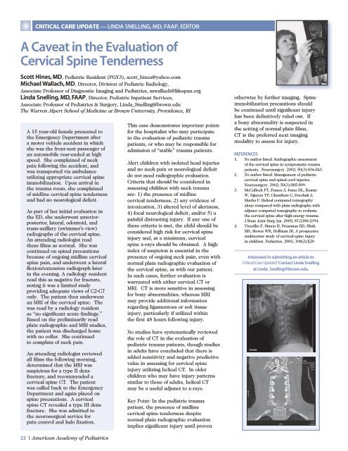
An attending radiologist reviewed<br />
all films the following morning,<br />
determined that the MRI was<br />
suspicious for a type II dens<br />
fracture, and recommended a<br />
cervical spine CT. The patient<br />
was called back to the Emergency<br />
Department and again placed on<br />
spine precautions. A cervical<br />
spine CT revealed a type III dens<br />
fracture. She was admitted to<br />
the neurosurgical service for<br />
pain control and halo fixation.<br />
22 | <strong>American</strong> <strong>Academy</strong> <strong>of</strong> <strong>Pediatrics</strong><br />
This case demonstrates important points<br />
for the hospitalist who may participate<br />
in the evaluation <strong>of</strong> pediatric trauma<br />
patients, or who may be responsible for<br />
admission <strong>of</strong> “stable” trauma patients.<br />
Alert children with isolated head injuries<br />
and no neck pain or neurological deficit<br />
do not need radiographic evaluation.<br />
Criteria that should be considered in<br />
assessing children with neck trauma<br />
are: 1) the presence <strong>of</strong> midline<br />
cervical tenderness, 2) any evidence <strong>of</strong><br />
intoxication, 3) altered level <strong>of</strong> alertness,<br />
4) focal neurological deficit, and/or 5) a<br />
painful distracting injury. If any one <strong>of</strong><br />
these criteria is met, the child should be<br />
considered high risk for cervical spine<br />
injury and, at a minimum, cervical<br />
spine x-rays should be obtained. A high<br />
index <strong>of</strong> suspicion is essential in the<br />
presence <strong>of</strong> ongoing neck pain, even with<br />
normal plain radiographic evaluation <strong>of</strong><br />
the cervical spine, as with our patient.<br />
In such cases, further evaluation is<br />
warranted with either cervical CT or<br />
MRI. CT is more sensitive in assessing<br />
for bony abnormalities, whereas MRI<br />
may provide additional information<br />
regarding ligamentous or s<strong>of</strong>t tissue<br />
injury, particularly if utilized within<br />
the first 48 hours following injury.<br />
No studies have systematically reviewed<br />
the role <strong>of</strong> CT in the evaluation <strong>of</strong><br />
pediatric trauma patients, though studies<br />
in adults have concluded that there is<br />
added sensitivity and negative predictive<br />
value in assessing for cervical spine<br />
injury utilizing helical CT. In older<br />
children who may have injury patterns<br />
similar to those <strong>of</strong> adults, helical CT<br />
may be a useful adjunct to x-rays.<br />
Key Point: In the pediatric trauma<br />
patient, the presence <strong>of</strong> midline<br />
cervical spine tenderness despite<br />
normal plain radiographic evaluation<br />
implies significant injury until proven<br />
otherwise by further imaging. Spine<br />
immobilization precautions should<br />
be continued until significant injury<br />
has been definitively ruled out. If<br />
a bony abnormality is suspected in<br />
the setting <strong>of</strong> normal plain films,<br />
CT is the preferred next imaging<br />
modality to assess for injury.<br />
REFERENCES<br />
1. No author listed. Radiographic assessment<br />
<strong>of</strong> the cervical spine in symptomatic trauma<br />
patients. Neurosurgery. 2002; 50(3):S36-S43.<br />
2. No author listed. Management <strong>of</strong> pediatric<br />
cervical spine and spinal cord injuries.<br />
Neurosurgery. 2002; 50(3):S85-S99.<br />
3. McCulloch PT, France J, Jones DL, Krantz<br />
W, Nguyen TP, Chambers C, Dorchak J,<br />
Mucha P. Helical computed tomography<br />
alone compared with plain radiographs with<br />
adjunct computed tomography to evaluate<br />
the cervical spine after high-energy trauma.<br />
J Bone Joint Surg Am. 2005; 87:2388-2394.<br />
4. Viccellio P, Simon H, Pressman BD, Shah<br />
MN, Mower WR, H<strong>of</strong>fman JR. A prospective<br />
multicenter study <strong>of</strong> cervical spine injury<br />
in children. <strong>Pediatrics</strong>. 2001; 108(2):E20<br />
Interested in submitting an article to<br />
Critical Care Update? Contact Linda Snelling<br />
at Linda_Snelling@brown.edu.


