

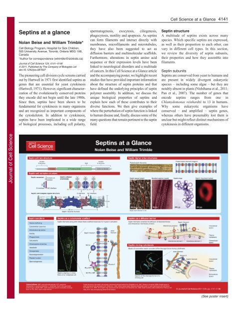
Septins at a glance - Journal of Cell Science - The Company of ...
Septins at a glance - Journal of Cell Science - The Company of ...
Septins at a glance - Journal of Cell Science - The Company of ...

You also want an ePaper? Increase the reach of your titles
YUMPU automatically turns print PDFs into web optimized ePapers that Google loves.
<strong>Journal</strong> <strong>of</strong> <strong>Cell</strong> <strong>Science</strong><br />
<strong>Septins</strong> <strong>at</strong> a <strong>glance</strong><br />
Nolan Beise and William Trimble*<br />
<strong>Cell</strong> Biology Program, Hospital for Sick Children,<br />
555 University Avenue, Toronto, Ontario M5G 1X8,<br />
Canada<br />
*Author for correspondence (wtrimble@sickkids.ca)<br />
<strong>Journal</strong> <strong>of</strong> <strong>Cell</strong> <strong>Science</strong> 124, 4141–4146<br />
© 2011. Published by <strong>The</strong> <strong>Company</strong> <strong>of</strong> Biologists Ltd<br />
doi:10.1242/jcs.087007<br />
<strong>The</strong> pioneering cell division cycle screens carried<br />
out by Hartwell in 1971 first identified septins as<br />
genes th<strong>at</strong> are essential for yeast cytokinesis<br />
(Hartwell, 1971). However, significant characteriz<strong>at</strong>ion<br />
<strong>of</strong> the evolutionarily conserved proteins<br />
they encode did not begin until the l<strong>at</strong>e 1980s.<br />
Since then, septins have been shown to be<br />
fundamental for cytokinesis in many organisms<br />
and are recognized as important components <strong>of</strong><br />
the cytoskeleton. In addition to cytokinesis,<br />
septins have been implic<strong>at</strong>ed in a wide range<br />
<strong>of</strong> biological processes, including cell polarity,<br />
sperm<strong>at</strong>ogenesis, exocytosis, ciliogenesis,<br />
phagocytosis, motility and apoptosis. As septins<br />
can form filaments and interact directly with<br />
membranes, micr<strong>of</strong>ilaments and microtubules,<br />
they have also been suggested to act as<br />
diffusion barriers and multimolecular scaffolds.<br />
Furthermore, alter<strong>at</strong>ions in septin amino acid<br />
sequence or their expression levels have been<br />
linked to neurological disorders and a multitude<br />
<strong>of</strong> cancers. In this <strong>Cell</strong> <strong>Science</strong> <strong>at</strong> a Glance article<br />
and the accompanying poster, we highlight recent<br />
studies th<strong>at</strong> have provided important inform<strong>at</strong>ion<br />
about the structure <strong>of</strong> septin proteins and th<strong>at</strong><br />
have defined the underlying principles <strong>of</strong> septin<br />
polymer assembly. In addition, we discuss the<br />
unique biological properties <strong>of</strong> septins and<br />
explain how each <strong>of</strong> these contributes to their<br />
diverse functions. We then give examples <strong>of</strong><br />
where the perturb<strong>at</strong>ion <strong>of</strong> septin function is linked<br />
to human disease and, finally, discuss some <strong>of</strong> the<br />
many questions th<strong>at</strong> remain pertinent to the septin<br />
field.<br />
<strong>Cell</strong> <strong>Science</strong> <strong>at</strong> a Glance 4141<br />
Septin structure<br />
A multitude <strong>of</strong> septins exists across many<br />
species. Which specific septins are expressed,<br />
as well as their proportion to each other, can<br />
vary in different cell types. In this section,<br />
we review the diversity <strong>of</strong> septin subunits,<br />
their properties and how they assemble into<br />
filaments.<br />
Septin subunits<br />
<strong>Septins</strong> are conserved from yeast to humans and<br />
are present in widely divergent eukaryotic<br />
species – including some algae – but they are<br />
notably absent in plants (Nishihama et al., 2011;<br />
Pan et al., 2007). <strong>The</strong> number <strong>of</strong> genes th<strong>at</strong><br />
encode septins ranges from one in<br />
Chlamydomonas reinhardtii to 13 in humans.<br />
Why some eukaryotic organisms have<br />
conserved – and amplified – septin genes,<br />
whereas others have presumably lost them is<br />
unclear but might reflect distinct mechanisms <strong>of</strong><br />
cytokinesis in different organisms.<br />
(See poster insert)
<strong>Journal</strong> <strong>of</strong> <strong>Cell</strong> <strong>Science</strong><br />
4142<br />
<strong>Journal</strong> <strong>of</strong> <strong>Cell</strong> <strong>Science</strong> 124 (24)<br />
Comparison <strong>of</strong> septins from many different<br />
species has revealed th<strong>at</strong> these proteins share<br />
several conserved domains and, in species with<br />
multiple septin genes, can be organized into<br />
subgroups. In mammals, four such distinct<br />
groups – or families – are represented by<br />
SEPT1, SEPT3, SEPT6 and SEPT7, and these<br />
share some structural similarities with the four<br />
main septins in yeast (Cao et al., 2007). Nearly<br />
all septins contain a GTPase domain th<strong>at</strong><br />
belongs to the GTPase superclass <strong>of</strong> P-loop<br />
nucleotide triphosph<strong>at</strong>ases, and whose other<br />
members include kinesin, myosin and Ras<br />
proteins (Leipe et al., 2002). However, the<br />
GTPase domain <strong>of</strong> septins is unique among this<br />
family in th<strong>at</strong> it contains a conserved sequence<br />
near its end – the septin unique element (SUE)<br />
(Pan et al., 2007; Steels et al., 2007; Versele et<br />
al., 2004). Most septins in yeast and metazoans<br />
can bind and hydrolyze GTP (Field et al., 1996;<br />
Mendoza et al., 2002; Sheffield et al., 2003;<br />
Versele and Thorner, 2004). In vitro, this<br />
hydrolysis is slow. For example, the apparent<br />
kc<strong>at</strong> value for SEPT2 is 2.7�10 –4 seconds –1<br />
(Huang et al., 2006), which is similar to th<strong>at</strong> <strong>of</strong><br />
the structurally rel<strong>at</strong>ed Ras GTPase (kc<strong>at</strong><br />
3.4�10 –4 seconds –1 ) (Neal et al., 1988) and the<br />
Cdc3–Cdc12 complex in yeast (apparent kc<strong>at</strong> <strong>of</strong><br />
3�10 –4 seconds –1 ) (Farkasovsky et al., 2005). In<br />
vivo, septins also display slow r<strong>at</strong>es <strong>of</strong> GTP<br />
exchange (Frazier et al., 1998; Huang et al.,<br />
2006; Vrabioiu et al., 2003).<br />
<strong>Septins</strong> have additional conserved elements<br />
th<strong>at</strong> flank the GTPase domain. N-terminal to the<br />
GTPase domain, most septins possess a<br />
polybasic region th<strong>at</strong> has been shown to bind<br />
phosphoinositides (Casamayor and Snyder,<br />
2003; Zhang et al., 1999). Moreover, the Cterminus<br />
<strong>of</strong> most septins contains an extension,<br />
which is predicted to form coiled coils th<strong>at</strong><br />
might be important for certain septin–septin and<br />
septin–substr<strong>at</strong>e interactions (Casamayor and<br />
Snyder, 2003; Versele and Thorner, 2005).<br />
<strong>Septins</strong> have two conserved interfaces th<strong>at</strong><br />
are involved in the form<strong>at</strong>ion <strong>of</strong> septin–septin<br />
interactions and subsequent complex assembly:<br />
the guanine nucleotide-binding domain (G<br />
interface), and the N- and C-terminal extensions<br />
(NC interface) (Sirajuddin et al., 2007).<br />
Interestingly, the SUE partly overlaps with the G<br />
interface, where it might also contribute to<br />
septin assembly into complexes (Sirajuddin et<br />
al., 2009; Versele et al., 2004). Within the<br />
cytosol, most septins are found assembled into<br />
hetero-oligomeric filaments th<strong>at</strong> are composed<br />
<strong>of</strong> octameric non-polar unit complexes (Bertin<br />
et al., 2008; John et al., 2007; Kinoshita et al.,<br />
2002; Sellin et al., 2011; Sheffield et al., 2003).<br />
In fact, mammalian septins exist solely as 6- to<br />
8-unit heteromeric complexes within the cell<br />
(Sellin et al., 2011).<br />
Unit complex<br />
Septin assembly into filaments begins with<br />
their incorpor<strong>at</strong>ion into unit complexes.<br />
Typically these complexes are heterooligomers<br />
th<strong>at</strong> are composed <strong>of</strong> four, six or<br />
eight septin monomers polymerized into a<br />
linear, non-polar polymer. Complexes <strong>of</strong> septin<br />
units were first identified when the initial<br />
purific<strong>at</strong>ion <strong>of</strong> septin filaments from<br />
Saccharomyces cerevisiae (Cdc3, Cdc10,<br />
Cdc11, Cdc12) and from Drosophila<br />
melanogaster (Pnut, Sep1, Sep2) showed th<strong>at</strong><br />
these proteins are present in near equal<br />
amounts, suggesting 1:1:1:1 and 1:1:1<br />
stoichiometries, respectively (Field et al.,<br />
1996; Frazier et al., 1998; Oegema et al.,<br />
1998). Electron microscopy studies <strong>of</strong> septin<br />
complexes in S. cerevisiae and Caenorhabditis<br />
elegans showed th<strong>at</strong> septin complexes<br />
(Cdc11–Cdc12–Cdc3–Cdc10–Cdc10–Cdc3–<br />
Cdc12–Cdc11 and Unc59–Unc-61–Unc61–<br />
Unc59, respectively) are non-polar (Bertin<br />
et al., 2008; John et al., 2007). In mammals, a<br />
septin complex consisting <strong>of</strong> SEPT2, SEPT4,<br />
SEPT6 and SEPT7 was first identified in brain<br />
tissue (Hsu et al., 1998). Two other groups<br />
were able to isol<strong>at</strong>e a complex from<br />
mammalian cell lines and mouse brain th<strong>at</strong><br />
contained SEPT2, SEPT6 and SEPT7 with a<br />
stoichiometry <strong>of</strong> approxim<strong>at</strong>ely 1:1:1 (Joberty<br />
et al., 2001; Kinoshita et al., 2002). Further<br />
structural characteriz<strong>at</strong>ion <strong>of</strong> recombinant<br />
complexes <strong>of</strong> these septins revealed them to be<br />
non-polar, rod-shaped oligomers in which the<br />
order <strong>of</strong> septins is SEPT7–SEPT6–SEPT2–<br />
SEPT2–SEPT6–SEPT7 (Sirajuddin et al.,<br />
2007). Most mammalian cells appear to<br />
express members <strong>of</strong> each <strong>of</strong> the four septin<br />
families. It is, therefore, unclear where<br />
members <strong>of</strong> the SEPT3 family fit within the<br />
unit complex. However, recent biochemical<br />
approaches have been used to show th<strong>at</strong>, in<br />
HeLa cells, the SEPT3 family member SEPT9<br />
localizes to the ends <strong>of</strong> these hexamers in<br />
octameric complexes (Kim et al., 2011a; Sellin<br />
et al., 2011). Interestingly, the composition <strong>of</strong><br />
the septin unit complex can be flexible because<br />
septins from the same subgroup, and even<br />
is<strong>of</strong>orms <strong>of</strong> the same septin, can substitute for<br />
each other within this structure (Sellin et al.,<br />
2011).<br />
Septin monomers within the unit complex<br />
interact through G–G and NC–NC interfaces,<br />
thereby pairing up with each other (Sirajuddin et<br />
al., 2007). <strong>The</strong>se G–G and NC–NC interactions<br />
altern<strong>at</strong>e along the unit complex, and are<br />
necessary for its form<strong>at</strong>ion (Sirajuddin et al.,<br />
2007). <strong>The</strong> coiled-coil C-termini <strong>of</strong> the septin<br />
monomers have also been implic<strong>at</strong>ed in the<br />
form<strong>at</strong>ion <strong>of</strong> septin unit complexes, by directly<br />
interacting with each other (Moshe S. Kim,<br />
Carol D. Froese and W. T., unpublished results)<br />
(Low and Macara, 2006; Shinoda et al., 2010).<br />
Unfortun<strong>at</strong>ely, the detailed structure <strong>of</strong> septin<br />
coiled-coil interactions in the mammalian septin<br />
complex has not yet been visualized, as these<br />
regions <strong>of</strong> the proteins were not resolved in the<br />
septin crystal structure (Sirajuddin et al., 2007).<br />
However, electron microscopy (EM) images <strong>of</strong><br />
the yeast septin complex show finger-like<br />
projections from the unit complex, which are<br />
consistent with the coiled-coil domains th<strong>at</strong><br />
project perpendicular to the septin complex<br />
(Bertin et al., 2008; Hsu et al., 1998).<br />
Guanine nucleotides also have a role in the<br />
form<strong>at</strong>ion and stability <strong>of</strong> septin unit complexes.<br />
<strong>The</strong>y fully s<strong>at</strong>ur<strong>at</strong>e recombinant septin octamers<br />
in yeast and septin hexamers in human, although<br />
the function <strong>of</strong> these nucleotides in the assembly<br />
<strong>of</strong> septin complexes is still not fully understood<br />
(Farkasovsky et al., 2005; Sirajuddin et al.,<br />
2007; Vrabioiu et al., 2003). Mut<strong>at</strong>ion <strong>of</strong><br />
residues th<strong>at</strong> contribute to the binding <strong>of</strong> septin<br />
to GTP alters the form<strong>at</strong>ion, appearance,<br />
localiz<strong>at</strong>ion and/or function <strong>of</strong> septin polymers<br />
(Casamayor and Snyder, 2003; Ding et al., 2008;<br />
Hanai et al., 2004; Kinoshita et al., 1997;<br />
Nagaraj et al., 2008; Robertson et al., 2004;<br />
Steels et al., 2007; Vega and Hsu, 2003).<br />
Structural studies <strong>of</strong> SEPT2 have shown th<strong>at</strong><br />
binding to guanine nucleotide (GTP or GDP)<br />
induces stable conform<strong>at</strong>ional changes in the G<br />
and NC surfaces th<strong>at</strong> – in turn – might regul<strong>at</strong>e<br />
septin–septin interactions (Sirajuddin et al.,<br />
2007; Sirajuddin et al., 2009). GTP hydrolysis<br />
might, therefore, act as a switch th<strong>at</strong> regul<strong>at</strong>es<br />
complex assembly (Sirajuddin et al., 2007;<br />
Sirajuddin et al., 2009).<br />
Interestingly, when expressed alone, SEPT2<br />
forms a dimer <strong>at</strong> its G interface, yet SEPT2-GDP<br />
dimers can interact through both G and NC<br />
interfaces and, in unit complexes, SEPT2 only<br />
interacts with other SEPT2 molecules through<br />
the NC interface (Moshe S. Kim, Carol D.<br />
Froese and W. T., unpublished results)<br />
(Sirajuddin et al., 2007). Indeed, we found th<strong>at</strong><br />
when any two septins are overexpressed<br />
together, they preferentially interact <strong>at</strong> their G<br />
interface, even though they might normally<br />
interact <strong>at</strong> their NC interface in the unit complex<br />
(Moshe S. Kim, Carol D. Froese and W. T.,<br />
unpublished results). Despite this apparent<br />
promiscuity, septin filaments assemble in an<br />
ordered manner, suggesting th<strong>at</strong> preferential<br />
pairwise G interface assembly is likely to<br />
precede the form<strong>at</strong>ion <strong>of</strong> septin unit complexes.<br />
Because SEPT2–SEPT6 and SEPT7–SEPT9<br />
pairs interact in the octamer <strong>at</strong> their G interfaces,<br />
these might assemble first as GTP-bound forms.<br />
Subsequent GTP hydrolysis might then trigger<br />
conform<strong>at</strong>ional changes <strong>at</strong> the NC interfaces to<br />
allow subsequent assembly <strong>of</strong> the octamer
<strong>Journal</strong> <strong>of</strong> <strong>Cell</strong> <strong>Science</strong><br />
(Moshe S. Kim, Carol D. Froese and W. T.,<br />
unpublished results) (Sirajuddin et al., 2009). In<br />
addition, interactions <strong>of</strong> SEPT6–SEPT7 and NC<br />
might also be promoted by the binding <strong>of</strong> Cdc42<br />
effector protein 5 (Borg3) to the septin coiledcoil<br />
domains (Sheffield et al., 2003). In the same<br />
way th<strong>at</strong> septin monomers are the building<br />
blocks <strong>of</strong> the unit complex, those unit<br />
complexes th<strong>at</strong> become polymerized end-to-end<br />
subsequently form the building blocks <strong>of</strong> septin<br />
filaments.<br />
Filaments<br />
Septin filaments are the predominant septin<br />
structure within the cell. As septin unit<br />
complexes vary in composition, septin<br />
filament composition is likely to change<br />
correspondingly. <strong>The</strong>se filaments primarily<br />
localize to the cleavage furrow in dividing cells<br />
and to stress fibers in interphase cells, but are<br />
also present <strong>at</strong> other structures, which rel<strong>at</strong>e to<br />
their functions. <strong>The</strong> basic septin filament is 4–5<br />
nm wide with varying lengths, and has the<br />
capacity to form rings, bundles, linear filaments<br />
and gauzes (Bertin et al., 2008; Kinoshita et al.,<br />
2002; Rodal et al., 2005). <strong>The</strong> mechanism <strong>of</strong><br />
septin assembly into this diverse array <strong>of</strong><br />
structures is not completely known, but there is<br />
some evidence th<strong>at</strong> links Cdc42, the Borg<br />
family, anillin, guanine nucleotides and the<br />
membrane associ<strong>at</strong>ion <strong>of</strong> septins to the<br />
regul<strong>at</strong>ion <strong>of</strong> septin filament assembly<br />
(Caviston et al., 2003; Gladfelter et al., 2002;<br />
Joberty et al., 2001; Kinoshita et al., 2002;<br />
Nagaraj et al., 2008; Smith et al., 2002; Tanaka-<br />
Takiguchi et al., 2009). In yeast, mut<strong>at</strong>ions in the<br />
Rho-type GTPase Cdc42p abrog<strong>at</strong>e ring<br />
form<strong>at</strong>ion by causing defects in the polarized<br />
localiz<strong>at</strong>ion <strong>of</strong> septins to the presumptive bud<br />
site (Gladfelter et al., 2001; Jeong et al., 2001).<br />
Joberty et al. have shown th<strong>at</strong> Cdc42-GTP is<br />
involved in neg<strong>at</strong>ively regul<strong>at</strong>ing Borgmedi<strong>at</strong>ed<br />
septin polymeriz<strong>at</strong>ion in mammals<br />
(Joberty et al., 2001). Additionally, defects in<br />
anillin cause loss <strong>of</strong> septin recruitment to the<br />
cleavage furrow, and overexpression <strong>of</strong> the<br />
septin-interacting fragment <strong>of</strong> anillin perturbs<br />
septin organiz<strong>at</strong>ion (Field et al., 2005; Kinoshita<br />
et al., 2002). As guanine nucleotides might<br />
regul<strong>at</strong>e unit complex form<strong>at</strong>ion, it would not be<br />
surprising if they also were to affect higher order<br />
septin filament assembly. In yeast, GTP binding<br />
by all four septins is necessary for the form<strong>at</strong>ion<br />
<strong>of</strong> the septin ring under stressful growth<br />
conditions (37°C), and a combin<strong>at</strong>ion <strong>of</strong> septin<br />
mutants th<strong>at</strong> are defective in GTP-binding is<br />
<strong>of</strong>ten lethal (Longtine et al., 1996; Nagaraj et al.,<br />
2008; Versele and Thorner, 2004). Both yeast<br />
and mammalian septins show increased r<strong>at</strong>es <strong>of</strong><br />
polymeriz<strong>at</strong>ion into filaments following their<br />
associ<strong>at</strong>ion with lipid bilayers (Bertin et al.,<br />
2010; Tanaka-Takiguchi et al., 2009). <strong>The</strong><br />
form<strong>at</strong>ion <strong>of</strong> septin filaments is required for<br />
stable associ<strong>at</strong>ion <strong>of</strong> septins with the plasma<br />
membrane and successful completion <strong>of</strong> yeast<br />
cytokinesis (McMurray et al., 2011). This<br />
suggests th<strong>at</strong> septin membrane associ<strong>at</strong>ions and<br />
septin filament assembly are inextricably linked<br />
(McMurray et al., 2011). Interestingly, despite<br />
septin unit complexes being very stable, septin<br />
filaments can be highly dynamic during certain<br />
stages <strong>of</strong> the cell cycle (Dobbelaere and<br />
Barral, 2004; Sellin et al., 2011; Vrabioiu<br />
and Mitchison, 2006). Septin filament<br />
characteristics also depend on the properties <strong>of</strong><br />
their monomer constituents, which indic<strong>at</strong>es<br />
th<strong>at</strong> the function <strong>of</strong> septin filaments varies<br />
depending on their composition.<br />
Septin functions<br />
<strong>Septins</strong> as scaffolds<br />
Yeast<br />
As diverse and rel<strong>at</strong>ively st<strong>at</strong>ic polymers,<br />
septins are able to interact with a large variety <strong>of</strong><br />
molecules. As such, they have largely been<br />
described as scaffolds th<strong>at</strong> recruit molecules to<br />
their sites <strong>of</strong> function and promote<br />
protein–protein interactions. In S. cerevisiae,<br />
more than 40 proteins localize to the mother-bud<br />
neck in a septin-dependent manner (Gladfelter<br />
et al., 2001); form<strong>at</strong>ion <strong>of</strong> a functional<br />
contractile ring <strong>at</strong> the mother-bud neck requires<br />
septin scaffolding <strong>of</strong> several proteins th<strong>at</strong> are<br />
essential for cytokinesis. For example,<br />
degrad<strong>at</strong>ion <strong>of</strong> the Wee1-family kinase Swe1p<br />
by its neg<strong>at</strong>ive regul<strong>at</strong>ors leads to progression <strong>of</strong><br />
mitosis, and occurs when septins provide a<br />
scaffold for the recruitment <strong>of</strong> a multi-protein<br />
kinase cascade to the mother-bud neck (Barral et<br />
al., 1999; Hanrahan and Snyder, 2003; Longtine<br />
et al., 2000; Sakchaisri et al., 2004; Szkotnicki et<br />
al., 2008). Interestingly, septin scaffolding in the<br />
form <strong>of</strong> microtubule capture <strong>at</strong> the cleavage<br />
plane ensures correct positioning <strong>of</strong> the spindle<br />
pole body and subsequent segreg<strong>at</strong>ion <strong>of</strong><br />
replic<strong>at</strong>ed chromosomes into mother and<br />
daughter cells (Kusch et al., 2002). Despite<br />
septins being essential for cytokinesis in<br />
budding yeast, they are not necessary for cell<br />
division in fission yeast S. pombe. In fact,<br />
deletion <strong>of</strong> all four veget<strong>at</strong>ively expressed<br />
septins results in only mild cell separ<strong>at</strong>ion<br />
defects in fission yeast (Berlin et al., 2003; Tasto<br />
et al., 2003). Thus, although they are<br />
dispensable for cytokinesis, septins are<br />
important for subsequent cell separ<strong>at</strong>ion in S.<br />
pombe (Wu et al., 2010).<br />
Metazoans<br />
Septin scaffolding <strong>of</strong> kinases and their substr<strong>at</strong>es<br />
also seems to occur in mammals, as<br />
demonstr<strong>at</strong>ed by phosphoryl<strong>at</strong>ion <strong>of</strong> myosin II <strong>at</strong><br />
<strong>Journal</strong> <strong>of</strong> <strong>Cell</strong> <strong>Science</strong> 124 (24)<br />
4143<br />
sites <strong>at</strong> which septin filaments are loc<strong>at</strong>ed near<br />
actin stress fibers (Joo et al., 2007). Because<br />
septins bind the septin-associ<strong>at</strong>ed Rho guanine<br />
nucleotide exchange factor (SA-Rho-GEF) and<br />
myosin (Joo et al., 2007; Nag<strong>at</strong>a and Inagaki,<br />
2004), the entire GEF–Rho–ROCK–myosin<br />
signaling kinase cascade, which is essential for<br />
full myosin activ<strong>at</strong>ion, might be scaffolded by<br />
septins on stress fibers. In addition, septin<br />
scaffolds <strong>at</strong> stress fibers are also important in<br />
regul<strong>at</strong>ing the DNA damage response by<br />
interacting with suppressor <strong>of</strong> cytokine signaling<br />
7 (SOCS7) (Kremer et al., 2007). As septins are<br />
conserved between fungal and animal species, it<br />
is not surprising th<strong>at</strong> many septin functions are<br />
also conserved among these species. An<br />
essential role for septins in cytokinesis has also<br />
been shown in D. melanogaster and mammals<br />
(Nag<strong>at</strong>a et al., 2003; Neufeld and Rubin, 1994;<br />
Surka et al., 2002). Interestingly, SEPT5containing<br />
scaffolds additionally regul<strong>at</strong>e<br />
neuronal exocytosis through interactions with<br />
syntaxin and SNARE proteins (Beites et al.,<br />
1999; Yang et al., 2010).<br />
<strong>Septins</strong> as membrane modul<strong>at</strong>ors<br />
Yeast<br />
In addition to acting as scaffolds, septins<br />
modul<strong>at</strong>e membrane dynamics by binding lipids<br />
through their polybasic domains. For example,<br />
septin filaments <strong>at</strong> the cortex <strong>of</strong> the mother-bud<br />
neck in yeast limit the diffusion <strong>of</strong> membraneassoci<strong>at</strong>ed<br />
components between daughter and<br />
mother cells (Barral et al., 2000; Takizawa et al.,<br />
2000). This diffusion-barrier-dependent<br />
polarized bud localiz<strong>at</strong>ion is essential for<br />
successful mitosis (Barral et al., 2000). In<br />
addition, the septin structure <strong>at</strong> the mother-bud<br />
neck transitions from an hourglass-shaped<br />
structure to two parallel rings on either side <strong>of</strong><br />
the mother-bud neck. Between them, the rings<br />
define a specialized cortical compartment th<strong>at</strong><br />
retains diffusable factors such as the exocyst and<br />
polarizome complexes <strong>at</strong> the mother-bud neck,<br />
which are important for cytokinesis (Dobbelaere<br />
and Barral, 2004). <strong>The</strong> yeast cleavage furrow<br />
septin ring also particip<strong>at</strong>es in limiting the<br />
diffusion <strong>of</strong> ER and nuclear envelope proteins<br />
through the bud neck by recruiting other<br />
membrane-associ<strong>at</strong>ed factors such as Bud6<br />
(Luedeke et al., 2005; Shcheprova et al., 2008).<br />
Interestingly, a septin diffusion barrier is not<br />
necessary for the successful completion <strong>of</strong><br />
cytokinesis, as shown in S. cerevisiae mutants<br />
th<strong>at</strong> cannot form a diffusion-limiting hourglass<br />
structure (Wloka et al., 2011).<br />
Metazoans<br />
In mammals, a diffusion barrier does exist <strong>at</strong> the<br />
cleavage furrow (and septins are found <strong>at</strong> this<br />
loc<strong>at</strong>ion), but it remains to be determined
<strong>Journal</strong> <strong>of</strong> <strong>Cell</strong> <strong>Science</strong><br />
4144<br />
<strong>Journal</strong> <strong>of</strong> <strong>Cell</strong> <strong>Science</strong> 124 (24)<br />
whether this barrier is septin-dependent<br />
(Schmidt and Nichols, 2004). Mouse SEPT2containing<br />
filaments <strong>at</strong> the base <strong>of</strong> primary cilia,<br />
however, have been shown to function as a<br />
diffusion barrier to ciliary membrane proteins<br />
th<strong>at</strong> are essential for correct ciliary structure and<br />
function (Hu et al., 2010). A septin diffusion<br />
barrier also forms a ring <strong>at</strong> the sperm annulus,<br />
which is required for the cortical organiz<strong>at</strong>ion,<br />
morphology and normal motility <strong>of</strong> sperm<br />
flagella (Ihara et al., 2005; Kissel et al., 2005;<br />
Lin et al., 2009). Phagocytosis requires septins<br />
<strong>at</strong> the base <strong>of</strong> the phagocytic cup and it is<br />
tempting to specul<strong>at</strong>e th<strong>at</strong>, in addition to<br />
scaffolding actomyosin components, this<br />
functions to segreg<strong>at</strong>e lipid distribution <strong>at</strong> this<br />
structure (Huang et al., 2008; Yeung et al., 2009;<br />
Yeung et al., 2006). Interestingly, septins also<br />
form another ring <strong>at</strong> the base <strong>of</strong> dendritic spines<br />
in neurons, which is essential for their<br />
morphology (Cho et al., 2011; Xie et al., 2007).<br />
Neuronal SEPT6-containing rings <strong>at</strong> dendritic<br />
branch points might act as diffusion barriers or<br />
membrane braces, thereby maintaining<br />
polarized molecular distributions and dendritic<br />
membrane structure (Cho et al., 2011).<br />
In addition to acting as diffusion barriers,<br />
filamentous, cortical septins are also able to<br />
modul<strong>at</strong>e cellular membrane rigidity as shown<br />
by studies in T-cells (Tooley et al., 2009).<br />
Interestingly, decreases in septin-medi<strong>at</strong>ed<br />
membrane bracing does not block motility, but<br />
cells become poorly persistent and<br />
uncoordin<strong>at</strong>ed (Tooley et al., 2009). This link is<br />
further supported by septin-dependent neuronal<br />
migr<strong>at</strong>ion (Shinoda et al., 2010). Additionally,<br />
Tanaka-Takiguchi and co-workers showed th<strong>at</strong><br />
septin-containing brain extracts are able to<br />
tubul<strong>at</strong>e giant liposomes following membraneassoci<strong>at</strong>ed<br />
septin filament form<strong>at</strong>ion, which<br />
further supports a role for septins as membrane<br />
modul<strong>at</strong>ors (Tanaka-Takiguchi et al., 2009).<br />
Additional roles for septins in metazoans<br />
In addition to their roles as molecular scaffolds<br />
and membrane modul<strong>at</strong>ors, septins have been<br />
shown to carry out other functions in metazoans.<br />
For example, mammalian septins are required<br />
for cell polarity, although they act through a<br />
different mechanism than in yeast. SEPT2<br />
particip<strong>at</strong>es in epithelial cell polarity by<br />
facilit<strong>at</strong>ing apical and basal factor vesicle<br />
transport along polyGlu microtubule tracks by<br />
preventing the binding <strong>of</strong> microtubuleassoci<strong>at</strong>ed<br />
protein 4 (MAP4) (Spiliotis et al.,<br />
2008). In host cell defence, ring-like septin<br />
structures form cages around intracellular<br />
bacteria, thereby restricting their prolifer<strong>at</strong>ion<br />
and facilit<strong>at</strong>ing their degrad<strong>at</strong>ion by autophagy<br />
(Mostowy et al., 2010). Finally, one specialized<br />
type <strong>of</strong> septin function involves an altern<strong>at</strong>ive<br />
transcript <strong>of</strong> SEPT4 called ARTS, which<br />
transloc<strong>at</strong>es from mitochondria to the nucleus<br />
upon exposure to an apoptotic agent and induces<br />
apoptosis by activ<strong>at</strong>ing caspase 3 (Gottfried et<br />
al., 2004; Larisch, 2004; Larisch et al., 2000).<br />
Septin-associ<strong>at</strong>ed diseases<br />
Because mammalian septins interact with a<br />
variety <strong>of</strong> molecules and are essential in many<br />
cellular processes, it is not surprising th<strong>at</strong> they<br />
are implic<strong>at</strong>ed in multiple human diseases.<br />
Several septins have been found to associ<strong>at</strong>e<br />
with protein aggreg<strong>at</strong>es th<strong>at</strong> are common in<br />
neurodegener<strong>at</strong>ive disorders such as Parkinson<br />
and Alzheimer disease (Garcia et al., 2006; Ihara<br />
et al., 2003; Kinoshita et al., 1998). <strong>Septins</strong> are<br />
also able to interact with parkin, an E3 ubiquitin<br />
ligase th<strong>at</strong> is involved in Parkinson disease<br />
(Choi et al., 2003; Zhang et al., 2000). Further<br />
evidence th<strong>at</strong> links septins to neurological<br />
disorders stems from mut<strong>at</strong>ions within SEPT9,<br />
causing hereditary neuralgic amyotrophy<br />
(HNA) (Hannibal et al., 2009; Kuhlenbaumer et<br />
al., 2005; Landsverk et al., 2009). As cytokinetic<br />
defects are linked to cancer (Sagona and<br />
Stenmark, 2010), it is not surprising th<strong>at</strong> septins,<br />
which are essential to this process, have also<br />
been linked to cancer (Russell and Hall, 2005).<br />
Several septins have been identified as mixed<br />
lineage leukemia (MLL) fusion partners th<strong>at</strong><br />
contribute to the p<strong>at</strong>hogenesis <strong>of</strong> leukemia<br />
(Borkhardt et al., 2001; Cerveira et al., 2006;<br />
Kojima et al., 2004; Osaka et al., 1999). Notably,<br />
septins are also being used in a diagnostic<br />
context, with the methyl<strong>at</strong>ion <strong>of</strong> the SEPT9<br />
promoter serving as an effective biomarker for<br />
colorectal cancer (deVos et al., 2009;<br />
Grutzmann et al., 2008; L<strong>of</strong>ton-Day et al.,<br />
2008). <strong>The</strong> role <strong>of</strong> septins in neurological<br />
disorders and cancer is complex and poorly<br />
understood. For more detail on this subject, we<br />
refer readers to a recent review by Peterson and<br />
Petty (Peterson and Petty, 2010).<br />
Perspectives<br />
Since the initial characteriz<strong>at</strong>ion <strong>of</strong> septins in the<br />
l<strong>at</strong>e 1980s, research in this field has grown<br />
impressively, especially when considering the<br />
number and diversity <strong>of</strong> septins th<strong>at</strong> have been<br />
characterized across species. This research has<br />
led to some very interesting questions rel<strong>at</strong>ed to<br />
septin structure and function. Determining the<br />
mechanism <strong>of</strong> both the septin unit complex and<br />
septin filament assembly will be paramount in<br />
elucid<strong>at</strong>ing the myriad <strong>of</strong> properties and<br />
functions th<strong>at</strong> septins exhibit within the cell.<br />
Specifically, it will be interesting to illumin<strong>at</strong>e<br />
how the variable N- and C-termini <strong>of</strong> septins<br />
regul<strong>at</strong>e filament polymeriz<strong>at</strong>ion, stability and<br />
scaffolding functions. Additionally, it will be<br />
exciting to elucid<strong>at</strong>e how the coiled-coil<br />
domains <strong>of</strong> septin proteins interact and function<br />
within the septin polymer and how septinassoci<strong>at</strong>ed<br />
factors influence septin filament<br />
assembly and disassembly. In conjunction with<br />
this it will be interesting to characterize<br />
heterogeneous septin filaments and the way in<br />
which varying filament composition rel<strong>at</strong>es to<br />
different properties and functions. Septin-lipid<br />
associ<strong>at</strong>ion is another relevant topic <strong>of</strong> future<br />
research. Not only are lipids implic<strong>at</strong>ed in<br />
promoting septin polymeriz<strong>at</strong>ion, but septin<br />
filaments might also inhibit the l<strong>at</strong>eral diffusion<br />
<strong>of</strong> lipids forming distinct domains. <strong>The</strong>se and<br />
many more questions will hopefully be resolved<br />
in the coming years, thus making it a very<br />
exciting time in septin research.<br />
Acknowledgement<br />
<strong>The</strong> authors like to acknowledge the many colleagues<br />
whose work was included in the poster, and especially<br />
Alfred Wittingh<strong>of</strong>er and Eva Nogales for providing<br />
high-resolution images.<br />
Funding<br />
This work was supported by Canadian Institutes <strong>of</strong><br />
Health Research (CIHR) grant MOP97950 to W.S.T.,<br />
and N.B. was supported by studentships from CIHR<br />
and the Hospital for Sick Children Found<strong>at</strong>ion.<br />
Individual poster panels are available as JPEG files <strong>at</strong><br />
http://jcs.biologists.org/cgi/content/full/124/24/4141/DC1<br />
References<br />
Barral, Y., Parra, M., Bidlingmaier, S. and Snyder, M.<br />
(1999). Nim1-rel<strong>at</strong>ed kinases coordin<strong>at</strong>e cell cycle<br />
progression with the organiz<strong>at</strong>ion <strong>of</strong> the peripheral<br />
cytoskeleton in yeast. Genes Dev. 13, 176-187.<br />
Barral, Y., Mermall, V., Mooseker, M. S. and Snyder, M.<br />
(2000). Compartmentaliz<strong>at</strong>ion <strong>of</strong> the cell cortex by septins<br />
is required for maintenance <strong>of</strong> cell polarity in yeast. Mol.<br />
<strong>Cell</strong> 5, 841-851.<br />
Beites, C. L., Xie, H., Bowser, R. and Trimble, W. S.<br />
(1999). <strong>The</strong> septin CDCrel-1 binds syntaxin and inhibits<br />
exocytosis. N<strong>at</strong>. Neurosci. 2, 434-439.<br />
Berlin, A., Paoletti, A. and Chang, F. (2003). Mid2p<br />
stabilizes septin rings during cytokinesis in fission yeast. J.<br />
<strong>Cell</strong> Biol. 160, 1083-1092.<br />
Bertin, A., McMurray, M. A., Grob, P., Park, S. S.,<br />
Garcia, G., P<strong>at</strong>anwala, I., Ng, H. l., Alber, T., Thorner,<br />
J. and Nogales, E. (2008). Saccharomyces cerevisiae<br />
septins: supramolecular organiz<strong>at</strong>ion <strong>of</strong> heterooligomers<br />
and the mechanism <strong>of</strong> filament assembly. Proc. N<strong>at</strong>l. Acad.<br />
Sci. USA 105, 8274-8279.<br />
Bertin, A., McMurray, M. A., Thai, L., Garcia, G., 3rd,<br />
Votin, V., Grob, P., Allyn, T., Thorner, J. and Nogales, E.<br />
(2010). Phosph<strong>at</strong>idylinositol-4,5-bisphosph<strong>at</strong>e promotes<br />
budding yeast septin filament assembly and organiz<strong>at</strong>ion. J.<br />
Mol. Biol. 404, 711-731.<br />
Borkhardt, A., Teigler-Schlegel, A., Fuchs, U., Keller, C.,<br />
Konig, M., Harbott, J. and Haas, O. A. (2001). An<br />
ins(X;11)(q24;q23) fuses the MLL and the Septin<br />
6/KIAA0128 gene in an infant with AML-M2. Genes<br />
Chromosomes Cancer 32, 82-88.<br />
Cao, L., Ding, X., Yu, W., Yang, X., Shen, S. and Yu, L.<br />
(2007). Phylogenetic and evolutionary analysis <strong>of</strong> the septin<br />
protein family in metazoan. FEBS Lett. 581, 5526-5532.<br />
Casamayor, A. and Snyder, M. (2003). Molecular<br />
dissection <strong>of</strong> a yeast septin: distinct domains are required<br />
for septin interaction, localiz<strong>at</strong>ion, and function. Mol. <strong>Cell</strong>.<br />
Biol. 23, 2762-2777.<br />
Caviston, J. P., Longtine, M., Pringle, J. R. and Bi, E.<br />
(2003). <strong>The</strong> role <strong>of</strong> Cdc42p GTPase-activ<strong>at</strong>ing proteins in
<strong>Journal</strong> <strong>of</strong> <strong>Cell</strong> <strong>Science</strong><br />
assembly <strong>of</strong> the septin ring in yeast. Mol. Biol. <strong>Cell</strong> 14,<br />
4051-4066.<br />
Cerveira, N., Correia, C., Bizarro, S., Pinto, C., Lisboa,<br />
S., Mariz, J. M., Marques, M. and Teixeira, M. R. (2006).<br />
SEPT2 is a new fusion partner <strong>of</strong> MLL in acute myeloid<br />
leukemia with t(2;11)(q37;q23). Oncogene 25, 6147-6152.<br />
Cho, S. J., Lee, H., Dutta, S., Song, J., Walikonis, R. and<br />
Moon, I. S. (2011). Septin 6 regul<strong>at</strong>es the cytoarchitecture<br />
<strong>of</strong> neurons through localiz<strong>at</strong>ion <strong>at</strong> dendritic branch points<br />
and bases <strong>of</strong> protrusions. Mol. <strong>Cell</strong>s 32, 89-98<br />
Choi, P., Snyder, H., Petrucelli, L., <strong>The</strong>isler, C., Chong,<br />
M., Zhang, Y., Lim, K., Chung, K. K., Kehoe, K.,<br />
D’Adamio, L. et al. (2003). SEPT5_v2 is a parkin-binding<br />
protein. Mol. Brain Res. 117, 179-189.<br />
deVos, T., Tetzner, R., Model, F., Weiss, G., Schuster, M.,<br />
Distler, J., Steiger, K. V., Grutzmann, R., Pilarsky, C.,<br />
Habermann, J. K. et al. (2009). Circul<strong>at</strong>ing methyl<strong>at</strong>ed<br />
SEPT9 DNA in plasma is a biomarker for colorectal cancer.<br />
Clin. Chem. 55, 1337-1346.<br />
Ding, X., Yu, W., Liu, M., Shen, S., Chen, F., Cao, L.,<br />
Wan, B. and Yu, L. (2008). GTP binding is required for<br />
SEPT12 to form filaments and to interact with SEPT11.<br />
Mol. <strong>Cell</strong>s 25, 385-389.<br />
Dobbelaere, J. and Barral, Y. (2004). Sp<strong>at</strong>ial coordin<strong>at</strong>ion<br />
<strong>of</strong> cytokinetic events by compartmentaliz<strong>at</strong>ion <strong>of</strong> the cell<br />
cortex. <strong>Science</strong> 305, 393-396.<br />
Farkasovsky, M., Herter, P., Voss, B. and Wittingh<strong>of</strong>er,<br />
A. (2005). Nucleotide binding and filament assembly <strong>of</strong><br />
recombinant yeast septin complexes. Biol. Chem. 386, 643-<br />
656.<br />
Field, C. M., al-Awar, O., Rosenbl<strong>at</strong>t, J., Wong, M. L.,<br />
Alberts, B. and Mitchison, T. J. (1996). A purified<br />
Drosophila septin complex forms filaments and exhibits<br />
GTPase activity. J. <strong>Cell</strong> Biol. 133, 605-616.<br />
Field, C. M., Coughlin, M. L., Doberstein, S., Marty, T.<br />
and Sullivan, W. (2005). Characteriz<strong>at</strong>ion <strong>of</strong> anillin<br />
mutants reveals essential roles in septin localiz<strong>at</strong>ion and<br />
plasma membrane integrity. Development 132, 2849-2860.<br />
Frazier, J. A., Wong, M. L., Longtine, M. S., Pringle, J.<br />
R., Mann, M., Mitchison, T. J. and Field, C. (1998).<br />
Polymeriz<strong>at</strong>ion <strong>of</strong> purified yeast septins: evidence th<strong>at</strong><br />
organized filament arrays may not be required for septin<br />
function. J. <strong>Cell</strong> Biol. 143, 737-749.<br />
Garcia, W., de Araujo, A. P., Neto Mde, O., Ballestero,<br />
M. R., Polikarpov, I., Tanaka, M., Tanaka, T. and<br />
Garr<strong>at</strong>t, R. C. (2006). Dissection <strong>of</strong> a human septin:<br />
definition and characteriz<strong>at</strong>ion <strong>of</strong> distinct domains within<br />
human SEPT4. Biochemistry 45, 13918-13931.<br />
Gladfelter, A. S., Pringle, J. R. and Lew, D. J. (2001). <strong>The</strong><br />
septin cortex <strong>at</strong> the yeast mother-bud neck. Curr. Opin.<br />
Microbiol. 4, 681-689.<br />
Gladfelter, A. S., Bose, I., Zyla, T. R., Bardes, E. S. G.<br />
and Lew, D. (2002). Septin ring assembly involves cycles<br />
<strong>of</strong> GTP loading and hydrolysis by Cdc42p. J. <strong>Cell</strong> Biol. 156,<br />
315-326.<br />
Gottfried, Y., Rotem, A., Lotan, R., Steller, H. and<br />
Larisch, S. (2004). <strong>The</strong> mitochondrial ARTS protein<br />
promotes apoptosis through targeting XIAP. EMBO J. 23,<br />
1627-1635.<br />
Grutzmann, R., Molnar, B., Pilarsky, C., Habermann, J.<br />
K., Schlag, P. M., Saeger, H. D., Miehlke, S., Stolz, T.,<br />
Model, F., Roblick, U. J. et al. (2008). Sensitive detection<br />
<strong>of</strong> colorectal cancer in peripheral blood by septin 9 DNA<br />
methyl<strong>at</strong>ion assay. PLoS ONE 3, e3759.<br />
Hanai, N., Nag<strong>at</strong>a, K., Kawajiri, A., Shiromizu, T.,<br />
Saitoh, N., Hasegawa, Y., Murakami, S. and Inagaki, M.<br />
(2004). Biochemical and cell biological characteriz<strong>at</strong>ion <strong>of</strong><br />
a mammalian septin, Sept11. FEBS Lett. 568, 83-88.<br />
Hannibal, M. C., Ruzzo, E. K., Miller, L. R., Betz, B.,<br />
Buchan, J. G., Knutzen, D. M., Barnett, K., Landsverk,<br />
M. L., Brice, A., LeGuern, E. et al. (2009). SEPT9 gene<br />
sequencing analysis reveals recurrent mut<strong>at</strong>ions in<br />
hereditary neuralgic amyotrophy. Neurology 72, 1755-1759.<br />
Hanrahan, J. and Snyder, M. (2003). Cytoskeletal<br />
activ<strong>at</strong>ion <strong>of</strong> a checkpoint kinase. Mol. <strong>Cell</strong> 12, 663-673.<br />
Hartwell, L. H. (1971). Genetic control <strong>of</strong> the cell division<br />
cycle in yeast. IV. Genes controlling bud emergence and<br />
cytokinesis. Exp. <strong>Cell</strong> Res. 69, 265-276.<br />
Hsu, S. C., Hazuka, C. D., Roth, R., Foletti, D. L.,<br />
Heuser, J. and Scheller, R. H. (1998). Subunit<br />
composition, protein interactions, and structures <strong>of</strong> the<br />
mammalian brain sec6/8 complex and septin filaments.<br />
Neuron 20, 1111-1122.<br />
Hu, Q., Milenkovic, L., Jin, H., Scott, M. P., Nachury, M.<br />
V., Spiliotis, E. T. and Nelson, W. J. (2010). A septin<br />
diffusion barrier <strong>at</strong> the base <strong>of</strong> the primary cilium maintains<br />
ciliary membrane protein distribution. <strong>Science</strong> 329, 436-<br />
439.<br />
Huang, Y.-W., Surka, M. C., Reynaud, D., Pace-Asciak,<br />
C. and Trimble, W. S. (2006). GTP binding and hydrolysis<br />
kinetics <strong>of</strong> human septin 2. FEBS J. 273, 3248-3260.<br />
Huang, Y. W., Yan, M., Collins, R. F., DiCiccio, J. E.,<br />
Grinstein, S. and Trimble, W. S. (2008). Mammalian<br />
septins are required for phagosome form<strong>at</strong>ion. Mol. Biol.<br />
<strong>Cell</strong> 19, 1717-1726.<br />
Ihara, M., Tomimoto, H., Kitayama, H., Morioka, Y.,<br />
Akiguchi, I., Shibasaki, H., Noda, M. and Kinoshita, M.<br />
(2003). Associ<strong>at</strong>ion <strong>of</strong> the cytoskeletal GTP-binding protein<br />
Sept4/H5 with cytoplasmic inclusions found in Parkinson’s<br />
disease and other synucleinop<strong>at</strong>hies. J. Biol. Chem. 278,<br />
24095-24102.<br />
Ihara, M., Kinoshita, A., Yamada, S., Tanaka, H.,<br />
Tanigaki, A., Kitano, A., Goto, M., Okubo, K.,<br />
Nishiyama, H. and Ogawa, O. (2005). Cortical<br />
organiz<strong>at</strong>ion by the septin cytoskeleton is essential for<br />
structural and mechanical integrity <strong>of</strong> mammalian<br />
sperm<strong>at</strong>ozoa. Dev. <strong>Cell</strong> 8, 343-352.<br />
Jeong, J. W., Kim, D. H., Choi, S. Y. and Kim, H. B.<br />
(2001). Characteriz<strong>at</strong>ion <strong>of</strong> the CDC10 product and the<br />
timing <strong>of</strong> events <strong>of</strong> the budding site <strong>of</strong> Saccharomyces<br />
cerevisiae. Mol. <strong>Cell</strong>s 12, 77-83.<br />
Joberty, G., Perlungher, R. R., Sheffield, P. J., Kinoshita,<br />
M., Noda, M., Haystead, T. and Macara, I. G. (2001).<br />
Borg proteins control septin organiz<strong>at</strong>ion and are neg<strong>at</strong>ively<br />
regul<strong>at</strong>ed by Cdc42. N<strong>at</strong>. <strong>Cell</strong> Biol. 3, 861-866.<br />
John, C. M., Hite, R. K., Weirich, C. S., Fitzgerald, D.<br />
J., Jawhari, H., F<strong>at</strong>y, M., Schlapfer, D., Kroschewski, R.,<br />
Winkler, F. K., Walz, T. et al. (2007). <strong>The</strong> Caenorhabditis<br />
elegans septin complex is nonpolar. EMBO J. 26, 3296-<br />
3307.<br />
Joo, E., Surka, M. C. and Trimble, W. S. (2007).<br />
Mammalian SEPT2 is required for scaffolding nonmuscle<br />
myosin II and its kinases. Dev. <strong>Cell</strong> 13, 677-690.<br />
Kim, M. S., Froese, C. D., Estey, M. P. and Trimble, W.<br />
(2011a). SEPT9 occupies the terminal positions in septin<br />
octamers and medi<strong>at</strong>es polymeriz<strong>at</strong>ion-dependent functions<br />
in abscission. J. <strong>Cell</strong> Biol. (in press).<br />
Kinoshita, A., Kinoshita, M., Akiyama, H., Tomimoto,<br />
H., Akiguchi, I., Kumar, S., Noda, M. and Kimura, J.<br />
(1998). Identific<strong>at</strong>ion <strong>of</strong> septins in neur<strong>of</strong>ibrillary tangles in<br />
Alzheimer’s disease. Am. J. P<strong>at</strong>hol. 153, 1551-1560.<br />
Kinoshita, M., Kumar, S., Mizoguchi, A., Ide, C.,<br />
Kinoshita, A., Haraguchi, T., Hiraoka, Y. and Noda, M.<br />
(1997). Nedd5, a mammalian septin, is a novel cytoskeletal<br />
component interacting with actin-based structures. Genes<br />
Dev. 11, 1535-1547.<br />
Kinoshita, M., Field, C. M., Coughlin, M. L., Straight,<br />
A. F. and Mitchison, T. J. (2002). Self- and actin-templ<strong>at</strong>ed<br />
assembly <strong>of</strong> Mammalian septins. Dev. <strong>Cell</strong> 3, 791-802.<br />
Kissel, H., Georgescu, M.-M., Larisch, S., Manova, K.,<br />
Hunnicutt, G. R. and Steller, H. (2005). <strong>The</strong> Sept4 septin<br />
locus Is required for sperm terminal differenti<strong>at</strong>ion in mice.<br />
Dev. <strong>Cell</strong> 8, 353-364.<br />
Kojima, K., Sakai, I., Hasegawa, A., Niiya, H., Azuma,<br />
T., M<strong>at</strong>suo, Y., Fujii, N., Tanimoto, M. and Fujita, S.<br />
(2004). FLJ10849, a septin family gene, fuses MLL in a<br />
novel leukemia cell line CNLBC1 derived from chronic<br />
neutrophilic leukemia in transform<strong>at</strong>ion with<br />
t(4;11)(q21;q23). Leukemia 18, 998-1005.<br />
Kremer, B. E., Adang, L. A. and Macara, I. G. (2007).<br />
<strong>Septins</strong> regul<strong>at</strong>e actin organiz<strong>at</strong>ion and cell-cycle arrest<br />
through nuclear accumul<strong>at</strong>ion <strong>of</strong> NCK medi<strong>at</strong>ed by SOCS7.<br />
<strong>Cell</strong> 130, 837-850.<br />
Kuhlenbaumer, G., Hannibal, M. C., Nelis, E.,<br />
Schirmacher, A., Verpoorten, N., Meuleman, J., W<strong>at</strong>ts,<br />
G. D., De Vriendt, E., Young, P., Stogbauer, F. et al.<br />
(2005). Mut<strong>at</strong>ions in SEPT9 cause hereditary neuralgic<br />
amyotrophy. N<strong>at</strong>. Genet. 37, 1044-1046.<br />
Kusch, J., Meyer, A., Snyder, M. P. and Barral, Y. (2002).<br />
Microtubule capture by the cleavage appar<strong>at</strong>us is required<br />
for proper spindle positioning in yeast. Genes Dev. 16, 1627-<br />
1639.<br />
Landsverk, M. L., Ruzzo, E. K., Mefford, H. C., Buysse,<br />
K., Buchan, J. G., Eichler, E. E., Petty, E. M., Peterson,<br />
E. A., Knutzen, D. M., Barnett, K. et al. (2009).<br />
Duplic<strong>at</strong>ion within the SEPT9 gene associ<strong>at</strong>ed with a<br />
<strong>Journal</strong> <strong>of</strong> <strong>Cell</strong> <strong>Science</strong> 124 (24)<br />
4145<br />
founder effect in North American families with hereditary<br />
neuralgic amyotrophy. Hum. Mol. Genet. 18, 1200-1208.<br />
Larisch, S. (2004). <strong>The</strong> ARTS connection: role <strong>of</strong> ARTS in<br />
apoptosis and cancer. <strong>Cell</strong> Cycle 3, 1021-1023.<br />
Larisch, S., Yi, Y., Lotan, R., Kerner, H., Eimerl, S., Tony<br />
Parks, W., Gottfried, Y., Birkey Reffey, S., de<br />
Caestecker, M. P., Danielpour, D. et al. (2000). A novel<br />
mitochondrial septin-like protein, ARTS, medi<strong>at</strong>es<br />
apoptosis dependent on its P-loop motif. N<strong>at</strong>. <strong>Cell</strong> Biol. 2,<br />
915-921.<br />
Leipe, D. D., Wolf, Y. I., Koonin, E. V. and Aravind, L.<br />
(2002). Classific<strong>at</strong>ion and evolution <strong>of</strong> P-loop GTPases and<br />
rel<strong>at</strong>ed ATPases. J. Mol. Biol. 317, 41-72.<br />
Lin, Y.-H., Lin, Y.-M., Wang, Y.-Y., Yu, I. S., Lin, Y.-W.,<br />
Wang, Y.-H., Wu, C.-M., Pan, H.-A., Chao, S.-C. and<br />
Yen, P. H. (2009). <strong>The</strong> expression level <strong>of</strong> septin12 is<br />
critical for spermiogenesis. Am. J. P<strong>at</strong>h. 174, 1857-1868.<br />
L<strong>of</strong>ton-Day, C., Model, F., Devos, T., Tetzner, R., Distler,<br />
J., Schuster, M., Song, X., Lesche, R., Liebenberg, V.,<br />
Ebert, M. et al. (2008). DNA methyl<strong>at</strong>ion biomarkers for<br />
blood-based colorectal cancer screening. Clin. Chem. 54,<br />
414-423.<br />
Longtine, M. S., DeMarini, D. J., Valencik, M. L., Al-<br />
Awar, O. S., Fares, H., De Virgilio, C. and Pringle, J. R.<br />
(1996). <strong>The</strong> septins: roles in cytokinesis and other<br />
processes. Curr. Opin. <strong>Cell</strong> Biol. 8, 106-119.<br />
Longtine, M. S., <strong>The</strong>esfeld, C. L., McMillan, J. N.,<br />
Weaver, E., Pringle, J. R. and Lew, D. J. (2000). Septindependent<br />
assembly <strong>of</strong> a cell cycle-regul<strong>at</strong>ory module in<br />
Saccharomyces cerevisiae. Mol. <strong>Cell</strong>. Biol. 20, 4049-4061.<br />
Low, C. and Macara, I. G. (2006). Structural analysis <strong>of</strong><br />
septin 2, 6, and 7 complexes. J. Biol. Chem. 281, 30697-<br />
30706.<br />
Luedeke, C., Frei, S. B., Sbalzarini, I., Schwarz, H.,<br />
Spang, A. and Barral, Y. (2005). Septin-dependent<br />
compartmentaliz<strong>at</strong>ion <strong>of</strong> the endoplasmic reticulum during<br />
yeast polarized growth. J. <strong>Cell</strong> Biol. 169, 897-908.<br />
McMurray, M. A., Bertin, A., Garcia, G., 3rd, Lam, L.,<br />
Nogales, E. and Thorner, J. (2011). Septin filament<br />
form<strong>at</strong>ion is essential in budding yeast. Dev. <strong>Cell</strong> 20, 540-<br />
549.<br />
Mendoza, M., Hyman, A. A. and Glotzer, M. (2002). GTP<br />
binding induces filament assembly <strong>of</strong> a recombinant septin.<br />
Curr. Biol. 12, 1858-1863.<br />
Mostowy, S., Bonazzi, M., Hamon, M. A., Tham, T. N.,<br />
Mallet, A., Lelek, M., Gouin, E., Demangel, C., Brosch,<br />
R., Zimmer, C. et al. (2010). Entrapment <strong>of</strong> intracytosolic<br />
bacteria by septin cage-like structures. <strong>Cell</strong> Host Microbe 8,<br />
433-444.<br />
Nagaraj, S., Rajendran, A., Jackson, C. E. and Longtine,<br />
M. S. (2008). Role <strong>of</strong> nucleotide binding in septin-septin<br />
interactions and septin localiz<strong>at</strong>ion in Saccharomyces<br />
cerevisiae. Mol. <strong>Cell</strong>. Biol. 28, 5120-5137.<br />
Nag<strong>at</strong>a, K.-I. and Inagaki, M. (2004). Cytoskeletal<br />
modific<strong>at</strong>ion <strong>of</strong> Rho guanine nucleotide exchange factor<br />
activity: identific<strong>at</strong>ion <strong>of</strong> a Rho guanine nucleotide<br />
exchange factor as a binding partner for Sept9b, a<br />
mammalian septin. Oncogene 24, 65-76.<br />
Nag<strong>at</strong>a, K. i., Kawajiri, A., M<strong>at</strong>sui, S., Takagishi, M.,<br />
Shiromizu, T., Saitoh, N., Izawa, I., Kiyono, T., Itoh, T.<br />
J., Hotani, H. et al. (2003). Filament form<strong>at</strong>ion <strong>of</strong> MSF-A,<br />
a mammalian septin, in human mammary epithelial cells<br />
depends on interactions with microtubules. J. Biol. Chem.<br />
278, 18538-18543.<br />
Neal, S. E., Eccleston, J. F., Hall, A. and Webb, M. R.<br />
(1988) Kinetic analysis <strong>of</strong> the hydrolysis <strong>of</strong> GTP by<br />
p21 N-Ras : <strong>The</strong> basal GTPase mechanism. J. Biol. Chem. 263,<br />
18718-18722.<br />
Neufeld, T. P. and Rubin, G. M. (1994). <strong>The</strong> Drosophila<br />
peanut gene is required for cytokinesis and encodes a<br />
protein similar to yeast put<strong>at</strong>ive bud neck filament proteins.<br />
<strong>Cell</strong> 77, 371-379.<br />
Nishihama, R., Onishi, M. and Pringle, J. R. (2011). New<br />
insights into the phylogenetic distribution and evolutionary<br />
origins <strong>of</strong> the septins. Biol. Chem. 392, 681-687.<br />
Oegema, K., Desai, A., Wong, M. L., Mitchison, T. J. and<br />
Field, C. M. (1998). Purific<strong>at</strong>ion and assay <strong>of</strong> a septin<br />
complex from Drosophila embryos. Methods Enzymol. 298,<br />
279-295.<br />
Osaka, M., Rowley, J. D. and Zeleznik-Le, N. J. (1999).<br />
MSF (MLL septin-like fusion), a fusion partner gene <strong>of</strong><br />
MLL, in a therapy-rel<strong>at</strong>ed acute myeloid leukemia with a<br />
t(11;17)(q23;q25). Proc. N<strong>at</strong>l. Acad. Sci. USA 96, 6428-<br />
6433.
<strong>Journal</strong> <strong>of</strong> <strong>Cell</strong> <strong>Science</strong><br />
4146<br />
<strong>Journal</strong> <strong>of</strong> <strong>Cell</strong> <strong>Science</strong> 124 (24)<br />
Pan, F., Malmberg, R. L. and Momany, M. (2007).<br />
Analysis <strong>of</strong> septins across kingdoms reveals orthology and<br />
new motifs. BMC Evol. Biol. 7, 103.<br />
Peterson, E. A. and Petty, E. M. (2010). Conquering the<br />
complex world <strong>of</strong> human septins: implic<strong>at</strong>ions for health<br />
and disease. Clin. Gen. 77, 511-524.<br />
Robertson, C., Church, S. W., Nagar, H. A., Price, J.,<br />
Hall, P. A. and Russell, S. E. H. (2004). Properties <strong>of</strong><br />
SEPT9 is<strong>of</strong>orms and the requirement for GTP binding. J.<br />
P<strong>at</strong>hol. 203, 519-527.<br />
Rodal, A. A., Kozubowski, L., Goode, B. L., Drubin, D.<br />
G. and Hartwig, J. H. (2005). Actin and septin<br />
ultrastructures <strong>at</strong> the budding yeast cell cortex. Mol. Biol.<br />
<strong>Cell</strong> 16, 372-384.<br />
Russell, S. E. H. and Hall, P. A. (2005). Do septins have<br />
a role in cancer? Br. J. Cancer 93, 499-503.<br />
Sagona, A. P. and Stenmark, H. (2010). Cytokinesis and<br />
cancer. FEBS Lett. 584, 2652-2661.<br />
Sakchaisri, K., Asano, S., Yu, L., Shulewitz, M. J., Park,<br />
C. J., Park, J., Cho, Y., Veenstra, T. D., Thorner, J. and<br />
Lee, K. S. (2004). Coupling morphogenesis to mitotic entry.<br />
Proc. N<strong>at</strong>l. Acad. Sci. USA 101, 4124-4129.<br />
Schmidt, K. and Nichols, B. J. (2004). A barrier to l<strong>at</strong>eral<br />
diffusion in the cleavage furrow <strong>of</strong> dividing mammalian<br />
cells. Curr. Biol. 14, 1002-1006.<br />
Sellin, M. E., Sandblad, L., Stenmark, S. and Gullberg,<br />
M. (2011). Deciphering the rules governing assembly order<br />
<strong>of</strong> mammalian septin complexes. Mol. Biol. <strong>Cell</strong>. 22, 3152-<br />
3164.<br />
Shcheprova, Z., Baldi, S., Frei, S. B., Gonnet, G. and<br />
Barral, Y. (2008). A mechanism for asymmetric<br />
segreg<strong>at</strong>ion <strong>of</strong> age during yeast budding. N<strong>at</strong>ure 454, 728-<br />
734.<br />
Sheffield, P. J., Oliver, C. J., Kremer, B. E., Sheng, S.,<br />
Shao, Z. and Macara, I. G. (2003). Borg/septin<br />
interactions and the assembly <strong>of</strong> mammalian septin<br />
heterodimers, trimers, and filaments. J. Biol. Chem. 278,<br />
3483-3488.<br />
Shinoda, T., Ito, H., Sudo, K., Iwamoto, I., Morishita, R.<br />
and Nag<strong>at</strong>a, K. (2010). Septin 14 is involved in cortical<br />
neuronal migr<strong>at</strong>ion via interaction with Septin 4. Mol. Biol.<br />
<strong>Cell</strong> 21, 1324-1334.<br />
Sirajuddin, M., Farkasovsky, M., Hauer, F., Kühlmann,<br />
D., Macara, I. G., Weyand, M., Stark, H. and<br />
Wittingh<strong>of</strong>er, A. (2007). Structural insight into filament<br />
form<strong>at</strong>ion by mammalian septins. N<strong>at</strong>ure 449, 311-315.<br />
Sirajuddin, M., Farkasovsky, M., Zent, E. and<br />
Wittingh<strong>of</strong>er, A. (2009). GTP-induced conform<strong>at</strong>ional<br />
changes in septins and implic<strong>at</strong>ions for function. Proc. N<strong>at</strong>l.<br />
Acad. Sci. USA 106, 16592-16597.<br />
Smith, G. R., Givan, S. A., Cullen, P. and Sprague, G. F.<br />
(2002). GTPase-activ<strong>at</strong>ing proteins for Cdc42. Euk. <strong>Cell</strong> 1,<br />
469-480.<br />
Spiliotis, E. T., Hunt, S. J., Hu, Q., Kinoshita, M. and<br />
Nelson, W. J. (2008). Epithelial polarity requires septin<br />
coupling <strong>of</strong> vesicle transport to polyglutamyl<strong>at</strong>ed<br />
microtubules. J. <strong>Cell</strong> Biol. 180, 295-303.<br />
Steels, J. D., Estey, M. P., Froese, C. D., Reynaud, D.,<br />
Pace-Asciak, C. and Trimble, W. S. (2007). Sept12 is a<br />
component <strong>of</strong> the mammalian sperm tail annulus. <strong>Cell</strong><br />
Motil. Cytoskeleton 64, 794-807.<br />
Surka, M. C., Tsang, C. W. and Trimble, W. (2002). <strong>The</strong><br />
mammalian septin MSF localizes with microtubules and is<br />
required for completion <strong>of</strong> cytokinesis. Mol. Biol. <strong>Cell</strong> 13,<br />
3532-3545.<br />
Szkotnicki, L., Crutchley, J. M., Zyla, T. R., Bardes, E.<br />
S. G. and Lew, D. J. (2008). <strong>The</strong> checkpoint kinase Hsl1p<br />
is activ<strong>at</strong>ed by Elm1p-dependent phosphoryl<strong>at</strong>ion. Mol.<br />
Biol. <strong>Cell</strong> 19, 4675-4686.<br />
Takizawa, P. A., DeRisi, J. L., Wilhelm, J. E. and Vale,<br />
R. D. (2000). Plasma membrane compartmentaliz<strong>at</strong>ion in<br />
yeast by messenger RNA transport and a septin diffusion<br />
barrier. <strong>Science</strong> 290, 341-344.<br />
Tanaka-Takiguchi, Y., Kinoshita, M. and Takiguchi, K.<br />
(2009). Septin-medi<strong>at</strong>ed uniform bracing <strong>of</strong> phospholipid<br />
membranes. Curr. Biol. 19, 140-145.<br />
Tasto, J. J., Morrell, J. L. and Gould, K. L. (2003). An<br />
anillin homologue, Mid2p, acts during fission yeast<br />
cytokinesis to organize the septin ring and promote cell<br />
separ<strong>at</strong>ion. J. <strong>Cell</strong> Biol. 160, 1093-1103.<br />
Tooley, A. J., Gilden, J., Jacobelli, J., Beemiller, P.,<br />
Trimble, W. S., Kinoshita, M. and Krummel, M. F.<br />
(2009). Amoeboid T lymphocytes require the septin<br />
cytoskeleton for cortical integrity and persistent motility.<br />
N<strong>at</strong>. <strong>Cell</strong> Biol. 11, 17-26.<br />
Vega, I. E. and Hsu, S. C. (2003). <strong>The</strong> septin protein Nedd5<br />
associ<strong>at</strong>es with both the exocyst complex and microtubules<br />
and disruption <strong>of</strong> its GTPase activity promotes aberrant<br />
neurite sprouting in PC12 cells. Neuroreport 14, 31-37.<br />
Versele, M. and Thorner, J. (2004). Septin collar<br />
form<strong>at</strong>ion in budding yeast requires GTP binding and direct<br />
phosphoryl<strong>at</strong>ion by the PAK, Cla4. J. <strong>Cell</strong> Biol. 164, 701-<br />
715.<br />
Versele, M. and Thorner, J. (2005). Some assembly<br />
required: yeast septins provide the instruction manual.<br />
Trends <strong>Cell</strong> Biol. 15, 414-424.<br />
Versele, M., Gullbrand, B., Shulewitz, M. J., Cid, V. J.,<br />
Bahmanyar, S., Chen, R. E., Barth, P., Alber, T. and<br />
Thorner, J. (2004). Protein-protein interactions governing<br />
septin heteropentamer assembly and septin filament<br />
organiz<strong>at</strong>ion in Saccharomyces cerevisiae. Mol. Biol. <strong>Cell</strong><br />
15, 4568-4583.<br />
Vrabioiu, A. M. and Mitchison, T. J. (2006). Structural<br />
insights into yeast septin organiz<strong>at</strong>ion from polarized<br />
fluorescence microscopy. N<strong>at</strong>ure 443, 466-469.<br />
Vrabioiu, A. M., Gerber, S. A., Gygi, S. P., Field, C. and<br />
Mitchison, T. J. (2003). <strong>The</strong> majority <strong>of</strong> the Saccharomyces<br />
cerevisiae septin complexes do not exchange guanine<br />
nucleotides. J. Biol. Chem. 279, 3111-3118.<br />
Wloka, C., Nishihama, R., Onishi, M., Oh, Y., Hanna, J.,<br />
Pringle, J. R., Krauss, M. and Bi, E. (2011). Evidence th<strong>at</strong><br />
a septin diffusion barrier is dispensable for cytokinesis in<br />
budding yeast. Biol. Chem. 392, 813-829.<br />
Wu, J. Q., Ye, Y., Wang, N., Pollard, T. D. and Pringle,<br />
J. R. (2010). Cooper<strong>at</strong>ion between the septins and the<br />
actomyosin ring and role <strong>of</strong> a cell-integrity p<strong>at</strong>hway during<br />
cell division in fission yeast. Genetics 186, 897-915.<br />
Xie, Y., Vessey, J. P., Konecna, A., Dahm, R., Macchi, P.<br />
and Kiebler, M. A. (2007). <strong>The</strong> GTP-binding protein septin<br />
7 is critical for dendrite branching and dendritic-spine<br />
morphology. Curr. Biol. 17, 1746-1751.<br />
Yang, Y. M., Fedchyshyn, M. J., Grande, G., Aitoubah,<br />
J., Tsang, C. W., Xie, H., Ackerley, C. A., Trimble, W. S.<br />
and Wang, L. Y. (2010). <strong>Septins</strong> regul<strong>at</strong>e developmental<br />
switching from microdomain to nanodomain coupling <strong>of</strong><br />
Ca(2+) influx to neurotransmitter release <strong>at</strong> a central<br />
synapse. Neuron 67, 100-115.<br />
Yeung, T., Terebiznik, M., Yu, L., Silvius, J., Abidi, W.<br />
M., Philips, M., Levine, T., Kapus, A. and Grinstein, S.<br />
(2006). Receptor activ<strong>at</strong>ion alters inner surface potential<br />
during phagocytosis. <strong>Science</strong> 313, 347-351.<br />
Yeung, T., Heit, B., Dubuisson, J. F., Fairn, G. D., Chiu,<br />
B., Inman, R., Kapus, A., Swanson, M. and Grinstein, S.<br />
(2009). Contribution <strong>of</strong> phosph<strong>at</strong>idylserine to membrane<br />
surface charge and protein targeting during phagosome<br />
m<strong>at</strong>ur<strong>at</strong>ion. J. <strong>Cell</strong> Biol. 185, 917-928.<br />
Zhang, J., Kong, C., Xie, H., McPherson, P. S., Grinstein,<br />
S. and Trimble, W. S. (1999). Phosph<strong>at</strong>idylinositol<br />
polyphosph<strong>at</strong>e binding to the mammalian septin H5 is<br />
modul<strong>at</strong>ed by GTP. Curr. Biol. 9, 1458-1467.<br />
Zhang, Y., Gao, J., Chung, K. K., Huang, H., Dawson,<br />
V. L. and Dawson, T. M. (2000). Parkin functions as an<br />
E2-dependent ubiquitin-protein ligase and promotes the<br />
degrad<strong>at</strong>ion <strong>of</strong> the synaptic vesicle-associ<strong>at</strong>ed protein,<br />
CDCrel-1. Proc. N<strong>at</strong>l. Acad. Sci. USA 97, 13354-13359.


