Dental Asia March/April 2022
For more than two decades, Dental Asia is the premium journal in linking dental innovators and manufacturers to its rightful audience. We devote ourselves in showcasing the latest dental technology and share evidence-based clinical philosophies to serve as an educational platform to dental professionals. Our combined portfolio of print and digital media also allows us to reach a wider market and secure our position as the leading dental media in the Asia Pacific region while facilitating global interactions among our readers.
For more than two decades, Dental Asia is the premium journal in linking dental innovators and manufacturers to its rightful audience. We devote ourselves in showcasing the latest dental technology and share evidence-based clinical philosophies to serve as an educational platform to dental professionals. Our combined portfolio of print and digital media also allows us to reach a wider market and secure our position as the leading dental media in the Asia Pacific region while facilitating global interactions among our readers.
- No tags were found...
Create successful ePaper yourself
Turn your PDF publications into a flip-book with our unique Google optimized e-Paper software.
www.dentalasia.net<br />
www.dentalasia.net<br />
MARCH<br />
MARCH /<br />
APRIL<br />
APRIL <strong>2022</strong><br />
<strong>2022</strong><br />
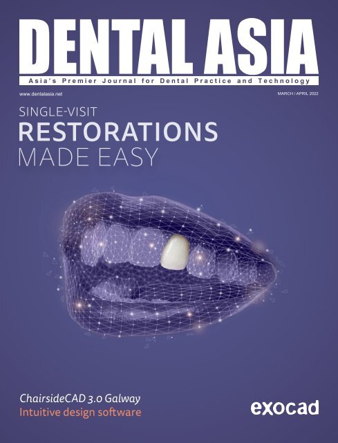
SINGLE-VISIT<br />
RESTORATIONS<br />
MADE EASY<br />
ChairsideCAD 3.0 Galway<br />
Intuitive design software
User Report<br />
DENTAL ASIA NOVEMBER / DECEMBER 2021 3
24<br />
49<br />
30<br />
58<br />
40<br />
BEHIND THE SCENES<br />
56 Combining digital and conventional denture<br />
workflows: An immediate denture case<br />
report<br />
58 exocad: comprehensive implant libraries in<br />
exoplan 3.0 Galway<br />
CONTENTS<br />
UNDER THE SPOTLIGHT<br />
24 Balancing practice and academic<br />
DENTAL PROFILE<br />
27 Coltene continues forward momentum<br />
towards innovation despite pandemic<br />
30 Straumann Group enables dental implants<br />
and orthodontics solutions for all<br />
CLINICAL FEATURE<br />
34 Minimally-invasive restoration of an incisal<br />
edge defect with CAD/CAM hybrid ceramic<br />
36 Osteoma of the maxilla<br />
40 Photobiomodulation: An updated literature<br />
review with a case report<br />
44 Class II orthodontic treatment using Invisalign<br />
treatment with mandibular advancement<br />
USER REPORT<br />
49 Correcting a single midline diastema<br />
52 Rehabilitation of an edentulous lower jaw<br />
presenting reduced residual bone crest with<br />
Anthogyr Mini Implant System<br />
54 Simultaneous GBR and GTR in the posterior<br />
mandible area<br />
IN DEPTH WITH<br />
61 Achieve gentle and effective treatment of<br />
periodontitis and periimplantitis with the<br />
Vector system<br />
62 Digital solutions lead the way into the dental<br />
practice<br />
63 Rolence <strong>Dental</strong> introduces latest digital<br />
image system and dental equipment<br />
64 The two sides of the W&H Lina steriliser<br />
65 BUSCH develops enhanced systems and<br />
tools<br />
SHOW PREVIEW<br />
75 IDEM returns for its 12th edition with new<br />
panels and conference<br />
76 Sino-<strong>Dental</strong> continues to be wonderful<br />
REGULARS<br />
4 Editor’s Note<br />
6 <strong>Dental</strong> Updates<br />
66 Product Highlights<br />
78 Giving Back to Society<br />
79 Events Calendar<br />
80 Advertiser’s Index<br />
2<br />
DENTAL ASIA MARCH / APRIL <strong>2022</strong>
User Report<br />
DENTAL ASIA NOVEMBER / DECEMBER 2021 3
EDITOR’S NOTE<br />
Riding the waves<br />
of progress<br />
PABLO SINGAPORE<br />
Publisher<br />
Publications Director<br />
Senior Editor<br />
William Pang<br />
williampang@pabloasia.com<br />
Jamie Tan<br />
jamietan@pabloasia.com<br />
Josephine Tan<br />
josephine@pabloasia.com<br />
The dental industry remains steadfast<br />
on its path to greater development<br />
and advancement. In this issue, we<br />
have prepared articles showcasing the<br />
community’s embracing of progress and<br />
advancement, through the relentless<br />
efforts of the practitioners adopting<br />
digital technology and streamlining their<br />
workflows.<br />
Speaking to <strong>Dental</strong> <strong>Asia</strong>, Dr Yu Na,<br />
Singapore’s first dental clinician to<br />
receive the National Medical Research<br />
Council’s Clinician Scientist Award in<br />
the Investigator Category, talks about<br />
her personal experiences and recent<br />
achievements in research, showcasing the<br />
fine footwork that goes into threading the<br />
tightrope between practice and academia.<br />
On the other hand, the adoption of<br />
digitalisation continues to be relevant to<br />
the dental community. On page 31, Vida<br />
Lau, commercial director of South East<br />
<strong>Asia</strong> at Straumann Group, stresses the<br />
importance of adopting digital workflows<br />
that can simplify complex treatment<br />
processes, benefitting both dentist and<br />
patient: “Ultimately, clinics that excel<br />
at digitalisation will gain considerable<br />
advantages in terms of outcome quality,<br />
as well as cost and time savings.”<br />
Likewise, further research in the<br />
community has also fuelled a new<br />
direction in dentistry. In a paper by Dr Kevin<br />
Ng and Dato’ Dr How Kim Chuan, it was<br />
demonstrated “that intraoral PBM could<br />
be used to decrease alignment treatment<br />
time and pain/discomfort, as well as<br />
encourage bone formation, promoting<br />
bone healings and osseointegration of<br />
dental implants” (pp.43).<br />
As we continue to usher in the first quarter<br />
of the year, <strong>Dental</strong> <strong>Asia</strong> is also proud<br />
to present a fresh new layout befitting<br />
its commitment to deliver the latest<br />
trends and developments in dentistry.<br />
We hope that this fresh start will deliver<br />
to you, our readers, fresh ideas and<br />
novel perspectives, and serve the wider<br />
community for the better.<br />
Agatha Wong<br />
Assistant Editor<br />
Scan for digital copy<br />
of <strong>Dental</strong> <strong>Asia</strong><br />
Assistant Editors<br />
Graphic Designer<br />
Circulation Manager<br />
PABLO BEIJING<br />
General Manager<br />
PABLO SHANGHAI<br />
Senior Editor<br />
Agatha Wong<br />
agatha@pabloasia.com<br />
Yap Shi Quan<br />
shiquan@pabloasia.com<br />
Czarmaine Masigla<br />
czarmaine@pabloasia.com<br />
Jolin Tan<br />
jolintan@pabloasia.com<br />
Shu Ai Ling<br />
circulation@pabloasia.com<br />
Ellen Gao<br />
pablobeijing@163.com<br />
Daisy Wang<br />
pabloshanghai@163.net<br />
HEAD OFFICE<br />
PABLO PUBLISHING &<br />
EXHIBITION PTE LTD<br />
3 Ang Mo Kio Street 62 #01-23<br />
Link@AMK, Singapore 569139<br />
Tel: (65) 62665512<br />
Email: info@pabloasia.com<br />
Website: www.dentalasia.net<br />
Company Registration No.: 200001473N<br />
Singapore MICA (P) No. 104/12/2021<br />
Malaysia KDN: PPS1528/07/2013 (022978)<br />
REGIONAL OFFICES<br />
PABLO BEIJING<br />
Tel: +86-10-6509-7728<br />
Email: pablobeijing@163.com<br />
PABLO SHANGHAI<br />
Tel: +86-21-52389737<br />
Email: pabloshanghai@163.net<br />
ADVISORY BOARD<br />
Dr William Cheung<br />
Dr Choo Teck Chuan<br />
Dr Chung Kong Mun<br />
Dr George Freedman<br />
Dr Fay Goldstep<br />
Dr Clarence Tam<br />
Prof Nigel M. King<br />
Dr Anand Narvekar<br />
Dr Kevin Ng<br />
Dr William O’Reilly<br />
A DENTAL ASIA MARCH / APRIL <strong>2022</strong><br />
Dr Wong Li Beng<br />
Dr Adrian U J Yap<br />
Dr Christopher Ho<br />
Dr How Kim Chuan<br />
Dr Derek Mahony<br />
Prof Alex Mersel
A<br />
Bone Level<br />
User Report<br />
REG & PX designs for<br />
biological integration<br />
With more than 30 years of experience<br />
in implantology, Anthogyr launched the<br />
Axiom® implant system 10 years ago to<br />
improve access to implantology by<br />
offering innovative and accessible<br />
solutions, a greater comfort for practitionners<br />
and performance in their<br />
everyday practice.<br />
DENTAL ASIA NOVEMBER / DECEMBER 2021 5
DENTAL UPDATES<br />
3Shape announces software updates to 3Shape Model Maker<br />
Users of 3Shape TRIOS intraoral scanner<br />
and 3Shape Studio applications can<br />
now automatically convert their digital<br />
impressions to dental models.<br />
The new Model Maker solution, Model<br />
Maker <strong>2022</strong>.1, uses an automated<br />
workflow powered by artificial intelligence<br />
(AI)-technology that converts scans to<br />
models in minutes.<br />
Full arch dental model optimised for 3D printing<br />
According to 3Shape, the model is<br />
automatically generated from the TRIOS<br />
scans with a single click and includes<br />
key-features to optimise 3D printing.<br />
The algorithm will create a model using<br />
hollowing and drain holes, thus optimising<br />
the use of resin and to ensure a good<br />
printing experience. Bevels are also added<br />
to the base structure of the dental model<br />
to ensure that models can be removed<br />
easily from the build platform of most<br />
common 3D printers.<br />
Additionally, an AI-powered algorithm in the<br />
cloud will convert TRIOS scans to .STL files<br />
which can then be exported and produced.<br />
The algorithm ensures an automated workflow<br />
requiring no input from the user. 3Shape<br />
reported that generating a model can take a<br />
couple of minutes depending on factors like<br />
local internet speed. While waiting to receive a<br />
model, users can exit the workflow using the<br />
‘home’ button and continue to work on other<br />
cases.<br />
Other updates include a Cross Section tool to<br />
inspect the model, and a progress overview<br />
page to verify if the model has finished<br />
generating.<br />
Model Maker <strong>2022</strong>.1 can only be accessed from<br />
the 3Shape Unite platform. Users can enter their<br />
workflow either from scratch as ‘new action’ in<br />
their TRIOS scanning workflow or by selecting<br />
relevant scans and images in the patient media<br />
library to create a model. ■<br />
3M reports Q4 and full 2021 performances<br />
3M has announced their performance for Q4<br />
and the whole of 2021. In particular, the total<br />
sales for the healthcare segment, which includes<br />
oral health, grew by 0.7% to US$2.3 billion in Q4<br />
2021, and the segment’s organic local-currency<br />
sales grew by 1.6% in the same period. The<br />
segment’s operating income was $536 million,<br />
a decrease of 1.6% year-on-year, and operating<br />
margins were 23.6%.<br />
On a geographic basis, total sales in Q4 2021<br />
grew 2.1% in the Americas, and decreased 0.2%<br />
in <strong>Asia</strong>-Pacific, and 4.5% in Europe, Middle East<br />
and Africa (EMEA). Organic local-currency sales<br />
grew 2.2% in the Americas, 1.4% in <strong>Asia</strong>-Pacific,<br />
and decreased 1.9% in EMEA for the same<br />
period.<br />
"3M delivered a solid fourth-quarter performance<br />
— with notable strength in December — as<br />
we maintained our relentless focus on<br />
serving customers in a challenging external<br />
environment," said Mike Roman, chairman and<br />
CEO of 3M. "Our team effectively managed<br />
supply chain disruptions, made good progress<br />
on pricing actions and controlled costs.<br />
"Throughout 2021 we performed well, delivering<br />
full-year sales growth of 10%, robust cash flow<br />
3M headquarters (Image: 3M)<br />
and a strong increase in EPS. We also returned<br />
significant cash to shareholders, reduced debt<br />
and helped the world respond to COVID-19. As<br />
we continue to actively manage our portfolio<br />
and improve our operations, we will prioritise<br />
investments in fast-growing end markets to<br />
drive long-term growth, as well as advance<br />
our commitment to sustainability. As we enter<br />
<strong>2022</strong>, I am confident we will continue to grow<br />
our business and find new ways to apply<br />
science to improve lives." ■<br />
For the whole of 2021 results, 3M recorded<br />
a 9.9% increase year-on-year to $35.4 billion.<br />
Organic local-currency sales increased 8.8%<br />
while acquisitions, net of divestitures, decreased<br />
sales by 0.5%. Foreign currency translation<br />
increased sales by 1.6% year-on-year.<br />
6 DENTAL ASIA MARCH / APRIL <strong>2022</strong>
DENTAL UPDATES<br />
DENTAL ASIA MARCH / APRIL <strong>2022</strong> 7
DENTAL UPDATES<br />
Align Technology promotes digital dentistry for interdisciplinary orthodontics<br />
Align Technology has reinforced its<br />
commitment to dental professionals in the<br />
region with its presence at the 16th Annual<br />
Conference of the Saudi Orthodontic Society<br />
(SOS) as a platinum sponsor of the event.<br />
The annual conference, which took place<br />
from 24-27 Feb <strong>2022</strong> in Riyadh, highlighted<br />
the importance of interdisciplinary<br />
orthodontics and brought together more than<br />
500 individuals from the orthodontic industry<br />
including doctors, laboratory practitioners<br />
and industry partners.<br />
Over the course of the conference, a range of<br />
lectures, panel discussions and workshops<br />
also took place with a focus on topics that<br />
promote interdisciplinary orthodontics and<br />
interdisciplinary collaboration.<br />
Angelo Maura, general manager of Middle<br />
East and Africa at Align Technology, said,<br />
“The 16th SOS Annual Conference will serve<br />
as a much-needed platform for leveraging the<br />
digital technologies that will move us toward<br />
the future of dentistry, as well as foster more<br />
collaboration between the different specialties<br />
in the field.”<br />
Align Technology hosted two key events<br />
during the conference, focusing on driving<br />
interdisciplinary orthodontics through the use<br />
of digital technologies. One of these was a<br />
lecture by Dr Waddah Sabouni, an orthodontist<br />
specialist, discussing “Alternative Approaches<br />
to Orthognathic Surgery with Clear Aligners”.<br />
The lecture covered the management of<br />
orthognathic surgery cases within the<br />
Invisalign system, explaining different<br />
approaches to the timing of the surgery<br />
within orthodontic treatment. Additionally,<br />
Dr Sabouni shared his perspective on the<br />
differences, benefits and considerations<br />
between the alternative approaches.<br />
Another key event being hosted by<br />
Align Technology was a four-hourlong<br />
workshop, also by Dr Sabouni, on<br />
“Aligner Treatment: Possibilities and<br />
Considerations”. Taking place on 27<br />
Feb, The workshop explored the broad<br />
aspects of the applicability of clear<br />
aligners within the orthodontic practice,<br />
including the scope of malocclusions that<br />
can be treated with aligners, as well as<br />
different interdisciplinary treatments such<br />
as cases with periodontal considerations,<br />
pre-prosthetic or pre-restorative<br />
treatments. ■<br />
CAD-Ray announces new partnerships with DOF, MaxxDigm and Akuretta<br />
CAD-Ray has expanded its technology<br />
offerings following the partnerships with<br />
another three manufacturers that share their<br />
vision for innovative, user-friendly dental<br />
technology.<br />
The union of CAD-Ray and DOF will bring the<br />
Craft 5X milling machine to computer-aided<br />
design (CAD)/computer-aided manufacturing<br />
(CAM) dental offices in the US market. The<br />
Craft 5X mill consists of a dust collector, water<br />
pump and compressor all in one tower-shaped<br />
housing. It is easy to install, user-friendly, and<br />
intuitive. CAD-Ray reported that the Craft 5X<br />
can work with any intraoral scanner and any<br />
CAD design platform, including both CLINUX<br />
by CAD-Ray and exocad.<br />
CAD-Ray has also established a partnership<br />
with MaxxDigm, a South Korean robotics<br />
company. CAD-Ray will be distributing the<br />
Chaiman four-axis chairside milling unit. The<br />
Chairman boasts 5kW servomotors, dual<br />
spindles and ultra-fast 10-minute milling<br />
times for glass ceramics and zirconia. It<br />
comes self-contained with everything needed<br />
to get started with chairside restorations,<br />
interfaces easily with any IOS and/or CAD<br />
system and has direct integrations with<br />
CLINUX by CAD-Ray and exocad Chairside<br />
CAD.<br />
Finally, CAD-Ray and Akuretta have<br />
announced a distribution partnership for the<br />
SOL <strong>Dental</strong> 3D Printing system. The SOL is a<br />
3D printer priced well below its competition<br />
in the dental space. The SOL is an LCD printer<br />
with a native resolution of 49 microns, and<br />
a chairside solution for high-precision, fast<br />
3D printing. SOL delivers accurate results<br />
with 54 LED lights that perform at 95%<br />
light uniformity. It can be paired with the<br />
SOL automatic wash/dry unit and Ultra-<br />
Fast UV cure box to complete the package.<br />
Furthermore, SOL is an open platform printer<br />
with resins available from Akuretta, and<br />
partner companies like Keystone, Dentca,<br />
Bego and more.<br />
A provider of dental CAD/CAM and conebeam<br />
computer tomography (CBCT)<br />
solutions, CAD-Ray is also a global distributor<br />
of MEDIT intraoral scanners, and has<br />
partnerships with technology manufacturers<br />
such as Sprint-Ray, Prexion, HDX-Will, Vita,<br />
exocad and more. The company’s own CAD<br />
software, CLINUX, is a browser-based CAD/<br />
CAM software for dentists. ■<br />
8 DENTAL ASIA MARCH / APRIL <strong>2022</strong>
DENTAL UPDATES<br />
Nexa3D partners Nowak <strong>Dental</strong><br />
Supplies to provide dental<br />
3D printing solutions<br />
Nexa3D, producer of resin 3D printers, announced that it has<br />
entered into a reseller partnership with Nowak <strong>Dental</strong> Supplies,<br />
a family-owned and -operated provider of dental devices and<br />
equipment. Nowak <strong>Dental</strong> Supplies will offer Nexa3D’s entire<br />
dental portfolio, including the NXD 200 dental 3D printer, NexaX<br />
software, the post-processing xWASH and xCURE systems, and<br />
the full range of Keystone validated dental 3D printing materials.<br />
The NXD 200, utilising Nexa3D’s lubricant sublayer photo-curing<br />
(LSPc) printing technology, features a 8.5-litre build volume,<br />
allowing for the printing of multiple parts simultaneously, as well<br />
as the fast speed for which Nexa3D printers are known.<br />
“The confidence that Nowak’s customers have in its team is<br />
critical to us,” said Jim Zarzour, head of dental solutions at<br />
Nexa3D. “That, in turn, gives us confidence, knowing that our<br />
products and joint customers will be fully supported. To have<br />
someone of Nowak <strong>Dental</strong> Supplies’ calibre as a reseller for the<br />
NXD 200 is something we’re very excited about.”<br />
PERFECTION IN<br />
BONE SURGERY<br />
→ YOUR SURGICAL<br />
APPROACH WILL CHANGE -<br />
THE PIEZOSURGERY® touch<br />
→ best cutting efficiency<br />
→ optimal intraoperative control<br />
→ perfect ergonomics<br />
→ made in Italy<br />
The NXD 200, with its modular design, enables easy repairs,<br />
part replacements, and technology upgrades, and is designed<br />
for use with the NexaX software. NexaX requires no advanced<br />
3D design or printing knowledge to utilise, making it ideal for<br />
experienced and novice users alike.<br />
“We at Nowak <strong>Dental</strong> are extremely excited about bringing<br />
this new printer to the market,” said Shawn Nowak, president<br />
of Nowak <strong>Dental</strong> Supplies. “With how quickly technology is<br />
changing, we are staying at the forefront of products to offer our<br />
customers. The Nexa3D brand of printers allows customers to<br />
stay competitive and bring the speed that is needed in today’s<br />
landscape.”<br />
Nowak <strong>Dental</strong> Supplies is a supplier to the dental industry, with a<br />
small-business model that emphasises personalised customer<br />
service and dedicated technical support. It works closely with<br />
both dental laboratories and dental offices, supplying from large<br />
lab equipment to small orthodontic accessories. ■<br />
→ www.mectron.com<br />
DENTAL ASIA MARCH / APRIL <strong>2022</strong> 9<br />
ad_PStouch_dental_asia_95x250_en_211214.indd 1 14.12.21 15:38
DENTAL UPDATES<br />
Dentsply Sirona partners with FDI and Smile Train to create cleft care protocols<br />
Dentsply Sirona has announced a three-way<br />
partnership with FDI World <strong>Dental</strong> Federation<br />
(FDI) and Smile Train, a cleft-focused<br />
organisation, to develop global standard<br />
protocols for digital cleft treatment.<br />
The protocols aim to increase access to digital<br />
treatments for patients with clefts and advance<br />
cleft care for the one in 700 babies born around<br />
the globe with cleft lip and or palate.<br />
According to the press release by Dentsply<br />
Sirona, the three partners will jointly work on<br />
integrating digital workflows and sustainable<br />
solutions into these new protocols as well<br />
as creating and providing the necessary<br />
clinical education infrastructure to oral health<br />
professionals around the world.<br />
The partnership also includes designing and<br />
setting up online courses and webinars to<br />
introduce dental professionals around the world<br />
to digital cleft care.<br />
Prior to this new partnership, Dentsply Sirona<br />
pledged a US$5 million donation to Smile Train<br />
as part of a five-year commitment to help<br />
children around the world gain access to<br />
cleft treatment.<br />
“At Dentsply Sirona, we live our sustainability<br />
strategy every day in numerous ways. One of<br />
the most rewarding aspects is working with<br />
Smile Train and FDI to be able to offer the<br />
best care possible to children with clefts,” said<br />
Jorge M. Gomez, CFO and head of the Dentsply<br />
Sirona Sustainability Programme. “Giving these<br />
children healthy smiles by utilising the most<br />
advanced digital technologies is part of our<br />
larger sustainability goal to improve global oral<br />
health care and create millions of healthy smiles<br />
around the world. We are happy to contribute our<br />
knowledge and technologies for this important<br />
cause.”<br />
“We are proud to be working with Dentsply<br />
Sirona and Smile Train to increase the global<br />
access to the best possible cleft care,” said<br />
Professor Ihsane Ben Yahya, president, FDI.<br />
“Cleft surgeries and cleft care benefit hugely<br />
from digitisation and together with our<br />
partners we work fervently on providing oral<br />
health professionals, especially in limitedresource<br />
settings and remote regions, with the<br />
Zoe was supported by Smile Train partner Hospital<br />
Pediatrico Dr Humberto J. Notti with presurgical<br />
orthopaedics, cleft lip, and cleft palate surgeries<br />
(Image: Dentsply Sirona)<br />
infrastructure and training necessary to correctly<br />
use these digital technologies.”<br />
“Children with clefts are more susceptible<br />
to poor oral health which can greatly impact<br />
their speech, ability to eat, and their overall<br />
well-being,” said Susannah Schaefer, president<br />
and CEO, Smile Train. “We are delighted that<br />
our new partnership with Dentsply Sirona and<br />
FDI ensures that Smile Train centres around<br />
the world will have access to digital treatment<br />
protocols that integrate the latest, newest<br />
technologies to provide best-in-class digital oral<br />
healthcare.” ■<br />
UnitedHealthcare <strong>Dental</strong> launches digital resource with quip to improve<br />
dental care<br />
UnitedHealthcare <strong>Dental</strong> has introduced several<br />
enhancements designed to help people improve<br />
their oral health and access dental care in a<br />
more convenient and informed way, with the aim<br />
of helping lower costs and improve satisfaction<br />
among members and dental professionals.<br />
According to the company, the latest offering<br />
expands access to 24/7 virtual dental visits to<br />
help members meet with a licensed dentist<br />
via phone or video, giving eligible members<br />
in many UnitedHealthcare <strong>Dental</strong> plans two<br />
virtual dental appointments with no cost<br />
sharing. The enhanced virtual dental care<br />
benefit is designed to improve access to<br />
dental advice anywhere and anytime, which<br />
may help avoid often-unnecessary visits to the<br />
emergency department for oral health issues.<br />
In addition, to help people improve their oral<br />
care habits, members of UnitedHealthcare<br />
<strong>Dental</strong> can now save up to 30% on smart<br />
electric toothbrushes, including toothpaste,<br />
refillable floss and refillable mouthwash from<br />
oral health company quip.<br />
UnitedHealthcare <strong>Dental</strong> members will also<br />
have access to an online resource to help bring<br />
greater transparency to treatment options<br />
and the cost of dental care, with the goal of<br />
helping prevent surprise costs. The Treatment<br />
Plan Calculator offers access to cost<br />
estimates based on actual contracted rates<br />
and the member’s plan, including real-time<br />
processing capabilities.<br />
“As more and more Americans adopt a<br />
digital-first mindset, these new resources<br />
are designed to help our members improve<br />
and maintain their dental health, which<br />
may contribute to overall well-being and<br />
help reduce the risk of certain chronic<br />
health conditions,” said Colleen Van Ham,<br />
CEO, UnitedHealthcare <strong>Dental</strong>. “These new<br />
initiatives advance UnitedHealthcare’s<br />
approach of using technology to improve<br />
access to quality, cost-effective medical and<br />
dental care, while empowering people with<br />
personalised information.” ■<br />
10 DENTAL ASIA MARCH / APRIL <strong>2022</strong>
DENTAL UPDATES<br />
DENTAL ASIA MARCH / APRIL <strong>2022</strong> 11
DENTAL UPDATES<br />
Partnership between Bien-Air <strong>Dental</strong> and Midmark to provide more complete<br />
operatory ecosystem<br />
Bien-Air <strong>Dental</strong> and Midmark have announced<br />
a partnership that combines the two<br />
companies’ dental technology into a simple,<br />
easy-to-use delivery care solution.<br />
<strong>Dental</strong> clinicians are exposed to health and<br />
safety risks on the job every day. Midmark<br />
and Bien-Air are working together to enhance<br />
safety, optimise ergonomics and improve both<br />
clinician and patient experiences. They will be<br />
integrating Bien-Air’s latest electric handpiece<br />
solutions into Midmark’s dental delivery<br />
equipment for a more complete operatory<br />
ecosystem.<br />
Now, dental clinicians can select Midmark’s<br />
Procenter and Elevance delivery units with<br />
Bien-Air’s newest micromotor and contra-angle<br />
handpieces for an operatory-ready solution.<br />
The solution simplifies workflow for a<br />
broad range of clinical procedures, provides<br />
ergonomic balance, lighter weight and smaller<br />
size for less strain, enable access to oral cavity,<br />
and reduce the risk of patient burn and crosscontamination.<br />
“Midmark is constantly looking for ways to help<br />
design better care in the clinical environment,<br />
which is what makes this partnership so<br />
exciting,” said Jon Wells, president and CEO<br />
for Midmark. “We are thrilled to be joining<br />
forces with another industry-leading company<br />
like ours that shares our strategic vision. Our<br />
collaboration reflects the emphasis both of our<br />
companies place on improving the lives and<br />
livelihoods of each dental team we serve and<br />
improving the experience at the point of care.”<br />
Edgar Schönbächler, CEO, Bien-Air <strong>Dental</strong>, also<br />
commented: “For more than 60 years, Bien-<br />
Air has been leading the way in electric motor<br />
systems for dental and surgical applications.<br />
Reliability and efficiency, typical Swiss values,<br />
have always guided our company. We are<br />
excited about this new step in our longterm<br />
partnership with Midmark. The seamless<br />
integration of our products is the result of a<br />
common understanding of the practitioner’s needs,<br />
helping to improve clinical efficiency and the<br />
patient’s journey.”<br />
As part of this new partnership, Midmark is also<br />
offering Bien-Air’s Lubricare 2 handpiece care and<br />
maintenance system for automatically cleaning<br />
and lubricating handpieces. This maintenance<br />
system integrates into the instrument processing<br />
workflow and is compatible with handpieces from<br />
a variety of manufacturers. ■<br />
Ivoclar Group launches new logo and visual identity<br />
The Ivoclar Group has launched a new visual<br />
identity to align with their goals of becoming<br />
more customer-oriented.<br />
In 2021, Ivoclar set new accents with targeted<br />
activities that are more strongly focused on<br />
the needs of the customer. In order to visually<br />
underline the new departure into a customeroriented,<br />
modern and innovative age, Ivoclar<br />
is starting <strong>2022</strong> with a new appearance and<br />
an adapted logo.<br />
Their most important change is the<br />
"Vivadent" in the brand name and logo has<br />
been dropped, as have some additional visual<br />
elements, so that in the future, the company's<br />
focus on the essentials will also be reflected<br />
in the logo.<br />
The reduction to the essentials, a lifestyleoriented,<br />
emotional visual language that puts<br />
people in the foreground contributes to the<br />
company’s mission of "Making People Smile".<br />
At Ivoclar, the external understanding of<br />
the brand rests on three supporting pillars:<br />
partnership and customer, innovation and<br />
technology, and family and friends.<br />
With its focus on putting people, partners and<br />
customers at the centre of its activities, Ivoclar<br />
underpins its claim to make the workflows in<br />
the daily work of dental technicians and dentists<br />
easier and more efficient, and likewise to make<br />
the patient experience as pleasant and personal<br />
as possible.<br />
The brand image is and remains a first point<br />
of contact with the company, which is why<br />
Ivoclar believes it is important that the brand's<br />
appearance also serves the function of a<br />
mission statement and reflects the innovative<br />
strength that is lived out.<br />
"With our long-standing tradition, our pioneering<br />
achievements and our constant innovation,<br />
we can rely on a strong corporate brand as a<br />
Ivoclar’s new logo, which is more minimalistic,<br />
and dropped the “Vivadent” in its previous logo<br />
foundation,” explained Diego Gabathuler, CEO of<br />
the Ivoclar Group. "Nevertheless, I am convinced<br />
that there is still a lot of unused potential for<br />
us here, which we want to fully exploit in the<br />
future. A clearly structured positioning, which<br />
is also expressed in the visual appearance, is<br />
particularly important with regard to a futureoriented<br />
alignment of the company, which leads<br />
to the innovation that places our customers and<br />
their patients at the centre of our actions and<br />
activities.”<br />
The new Ivoclar logo and corporate design will<br />
be used globally in all communication channels<br />
from now on — where this has not yet been<br />
incorporated — and will also be gradually applied<br />
to brochures and other printed collateral. ■<br />
12 DENTAL ASIA MARCH / APRIL <strong>2022</strong>
DENTAL UPDATES<br />
FOR DIGITAL TEAMPLAYERS.<br />
The new dimension of united dentistry<br />
in laboratory and practice.<br />
CASE SHARING<br />
CONNECTION KIT PRODUCTION KIT HIGH-SPEED ZIRCONIA KIT<br />
Intraoral scanner, software and AG.Live<br />
Case Sharing for Same Day Dentistry<br />
www.ceramill-drs.com<br />
Up to 3-pontic bridges directly in<br />
the practice within one session<br />
H<br />
20<br />
I G H<br />
min<br />
I N G<br />
S<br />
- S P E E D<br />
Sintering zirconia in just 20 minutes<br />
with 16 perfectly matched VITA shades<br />
Amann Girrbach <strong>Asia</strong><br />
Fon +65 6592 5190<br />
singapore@amanngirrbach.com<br />
DENTAL ASIA MARCH / APRIL <strong>2022</strong> 13<br />
www.amanngirrbach.com<br />
I N T E R<br />
Available exclusively in selected markets for the time being.<br />
Interested parties outside Germany please contact the responsible Amann Girrbach dealer.
DENTAL UPDATES<br />
Colgate-Palmolive introduces global Know Your OQ Campaign as part of Oral<br />
Health Commitment<br />
Colgate-Palmolive has announced the<br />
launch of a public health initiative, dubbed<br />
Know Your OQ, to empower people to<br />
understand and improve their oral health<br />
quotient.<br />
Similar to intelligence and emotional<br />
quotient, Colgate hopes for people to know<br />
their oral health quotient and understand the<br />
links between oral health and overall health<br />
and well-being. The company will commit<br />
more than US$100 million over the next<br />
five years to address a global health crisis<br />
affecting nearly half the world’s population<br />
and ensure oral health is incorporated into<br />
broader public health strategies.<br />
“As the worldwide leader in oral care and<br />
with our trusted Colgate brand in more<br />
homes than any other, Colgate-Palmolive<br />
has the opportunity to address a global<br />
health crisis that has far-reaching and<br />
significant impacts,” said Noel Wallace,<br />
chairman, president and CEO of Colgate-<br />
Palmolive. “We’ve got the team, the partners,<br />
the innovations and the motivation to<br />
reimagine a healthier future for all.”<br />
Oral health is often overlooked, even though<br />
an estimated 3.5 billion people currently<br />
suffer from oral diseases. Despite there<br />
being proven strategies for prevention,<br />
cavities remain the most prevalent chronic<br />
disease among adults and children, and it is<br />
estimated that 2.3 billion people suffer from<br />
tooth decay.<br />
Periodontal disease is among the most<br />
common ailments, with severe gum diseases,<br />
which may result in tooth loss, affecting<br />
10% of the global population. The crisis of<br />
oral disease has significant consequences,<br />
since oral health has impacts for physical<br />
health and emotional well-being: research<br />
has shown that oral health is linked to other<br />
physical health conditions, and a global<br />
Colgate study has found that childhood cavities<br />
lead to worry, anxiety and sadness in both kids<br />
and their parents.<br />
“Research has consistently shown that oral<br />
health is a window to overall health, yet oral<br />
health literacy is very low,” said Maria Ryan,<br />
vice-president and chief clinical officer at<br />
Colgate-Palmolive. “That’s why we’re on a<br />
mission to help people increase their oral<br />
health knowledge with: Know Your OQ. If we all<br />
understand the importance of oral health and<br />
embrace simple, proven preventative strategies,<br />
we can help decrease risk for oral diseases and<br />
empower people worldwide to join in the fight<br />
against oral diseases that impact overall health<br />
and well-being.”<br />
At the Know Your OQ website, people can take a<br />
free, interactive assessment to determine their<br />
OQ score. The website also includes information<br />
about the depth and breadth of the global<br />
oral health crisis and provides educational<br />
resources for primary care physicians, nurses<br />
and educational leaders as well as consumers to<br />
improve oral hygiene, encourage healthier habits<br />
and promote overall systemic health. ■<br />
Planmeca Romexis implant library installation packages available for download<br />
Planmeca Romexis has announced<br />
an improvement to its implant library.<br />
The individual installation packages for<br />
each implant manufacturer can now be<br />
downloaded directly on the Planmeca<br />
website. The packages include installers for<br />
both Windows and macOS, and the installers<br />
are compatible with Romexis 5.0 or newer.<br />
From now on, a specific implant<br />
manufacturer’s implants, sleeves, and<br />
others, can be individually installed into<br />
the Planmeca Romexis implant library,<br />
instead of installing the full Romexis implant<br />
library which contains implants from over a<br />
hundred manufacturers.<br />
Customers with the Romexis 3D Implant<br />
module will be able to download and<br />
install the packages to their implant library<br />
themselves on the Romexis server computer.<br />
Benefits of individual installation packages<br />
and direct download include: shorter timeto-market<br />
for new implants and updates;<br />
immediate access to installation packages<br />
after release; package size of 30-60MB,<br />
compared to extensive implant library<br />
package; faster installation when installing<br />
one manufacturer instead of the full library;<br />
easier use of the library list; and contents<br />
of each manufacturer-specific installation<br />
package on the library website. ■<br />
14 DENTAL ASIA MARCH / APRIL <strong>2022</strong>
DENTAL UPDATES<br />
DENTAL ASIA MARCH / APRIL <strong>2022</strong> 15
DENTAL UPDATES<br />
Carestream <strong>Dental</strong> announces sale of Scanning Technology Business<br />
Affiliates of Carestream <strong>Dental</strong> have<br />
entered into an agreement to sell<br />
Carestream <strong>Dental</strong>’s Scanning Technology<br />
business to Envista, a global dental products<br />
company for US$600 million. The Scanning<br />
Technology business is composed of<br />
Carestream <strong>Dental</strong>’s intraoral scanner<br />
equipment such as CS 3600, CS 3700, and<br />
CS 3800 and related software.<br />
Carestream <strong>Dental</strong> will continue to operate<br />
its imaging technology, clinical software and<br />
practice management software businesses<br />
which provide innovative solutions for<br />
dental practices, groups, dental support<br />
organisations (DSOs) and industry partners.<br />
The sale of the Scanning Technology<br />
business will not only deliver proceeds<br />
representing the value of the company’s<br />
innovation processes, but also allow<br />
Carestream <strong>Dental</strong> to focus its investments<br />
and efforts in the growing dental cloud and<br />
technology solutions market. Its recent<br />
investments in Sensei Cloud practice<br />
management and Swissmeda’s suite of<br />
clinical software solutions complement<br />
device innovation with new growing<br />
Software-as-a-Service (SaaS) applications<br />
focused on helping practices and groups<br />
build new revenue, profits and patient<br />
flows.<br />
“This is an exciting time for Carestream<br />
<strong>Dental</strong> and our customers,” said Lisa<br />
Ashby, CEO of Carestream <strong>Dental</strong>. “We’ve<br />
had a multi-year investment plan in<br />
cloud solutions and technology, and this<br />
transaction allows us to better focus<br />
and accelerate our innovation in AI tools,<br />
clinical cloud applications and cloud<br />
practice management for GPs, DSOs, and<br />
specialty practices. We will continue to work<br />
together with our customers and partners<br />
to create innovation which delivers both<br />
clinical and operational value, and think<br />
Envista represents a great home for our<br />
Scanning Technology business under which<br />
our employees and customers will thrive.”<br />
Subject to legal, regulatory and employee<br />
consultation requirements, it is anticipated<br />
that the transaction will close in late Q1 or<br />
early Q2 of <strong>2022</strong>. ■<br />
Henry Schein releases new webinar series to address complex oral health needs<br />
Henry Schein <strong>Dental</strong> recently announced its<br />
<strong>2022</strong> webinar series, Breaking Down Barriers<br />
to Care, designed to enhance access to oral<br />
care among vulnerable patient populations,<br />
including paediatric and older adult patients,<br />
and those with diverse health conditions and<br />
disabilities.<br />
The programme, which kicked off earlier<br />
this year, features speakers from a variety of<br />
clinical backgrounds, who address treatment<br />
for patients with HIV, cancer, cardiac issues,<br />
and more.<br />
The <strong>2022</strong> webinar series includes:<br />
• 26 May at 18:00 EDT: “A Lifetime of<br />
Optimum Oral Health Begins with<br />
Paediatric Preventive Care”<br />
• 27 Jun at 20:00 EDT: “Treating Patients Living<br />
With HIV+”<br />
Dr Gary Severance, executive leader of<br />
Professional Services, Henry Schein <strong>Dental</strong>,<br />
commented: “At Henry Schein, we remain<br />
committed to improving health equity for<br />
all populations, and offering education that<br />
customers can rely on to deliver oral care for<br />
better patient outcomes.<br />
“We are pleased to provide a programme that will<br />
help customers tackle areas of dentistry that are<br />
often overlooked and the complexities of diverse<br />
patient needs. By breaking down barriers to care,<br />
we can help health happen.”<br />
Additional webinars are in development for<br />
September, October and November, and will address<br />
breaking down barriers to care for patients with<br />
disabilities, diabetes and cardiac conditions. ■<br />
16 DENTAL ASIA MARCH / APRIL <strong>2022</strong>
DENTAL UPDATES<br />
DENTAL ASIA MARCH / APRIL <strong>2022</strong> 17
DENTAL UPDATES<br />
W&H provides inspiration for sustainability in dental practices<br />
Medical technology company W&H is calling<br />
on dental practices to get involved as part of<br />
its international campaign, #dentalsunited.<br />
The campaign, “#dentalsunited – UNITED<br />
we go green”, has the objective of setting<br />
out common steps toward environmental<br />
protection. The campaign provides a wealth<br />
of practical tips encouraging dental practices<br />
to think and act green.<br />
As Peter Malata, managing director of W&H,<br />
said: “Sustainability is not a vision for the<br />
future, but rather a necessity for the present.”<br />
When it comes to the environment, there<br />
are various opportunities for improvement<br />
in the medical and dental industry. Singleuse<br />
and plastics are often still commonly<br />
used in order to comply with the necessary<br />
hygiene standards. In addition to this, a lot<br />
of material is used for equipment as well as<br />
energy.<br />
This is where W&H comes in. What specific<br />
steps can dentists take in their practice<br />
to be a part of the solution? From energy<br />
consumption to conserving resources to<br />
consistent recycling, firms will find out what<br />
industry-specific action is required.<br />
FROM RAISING AWARENESS TO<br />
GREEN DENTAL PRACTICES<br />
Since October, the medical technology<br />
company has been providing its online<br />
audience with green suggestions and the<br />
opportunity to share their ideas once a week.<br />
Social media posts and further blog<br />
contributions are creating more awareness<br />
around the important topic of environmental<br />
protection. Sources of problems are identified<br />
and solutions provided for more sustainability.<br />
W&H also supports the quality seal ‘Die Grüne<br />
Praxis’, develops and creates sustainable<br />
products, and takes responsibility for the<br />
environment at its head office in Bürmoos,<br />
Salzburg, Austria, and in the W&H Group with a<br />
range of activities.<br />
Malata concluded: “As a manufacturer of<br />
medical devices, we have always been focused<br />
on innovation and advancement. Progress<br />
always requires change. With this in mind, we<br />
are committed to starting a movement in the<br />
medical and dental industry, in which we raise<br />
awareness, inspire and support each other<br />
along the path to more sustainability.” ■<br />
Grin to expand teledentistry platform with Oral-B<br />
Grin, a digital platform that provides oral<br />
health solutions with local dentists and<br />
orthodontists, has announced a partnership<br />
with Oral-B. At CES <strong>2022</strong>, the companies<br />
unveiled the latest dental technology which<br />
creates new industry standards in assisting<br />
patients in understanding their oral health.<br />
Grin’s remote monitoring and consultation<br />
capabilities, combined with Oral-B’s<br />
products, will ensure that their customers<br />
have access to the latest personalised oral<br />
health insights and at-home professional<br />
consultations.<br />
This means consumers can understand their<br />
brushing and flossing habits, select the right<br />
Oral-B products, improve behaviours for<br />
better smile results, and have more frequent,<br />
informed conversations with their local<br />
doctor, all in an easy-to-use application.<br />
Image: Daniel Frank/Unsplash<br />
According to Grin, the Grin experience<br />
consists of the Grin App, the Grin Scope, and<br />
Grin Scope mini — the US Food and Drug<br />
Adminstration (FDA)-listed medical devices<br />
which attach to any smartphone camera and<br />
take scans to measure changes to patients’<br />
teeth. Under the supervision of partner<br />
doctors, these technologies allow providers to<br />
transition patient check-ins from in-office to<br />
virtual by remotely monitoring teeth movement,<br />
gum health, and oral hygiene.<br />
With the burden of office visits reduced, oral<br />
health and teeth straightening under the care of<br />
a local doctor is more accessible.<br />
“I created Grin to transform the entire dental<br />
health industry by using technology that<br />
connects professionals and patients to<br />
bring better oral care directly to everyone,”<br />
commented Adam Schulhof, DMD, CEO and<br />
co-founder of Grin. “Partnering with one of the<br />
most trusted consumer product brands like<br />
Oral-B clearly attests to our commitment.<br />
“Grin will continue its pledge to drive innovation,<br />
and by joining ranks with Oral-B, we will ensure<br />
more people can have better oral health.” ■<br />
18 DENTAL ASIA MARCH / APRIL <strong>2022</strong>
DENTAL UPDATES<br />
DENTAL ASIA MARCH / APRIL <strong>2022</strong> 19
DENTAL UPDATES<br />
BEGO Group receives TOP 100 seal of approval<br />
The BEGO Group of companies from<br />
Bremen, Germany, has announced that they<br />
are included in the 29th annual TOP 100<br />
competition, having been awarded the TOP<br />
100 <strong>2022</strong> seal.<br />
At the heart of the TOP 100 innovation<br />
competition is a scientific selection process<br />
that participants must undergo. On behalf of<br />
compamedia, a media organisation based in<br />
Germany, the organiser of the competition,<br />
innovation researcher Nikolaus Franke,<br />
and his team examined the BEGO Group<br />
on the basis of more than 100 indicators<br />
from five categories: “Top Management<br />
Promoting Innovation”, “Innovation Climate”,<br />
“Innovative Processes and Organisation”,<br />
“External Orientation/Open Innovation” and<br />
“Innovation Success”.<br />
“How focused is a company on innovation?<br />
How consistently do its structures follow<br />
this goal? With TOP 100, we examine this,”<br />
explained Franke. “The most innovative<br />
medium-sized companies receive the seal.<br />
It shows that they are excellently equipped<br />
to meet future challenges.”<br />
On 24 Jun <strong>2022</strong>, BEGO will be honoured<br />
for these achievements in person by the<br />
competition’s mentor, science journalist<br />
Ranga Yogeshwar.<br />
BEGO is one of the top innovators for<br />
the second time. Founded in 1890, the<br />
German company offers dental technicians<br />
and dentists innovative equipment,<br />
instruments, materials, implants, services,<br />
and processes for the manufacture and<br />
processing of dental restorations.<br />
In 2015, BEGO launched a 3D printing<br />
system called Varseo, developed in-house<br />
with and for dental laboratories, for the<br />
laboratory production of a range of dental<br />
restorations from high-performance resins.<br />
In early 2020, it presented VarseoSmile<br />
Crown plus, a tooth-coloured, ceramic-filled<br />
hybrid material approved as a Class IIa<br />
medical device for 3D printing of permanent<br />
single crowns, inlays, onlays and veneers. ■<br />
The TOP 100 award<br />
Zest <strong>Dental</strong> Solutions to partner with <strong>Dental</strong> Whale<br />
Zest <strong>Dental</strong> Solutions has announced its<br />
newest partnership with the <strong>Dental</strong> Whale<br />
Membership. Effective immediately, the<br />
three-year agreement with <strong>Dental</strong> Whale<br />
supports over 19,000 <strong>Dental</strong> Whale offices<br />
across the US.<br />
buy LOCATOR and LOCATOR R-Tx abutments<br />
directly from Zest to support implant systems<br />
used in each member’s office. Additionally,<br />
<strong>Dental</strong> Whale will be utilising the LOCATOR and<br />
LOCATOR R-Tx Scan Bodies to support digital<br />
initiatives.<br />
Zest <strong>Dental</strong> Solutions as a key manufacturer<br />
in the implant locator and overdenture fields.<br />
We are excited to collaborate with bringing<br />
quality offerings and education to the <strong>Dental</strong><br />
Whale membership.” ■<br />
As part of this agreement, Zest will be part<br />
of <strong>Dental</strong> Whale’s preferred network for<br />
LOCATOR, LOCATOR R-Tx, and LOCATOR<br />
Overdenture Implant Solutions. In addition<br />
to local office support, Zest will also be<br />
partnering with the clinical support team<br />
to launch <strong>Dental</strong> Whale’s ORCA platform<br />
supporting Removable Overdenture<br />
surgical, prosthetic, and hands-on training<br />
programmes being hosted in-person and<br />
remotely across North America.<br />
Zest <strong>Dental</strong> LOCATOR family products are<br />
now part of <strong>Dental</strong> Whale’s network and<br />
are available to <strong>Dental</strong> Whale members.<br />
<strong>Dental</strong> Whale offices will have the ability to<br />
“Zest <strong>Dental</strong> is excited to have <strong>Dental</strong> Whale<br />
as part of our dental support organisations<br />
(DSO) member family,” said Tom Stratton, CEO<br />
of Zest <strong>Dental</strong> Solutions. “We are committed<br />
to supporting <strong>Dental</strong> Whale member offices<br />
with preferred access to the LOCATOR product<br />
family, and helping their clinicians grow their<br />
practices and create stellar smiles.”<br />
Phil Cruz, vice-president of procurement,<br />
<strong>Dental</strong> Whale, also commented: “<strong>Dental</strong> Whale<br />
looks forward to continuing its rapid growth<br />
with the addition of Zest <strong>Dental</strong> Solutions in the<br />
preferred supplier network. As part of <strong>Dental</strong><br />
Whale’s mission to provide solutions that<br />
simplify dentistry, we see the partnership with<br />
Zest <strong>Dental</strong> LOCATOR family products<br />
20 DENTAL ASIA MARCH / APRIL <strong>2022</strong>
DUAL CURING CORE & RESIN CEMENT<br />
DENTAL UPDATES<br />
3 Indications – 1 Material<br />
Ò Optimal monoblock interface<br />
Ò Two setting times<br />
Ò Outstanding sealing and protection<br />
Ò Excellent retention & durable strength<br />
POSTS FOR DIRECT AND INDIRECT INDICATIONS<br />
ParaPost ® System<br />
Ò A versatile range of fiber posts, metal posts<br />
and prefabricated casting post components<br />
Ò One-office-visit and laboratory techniques<br />
Ò More than 55 years of expertise<br />
Ò Proven clinical success with > 500 studies<br />
Ò Endo meets Resto – complete system with<br />
core-build-ups and cements<br />
005024 09.19<br />
https://www.facebook.com/COLTENE.<strong>Asia</strong>Pacific<br />
DENTAL ASIA MARCH / APRIL <strong>2022</strong> 21
DENTAL UPDATES<br />
Virtual event by Dentsply Sirona to announce collaboration with Google Cloud<br />
and Primeprint<br />
Dentsply Sirona has announced details about a<br />
collaboration with Google Cloud and a separate<br />
new 3D printing solution, Primeprint, in a virtual<br />
global on 4 Mar <strong>2022</strong>.<br />
The company will introduce a series of<br />
innovations designed to enhance digital<br />
workflows with benefits for dentists, dental<br />
labs and patients globally. A highlight of this<br />
event will be interviews with Urs Hoelzle, senior<br />
vice-president in engineering at Google, and<br />
Don Casey, CEO of Dentsply Sirona, who will be<br />
announcing a new collaboration between the<br />
two technology companies.<br />
GOOGLE CLOUD<br />
The collaboration with Google Cloud will help<br />
dentists and dental labs to unlock benefits of<br />
digital dentistry – whether they are continuing or<br />
just starting on their digital journey.<br />
The new digital dentistry solutions will be<br />
based on six key principles: enabling high-value<br />
dental care; next-generation digital featuring<br />
secure and seamless sharing of data with<br />
laboratories and other dental practitioners; easy<br />
access to data whenever needed; high-quality<br />
3D visualisation of dental imagery; stringent<br />
data protection and security standards; and<br />
an innovative environment for software, data<br />
integrity and storage.<br />
Christian Martin, managing director of Alps<br />
region at Google Cloud, commented: “Dentsply<br />
Sirona is transforming the dental industry. At<br />
Google Cloud we believe that we have the right<br />
expertise, capabilities and services to strongly<br />
support Dentsply Sirona in its vision for the<br />
future of oral healthcare.”<br />
Casey added: “This exciting collaboration will<br />
allow us to deliver on our promise: empowering<br />
millions of customers by proudly creating<br />
innovative solutions for healthy smiles. We will<br />
succeed in this by giving dentists an innovative<br />
platform to underpin the digital infrastructure<br />
of their practice and enabling a seamless<br />
workflow that helps dentists to focus on what<br />
matters most: treating their patients.”<br />
PRIMEPRINT<br />
Separate from the collaboration with Google<br />
Cloud, Dentsply Sirona will also launch<br />
Primeprint, a medical-grade 3D printing<br />
system for dental practices and dental labs.<br />
Primeprint is a smart hardware and software<br />
solution that is optimised for dental<br />
applications. It runs the entire<br />
printing process, including<br />
post processing, and<br />
delivers “reproducible and<br />
accurate” results with strictly<br />
biocompatible materials.<br />
The printing process meets<br />
high regulatory<br />
requirements for<br />
medical products.<br />
■<br />
New SLS powder: Glass filled Nylon 12 for strong, heat-resistant parts<br />
Formlabs has introduced the Nylon 12 GF<br />
Powder, a glass-reinforced plastic powder ideal<br />
for producing stiff, thermally stable parts on the<br />
Fuse 1 selective laser sintering (SLS) printer.<br />
The Nylon 12 GF Powder is the third material<br />
designed specifically for the Fuse 1 ecosystem,<br />
providing Formlabs customers with options to<br />
scale in-house production affordably. The new<br />
material joins Nylon 12 Powder and Nylon 11<br />
Powder in the Formlabs SLS materials library,<br />
enabling customers to diversify their operations<br />
and scale production with varied material<br />
properties.<br />
This new material combines the utility of<br />
Nylon 12 with the rigidity and heat resistance<br />
of glass filler, making it ideal for any application<br />
requiring sustained load-bearing ability and<br />
elevated thermal resistance. Fuse 1 users can now<br />
add Nylon 12 GF Powder to their catalogue if they<br />
need stiff, functional prototypes, robust jigs and<br />
fixtures, replacement parts, and parts with threads<br />
or sockets.<br />
The Nylon 12 GF Powder can be used for strong,<br />
stiff, stable medical devices and replacement<br />
parts in the healthcare industry, particularly<br />
for customisable end-user medical device<br />
components.<br />
Stiff, static jigs and fixtures, brackets, mounts,<br />
and casings are all product types that would<br />
benefit from the strong, rigid mechanical<br />
properties of this new powder.<br />
Nylon 12 GF Powder also withstands<br />
elevated temperatures, with a heat deflection<br />
temperature of 113°C. Many materials can<br />
deform when exposed to high heat, and lose<br />
their dimensional accuracy, especially when<br />
the heat is combined with a load-bearing<br />
application. Nylon 12 GF Powder has the<br />
thermal stability to withstand applications<br />
where other SLS powders would show signs of<br />
elongation and deformation.<br />
HIGH PERFORMANCE VERSATILE<br />
MATERIALS<br />
The Nylon 12 GF Powder’s tensile modulus is more<br />
than 50% higher than that of Nylon 12 Powder,<br />
and almost 75% higher than Nylon 11 Powder.<br />
Customers should choose Nylon 12 GF Powder<br />
when printing parts that need to bear heavy loads<br />
for a long time without any bending or elongation.<br />
Other benefits of Nylon 12 GF Powder include:<br />
a minimum refresh rate which ranges from 30-<br />
50%, and can be adjusted to minimise material<br />
waste, optimise cost per part, or improve part<br />
finish; a simplified material changeover process,<br />
which reduces downtime and enables more<br />
in-house, multi-material production with a single<br />
printer unit; among others. ■<br />
22 DENTAL ASIA MARCH / APRIL <strong>2022</strong>
DENTAL UPDATES<br />
exocad launches community blog<br />
exocad, an Align Technology company and<br />
a dental computer-aided design (CAD)/<br />
computer-aided manufacturing (CAM)<br />
software provider, has announced the launch<br />
of exoBlog, a community-based blog that<br />
will feature educational interviews with<br />
practitioners, dental technicians and thought<br />
leaders in the dental industry.<br />
exoBlog was launched on 23 Feb <strong>2022</strong>.<br />
The initial set of blog posts touched on<br />
first impressions of the recently released<br />
ChairsideCAD 3.0 Galway, providing first-hand<br />
experiences of implementing digital solutions<br />
in a dental practice and highlighting the<br />
benefits of digital workflows that can enable<br />
collaboration between laboratory and dental<br />
practices.<br />
“Community dialogue is extremely<br />
important to us at exocad,” said Christine<br />
McClymont, global head of marketing and<br />
communications at exocad. “With this new<br />
platform, we aim to bring together both<br />
newcomers to the field of digital dentistry<br />
and experienced professionals, to learn from<br />
each other and share practical tips to make<br />
their daily workflows easier.”<br />
The launch of exoBlog follows a broader<br />
expansion of exocad’s digital community<br />
resources. Users can now access information<br />
on exocad’s product releases, upcoming<br />
events, and tips and tricks on Facebook,<br />
Instagram, WeChat, YouTube, LinkedIn and,<br />
most recently, TikTok. ■<br />
Medical Protection Society launches research initiative to improve patient<br />
safety and clinician well-being<br />
Medical Protection Society (MPS), of which<br />
<strong>Dental</strong> Protection is a part, has launched the<br />
MPS Foundation: a new not-for-profit research<br />
initiative to help shape the future of patient<br />
safety through the funding of healthcare and<br />
dental research.<br />
The foundation will invest in research and<br />
analysis, with the key focus being on areas of<br />
patient safety and clinician well-being, both<br />
medical and dental. It is part of MPS, which<br />
currently supports more than 300,000 doctors,<br />
dentists and healthcare professionals, and<br />
has almost 130 years of global healthcare<br />
experience.<br />
The MPS Foundation is being launched<br />
across each of the countries and regions<br />
where MPS has a member base, including<br />
Malaysia, Singapore, Hong Kong, the<br />
UK, Ireland, South Africa, Australia, New<br />
Zealand and the Caribbean, and is open to<br />
applications from both members and nonmembers.<br />
The research projects supported by the MPS<br />
Foundation will have to be academically<br />
robust and evidence-based. Available<br />
funding will range from £5,000-200,000, or<br />
the equivalent in local currency, depending<br />
on the scale, focus and duration of the<br />
proposal.<br />
“I am proud to launch the MPS Foundation,<br />
which will help both members and other<br />
healthcare professionals navigate the<br />
challenges of modern practice and find<br />
research solutions.”<br />
MPS Foundation funding is focused in four<br />
main areas. First, the impact of human<br />
factors on patient safety, outcomes and<br />
risk. Second, the impact of processes<br />
and delivery models on patient safety,<br />
outcomes and risk. Third, the personal and<br />
professional wellbeing and development of<br />
clinicians, and lastly, the impact of digital<br />
integration and technology on patient safety,<br />
outcomes and risk.<br />
Dr Graham Stokes, dentist and MPS<br />
Foundation chair, said: “As the world’s<br />
leading protection organisation, our aim is<br />
simple: to create sustainable global change<br />
through ambitious research. The MPS<br />
Foundation will develop an international<br />
knowledge base that can be applied locally,<br />
leveraging our organisation’s global reach.<br />
The funding is eligible to universities, medical<br />
and dental schools, third sector and private<br />
research institutions and organisations,<br />
private or public hospitals or hospital groups,<br />
local hospital networks, primary health<br />
networks, dental practices, dental groups,<br />
MPS members and clients, and non-MPS<br />
individuals and organisations. ■<br />
DENTAL ASIA MARCH / APRIL <strong>2022</strong> 23
UNDER THE SPOTLIGHT<br />
Balancing practice<br />
and academic<br />
As Singapore’s first dental clinician to receive the<br />
National Medical Research Council’s Clinician Scientist<br />
Award – Investigator Category, Dr Yu Na is no stranger<br />
to further research in dentistry. <strong>Dental</strong> <strong>Asia</strong> spoke with<br />
the senior dental surgeon and clinician-scientist at<br />
National <strong>Dental</strong> Centre Singapore on her experience<br />
and expertise on the trends and advancements that<br />
will lead the industry in the coming years.<br />
EARLY EXPERIENCE AND<br />
INSPIRATION<br />
Can you share with us your<br />
beginnings as a dental<br />
professional, and what are your<br />
early influences in pursuing a<br />
career in this field?<br />
Dr Yu Na: When I was young, I<br />
remember going through braces<br />
and being curious about the<br />
mechanisms. I also remember<br />
trying to adjust the wires myself<br />
and being fascinated by the devices<br />
and instruments every time I went<br />
to the dentist. During high school, I<br />
chose to major in biology, and this<br />
eventually led me to dentistry.<br />
What are the reasons behind<br />
your decision to specialise in<br />
prosthodontics, and what inspired<br />
you to pursue your PhD studies<br />
focusing on cell-based strategies<br />
to regenerate periodontal tissues?<br />
Dr Yu: Each dental specialty has<br />
its attraction. Since my time as a<br />
young dentist, I have always felt a<br />
sense of satisfaction from seeing<br />
the smiles on my patients after<br />
their prosthodontic treatment. To<br />
me, that was the deciding factor<br />
for choosing prosthodontics. In<br />
prosthodontics, I enjoy the precise<br />
control in operative dentistry, the<br />
process of creating a device, and the<br />
delivery of an end product that we<br />
pass to our patients.<br />
I was looking for more fundamental<br />
biology topics to deepen my dental<br />
knowledge for my PhD studies. I<br />
was able to build my foundation<br />
in critical thinking and learnt the<br />
mental framework in scientific<br />
investigation. My research training<br />
also taught me to assess a<br />
clinical problem from scientific<br />
perspectives and seek solutions<br />
using an evidence-based approach.<br />
Regenerative periodontology is one<br />
of the most challenging domains in<br />
dentistry while cell therapy, tissue<br />
engineering and biomaterials are in<br />
the fields of cutting-edge science<br />
and help further shape my mindset<br />
of scientific thinking.<br />
How will you describe your typical<br />
day in the workplace as you juggle<br />
between clinical and academic<br />
commitments?<br />
Dr Yu: I take on various roles as a<br />
scientist, clinician, and manager on<br />
a typical day. I start my mornings<br />
with academic work which includes<br />
replying to emails, panel reviews,<br />
reports and drafting of papers.<br />
This is followed by project-related<br />
meetings where I review our<br />
projects, brainstorm, and gather<br />
progress updates with engineers<br />
and scientists. Over lunch, we are<br />
on Zoom calls with the clinical<br />
team. I start my clinics after lunch<br />
and attend to about five patients till<br />
evening. Thereafter, I will meet with<br />
a clinical educator on structuring a<br />
conference lecture.<br />
My day is packed, but each role<br />
is a conduit to the other. I see<br />
myself as a bridge between clinical<br />
24 DENTAL ASIA MARCH / APRIL <strong>2022</strong>
UNDER THE SPOTLIGHT<br />
problems, technical solutions, and<br />
using that to educate others. I<br />
also find it a privilege to be able to<br />
switch between my roles between a<br />
researcher and clinician.<br />
PERSONAL ACHIEVEMENT<br />
AND RESEARCH<br />
Can you briefly discuss your<br />
research proposal on clinically<br />
integrated smart digital workflow to<br />
improve the quality and efficiency in<br />
the production of removable partial<br />
dentures (RPD) for Singapore’s<br />
ageing population? In particular,<br />
what were the motivations behind<br />
this study, and what are the<br />
potential long-term implications?<br />
Dr Yu: Working with a team of<br />
dental laboratory technicians, I<br />
witness the challenges of manual<br />
fabrication and the dependency of<br />
skilled workmanship to produce<br />
a well-fabricated prosthetic. With<br />
the ageing population in Singapore,<br />
there is a need for radical solutions<br />
in dentistry. I saw a window of<br />
opportunity to innovate through<br />
digitalisation and I hope to leverage<br />
my position as a clinician-scientist<br />
to build an ecosystem in digital<br />
dentistry. It was with all these in<br />
mind that I initiated the digital<br />
solution with automatic design<br />
software and 3D printing for the<br />
mass manufacturing of RPDs.<br />
What other opportunities do you<br />
see in extending your work in this<br />
research, and what advice will you<br />
like to give to dental researchers to<br />
inspire them to push their interest in<br />
oral science?<br />
Dr Yu: Digital dentistry is disrupting<br />
conventional dental practice with<br />
its role in diagnosis and treatment<br />
planning, treatment, laboratory<br />
aid, patient motivation, practice<br />
management, training and research.<br />
There are numerous benefits to<br />
digital dental solutions, which<br />
include higher efficiency of cost and<br />
time, flexible workflow, improved<br />
accuracy, higher predictability of<br />
outcomes, fewer visits to the dentist,<br />
early detection of dental diseases,<br />
shorter recovery time, better quality<br />
images and reduced exposure<br />
to x-rays. To me, these long-term<br />
benefits outweigh the initial high<br />
capital costs and investment<br />
involved.<br />
THE FUTURE AND BEYOND<br />
Can you provide us with an<br />
outlook on digital technology in<br />
prosthodontics – what can be<br />
its limitations when it comes to<br />
providing dental care to patients,<br />
and how are you overcoming this<br />
challenge? More crucially, what<br />
advice will you give to fellow<br />
DENTAL ASIA MARCH / APRIL <strong>2022</strong> 25
UNDER THE SPOTLIGHT<br />
practitioners who are hesitant to<br />
adopt digital technology?<br />
Dr Yu: In recent years, we have seen<br />
an increase in the demand for dental<br />
procedures. With digital technology,<br />
we are better equipped to provide<br />
solutions for dental care, and we<br />
can improve our patient experience<br />
and enhance our dental workflow.<br />
The adoption of technology will<br />
also drive the dentistry industry<br />
forward. However, the expense and<br />
special training that comes with the<br />
solutions often result in dentists<br />
being reluctant to adopt such new<br />
technologies.<br />
Digitalisation will be the trend for<br />
future dentistry, I would encourage<br />
fellow practitioners to upskill their<br />
digital knowledge to prepare for the<br />
new service model.<br />
What other trends do you foresee<br />
happening in the dental field in<br />
the next five to 10 years, and how<br />
can dental professionals prepare<br />
themselves better as the world<br />
embarks on its recovery road from<br />
the pandemic?<br />
Dr Yu: The trend of digital<br />
transformation in dentistry is evident<br />
in the past few years. Digital dental<br />
solutions provide better comfort and<br />
experience during dental visits. They<br />
save time for both the patient and<br />
the dentist.<br />
These solutions help dental<br />
professionals manage their practice<br />
more efficiently by streamlining the<br />
workflow. As these solutions can<br />
potentially increase the profitability<br />
of dental practice, the adoption<br />
rate is increasing. It will continue<br />
to increase in the years to come as<br />
more dental professionals realise<br />
the value these solutions have for<br />
the success of their practice.<br />
As more and more patients start to<br />
demand better experience and more<br />
time-efficient procedures, I believe<br />
that these digital solutions will<br />
become the norm. DA<br />
26 DENTAL ASIA MARCH / APRIL <strong>2022</strong>
DENTAL PROFILE<br />
Coltene continues forward<br />
momentum towards innovation<br />
despite pandemic<br />
Despite the COVID-19 pandemic disrupting business operations globally,<br />
Coltene directs its strategies towards growth and operations through<br />
maintaining communication with its customers and continued product<br />
innovation.<br />
In the middle of difficulty lies<br />
opportunity – what Albert Einstein once<br />
said and can still be applied in today’s<br />
world that looms under the COVID-19<br />
pandemic. One company that has been<br />
focusing on growth while continuing<br />
to innovate in its products to better<br />
support the dental community during<br />
these challenging times is Coltene,<br />
which has been proactive in maintaining<br />
communication with its partners during<br />
the pandemic.<br />
Speaking with <strong>Dental</strong> <strong>Asia</strong>, Rodolfo Frei,<br />
director of Coltene <strong>Asia</strong>-Pacific, said:<br />
“This communication has allowed us<br />
to remain close to the developing and<br />
changing needs of the market as a<br />
result of the pandemic to serve them<br />
best. This has been possible, thanks to<br />
the fact that we have great coverage<br />
with Coltene managers around <strong>Asia</strong>-<br />
Pacific.”<br />
Understanding local markets and<br />
their different needs is still key to the<br />
success of any company. Frei shared<br />
that Coltene has been able to maintain<br />
dedicated and personalised contact<br />
with local partners through its local<br />
sales force, but at the same time<br />
also connected to its headquarters in<br />
Switzerland with a global message.<br />
“This period has also allowed us an<br />
opportunity to review certain strategies<br />
to discontinue non-strategic products<br />
and focus on key segments which, in<br />
turn, means that our customers get<br />
the products and services that they<br />
want, and have come to expect from<br />
Coltene,” he added.<br />
Beyond communication, Coltene<br />
has also compiled and continuously<br />
updated tools and resources on its<br />
website to support the healthcare<br />
professionals in managing the<br />
impact of COVID-19. In addition,<br />
the company offers various<br />
webinars on the topic<br />
of infection control to<br />
provide its customers<br />
with in-depth training<br />
in this area looking<br />
at aspects such<br />
as hand hygiene,<br />
surface cleaning and<br />
disinfection to name<br />
but a few.<br />
He continued:<br />
“Many companies<br />
started trying<br />
online training for<br />
the first time<br />
due to contact<br />
restrictions.<br />
Congresses<br />
and fairs took<br />
place virtually<br />
and Zoom<br />
meetings<br />
suddenly<br />
became part<br />
of everyday<br />
Rodolfo Frei,<br />
Director of Coltene <strong>Asia</strong>-Pacific<br />
DENTAL ASIA MARCH / APRIL <strong>2022</strong> 27
DENTAL PROFILE<br />
life. Due to the high acceptance of<br />
this form of communication, some<br />
activities will certainly still take place<br />
online after COVID-19. Nevertheless,<br />
the dental community is now looking<br />
forward to face-to-face contact again.<br />
“A somewhat surprising finding<br />
is that in markets with very strict<br />
hygiene regulations, the demand for<br />
disinfectants has not increased as<br />
much as one would generally expect.<br />
From this, we conclude that, in general,<br />
dental practices were already doing<br />
a great deal for patient and practice<br />
team safety before the pandemic.”<br />
CONTINUING PRODUCT<br />
INNOVATION<br />
With the information gathered from<br />
their customers, Coltene was able<br />
to revise and release new products<br />
for their customers. Undeterred by<br />
COVID-19, the company has been<br />
consistently introducing new products<br />
and innovations to the industry.<br />
A product Coltene has developed is<br />
SUPERKRAFT which combines two<br />
solutions – SoloCem and ONE COAT 7<br />
UNIVERSAL – into one product. The<br />
former is a self-adhesive cement<br />
and the latter is a light-cured,<br />
one-component bonding agent.<br />
Frei explained: “The dentist should<br />
have a cement that can do both –<br />
lute a restoration self-adhesively<br />
whenever the indication allows it,<br />
and lute a restoration adhesively in<br />
combination with a bonding agent<br />
whenever a ‘bonding-boost’ is<br />
required.”<br />
Besides SUPERKRAFT, Coltene has<br />
also released the BRILLIANT family,<br />
a line of composites focusing on<br />
efficiency and aesthetics.<br />
“Our base material BRILLIANT<br />
EverGlow is known for its good<br />
polishability and gloss retention. At<br />
the same time, handling properties<br />
are highly appreciated by dentists,”<br />
he remarked. “The CAD/CAM<br />
composite blocks BRILLIANT Crios<br />
and the composite shells BRILLIANT<br />
COMPONEER are based on the same<br />
material as BRILLIANT EverGlow.<br />
This means that you have a perfectly<br />
matching system to make great<br />
looking restorations out of one<br />
material.”<br />
The duo shade system offered by<br />
the BRILLIANT range, he added,<br />
contains seven basic shades which<br />
cover the VITA range. In conjunction<br />
with the translucent and opaque<br />
shades, dentists will have the kit<br />
required to meet the demands of<br />
aesthetic restorations.<br />
Another new launch by Coltene is<br />
the CanalPro Jeni endo-motor. Fully<br />
automatic and electric controlled<br />
with an integrated apex locator,<br />
the Jeni aids with smoothening<br />
root canal preparation. The<br />
Jeni assistance system also<br />
continuously adapts to the<br />
individual root canal anatomy<br />
and guides the mechanical and<br />
chemical preparation step by step.<br />
Citing a quote from Dr Nicolas<br />
Gutierrez from Madrid, who said<br />
that it was “amazing how the endo<br />
assisted algorithm worked in the<br />
hands of a prosthodontist”, Frei<br />
commented that dentists were<br />
28 DENTAL ASIA MARCH / APRIL <strong>2022</strong>
DENTAL PROFILE<br />
“enthusiastic about the possibilities<br />
for everyday use of the Jeni Motor”.<br />
With these new product launches,<br />
Coltene was able to respond to the<br />
needs of the market in spite of a<br />
global pandemic crisis.<br />
Nevertheless, Frei emphasised the<br />
need for a hands-on approach in<br />
experiencing their new products. He<br />
raised the Jeni system as an example<br />
and said: “During COVID-19, we were<br />
forced to rely mainly on webinars but<br />
the true power of the endo-motor is<br />
only revealed when you can practice<br />
with it yourself. Therefore, now that<br />
the measures have been relaxed<br />
somewhat, we are relying on face-toface<br />
training in the form of our Europewide<br />
‘Jeni City Tour’.”<br />
THE PATH FORWARD<br />
Having joined the Coltene family in<br />
2009, Frei has had a front-row seat in<br />
observing the company’s growth for<br />
the last decade. As the dental industry<br />
continues to evolve and demands<br />
continue to change, its leadership has<br />
allowed Coltene to continue offering<br />
new products to its customers,<br />
establishing itself as a provider and<br />
manufacturer of consumables and<br />
small-size equipment for dental<br />
treatment applications.<br />
Frei lauds the arrival of Martin<br />
Schaufelberger, who joined Coltene as<br />
CEO in 2012, and said: “His vision and<br />
leadership has helped the group to be<br />
recognised worldwide, in the areas of<br />
infection control, dental preservation<br />
and efficient treatment.”<br />
Particularly in the area of infection<br />
control, Coltene acquired SciCan, a<br />
provider of infection control solutions,<br />
and Micro-Mega, a manufacturer of<br />
endodontic instruments. The merger<br />
with Coltene strengthens the market<br />
position and product range under<br />
this new group, opening up synergies<br />
and expanding the range in the joint<br />
business fields of infection control<br />
and endodontics. With its combined<br />
resources, the group is said to be<br />
in an even better position to address<br />
compliance and regulation standards.<br />
“With the integration of SciCan into<br />
the group, we were able to meet the<br />
ever-increasing demand in hygiene and<br />
disinfection protocols. Our innovations<br />
in this field meant that we remained<br />
at the forefront with solutions that<br />
the pandemic demanded. It is a move<br />
that has enabled us to strengthen our<br />
presence as the true infection control<br />
specialists,” Frei elaborated.<br />
As the Coltene team continues to deliver<br />
new products to its customers, Frei is<br />
grateful for the partnerships formed<br />
during this trying period, and concluded:<br />
“We forge and recognise partnerships<br />
during difficult moments, and we are<br />
delighted and honoured to have such<br />
loyal, enthusiastic, proactive customers<br />
in the whole region.<br />
“We, the Coltene team are excited, and<br />
look forward to being able to continue<br />
sharing our expertise and passion with<br />
the dental community in <strong>Asia</strong>.” DA<br />
DENTAL ASIA MARCH / APRIL <strong>2022</strong> 29
DENTAL PROFILE<br />
Straumann Group enables<br />
dental implants and<br />
orthodontics solutions for all<br />
With a slate of products and service geared toward aesthetic<br />
dentistry, Straumann Group and its united brands are enabling<br />
dentists and patients around the world to achieve valued results<br />
using the latest in dental treatment for the <strong>Asia</strong>-Pacific region.<br />
With a slate of products and services<br />
geared toward aesthetic dentistry,<br />
Straumman Group and its brands are<br />
enabling dentists and patients around<br />
the world to achieve valued results<br />
using the latest in dental treatment<br />
for the <strong>Asia</strong>-Pacific region.<br />
Headquartered in Basel, Switzerland,<br />
the Straumann Group and its brands<br />
– including Straumann, Neodent,<br />
ClearCorrect, <strong>Dental</strong> Wings and<br />
other fully- or partly-owned<br />
companies and partners<br />
– are committed to<br />
delivering excellence,<br />
innovation and quality in<br />
replacement, corrective<br />
and digital dentistry.<br />
With more than 9,000<br />
employees worldwide,<br />
the group develops,<br />
manufactures<br />
and supplies<br />
dental implants,<br />
instruments,<br />
biomaterials, CAD/<br />
CAM prosthetics,<br />
digital equipment,<br />
software,<br />
and clear<br />
aligners for<br />
applications<br />
in replacement, restorative, orthodontic<br />
and preventive dentistry.<br />
Vida Lau, commercial director of<br />
South East <strong>Asia</strong> at Straumann, told<br />
<strong>Dental</strong> <strong>Asia</strong>: “Straumann Group offers<br />
Neodent and Straumann implant<br />
solutions as well as ClearCorrect<br />
aligners which are comfortable,<br />
removable, nearly invisible, and, best of<br />
all, they work without impacting your<br />
daily life.<br />
“Straumann delivers innovative<br />
systems that are connected to digital<br />
solutions, services, equipment with<br />
partners and third parties providing<br />
digital solutions such as scanners<br />
and workflows for dentists that will<br />
optimise treatment outcomes for<br />
patients. Partnering with the largest<br />
global scientific network we create<br />
a leading ecosystem for aesthetic<br />
dentistry within the world, and we<br />
are considered by many clinicians in<br />
providing dental implants to be the<br />
standard of care for replacing missing<br />
teeth.”<br />
Available in Malaysia since 2004<br />
through its suppliers and stakeholders,<br />
Straumann’s products and services<br />
achieved prominence in its home<br />
country and the countries they<br />
Vida Lau, Commercial Director,<br />
South East <strong>Asia</strong>, Straumann<br />
30 DENTAL ASIA MARCH / APRIL <strong>2022</strong>
DENTAL PROFILE<br />
Straumann Group offers a wide selection of products and services for the dental community<br />
operate in. Being one of the world’s<br />
top distributors has enhanced the<br />
company’s exposure within the<br />
market, giving them an edge in terms<br />
of speed and efficiency, she added.<br />
“We are proud to offer up-to-date<br />
technology as part of our service. Our<br />
partners can expect modern care and<br />
treatment in all your procedures with<br />
our dental technology.”<br />
GOING DIGITAL WITH<br />
DENTISTRY<br />
Digital innovation has been reshaping<br />
administrative and clinical processes<br />
for several years now, according to<br />
Vida, on top of generational changes<br />
that also reflect a changing consumer<br />
attitude towards implants. She<br />
elaborated: “Several fundamental<br />
and reinforcing trends drive deep<br />
structural changes, and as we move<br />
along in embracing advanced digital<br />
solutions, it is now more than ever<br />
to remain resilient to stay ahead of<br />
the competition. Instantaneously,<br />
technological advancements,<br />
low-cost alternatives, a lack of<br />
qualified personnel, changing patient<br />
preferences and dental school<br />
curricula, as well as new treatment<br />
standards are further accelerating the<br />
adoption of digital solutions.”<br />
These advanced solutions, she added,<br />
will eventually cover the entire dental<br />
workflow, delivering quantifiable<br />
benefits for dentists, technicians and<br />
more importantly, patients.<br />
“Ultimately, clinics that excel at<br />
digitalisation will gain considerable<br />
advantages in terms of outcome<br />
quality, as well as cost and time<br />
savings,” Vida said. “With that,<br />
Straumann Group has been one of the<br />
partners of choice that is preferred by<br />
suppliers, stakeholders and patients<br />
in the aesthetic dentistry in Malaysia<br />
since 2004.”<br />
DRIVING PROGRESS IN THE<br />
TOOTH REPLACEMENT MARKET<br />
<strong>Dental</strong> implants often lead to longlasting<br />
restorations. In recent years,<br />
dental practices have adapted to<br />
new safety standards. Vida stressed<br />
that Straumann’s strategy is devoted<br />
to further creating smiles, restoring<br />
confidence and shaping their future,<br />
which in return generates value for their<br />
company, the general public and dental<br />
practitioners.<br />
She elaborated: “Many patients search<br />
the web for healthcare services,<br />
including dental care, every year. Be it a<br />
general dentist or a specialist, the vital<br />
driving force behind implants’ growth<br />
in dentistry is the value patients place<br />
on their teeth and smile combined<br />
DENTAL ASIA MARCH / APRIL <strong>2022</strong> 31
DENTAL PROFILE<br />
with a growing ageing population,<br />
increasing healthcare awareness,<br />
and expenditure on dental care.<br />
<strong>Dental</strong> implants are also a smart<br />
choice for adults of all ages, whether<br />
they were born without a tooth or<br />
have had to have teeth removed due<br />
to injury, infection or decay. They can<br />
also be an option for adolescents,<br />
once facial growth and development<br />
has been completed.<br />
“We know that patients are looking<br />
for safer and less invasive solutions,<br />
as well as metal-free alternatives for<br />
replacement procedures. Hence the<br />
increased efficiency, effective cost<br />
control, and time-saving nature of<br />
digital technologies will propel the<br />
evolution of the dental healthcare<br />
industry.”<br />
Through continuous innovation and<br />
technological advances, Straumann<br />
Group has been working towards<br />
creating reliable implant solutions<br />
while partnering with dental<br />
professionals to increase a patient’s<br />
chances for a healthy recovery and<br />
comfort.<br />
only. In countries where dental<br />
services were continued, patient<br />
visits dropped, making it a very tough<br />
time for dental professionals and the<br />
industry in general. Our key focus was<br />
on safety first and doing all we can to<br />
protect people, prevent the disease<br />
from spreading, and slow the spread<br />
of COVID-19. Our second priority was<br />
looking after our businesses so that<br />
we can offer an even better service<br />
when normal life returns,” Vida said.<br />
The Straumann Group has taken<br />
several initiatives to provide<br />
additional help and support to<br />
customers at this time. By ensuring<br />
that all their services and solutions<br />
remained available, the group was<br />
able to provide sufficient stock to<br />
meet demands. Moreover, their sales<br />
teams and customer services were<br />
available online or by phone to help<br />
with any requirements or concerns.<br />
“We also took the time to share<br />
valuable business insights and<br />
recovery plans to help when things<br />
return to normal. Many dental<br />
professionals were using the break<br />
from normal work to catch up on the<br />
latest techniques, new treatment<br />
solutions from remote monitoring,<br />
intraoral scanning, and implant<br />
immediacy protocols. Straumann’s<br />
SMART education platform was<br />
particularly popular with general<br />
dentists. Offered free-of-charge, it<br />
featured a large number of live and<br />
archived webinars with global key<br />
opinion leaders. On top of this, our<br />
academic partner, the ITI, offered<br />
free access to the online academy,”<br />
she revealed. DA<br />
“Our award-winning technologies<br />
are premium solutions designed to<br />
improve patient treatment, shorten<br />
treatment times, and make tooth<br />
replacement less intrusive all the<br />
while increasing patient’s chances<br />
for a healthy recovery and comfort<br />
with a portfolio of design, materials<br />
and surfaces, including groundbreaking<br />
technologies,” she added.<br />
TRULY LIVING IN<br />
UNPRECEDENTED TIMES<br />
The COVID-19 pandemic has<br />
disrupted business operations<br />
around the world, and the medical<br />
realm is no exception. Yet, even in<br />
times of crisis, Straumann Group<br />
remained purpose-driven and<br />
motivated to serve the community.<br />
“Many dental practises had to either<br />
shut down completely or were<br />
restricted to emergency procedures<br />
32 DENTAL ASIA MARCH / APRIL <strong>2022</strong>
CLINICAL FEATURE<br />
Minimally-invasive restoration of an<br />
incisal edge defect with CAD/CAM<br />
hybrid ceramic<br />
With the CAD/CAM hybrid ceramic VITA ENAMIC, dentists can provide a<br />
minimally-invasive restoration for patients, while also delivering a seamless<br />
integration into the natural tooth structure.<br />
By Dr Sheng Fang and dental technician Feng Li<br />
The computer-aided design (CAD)<br />
and computer-aided manufacturing<br />
(CAM) hybrid ceramic VITA ENAMIC<br />
consists of a porous, pre-sintered,<br />
fine structure feldspar ceramic that<br />
is infiltrated with polymer. The dual<br />
ceramic-polymer network allows for<br />
very delicate reconstruction with<br />
wafer-thin, precise marginal areas of up<br />
to 0.2mm. With its dentin-like elasticity,<br />
its enamel-like abrasion behaviour and<br />
its natural light transmission, the CAD/<br />
CAM material exhibits functional and<br />
aesthetic integration into the natural<br />
tooth structure. In the following case<br />
study, dentist Dr Sheng Fang and dental<br />
technician Feng Li demonstrate the<br />
restoration of an incisal edge defect<br />
with a minimally-invasively process<br />
on the central maxillary anterior tooth<br />
using the hybrid ceramic VITA ENAMIC.<br />
THE PATIENT’S PROFILE<br />
A 21-year-old patient presented a<br />
fracture with the composite filling<br />
of her distal corner of tooth 21 from<br />
secondary caries. She wanted a<br />
long-term, permanent restoration to<br />
be integrated harmoniously into her<br />
tooth structure. As this restoration<br />
was scheduled to be minimally invasive<br />
and required a reconstruction with<br />
Fig. 1<br />
Fig. 1: The initial situation with fractured tooth 21 when the patient first presented<br />
at the dental practice<br />
thin walls, the dentist and dental care<br />
team opted for a CAD/CAM-supported<br />
reconstruction using the hybrid<br />
ceramic VITA ENAMIC.<br />
TOOTH SHADE DETERMINATION<br />
AND MATERIAL SELECTION<br />
Precise colour information is essential<br />
in making the correct choice when<br />
it comes to shade-matching the<br />
material blank. To ensure optimal<br />
shade integration of the existing incisal<br />
edge defect on the reconstruction, the<br />
tooth shade was determined using the<br />
VITA Linearguide 3D-MASTER shade<br />
guide after applying local anaesthesia.<br />
Systematic tooth shade determination<br />
was carried out in two steps: In the<br />
first step, a brightness level from zero<br />
to five was determined using the VITA<br />
Valueguide 3D-MASTER shade guide.<br />
The colour intensity and shade were<br />
then determined using the appropriate<br />
shade guide from the VITA Chroma/<br />
Hueguide 3D-MASTER. Tooth shade<br />
1M2 was then selected. Since this<br />
primarily involved a restoration of the<br />
translucent enamel area, a translucent<br />
HT blank in the colour 1M2 was<br />
selected for the CAD/CAM-supported<br />
34 DENTAL ASIA MARCH / APRIL <strong>2022</strong>
CLINICAL FEATURE<br />
Fig. 2<br />
Fig. 3<br />
Fig. 4 Fig. 5<br />
Fig. 6<br />
Fig. 2: Secondary caries had formed under a direct composite<br />
filling, which led to a filling fracture<br />
Fig. 3: The tooth shade was systematically determined in two<br />
steps using the VITA Linearguide 3D-MASTER<br />
Fig. 4: The caries was removed under local anaesthesia and the<br />
edge areas in the enamel were slightly tapered<br />
Fig. 5: The CAD/CAM-supported finished restoration made of<br />
VITA ENAMIC with wafer-thin edges<br />
Fig. 6: The final result after the fully adhesive integration of VITA<br />
ENAMIC using composite cement<br />
fabrication. To prepare for the<br />
digital impression, only the caries<br />
was removed and the enamel edges<br />
of the defect were slightly tapered.<br />
CAD/CAM FABRICATION AND<br />
FINISHING<br />
An intraoral scan with the CEREC<br />
Omnicam 4.2 and the virtual design<br />
of the restoration using the CAD<br />
software inLab CAD 15.2 was then<br />
carried out. The order was sent to<br />
the inLab MC XL milling unit and<br />
executed. Afterwards, the sprue<br />
was removed and the restoration<br />
was finished with fine diamond<br />
instruments. Lastly, the final<br />
finishing was done using the VITA<br />
ENAMIC Polishing Set technical.<br />
During the try-in, the partial<br />
restoration was a perfect fit and<br />
could be etched on the adhesive<br />
surfaces using hydrofluoric acid and<br />
then salinised. The tooth substance<br />
was pre-treated using the acid etching<br />
technique and then an adhesive was<br />
applied. This was followed by final<br />
seating using composite cement.<br />
FINALISATION AND SUMMARY<br />
After the cement residues were<br />
removed, the transition between tooth<br />
and restoration were evened out using<br />
the VITA ENAMIC Polishing Set. The<br />
delicate restoration showed a very<br />
harmonious integration in the natural<br />
tooth structure, thanks to its natural<br />
play of colour and light. Due to the<br />
comparatively low brittleness of hybrid<br />
ceramic — even with very thin wall<br />
thicknesses — and its thinly tapered<br />
edges, hybrid ceramic can be precisely<br />
processed and the patient provided<br />
with minimally invasive treatment.<br />
Using the digital workflow to create<br />
an efficient fabrication of the indirect<br />
restoration, it was possible to treat<br />
the patient in one session. The<br />
dental team and the patient were<br />
completely satisfied with the results<br />
of the final restoration. DA<br />
This article was published with<br />
VITA’s approval.<br />
ABOUT THE AUTHORS<br />
Dr Sheng Fang<br />
Feng Li<br />
<strong>Dental</strong> technician<br />
DENTAL ASIA MARCH / APRIL <strong>2022</strong> 35
CLINICAL FEATURE<br />
Osteoma of the maxilla<br />
A rare and benign bony lesion in the oral maxillofacial<br />
region, osteomas may lead to facial deformity and other<br />
debilitating issues which cause pain and discomfort to<br />
individuals.<br />
By Dr Lordjie Marr O. Morilla<br />
Fig 1a<br />
In 1935, Jaffe described osteoma as<br />
a specific entity. Since then, there<br />
are hundreds of published cases<br />
of osteoma. Jaffe has the following<br />
criteria for osteoma: (1) the lesion is<br />
a benign neoplasm; (2) forms a large<br />
amount of osteoid which become<br />
calcified; (3) has little or no evidence<br />
that the lesion was an inflammatory<br />
process; (4) has characteristic<br />
radiographic changes; and (5)<br />
presence of pain 5 .<br />
Osteoma is a benign, osteogenic<br />
lesion. It can grow to a large mass<br />
that can cause facial deformity<br />
or dysfunction. Some authors<br />
consider it as a true neoplasm, while<br />
others noted it as a developmental<br />
anomaly 1,3,8 . Osteomas that occur in<br />
the oral maxillofacial region, when<br />
associated with sebaceous cysts,<br />
multiple supernumerary teeth, and<br />
colorectal polyposis, can be a sign of<br />
Gardner’s syndrome.<br />
The purpose of this report is to<br />
present a case of an osteoma that<br />
occurred in the maxilla, which<br />
includes clinical, radiographical<br />
descriptions, and treatment.<br />
CASE REPORT<br />
A 43-year-old female patient<br />
consulted for the assessment and<br />
management of swelling on her<br />
maxillary right posterior residual<br />
ridge area.<br />
The patient had been aware of the<br />
slow but steady enlargement of the<br />
mass for 10 months to its current<br />
size. The lesion was associated with<br />
pain, but there was no problem with<br />
the mouth opening.<br />
She had no previous facial trauma<br />
nor significant medical, familial, or<br />
social history related to the lesion.<br />
The patient had already experienced<br />
tooth extraction but no adverse<br />
reaction to local anaesthesia nor<br />
complications post-extraction.<br />
Extraoral examination revealed<br />
neither facial asymmetry nor<br />
perioral lesions. Lymph node<br />
examination was insignificant (Figs.<br />
1a-b).<br />
Intraoral examination revealed<br />
swelling on her maxillary right<br />
posterior residual ridge area that<br />
extended from her right upper<br />
premolar area up to the right upper<br />
molar area (Fig. 2).<br />
Mucosa overlying the swelling has<br />
the same colour as the surrounding.<br />
Torus palatinus and mandibularis<br />
were also observed on the patient.<br />
On palpation, swelling is bony-hard,<br />
Fig 1b<br />
Fig 2<br />
Figs. 1a-b: Extraoral photographs show<br />
no facial deformity nor perioral lesion:<br />
(a) Facial profile (b) Top view profile<br />
Fig. 2: Intraoral view of the maxillary<br />
arch showing the lesion on the right<br />
posterior area of the patient (yellow<br />
arrow) and the torus palatinus (blue<br />
arrow)<br />
36 DENTAL ASIA MARCH / APRIL <strong>2022</strong>
CLINICAL FEATURE<br />
Fig 3b<br />
Fig 3a<br />
Fig 3c<br />
Fig 3d<br />
Figs. 3a-d: (a) Panoramic radiograph (b) Periapical radiograph (c,d) CBCT radiographs show a radiopacity on the right maxillary area.<br />
Small pedunculated protuberance can be seen on the palatal side of the right maxillary alveolar ridge (orange circles)<br />
non-compressible, non-fluctuant<br />
and non-pulsatile but presents dull<br />
pain.<br />
Intraoral and panoramic<br />
radiographs together with conebeam<br />
computed tomography (CBCT)<br />
revealed a diffused radiopacity on<br />
the area of the patient’s maxillary<br />
right posterior residual ridge area<br />
(Figs. 3a-d). Provisional diagnosis<br />
of osteoma, osteoblastoma, and<br />
exostosis was made.<br />
After case presentation and<br />
discussion, it was decided that<br />
an incisional biopsy be done first.<br />
Under local anaesthesia, a flap was<br />
made crestal to the lesion enough<br />
to have good access. Upon flap<br />
opening, several bony protrusions<br />
from the periosteum were seen<br />
(Figs. 4a-b).<br />
These protrusions from the buccal<br />
and lingual sides of the lesion, which<br />
are bony hard and round, were<br />
removed using a chisel and rotary<br />
instrument.<br />
The superficial surfaces of the<br />
specimens appeared pale and<br />
smooth, whereas the cut surface was<br />
rough. These specimens were fixed<br />
in 10% formaldehyde, decalcified,<br />
and were routinely processed. The<br />
final histopathological diagnosis<br />
revealed the lesion to be an<br />
osteoma.<br />
Because the lesion was making<br />
the patient uncomfortable while<br />
eating, the patient was prepared<br />
for definitive surgical treatment,<br />
excisional removal of the lesions,<br />
bone recontouring, and curettage.<br />
DISCUSSION<br />
Osteomas are benign, osteogenic<br />
lesions characterised by excessive<br />
and persistent but slow proliferation<br />
of bone 1,3,9,10 . These lesions may arise<br />
from the proliferation of cancellous,<br />
compact, or a combination of both<br />
types of bone 1,2,5 .<br />
Three different types are present<br />
depending on the location. It can be<br />
central, peripheral, and extraskeletal<br />
osteoma 1,2,3,5,12,22 .<br />
Central osteomas arise from the<br />
endosteum, peripheral osteomas<br />
DENTAL ASIA MARCH / APRIL <strong>2022</strong> 37
CLINICAL FEATURE<br />
Fig 4a<br />
Fig 4b<br />
Figs. 4a-b: (a) Flap opening to access the lesion (b) Intraoral appearance of the lesion after flap opening. Small pedunculated<br />
protuberances can be observed<br />
from the periosteum, and<br />
extraskeletal osteomas develop<br />
within a muscle 1,8,13 . Peripheral<br />
osteomas clinically appear on<br />
the surface of the bone as a<br />
sessile mass, while central and<br />
extraskeletal osteomas are usually<br />
asymptomatic 5,14,22 .<br />
Osteomas occur at any age but<br />
are found frequently in individuals<br />
older than 40 years old 1,2,15 .<br />
Osteomas are more frequent in<br />
males than in females 1,5,9,11,13 but<br />
some authors said that it occurs<br />
more often in women than men,<br />
and others have reported both<br />
female and male predilection<br />
in case series 2,3,16,17 . Children are<br />
rarely affected unless they have<br />
Gardner’s syndrome 5,18 .<br />
In the maxillofacial region,<br />
osteomas frequently occur in<br />
sinuses, more commonly in the<br />
frontal sinus 1,2,3,14 . It commonly<br />
affects the mandible than the<br />
maxilla 1,3,4,8,17 . When present in the<br />
maxilla, it is commonly found in<br />
the alveolar process 3 .<br />
In many cases, the diagnosis<br />
of osteoma is incidental since<br />
it is commonly asymptomatic 1,2,3 .<br />
Depending on the size and location,<br />
it can cause facial deformity,<br />
local sensitivity, deviation of the<br />
mandible, headache, bone pain,<br />
dysphagia, recurrent sinusitis, or<br />
ophthalmologic complaints 1,3,16,17 .<br />
The most common symptom when<br />
present is pain 1,19 . Osteomas are<br />
commonly solitary unless the patient<br />
has Gardner’s syndrome 8 .<br />
Conventional radiographic<br />
examinations, including panoramic<br />
and periapical radiographs, are<br />
generally sufficient to diagnose an<br />
osteoma. The use of CBCT results in<br />
better resolution can determine the<br />
precise localisation and extension of<br />
the lesion 1,4 .<br />
Peripheral osteomas appear as<br />
well-circumscribed, densely<br />
sclerotic, and radiopaque mass.<br />
Central and extraskeletal osteomas<br />
are commonly identified on routine<br />
radiographic examination, which<br />
can be seen as dense, circumscribed<br />
radiopacity 5,8 .<br />
The radiographic appearance<br />
with the clinical aspects is usually<br />
compatible with osteoma.<br />
However, a conclusive diagnosis<br />
is determined by microscopic<br />
histological examination. Routine<br />
clinical and radiographic follow-up<br />
should be performed, considering<br />
that most of the tumours are<br />
asymptomatic.<br />
The pathogenesis of osteoma is<br />
still unclear. Various hypotheses<br />
consider osteoma a true neoplasm,<br />
a developmental anomaly, or<br />
congenital and hereditary<br />
disorder. The possibility that<br />
peripheral osteomas may be a<br />
reactive mechanism triggered by<br />
trauma or infection, has also been<br />
suggested 1,2,3,4,5,8 .<br />
However, we have no information<br />
about the possible cause in the<br />
case presented in this report since<br />
there is no history of trauma or<br />
infection.<br />
Osteoma should be differentiated<br />
from several pathologies, including<br />
osteochondroma, fibrous<br />
dysplasia, chondroma, ossifying<br />
fibroma, condensing osteitis. Bony<br />
exostoses, except tori, tend to<br />
appear on buccal or facial aspects<br />
38 DENTAL ASIA MARCH / APRIL <strong>2022</strong>
CLINICAL FEATURE<br />
of alveolar bone and stop growing<br />
after puberty in contrast to osteoma,<br />
which generally occurs on lingual or<br />
palatal aspects that continue to grow<br />
post-puberty 1,3,4,17,20 .<br />
Focal sclerosing osteomyelitis,<br />
osteosarcoma, peripheral ossifying<br />
fibroma, chondroma, Paget’s disease,<br />
monostotic fibrous dysplasia, calcified<br />
meningioma, and odontoma should<br />
also be considered as differential<br />
diagnosis 1 . Osteoblastoma and osteoid<br />
osteoma usually present more pain<br />
and have a greater growth rate than<br />
osteomas 1,3,4,17.<br />
Surgery, which includes excision with<br />
bone recontouring and curettage,<br />
is not generally needed unless<br />
there are symptoms, deformity, has<br />
active growth, or other secondary<br />
problems 1,2,3,4,17,21 . Recurrence after<br />
surgery is rare, and no known reports<br />
are associated with malignant<br />
change 1,2,3,16 .<br />
The presence of osteomas may signify<br />
Gardner’s syndrome, an autosomaldominant<br />
disorder characterised by a<br />
triad of colorectal polyposis, skeletal<br />
abnormalities, and multiple impacted<br />
supernumerary teeth 1,2,8,9 .<br />
Since osteomas develop before<br />
colorectal polyposis, early detection<br />
of the syndrome is paramount to the<br />
prognosis of the disease. Thus, dental<br />
practitioners should be aware of the<br />
triad of Gardner’s disease 1,5,6,7 .<br />
CONCLUSION<br />
Osteomas are rare, benign,<br />
slow-growing, bony lesions that<br />
occur in the oral maxillofacial region.<br />
It should always be considered a<br />
differential diagnosis for any bony<br />
hard, slowly growing, with or without<br />
pain swelling on the oral maxillofacial<br />
region.<br />
Exact aetiology and pathogenesis<br />
remain unknown, but it is suggested<br />
that molecular and genetic<br />
research be done to improve the<br />
understanding of this lesion.<br />
Osteomas can cause facial<br />
deformity, limitation or deviation<br />
on the movement of the mandible,<br />
headache, bone pain, dysphagia,<br />
sinusitis, or ophthalmologic<br />
complaint; hence, dental<br />
practitioners should be aware of<br />
these signs and symptoms during an<br />
examination. DA<br />
References<br />
1. Durão, I. Chilvarquer, J.E Hayek, M.<br />
Provenzano and M.R Kendall. “Osteoma<br />
of the zygomatic arch and mandible,<br />
Report of two cases,” Revista Portuguesa<br />
Estomatologia Medicina Dentaria e<br />
Cirurgia Maxilofacial. 53:103-7, 2012<br />
2. CE de Andrea and PCW Hogendoorn.<br />
“Bone: Osteoma”. Atlas of Genetics<br />
and Cytogenetics in Oncology and<br />
Haematology. February 2009.<br />
3. K Sah, A Kale, H Seema, V Kotrashetti<br />
and BJ Pramod. “Peripheral osteoma<br />
of the maxilla: A rare case report,”<br />
Contemporary Clinical Dentistry 2:49-52,<br />
2011<br />
4. E Bulut, A Acikgoz, B Ozan and O<br />
Gunhan. “Large Peripheral Osteoma<br />
of theMandible: A Case Report,”<br />
International Journal of Dentistry Volume,<br />
2010<br />
5. G Gayathri, R RaviKumar, GA Manjunath<br />
and M Jyothi. “Osteoma of The Mandible,”<br />
Journal of <strong>Dental</strong> Sciences & Research<br />
2:1: Pages 116-122, 2011<br />
6. KE Verweij, HJH Engelkens, CA<br />
Bertheux and A Dees. “Multiple lesions in<br />
upper jaw,” The Netherlands Journal of<br />
Medicine, Vol 69, No. 7-8, 347-348, 2011<br />
7. G Basarana and M Erkanb. “One of<br />
the Rarest Syndromes in Dentistry:<br />
Gardner Syndrome,” European Journal of<br />
Dentistry, 2: 208–212, 2008<br />
8. J Regezi, J Sciubba and R Jordan.<br />
Oral Pathology – Clinical Pathologic<br />
Correlations 4th ed, p296-297, Elsevier,<br />
Singapore. 2004<br />
9. SC White and MJ Pharoah. “Benign<br />
Tumors of the Jaws,” Oral radiology. 2009.<br />
10. E Whaites. “Differential Diagnosis of<br />
Lesions of Variable Radiopacity in the<br />
Jaws,” 2007<br />
11. M Ida, T Kurabayashi, Y Takahashi, M<br />
Takagi and T Sasaki. “Osteoid Osteoma<br />
in the Mandible,” Dentomaxillofacial<br />
Radiology. 31:385-7,2002<br />
12. AF Durighetto, FM Ramos, MA Rocha<br />
and DE Perez. “Peripheral osteoma of the<br />
maxilla: report of a case.” Dentomaxillofac<br />
Radiol. 2007;36:308-10.<br />
13. A Kerckhaert, E Wolvius, K van<br />
der Wal and JW Oosterhuis. “A giant<br />
osteoma of the mandible: case report”. J<br />
Craniomaxillofac Surg. 2005;33:282.<br />
14. Fonseca, Marciani and Turvey. Oral and<br />
maxillofacial surgery : 2nd edn : 2008 :<br />
604-5<br />
15. A Mittal and I Nageshwar. “Large<br />
peripheral osteoma of the mandible,” Oral<br />
Radiology. 2008;24:39-41.<br />
16. Y Woldenberg, M Nash and L Bodner.<br />
“Peripheral osteoma of the maxillofacial<br />
region: Diagnosis and management: A<br />
study of 14 cases,” Med Oral Pathol Oral Cir<br />
Bucal 2005;10:E139-42.<br />
17. NB Sayan, C Ucok, HA Karasu and<br />
O Gunhau. “Peripheral osteoma of the<br />
Oral and Maxillofacial region: A study of<br />
35 new cases,” J Oral Maxillofac Surg<br />
2002;60:1299-301<br />
18. RE Marx and D Stern. Oral and<br />
maxillofacial pathology: 2003: 771-2<br />
19. M Chaudhary and M Kulkarni. “Osteoid<br />
osteoma of mandible,” J Oral Maxillofac<br />
Pathol. 2007;11:52-5<br />
20. PE Richardson, DM Arendt, JE Fidler<br />
and CM Webber. “Radiopaque mass in the<br />
submandibular region,” Journal of Oral<br />
and Maxillofacial Surgery, vol. 57, no. 6, pp.<br />
709–713, 1999.<br />
21. N Larrea-Oyarbide and E Valmaseda-<br />
Castell on, L. Berini-Ayt´es, and C. Gay-<br />
Escoda, “Osteomas of the craniofacial<br />
region. Review of 106 cases,” Journal of<br />
Oral Pathology and Medicine,vol. 37, no. 1,<br />
pp. 38-42, 2008.<br />
22. V Nayak, PK Rao, R Kini and U Shetty,<br />
“Peripheral osteoma of the mandible,” BMJ<br />
Case Rep 2020;13:e238225.<br />
ABOUT THE AUTHOR<br />
Dr Lordjie Marr<br />
Morilla received<br />
his Doctor of<br />
<strong>Dental</strong> Medicine<br />
degree from the<br />
University of the<br />
Philippines and<br />
pursued a Master of Science in<br />
Dentistry specialising in Oral Diagnostic<br />
Sciences at Khon Kaen University,<br />
Thailand. Councillor to the Philippine<br />
section of the <strong>Asia</strong>n Society of Oral and<br />
Maxillofacial Pathology, he also gives<br />
lectures on oral diagnosis and pathology<br />
and has presented his research<br />
at various local and international<br />
conferences. At present, he is a PhD<br />
student at The Hong Kong Polytechnic<br />
University and his research area focuses<br />
on genetics and associated mutations<br />
of complex diseases.<br />
DENTAL ASIA MARCH / APRIL <strong>2022</strong> 39
CLINICAL FEATURE<br />
Photobiomodulation: An updated<br />
literature review with a case report<br />
Garnering positive feedback and support across various literature,<br />
photobiomodulation can serve as an effective aid to help patients with<br />
reduced pain and discomfort during treatment.<br />
By Dr Kevin Ng, Dato’ Dr How Kim Chuan<br />
Orthodontic tooth movements cause<br />
pain and discomfort. It also takes a<br />
long time to complete, the average<br />
treatment time being usually two to<br />
three years to achieve satisfactory<br />
results. These factors hinder patients<br />
from seeking treatment. To avoid<br />
long-term suffering and to enhance<br />
patient co-operation and compliance,<br />
photobiomodulation (PBM) offers the<br />
possibility of shortening treatment<br />
times. Although different clinicians<br />
hold different views regarding the<br />
application of PBM to influence<br />
orthodontics effects and outcomes,<br />
a case is reported here with<br />
satisfactory patient feedback and<br />
acceptable clinical outcome. The<br />
PBM device was used contained 42<br />
LED cores that produced 828nm<br />
wavelength light and stimulate four<br />
minutes on each arch daily. It was<br />
found that 50% of the treatment<br />
time for the entire Invisalign course<br />
reported improved results with no<br />
pain.<br />
THE PBM DEVICE<br />
The bite plane device consists of<br />
42 LED cores and a power input of<br />
2.1V. The current of each LED core<br />
is 30mA. The LED Cores produced<br />
855nm wavelengths to stimulate the<br />
target sites. The energy absorbed<br />
after four minutes per arch was 249.9<br />
joules. The manufacturer claimed that<br />
PBM could enhance the biology of<br />
the treatment area and improve the<br />
healing of bone and soft tissues and<br />
further enhance tooth movement.<br />
CASE REPORT<br />
A male patient CST, aged 20, was<br />
presented with Class I Molars,<br />
moderate anterior crowding of upper<br />
and lower arches, and rotation of<br />
12, 23, 33 and 43. There was a lower<br />
mid-line shift to the left about 2mm,<br />
in addition to an overjet of 1mm, and<br />
an overbite of 3mm.<br />
The patient did not want an<br />
extraction and requested the shortest<br />
possible time to finish the treatment.<br />
An iTero scanning was performed,<br />
and arch expansions were designed<br />
to improve the arches form to create<br />
spaces for alignments. Forty-five<br />
40 DENTAL ASIA MARCH / APRIL <strong>2022</strong>
CLINICAL FEATURE<br />
sets of aligners were prepared and<br />
treatment time was to be about a<br />
year, with one aligner each week.<br />
PBM lights were prescribed, and two<br />
aligners were used for each week.<br />
The 45 aligners should be finished<br />
wearing by 23 weeks. The PBM device<br />
was applied for four minutes daily on<br />
each arch, and the patient was advised<br />
to return to the clinic monthly to<br />
check and ensure fittings. The patient<br />
was to record the degree of pain<br />
or discomfort monthly in the given<br />
form during the aligner treatment.<br />
The feedback pain scores were rated<br />
two to three for the whole period.<br />
Orthopantomograms (OPGs) were<br />
taken before and after treatment and<br />
no root absorption occurred.<br />
DISCUSSION<br />
Surgical corticotomy was used<br />
to accelerate orthodontic tooth<br />
movement before the application of<br />
PBM. However, post-operative pain<br />
and complications were concerns to<br />
the patient.<br />
In 2013, Kau assessed 73 patients and<br />
17 controls fitted with traditional<br />
orthodontic brackets and wires and<br />
treated with 850nm wavelength,<br />
near-infrared light with a power<br />
density of 60mW/cm 2 for 20-30<br />
minutes per day. He observed that<br />
PBM achieved clinically significant<br />
accelerated tooth movements<br />
compared to the control group.<br />
Lao proposed PBM as a non-invasive<br />
stimulation of the dentoalveolar<br />
complex with mitochondrial<br />
adenosine triphosphate (ATP)<br />
production at the mitochondrial<br />
cells. Due to the increased level of<br />
metabolic activity, ATP is increased at<br />
a localised level. Infrared light doubles<br />
cytochrome oxidase levels, mediating<br />
ATP production. Higher ATP<br />
availability accelerates cell turnover,<br />
resulting in a faster remodelling<br />
process and tooth movement. A<br />
1.12mm movement per week for the<br />
DENTAL ASIA MARCH / APRIL <strong>2022</strong> 41
CLINICAL FEATURE<br />
P re -t re a tment OPG<br />
In another study, Ozturk in 2020 found<br />
PBM applications displayed inhibitory<br />
and reparative effects on OIIRR by<br />
modulating the RANKL and COX-2<br />
expression levels. Resorption lacunae<br />
volume (p & lt; 0.001), resorption<br />
lacunae numbers (p & lt; 0.05), and<br />
percentage of the resorption (PR)<br />
lacunae (p & lt; 0.001) – these levels<br />
decreased with PBM applications when<br />
compared with the positive control<br />
groups. They also found the effects of<br />
the different wavelengths were similar 7 .<br />
Sfondrini et al in 2020 studied the pain<br />
experience on subjects with banded<br />
maxillary first molars. Each molar<br />
received one session of PBM treatment<br />
on two buccal and two palatal points.<br />
In the trial group, PBM treatment<br />
showed decreased pain compared to<br />
the control group 11 .<br />
Post-treatment OPG<br />
PBM group was observed compared to<br />
0.49mm in the control group 13 .<br />
The mechanism of PBM is due to certain<br />
biological wavelengths elicited by the LED<br />
cores, generating a therapeutic effect at<br />
the cellular level. The photon source after<br />
LED produced favourable effects both in<br />
animal 3 and human clinical studies 12, 14, 15 .<br />
The early clinical trial started with pain<br />
control, tooth movement acceleration, and<br />
increase bone remodelling and quality.<br />
Recently, this therapeutic technology had<br />
reported success in the improvement of<br />
mini-screw stability, temporomandibular<br />
disorders (TMD) disorders, root resorption,<br />
bone consolidation during maxillary<br />
expansion and distraction osteogenesis. As<br />
it was easy to operate and non-invasive,<br />
it became widely adopted for clinical use.<br />
Shan et al in 2021 subsequently found that<br />
PBM had a promising effect on postorthodontic<br />
root resorption rehabilitation 1 .<br />
Brawn et al in 2017 investigated the use<br />
of Biolux LED phototherapy daily for<br />
21 days on an extracted socket with<br />
hydroxyapatite (HA) grafting. They found<br />
that there were accelerated bone healing<br />
in the phototherapy-treated HA socket<br />
graft. This may provide faster implant<br />
osseointegration and healing compared<br />
to the control group 17 .<br />
Ekizer et al in 2016 studies found PBM<br />
had the potential of accelerating tooth<br />
movement and had a positive effect on<br />
mini-screws 4,9 and these results were<br />
agreed by Al Shahrani et al in 2019. They<br />
found there was a statistically significant<br />
difference between PBM therapy<br />
compared to a non-stimulated group<br />
with a mean difference of 0.59 8 . It was<br />
proposed that the action of LPT IL-1B<br />
attracted leucocytes and stimulated<br />
fibroblasts, endothelial cells, osteoclasts,<br />
causing the effects 9 .<br />
Reis et al in 2021 suggested that<br />
during orthodontic tooth movement,<br />
cytokines were released in the gingival<br />
crevice, affecting bone remodelling.<br />
PBM stimulations increased the levels<br />
of IL-1β, IL-8, OPN, and PGE2 LLLT was<br />
statistically related to an increase of<br />
IL-1β levels 10 .<br />
Abellán in 2021 used an intraoral<br />
scanner and cone-beam computed<br />
tomography (CBCT) to study molar<br />
intrusion using a mini screw as<br />
anchorage. Again, they found the<br />
technique was effective in accurately<br />
monitoring the intrusion distance<br />
(p > 0.05). CBCT records allowed<br />
volumetric evaluation of the root<br />
resorption process, and it was found<br />
to be less in the PBM group. They also<br />
suggested that the “application<br />
of PBM may provide better periodontal<br />
records and lower progression of root<br />
resorption at the expense of little<br />
lower intrusion distance and velocity’’ 5 .<br />
Finally, Caccianiga in <strong>2022</strong> reported<br />
30 patients treated with rapid palatal<br />
expansion. The pain was significantly<br />
lower in the experimental group 16 . PBM<br />
42 DENTAL ASIA MARCH / APRIL <strong>2022</strong>
CLINICAL FEATURE<br />
was used in orthodontics together with<br />
anchoring TAD micro-screws as stability.<br />
After 60 days post-treatment, significantly<br />
higher stability was noted in the PBM<br />
group, and the authors had concluded<br />
that application of the 808nm diode laser<br />
increased micro-implant stability clinically.<br />
PBM also reduced the pain felt by patients 2 .<br />
CONCLUSION<br />
With the updated information suggested<br />
above, it is possible to apply PBM safely<br />
to assist the orthodontic patient and even<br />
to improve treatment time and outcome<br />
for patients who have received implant<br />
treatments. The findings here suggest<br />
that intraoral PBM could be used to<br />
decrease alignment treatment time and<br />
pain or discomfort, as well as encourage<br />
bone formation, promoting bone healings<br />
and osseointegration of dental implants.<br />
However, due to research limitations, a<br />
further extensive study by multi-centred,<br />
randomised clinical trials is suggested.<br />
References<br />
1<br />
Shan Z., Wong K., McGrath C. et al<br />
(2021). “Comprehensive Effects of<br />
Photobiomodulation Therapy as an<br />
Adjunct to Post-orthodontic Treatment<br />
Care: A Systematic Review”. Oral Health &<br />
Preventive Dent 19:203-216. quintpub.com.<br />
2021<br />
2<br />
Matys, J.; Jaszczak, E.; Flieger, R.;<br />
Kostrzewska-Kaminiarz, K.; Grzech-Leśniak,<br />
K.; Dominiak, M. “Effect of ozone and diode<br />
laser (635 nm) in reducing orthodontic pain<br />
in the maxillary arch—a randomised clinical<br />
controlled trial”. Lasers Med. Sci. 2020, 35,<br />
487–496<br />
3<br />
Melo Conti C., Suzuki H. et al (2019). “Effect<br />
of PBM on root resorption induced by<br />
orthodontic tooth movement and RANKL/<br />
OPG expression in rats”. Photochemistry<br />
and Wiley Online Library<br />
4<br />
Atsawasuwan P., Shirazi S. (2018).<br />
“Advances in orthodontic tooth movement:<br />
gene therapy and molecular biology aspect”.<br />
Current approaches in Orthodontics, pp 41-<br />
68 2018-books.google.com<br />
5<br />
Abellan R., Gomez C., Palma J. (2021).<br />
“Effect of PBM on the Upper first molar<br />
intrusion movement using mini-screws<br />
anchorage: A Randomised Controlled Trial”<br />
Photobiomodulation, Photomedicine, and<br />
Laser Surgery vol. 39, No. 8-liebertpub.com<br />
6<br />
Kumar A.N., Jadhav V. et al (2021) “Light<br />
Emitting Diode Mediated PBM Therapy in<br />
Orthodontics-A Review of Contemporary<br />
Literature”. Journal of Evolution of Medical<br />
and <strong>Dental</strong> Sciences vol. 10, Issue 32)<br />
7<br />
Ozturk T., Amuk G.N. (2020). “Effect of PBM<br />
at different wavelengths on orthodontically<br />
induced root resorption in orthodontic<br />
retention period: a micro-CT and RT-PCR<br />
study”. Lasers Med Sci 2020 35: 1419-29<br />
8<br />
Al Shahrani I., Togoo R.A., Hosmani J.<br />
(2019). “PBM in acceleration of orthodontic<br />
tooth movement: A systemic review and<br />
meta analysis”. Complementary Therapies in<br />
medicine Vol 47 2019 –Elsevier<br />
9<br />
Ekizer A., Türker G., Uysal T., Güray E.,<br />
Taşdemir Z. (2016). “Light emitting diode<br />
mediated photobiomodulation therapy<br />
improves orthodontic tooth movement<br />
and mini-screw stability: a randomized<br />
controlled clinical trial”. Lasers Surg Med<br />
48(10):936–943<br />
10<br />
Reis C., Furtado T., Mendes W. (2021).<br />
“PBM impacts the levels of inflammatory<br />
mediators during orthodontic tooth<br />
movement? A Systemic review with<br />
meta-analysis”. Lasers in Medical Sc 2021<br />
Springer<br />
11<br />
Sfondrini M.F., Vitale M., Pinheiro A.L.B.<br />
(2020). “PBM and pain reduction in patients<br />
requiring orthodontic band application:<br />
randomized clinical trial”. BioMed research<br />
international. Article ID 7460938 2020 –<br />
hindawi.com<br />
12<br />
Heravi F., Moradi A., and Ahrari F., (2014).<br />
“The effect of low level laser therapy on<br />
the rate of tooth movement and pain<br />
perception during canine retraction”.<br />
Oral Health and <strong>Dental</strong> Management,<br />
vol. 13, no. 2, pp. 183–188<br />
13<br />
Shaughnesy T., Kantarci A. et al<br />
(2016). “Intraoral photobiomodulationinduced<br />
orthodontic tooth alignment:<br />
a preliminary study”. BMC Oral Health,<br />
Volume 16, Article number: 3<br />
14<br />
Kau C.H., Kantarci A., Shaughnessy<br />
T., et al (2013). “Photobiomodulation<br />
accelerates orthodontic alignment in<br />
the early phase of treatment”. Progress<br />
in Orthodontics, vol. 14, Article number:<br />
30<br />
15<br />
Deana N.F., Zaror C., Sandoval P.,<br />
Alves N. (2017). “Effectiveness of<br />
Low-Level Laser Therapy in Reducing<br />
Orthodontic Pain: A Systematic Review<br />
and Meta-Analysis”. Pain research &<br />
management vol. 2017 (2017): 8560652.<br />
doi:10.1155/2017/8560652<br />
16<br />
Caccianiga G. et al (<strong>2022</strong>).<br />
“Pain Reduction during Rapid<br />
Palatal Expansion Due to LED<br />
Photobiomodulation Irradiation: A<br />
Randomized Clinical Trial”. Life <strong>2022</strong>,<br />
12, 37.<br />
17<br />
Brawn P., Kwong A. (2007). “Histologic<br />
comparison of light emitting diode<br />
phototherapy-treated hydroxyapatitegrafted<br />
extraction sockets: a samemouth<br />
case study”. Implant Dent 2007<br />
Jun;16(2):204-11<br />
ABOUT THE AUTHORS<br />
Dr Kevin Ng is<br />
a specialist in<br />
community dentistry.<br />
He was a visiting<br />
professor at<br />
Guangzhou Medical<br />
University, and a Hon.<br />
a/Clinical Professor at the University of<br />
Hong Kong from 2017-2019.<br />
Dato’ Dr How<br />
Kim Chuan is the<br />
president of ICD<br />
Section XV and the<br />
Osseointegration<br />
Society of<br />
Malaysia. He was<br />
a visiting professor at Zhejiang Chinese<br />
Medical University, and a professor at<br />
both the Air Force University and Hong<br />
Kong University.<br />
DENTAL ASIA MARCH / APRIL <strong>2022</strong> 43
CLINICAL FEATURE<br />
Class II orthodontic treatment using<br />
Invisalign treatment with mandibular<br />
advancement<br />
Treatment of a moderate Class II, division 1 malocclusion and ectopic permanent<br />
canines using Invisalign aligners with precision wings and extrusion elastics.<br />
By Dr Bart Iwasiuk<br />
The patient was a 13-year-, one-monthold<br />
male. His parents had noticed that his<br />
upper teeth, the permanent canines, were<br />
not coming in correctly.<br />
A diagnosis revealed that he had permanent<br />
dentition with a straight facial profile,<br />
severe Class II relationship on the right and<br />
left dental, a 5-6mm overbite, constricted<br />
upper arch with blocked-out upper canines,<br />
mild lower anterior crowding, hypocalcified<br />
spots on the permanent upper incisors, and<br />
agenesis of all third molars.<br />
TREATMENT GOALS<br />
The treatment goals were thus to bring the<br />
permanent upper canines into the arch<br />
and align all the teeth. The bite was to<br />
be corrected to Class I molar and canine<br />
without extractions. In addition, the deep<br />
bite was to be reduced, the arches widened<br />
and the smile broaded. Furthermore, the<br />
patient was to maintain good oral hygiene<br />
during and after orthodontic treatment.<br />
The treatment plan consisted of the<br />
following steps:<br />
1. First, create proper mesial-distal space<br />
for the upper permanent canines with<br />
Invisalign aligners and weekly aligner<br />
changes, using distalisation mechanics<br />
supported with Class II elastics. Level the<br />
lower arch during this time.<br />
2. Connect Class II elastics to buttons<br />
CEPHALOMETRIC VALUES<br />
Measurement Value Norm StDev<br />
SNA 76.8 82 3<br />
SNB 75.2 79 3<br />
ANB 1.6 3 2<br />
U1-SN 102.4 103 6<br />
L1-MP 92.9 90 5<br />
Interincisal angle 134.2 135 11<br />
FH-MP (FMA) 23.1 24 3<br />
bonded to the upper canines to help<br />
them extrude (3/16” 4.5 oz. elastics<br />
full-time). This step can wait so long as<br />
the canines continue to erupt while the<br />
teeth have settled and the occlusion has<br />
stabilised. Form lingual bite ramps into<br />
the upper clear retainer if the deep bite<br />
starts to relapse.*<br />
space is being generated.<br />
3. Re-scan the patient and add precision<br />
wings to the aligners for mandibular<br />
advancement once the canines have<br />
erupted into the arch.<br />
4. Detail and finish the occlusion with<br />
additional aligners as needed.<br />
5. Retain the teeth with a bonded lingual<br />
wire on upper 2-2 and lower 3-3. Add a<br />
clear retainer to the upper arch up to 45<br />
days later, for night-time wear, once the<br />
The following Invisalign aligner features<br />
were used:<br />
• Upper canine eruption compensation<br />
feature for structural integrity of the<br />
aligner, since the actual canines are<br />
ectopic.<br />
• Optimised attachments for various tooth<br />
movements in the set-up.<br />
• Upper lingual precision bite ramps to<br />
help level the lower arch.<br />
44 DENTAL ASIA MARCH / APRIL <strong>2022</strong>
CLINICAL FEATURE<br />
Initial records<br />
ClinCheck software set-up and staging for<br />
the initial aligners<br />
TREATMENT SUMMARY<br />
• Number of aligners used:<br />
• Upper: 27 (without precision wings)<br />
+36 (MA) +12 additional aligners (for<br />
detailing)<br />
• Lower: 29 (without precision wings)<br />
+36 (MA) +12 additional aligners (for<br />
detailing)<br />
Aligner change interval was conducted<br />
on a weekly basis.<br />
Initial aligners<br />
• Precision button cut-outs on the lower<br />
6s for the Class II elastics.<br />
• Conventional attachments for aligner<br />
retention.<br />
• Precision wings for mandibular<br />
advancement after the permanent<br />
canines erupt.<br />
CLINCHECK SOFTWARE SET-UP AND<br />
STAGING INITIAL ALIGNERS<br />
To prepare adequate space for the<br />
permanent upper canines with minimal<br />
upper incisor flaring, the upper<br />
posterior teeth were distalised in the<br />
set-up for the initial Invisalign aligners.<br />
The ectopic canines were not included<br />
in the set-up.<br />
Instead, eruption compensation features<br />
were used, with buttons for Class II<br />
elastics bonded to the permanent<br />
canines as they erupted.<br />
The total active aligner treatment time<br />
took 18 months. This time does not<br />
include a two-month break between<br />
each aligner series to let the teeth settle<br />
into occlusion, and four weeks to order<br />
and deliver the aligners — thus, three<br />
months total between each aligner<br />
phase.<br />
In addition, buttons for Class II/canine<br />
extrusion elastics were bonded to the<br />
permanent upper canines after the first<br />
3 months of aligner wear. The 3/16” 4.5<br />
oz. Class II elastics were worn full time.<br />
DENTAL ASIA MARCH / APRIL <strong>2022</strong> 45
CLINICAL FEATURE<br />
Mandibular advancement aligners<br />
Initial aligners with buttons bonded to the upper canines and lower first molars for 3/16” 4.5 oz. extrusion/<br />
Class II elastics once adequate space has been re-captured<br />
ClinCheck software set-up and staging for<br />
the mandibular advancement aligners, shown<br />
with precision wings (top row) and without the<br />
wings (middle row). The overbite is set to an<br />
overcorrected position of one to 1.5mm open.<br />
The mandile was positioned 2mm past edge-toedge<br />
which is slightly Class III with the wings in<br />
place<br />
After aligner #27 of 29 of the first series,<br />
the upper aligners were discontinued,<br />
without retention, and the remaining<br />
lower aligners were worn only at<br />
night for one month each, to allow the<br />
permanent upper canines to further<br />
erupt. Afterwards, new intraoral scans of<br />
the arches were taken for the Invisalign<br />
treatment with mandibular advancement.<br />
APPOINTMENT SCHEDULE<br />
For aligners without the mandibular<br />
advancement feature, the patient was<br />
seen every three months. All the initial<br />
“non-MA” Invisalign aligners were<br />
delivered to the patient all at once, so that<br />
they could continue to make progress<br />
with their treatment even if they missed<br />
an appointment.<br />
After the initial aligners were completed (27 upper + 27 lower aligners, no precision wings)<br />
For aligners with precision wings, the<br />
patient was seen every eight weeks which<br />
is before every bite jump increment. This<br />
46 DENTAL ASIA MARCH / APRIL <strong>2022</strong>
CLINICAL FEATURE<br />
was done to ensure that the patient could<br />
properly engage the precision wings at each<br />
new jump. This step is a critical checkpoint<br />
because if not enough growth has occurred<br />
since the last increment, the precision<br />
wings might not engage correctly and the<br />
patient might crush the precision wings<br />
with their bite, which can lead to unwanted<br />
torqueing of the teeth around the precision<br />
wings. Should the patient be unable to<br />
engage the precision wings of the new<br />
bite jump increment, they can step back<br />
one or two aligners and wear them for an<br />
additional week or two each. This additional<br />
six to eight weeks in the previous aligner<br />
stages is usually enough to allow the bite to<br />
comfortably advance to the next bite-jump<br />
increment.<br />
After the mandibular advancement (MA) aligner phase was completed (36 upper + 36 lower aligners with<br />
precision wings). Notice the normal overbite achieved despite the one to 1.5mm open bite in the anterior<br />
segment of the ClinCheck software set-up<br />
CLINICAL DISCUSSION<br />
The primary goal of the initial aligners<br />
was to make space for the permanent<br />
upper canines and capture them into the<br />
arch. To avoid flaring the upper incisors,<br />
distalisation mechanics with Class II elastics<br />
for anchorage was planned. This approach<br />
cannot be used with the mandibular<br />
advancement feature in the aligners, so<br />
this phase was completed first before the<br />
mandibular advancement phase. Invisalign<br />
aligners sequential or modified sequential<br />
distalisation is effective for first molar<br />
distalisation. Although several distalisation<br />
staging patterns are available 1 , a standard<br />
approach of distalising two teeth at any<br />
given time was utilised in this specific case.<br />
Since the canines were ectopic, they were<br />
not included in the ClinCheck software<br />
set-up. An eruption compensation feature<br />
for the canines was requested instead, so<br />
that the aligners would still be structurally<br />
sound.<br />
After 12 additional U/L aligners for detailing, a solid Class I bite was achieved, along with ideal overbite<br />
and overjet. No final radiographs were taken due to a lack of medical justification for these per Canada’s<br />
guidelines on dental radiographs<br />
Bonded buttons and Class II elastics were<br />
used to help guide the canines into place.<br />
The upper cuspids tipped back and rotated<br />
during this phase as a result, but the elastic<br />
side effect was resolved with the second<br />
set of aligners during the mandibular<br />
advancement phase, which allowed<br />
simultaneous Class II correction and full<br />
control over canine alignment. Once the<br />
DENTAL ASIA MARCH / APRIL <strong>2022</strong> 47
CLINICAL FEATURE<br />
implemented during the post-MA finishing<br />
phase for any additional A-P correction needed<br />
on U3 precision-cut hooks to L6 bonded<br />
buttons. Aside from the overbite correction, an<br />
overcorrection for individual tooth alignment<br />
such as rotations, was not built into the<br />
mandibular advancement aligners, as detailing<br />
the occlusion with additional aligners was to<br />
be done after the mandibular advancement<br />
phase is completed.<br />
The goal of the detailing aligners after the<br />
mandibular advancement phase was to improve<br />
individual tooth positions and the curve of<br />
Wilson around the bicuspids and the first<br />
molars. Class II elastics were worn for 10 weeks<br />
at night only, and then on the right side full time<br />
during the last two aligners in order to improve<br />
the midline. Bonded lingual retention was placed<br />
on upper 2-2 and lower 3-3. A clear upper<br />
retainer was delivered 1.5 months later, after the<br />
occlusion had settled.<br />
Final records post settling taken two months after the completion of treatment. No final radiographs<br />
were taken due to a lack of medical justification for these per Canada’s guidelines on dental radiographs<br />
permanent canines were located close to the<br />
desired position, a settling phase was started<br />
prior to a new intraoral scan being taken for<br />
the mandibular advancement phase.<br />
The goal of the mandibular advancement<br />
phase was to coordinate the arches and<br />
remove any anterior dental interferences<br />
preventing the posterior teeth from<br />
occluding in a solid Class I relationship.<br />
The precision wings were used to disclude<br />
the bite and position the mandible forward<br />
without needing elastics. Good aligner<br />
adaptation is especially important during<br />
this phase, so any added features that will<br />
reduce the plastic-to-tooth contact area<br />
such as button cutouts was avoided. Having<br />
two retentive attachments per quadrant<br />
was preferred during this phase, and lingual<br />
attachments** on the permanent first<br />
molars were used if the location of the<br />
precision wings prevented the placing of<br />
retention attachments buccally, which is<br />
our first choice. For retention, occlusallybeveled<br />
horizontal attachments which is 3<br />
or 4mm depending on size of crown, were<br />
used. Avoid placing these attachments in the<br />
interproximal zones.<br />
By intruding the upper and lower incisors<br />
and using attachments on the canines and<br />
bicuspids to help anchor the aligners onto the<br />
teeth, the anterior interferences were<br />
removed. The arch widths were also optimised<br />
for a Class I bite during this phase, and<br />
anterior lingual precision bite ramps in the<br />
upper aligners helped generate intrusion<br />
forces in the anterior segments.<br />
At aligner #31 of 36 (MA phase), the patient<br />
had difficulty engaging the precision wings on<br />
the right side, so all the precision wings were<br />
cut off and the patient wore their aligners<br />
on 4-4 from stages 32 to 36. This allowed for<br />
passive eruption of the permanent first molars<br />
back into occlusion, as a common side effect<br />
seen with use of the precision wings, as well as<br />
other appliances with full-crown coverage like<br />
a Herbst, is disclusion of the posterior teeth.<br />
Rather than initiate a second mandibular<br />
advancement phase, Class II elastics were<br />
* Vivera retainers can be ordered with precision<br />
bite ramps as a feature.<br />
** Since this case has been treated, lingual<br />
attachments have now become available and<br />
can be requested in special instructions in<br />
prescription form on molars or premolars<br />
for Invisalign treatment with mandibular<br />
advancement. One attachment per quadrant is<br />
recommended to provide sufficient retention.<br />
DA<br />
This article was published with Align<br />
Technology’s approval.<br />
References<br />
1<br />
Data on file at Align Technology, as of July 16, 2019.<br />
ABOUT THE AUTHOR<br />
Dr Bart Iwasiuk has been in<br />
private practice since 2002<br />
and has treated patients with<br />
Invisalign aligners since his<br />
orthodontic residency training.<br />
He achieved Elite Provider<br />
status in 2012, and continues<br />
to push the limits of what can be accomplished<br />
with clear aligners. He received his DDS from<br />
the University of Toronto and his orthodontic<br />
certificate from the University of Rochester.<br />
48 DENTAL ASIA MARCH / APRIL <strong>2022</strong>
USER REPORT<br />
Correcting a single midline diastema<br />
An ethical treatment plan; an impressive aesthetic result.<br />
By Dr Minesh Patel<br />
INITIAL PRESENTATION<br />
Mr MB is a long-standing patient with<br />
excellent oral health and a proven history<br />
of six-monthly examination and hygiene<br />
appointments with low caries, periodontal<br />
and tooth wear risk. After engaging in a<br />
discussion regarding the patient’s tooth<br />
position, he was delighted to hear that<br />
modern dentistry would allow closure of the<br />
gap between his front teeth (Fig. 1), without<br />
tooth damage and the need for orthodontics,<br />
which for him was too involved and<br />
time-consuming.<br />
Fig. 1<br />
After discussing alternative options, including<br />
ceramic veneers, composite bonding was<br />
the most ethical option for this patient.<br />
When treating anterior teeth with any form<br />
of restorative dentistry, it is important to<br />
discuss tooth whitening to ensure the patient<br />
is aware that restorative materials, once<br />
selected, will match the existing tooth shade<br />
and cannot be altered in the future.<br />
Fig. 2 Fig. 3<br />
ACTIVE CONCERNS<br />
Long-standing midline diastema, with scope<br />
for visual aesthetic improvement (Fig. 2).<br />
TREATMENT OBJECTIVE<br />
Minimally invasive aesthetic enhancement<br />
using home tooth whitening and correction<br />
of a single midline diastema using direct<br />
composite bonding with Coltene Brilliant<br />
EverGlow.<br />
TREATMENT PLAN<br />
1. Four weeks of home tooth whitening<br />
using custom trays:<br />
• Two weeks 10% carbamide peroxide<br />
overnight use<br />
• Two weeks 16% carbamide peroxide<br />
overnight use<br />
2. One week use of 6% hydrogen peroxide,<br />
for optional one-hour boosts<br />
3. Fig. Two-week 3 review to allow for rebound<br />
and composite try-in<br />
4. Freehand partial coverage, bonding UR1<br />
molar and UL1 molar<br />
5. Replacement upper “top up” whitening<br />
tray<br />
TREATMENT PROTOCOL<br />
To maximise the aesthetic gain planned<br />
Fig. 5<br />
with composite bonding, it was agreed to<br />
complete a period of home tooth whitening<br />
to lift the base shade of all teeth which was<br />
currently close to an A2 (Fig. 3).<br />
Upper and lower good-quality alginates<br />
were taken and a period of four weeks’<br />
home tooth whitening was performed,<br />
according to the aforementioned<br />
protocol. It is important to allow two<br />
weeks of no whitening, for the shade<br />
to settle and allow bond strengths to<br />
return to normal.<br />
At the review appointment, the<br />
patient and I were satisfied with the<br />
whitening progress. Shade BL was<br />
selected as the new and improved<br />
tooth colour, which was verified<br />
and accepted by the patient. A quick<br />
unbounded composite try-in was<br />
performed, using Coltene Brilliant<br />
EverGlow shade BL, while the teeth<br />
were at normal hydration to confirm<br />
the material selection.<br />
DENTAL ASIA MARCH / APRIL <strong>2022</strong> 49
USER REPORT<br />
On the day of treatment,<br />
complete anterior isolation was<br />
then performed using a heavy<br />
gauge latex rubber dam, with<br />
W2A clamps attached to the<br />
premolars to secure the dam.<br />
Floss ligatures were placed and<br />
tightened around both central<br />
incisors to further retract the<br />
dam into the gingival sulcus<br />
with the benefit of suppressing<br />
the papilla, which is extremely<br />
useful when closing diastemas<br />
with direct composite bonding<br />
(Fig. 4).<br />
Fig. 4<br />
Fig. 6<br />
Fig. 5<br />
Fig. 7<br />
The teeth were cleaned with<br />
an Enhance polishing cone,<br />
followed by air abrasion using<br />
a Rønvig sandblaster with<br />
30-micron aluminium oxide<br />
particles. The teeth were then<br />
etched, followed by thorough<br />
rinsing, drying and application<br />
of a bonding agent that was<br />
carefully air dispersed.<br />
Fig. 8<br />
Fig. 9<br />
A pre-rolled composite<br />
increment was first applied to<br />
the UR1 and adapted freehand<br />
from all directions to achieve the<br />
ideal initial starting proportions.<br />
Care was taken to adapt the<br />
gingival portion to avoid gross<br />
overhangs. Good isolation made<br />
this much easier (Fig. 5).<br />
This increment was then cured<br />
and refined using interproximal<br />
strips to remove gingival excess<br />
(Fig. 6) and a Sof-Lex disc to<br />
gently contour the contact point<br />
to a smooth convex profile (Fig.<br />
7).<br />
Polytetrafluoroethylene<br />
(PTFE) tape was then placed<br />
over the UR1 and the process<br />
was repeated on the UL1. For<br />
optimal adaption of composite<br />
placement and smooth<br />
transitional junctions from<br />
composite to the tooth, a GC<br />
Fig. 10<br />
Fig. 12<br />
sculpting brush (Fig. 8) and<br />
Optrasculpt (Fig. 9) modelling<br />
instruments were employed to<br />
eliminate instrument indentations<br />
from being introduced into the<br />
composite increments (Fig. 10).<br />
Following final curing, the palatal<br />
aspect was re-checked (Fig.<br />
11) and a small overhang was<br />
removed by using an ultra-thin<br />
metal polishing strip passed<br />
under the interproximal contact<br />
(Fig. 12).<br />
Fig. 11<br />
Fig. 13<br />
Once again, effective isolation<br />
made this possible and is almost<br />
mandatory when attempting<br />
such cases freehand. Following<br />
rubber dam removal, the primary<br />
form was lightly adjusted using a<br />
coarse Sof-Lex disc. An Enhance<br />
polishing cone was used to<br />
reductively polish the marginal<br />
junctions. A medium grit flame bur<br />
was used in an electric handpiece<br />
in a light feathering motion to<br />
introduce tertiary anatomy into<br />
the restorations (Fig. 13).<br />
50 DENTAL ASIA MARCH / APRIL <strong>2022</strong>
USER REPORT<br />
Fig. 14<br />
Fig. 15<br />
Fig. 16<br />
Fig. 17<br />
Fig. 18<br />
ABOUT THE AUTHOR<br />
Fig. 19<br />
The two-step Diatech polishing<br />
wheels from Coltene (Fig. 14)<br />
were then used on all surfaces of<br />
the restorations until a final high<br />
lustre was established (Fig. 15).<br />
Occlusal checks were performed<br />
as standard, to ensure the new<br />
restorations conformed to the<br />
existing occlusion. The close<br />
palatal inspection was performed<br />
under high magnification to<br />
ensure a smooth, ledge-free<br />
transition of restorative material<br />
towards the gingiva to respect the<br />
soft tissues (Fig. 16).<br />
FINAL APPRAISAL<br />
Following two-and-a-half weeks<br />
of healing, the patient was<br />
reviewed and the papilla had reestablished<br />
to confirm a hygienic<br />
result that blended well with the<br />
existing anatomy and soft tissues.<br />
The patient has since been<br />
seen for a one-year recall visit<br />
where the restorations have<br />
demonstrated excellent retention<br />
of polish (Fig. 17) and an extremely<br />
healthy soft tissue outcome<br />
(Figs. 18-19).<br />
TOP TIPS<br />
1. Use a mock-up. Place<br />
composite without etch and bond,<br />
and shape roughly to determine<br />
the outcome and ensure it fits<br />
within the natural envelope of<br />
function. Check excursions and<br />
guidance at this stage. This is<br />
increasingly important when<br />
lengthening teeth or broadening<br />
and elongating lateral incisors<br />
which may incur a lateral<br />
interference in some cases,<br />
causing early failure. A mock-up<br />
also allows clinicians to assess<br />
and control patient expectations<br />
and is an excellent tool to choose<br />
the final restorative method<br />
and shade, and aid in informed<br />
consent.<br />
2. Isolate like a pro. For diastema<br />
closures, use a medium or heavy<br />
dam secured with self-tightening<br />
floss ligatures. This will ensure<br />
the dam is retracted to the soft<br />
tissue junction and suppress the<br />
papilla, which is useful for median<br />
diastemas. Expect the papilla to<br />
rebound within a few weeks.<br />
3. Sculpt like an artist. Use the<br />
correct instruments for labial<br />
increments. The final appearance<br />
and polish are determined earlier<br />
on during placement and a good<br />
polish is an outcome of great<br />
finishing. Use a broad, softer<br />
instrument to adapt the final<br />
labial increment to the tooth to<br />
ensure seamless margination and<br />
a final layer free of instrument<br />
indentations and irregularities.<br />
4. Use the light. Following initial<br />
placement and gross finishing,<br />
take a photo of the direct anterior<br />
restorations. Schedule a second<br />
appointment for final refinements<br />
and high-gloss polishing two weeks<br />
later. Assess this photo in the<br />
meantime and the way the light<br />
hits the line angles of the<br />
restorations, as well as the axial<br />
inclination of the restorations.<br />
Aim to see even, straight-line<br />
light reflections on the mesial<br />
and distal line angles, as well<br />
as an axial inclination pointing<br />
down and towards the naval.<br />
Plan the refinements to achieve<br />
these optical properties at the<br />
next visit. A twin flash system is<br />
recommended for anterior work.<br />
DA<br />
ABOUT THE AUTHOR<br />
DENTAL ASIA MARCH / APRIL <strong>2022</strong> 51
USER REPORT<br />
Rehabilitation of an edentulous lower jaw<br />
presenting reduced residual bone crest<br />
with Anthogyr Mini Implant System<br />
Designed to deliver retentive connection, reliability and efficacy, mini implants<br />
are an alternative solutions who are unable to receive conventional implants.<br />
By Dr Sergio Salina<br />
Fig. 1<br />
Fig. 2<br />
Fig. 3<br />
Fig. 4<br />
Fig. 5<br />
Fig. 6<br />
Fig. 7 Fig. 8<br />
Fig. 9<br />
Fig. 1: Initial situation<br />
Fig. 2: Pre-operative view of the edentulous mandibular crest<br />
Fig. 3. Pre-operative panoramic x-ray<br />
Fig. 4: Use of the depth and alignment gauge to check the correct placement of the drilling and alignment for the two first implant sites<br />
Fig. 5: Post-operative picture following placement of four mini implants<br />
Fig. 6: Post-operative panoramic x-ray<br />
Figs. 7-8: Immediate post-operative situation following surgery: existing denture adaptation with soft relining material<br />
Fig. 9: Mini implants follow-up at 30 days following surgery<br />
52 DENTAL ASIA MARCH / APRIL <strong>2022</strong>
USER REPORT<br />
A non-smoker and -diabetic 74-year-old<br />
female patient presented with a severe<br />
grade of multiple sclerosis and rheumatoid<br />
arthritis. The patient was cared for in her<br />
wheelchair. Over the last 30 years, she lost a<br />
great part of her lower dentition, except the<br />
third molar. The upper jaw consisted of an<br />
old bridge and a removable overdenture.<br />
Fig. 10 Fig. 11<br />
The project, taking into account the critical<br />
economic condition of the patient, was to<br />
extract the root 43 and insert four mini<br />
implants in the lower jaw, in accordance with<br />
digital planning based on her cone-beam<br />
computed tomography (CBCT) scan.<br />
CONCLUSION<br />
The Anthogyr Mini Implants System was<br />
optimised for rehabilitating this patient with<br />
disability problems in a simple and effective<br />
way.<br />
It is an affordable, less invasive, user-friendly<br />
solution for all patients who are unable<br />
to receive rehabilitations supported by<br />
conventional implants, for anatomical or<br />
economic reasons, or poor general health<br />
conditions. The main indication is elderly<br />
and special patients who need to anchor<br />
their removable prostheses and have<br />
sufficient residual bone crest to receive a<br />
minimum implant size of 10mm in length and<br />
Ø2.6mm in width.<br />
Anthogyr Mini Implant System offers new<br />
treatment possibilities for edentulous<br />
patients as it is much more solid and reliable<br />
than the old and conventional mini implants<br />
which have been used so far. The Optiloc<br />
retentive connection is also very compact<br />
and performant.<br />
The surgical protocol for these implants is<br />
easy and less invasive: in most patients, the<br />
procedure involves a flapless surgery, with<br />
short surgical times and with no or very<br />
little post-operative discomfort.<br />
With regards to the Optiloc prosthetic<br />
system, it is worth noting that a good<br />
impression was made despite not having<br />
a long follow-up. The Optiloc retentive<br />
attachment performed much better<br />
Fig. 12a<br />
Fig. 12b<br />
Fig. 14<br />
Fig. 15<br />
than conventional ball attachment and it<br />
required a minimum amount of space in the<br />
prosthesis when compared to these systems.<br />
Hence, the Anthogyr Mini Implant System<br />
has been adopted as a key alternative<br />
treatment solution within the treatment<br />
panel for all patients who need to support<br />
a removable denture and who cannot<br />
receive “conventional” implants. Overall, this<br />
system is easy to use, fast, with excellent<br />
retentive connection, and in many cases<br />
also convenient, because it allows the<br />
maintenance of the patient’s prosthesis<br />
with small and easy adaptations. DA<br />
Fig. 13<br />
Fig. 10: Checking of the Optiloc retention<br />
components and denture adaptation. Red and<br />
extra-light retention inserts will be placed initially<br />
Fig. 11: For the preparation of mini implants<br />
for definitive denture relining and installation,<br />
mounting collars were placed on each Optiloc<br />
connection to protect the implant neck from<br />
any resin or adhesive residue. Matrix housings<br />
including retention inserts were then placed to<br />
exert a slight compression between the mounting<br />
collar and the matrix housing<br />
Fig. 12a: The base of the existing denture is<br />
excavated in the areas where the Optiloc matrix<br />
housings will be located, using a contra-angle<br />
and a resin bur<br />
Fig. 12b: A silicone impression is taken to confirm<br />
that there is sufficient space between the matrix<br />
housings and the base of the prosthesis<br />
Fig. 13: Denture prepared with cavities, using a<br />
Fig. 15<br />
monomer, for placement on the matrix housings<br />
Fig. 14: After final positioning and occlusion check,<br />
the denture, with settled matrix housings, is ready<br />
for mounting collars removal and finishing<br />
Fig. 15: Final clinical situation with the removable<br />
denture successfully stabilised by Optiloc system<br />
on Anthogyr Mini Implants<br />
ABOUT THE AUTHOR<br />
Dr Sergio Salina<br />
graduated with a degree<br />
in dentistry in 1995 and<br />
a specialisation in oral<br />
surgery with honours in<br />
2007 at the University<br />
of Milan; he subsequent<br />
took an advanced in periodontology<br />
at the University of Verona. A teacher<br />
and coordinator of EMDOLA Master at<br />
University of Parma, he is currently an<br />
active member of the Italian Academy of<br />
Osseointegration (IAO).<br />
DENTAL ASIA MARCH / APRIL <strong>2022</strong> 53
USER REPORT<br />
Simultaneous GBR and GTR in<br />
the posterior mandible area<br />
By Dr Cheng-Hsiang Hsu<br />
EXAMINATION<br />
A 58-year-old male patient had lost his<br />
lower-right first molar one month ago.<br />
It was extracted due to severe mobile<br />
and discomfort. He was told that a<br />
second premolar was to be taken to<br />
restore the posterior dentition with<br />
dental implants. He came to my clinic for<br />
a second opinion.<br />
The probing depths from mesial to distal<br />
were 3,2,3mm on the buccal surface and<br />
3,3,5mm on the lingual surface of the<br />
second premolar. A probing depth of<br />
7mm at the middle site of the posterior<br />
surface was also found. No mobility of<br />
teeth was found at this area although<br />
the attrition of the buccal cusp of the<br />
second premolar was found. No other<br />
symptoms and signs of inflammation<br />
were noted.<br />
Fig. 1<br />
The periapical films show the severe<br />
destruction of the supporting bone at<br />
the distal side of the second premolar.<br />
The intact mesial bony support of this<br />
tooth was also found (Fig. 1).<br />
According to the clinical information list,<br />
the destruction of the alveolar process<br />
should be associated with the missing<br />
first molar, and the prognosis of the<br />
second premolar would be fair.<br />
SURGICAL PROCEDURES<br />
The upper-left corner of Fig. 2 shows<br />
the intact buccal wall and the destructed<br />
bone of the distal side of the second<br />
premolar.<br />
After implant placement, some rough<br />
surfaces and threads were exposed<br />
without bony support, as shown in the<br />
Fig. 2<br />
54 DENTAL ASIA MARCH / APRIL <strong>2022</strong>
USER REPORT<br />
upper-right corner of Fig. 2. The most<br />
apical level of the bony destruction was<br />
about apical third-deep from the second<br />
premolar.<br />
From the occlusal view, in the lower left<br />
corner of Fig. 2, the lingual bony wall was<br />
not totally damaged, resulting in the twowall<br />
defect within the bony architecture.<br />
This was a good environment for the guided<br />
bone regeneration of the newly placed<br />
implant and guided tissue regeneration of<br />
the second premolar.<br />
Fig. 3<br />
The buccal and lingual surface of the<br />
bony support of the Second premolar<br />
were not damaged, as observed in the<br />
lower right corner of Fig. 2, but the root<br />
surface was exposed in the middle of the<br />
distal side, creating the V-shape of the<br />
periodontal destruction.<br />
The defect was filled with 100% Puros ®<br />
allograft and completely covered with a<br />
Biomend collagen membrane. The<br />
membrane was shaped like a saddle for<br />
complete coverage and fully seated over<br />
the ridge. Guided bone regeneration and<br />
guided tissue regeneration were<br />
performed at the same surgery, the wound<br />
was closed by 4-0 Vicryl sutures (Fig. 3).<br />
Fig. 4 Fig. 5<br />
FINAL RESULTS<br />
The six-month healing process went<br />
smoothly and no other symptoms were<br />
noted. The periapical films showed the<br />
radio-opaque area between the second<br />
premolar and the dental implant (Fig. 4).<br />
After six months, healing the regenerated<br />
bone filled the defect between the<br />
second premolar and dental implant.<br />
No remaining bone particles were seen.<br />
All rough surface was fully covered by the<br />
alveolar bone without exposed threads.<br />
The clinical photos and periapical films<br />
(Figs. 4-7) showed the differences before<br />
and after the treatment. The alveolar<br />
bone was regenerated. The patient<br />
accepted another implant and the<br />
regenerated bone was still in good<br />
condition five years after the treatment. DA<br />
Fig. 6 Fig. 7<br />
ABOUT THE AUTHOR<br />
Dr Hsu received his B.D.S degree from<br />
the Taipei Medical University (TMU)<br />
in Taiwan in 2000 and MS degree<br />
from the National Defense Medical<br />
College in Taipei, Taiwan, in 2004. He<br />
currently teaches periodontology and<br />
implant dentistry in the undergraduate<br />
and graduate programme at National<br />
Defense Medical College, and was<br />
appointed head of dental department<br />
at E.C.K. Hospital from 2010-2012<br />
and Taiwan Adventist’s Hospital from<br />
2014-2017. Dr Hsu was the president of<br />
Taiwan Academy of Osseointegration<br />
from 2016-2018 and the president of<br />
TMU <strong>Dental</strong> Taipei Alumni Association<br />
from 2018-2020. He is also the director<br />
of Taipei dental associates from 2018<br />
to now. He has had his own private<br />
practice since 2018 in Taipei, Taiwan.<br />
DENTAL ASIA MARCH / APRIL <strong>2022</strong> 55
BEHIND THE SCENES<br />
Combining digital and conventional<br />
denture workflows: An immediate<br />
denture case report<br />
By Wendy Auclair Clark, DDS, MS; Ibrahim Duqum DDS, MS; with Chris Love, laboratory technician, CDT; at Absolute <strong>Dental</strong> Lab, Durham NC<br />
Emerging technologies and<br />
developing workflows have completely<br />
changed the way many clinicians<br />
and dental laboratory technicians<br />
approach complete dentures in<br />
the last decade. It is important<br />
to remember, however, that<br />
fundamental prosthodontic concepts<br />
have not changed. One of the most<br />
exciting things about digital dentures<br />
is the flexibility of the workflows; once<br />
you grasp both the conventional and<br />
digital complete denture concepts,<br />
your toolbox becomes vast. This<br />
immediate denture case exemplifies<br />
the integration of conventional and<br />
digital complete denture concepts.<br />
A 75-year-old patient presented with<br />
a maxillary partial denture. It had<br />
been repaired many times, and his<br />
chief concern was replacement due<br />
to a fractured tooth of the maxillary<br />
left canine (Fig. 1a-c). After clinical<br />
evaluation, it was determined that the<br />
remaining maxillary teeth were not<br />
sufficient to predictably support a<br />
removable partial denture long term.<br />
After discussion, the treatment plan<br />
was formed: immediate complete<br />
maxillary denture and mandibular<br />
immediate acrylic partial denture.<br />
Both will be replaced with definitive<br />
prostheses after healing. The patient<br />
had a deep, V-shaped palate – a<br />
possible challenge to process and<br />
maintain even thickness and patient<br />
comfort. A digital denture was<br />
selected for the final product to<br />
increase the predictability of the fit of<br />
the base.<br />
While we often begin our removable<br />
cases with an intraoral scan, capturing<br />
the anatomy in the mandibular space,<br />
with its long span distal extensions,<br />
tends to present a clinical challenge.<br />
As such, we began this case with<br />
conventional impressions and utilised<br />
a record base and contoured occlusal<br />
rim to establish ideal tooth position,<br />
soft tissue support, the vertical<br />
dimension of occlusion and centric<br />
relation (Fig. 2).<br />
The case was then digitised by our<br />
laboratory partner, Chris Love, CDT.<br />
Using the wax rim in the occlusion<br />
scan allows the transfer of clinical data<br />
for tooth position. A proposed tooth<br />
arrangement was completed using<br />
3Shape <strong>Dental</strong> System with Full<br />
Denture Design Module.<br />
Digital communication allowed the<br />
clinician to view the setup before<br />
the monolithic try-in was printed<br />
on the Carbon printer with Lucitone<br />
Digital Try-In 3D Trial Placement<br />
Resin, shade A2. A window was open<br />
with an e-cutter in the areas where<br />
teeth remained (Fig. 4a). This allowed<br />
the patient and clinician to approve<br />
aesthetics, speech, occlusion, and<br />
border extension before finalising<br />
(Fig. 4b).<br />
Fig. 1a<br />
Fig. 1b<br />
Fig. 1c<br />
Fig. 1d<br />
Fig. 1a: Pre-treatment presentation panoramic radiograph<br />
Fig. 1b: High smile with existing prostheses<br />
Fig. 1c: Image without prostheses<br />
Fig. 2: A conventional record base and the occlusal wax rim was utilised<br />
to record vertical dimension of occlusion, centric relation, incisal edge<br />
and midline position.<br />
56 DENTAL ASIA MARCH / APRIL <strong>2022</strong>
BEHIND THE SCENES<br />
prosthesis to allow for prosthetically guided<br />
treatment.<br />
Of all the digital denture workflows we have<br />
integrated into our practice, the immediate<br />
denture workflow is one of our favourites.<br />
The fit, aesthetics, and patient acceptance<br />
of immediate dentures are unpredictable at<br />
Fig. 4a<br />
the time of extraction and placement. The<br />
preservation and overlay of pre-extraction<br />
records and the enhanced communication<br />
between the clinician and dental laboratory<br />
technician have drastically improved our<br />
experience and allowed us to provide higher<br />
quality, more patient-centred care. DA<br />
Fig. 3<br />
Fig. 4b<br />
ABOUT THE AUTHORS<br />
Dr Wendy AuClair Clark is<br />
full-time prosthodontic faculty<br />
at the University of North<br />
Carolina Chapel Hill. She<br />
earned her doctoral degree<br />
from the Marquette University<br />
in Milwaukee, Wisconsin, and a Master’s<br />
degree and certificate in prosthodontics from<br />
the University of Alabama at Birmingham. She<br />
Fig. 6a<br />
Fig. 5<br />
Fig. 6b<br />
is currently pursuing certification from the<br />
American Board of Prosthodontics. Dr Clark<br />
practised prosthodontics for seven years with<br />
Team Atlanta (Goldstein, Garber & Salama).<br />
She lectures nationally on a variety of topics,<br />
has co-authored several articles in numerous<br />
peer-reviewed journals, and was named a<br />
Leader in Continuing Education by Dentistry<br />
Today in 2017.<br />
Fig. 3: Proposed design, overlaid with existing dentition<br />
Fig. 4a: Monolithic printed try-in, with windows open to allow clinical try-in<br />
Fig. 4b: Clinical monolithic try-in<br />
Fig. 5: Immediate denture design – note extension of deep, V-shaped palate<br />
Fig. 6a, b: Immediate prostheses, one-week post-insertion<br />
Requested changes were communicated<br />
with digital photographs to the technician<br />
and incorporated in the final design (Fig. 5).<br />
The denture base was printed on the<br />
Carbon printer with Lucitone Digital Print<br />
3D Denture Base with fused IPN 3D Digital<br />
Denture Teeth. The immediate prostheses<br />
were inserted at the time of extractions,<br />
with no necessary reline (Fig. 6).<br />
Since the patient is pleased with the<br />
aesthetics and function, a reline impression<br />
will be made inside the denture six to nine<br />
months post-extraction to fabricate a<br />
conventional denture. Moreover, the patient<br />
is now considering a maxillary implant<br />
overdenture; the current denture design<br />
can be used for implant planning, guided<br />
surgery and fabrication of a new definitive<br />
Dr Ibrahim Duqum is<br />
a clinical associate<br />
professor at the University<br />
of North Carolina’s Adams<br />
School of Dentistry. He is a<br />
Fellow of the International<br />
College of Dentists and a<br />
member of numerous dental organisations.<br />
A recipient of the 2014 Richard Hunt<br />
Memorial Award for Teaching Excellence<br />
and the 2015 UNC-Chapel Hill nominee for<br />
the OKU Charles Craig National Award for<br />
the best innovative dental educator, Duqum<br />
serves as an editorial board member and<br />
reviewer for multiple dental journals.<br />
DENTAL ASIA MARCH / APRIL <strong>2022</strong> 57
BEHIND THE SCENES<br />
exocad: comprehensive implant<br />
libraries in exoplan 3.0 Galway<br />
In the exoplan 3.0 Galway* implant libraries, users can now find<br />
more than 10,000 implants from approximately 100 manufacturers.<br />
Moreover, the comprehensive libraries contain more validated<br />
information that enables exoplan users to benefit from a seamless<br />
digital workflow. Akira Schüttler, system integration manager at<br />
exocad, explains the details.<br />
How comprehensive is the implant<br />
library for exoplan?<br />
Akira Schüttler: With exoplan, we<br />
currently support approximately<br />
600 implant systems with<br />
over 10,000 implants from<br />
approximately 100 implant<br />
manufacturers worldwide** (Fig.<br />
1). This means that exocad offers<br />
a leading selection of implant<br />
systems that are available for<br />
implant planning all around the<br />
world, where an implant system<br />
can be incorporated regardless of<br />
the treatment planning location,<br />
even when the system is not sold<br />
locally. Therefore, implantology<br />
teams comprised of dentists, dental<br />
technicians and surgeons can work<br />
together on a case regardless of<br />
their physical location – even if<br />
they are in different countries or<br />
continents.<br />
What information is available to<br />
exoplan users about the implants?<br />
Schüttler: Our goal is to include<br />
all information from the official<br />
manufacturer documentation<br />
in the libraries where possible.<br />
Numerous parameters are<br />
Fig. 1<br />
stored for every implant, ranging<br />
from the manufacturer’s article<br />
number, through the interface and<br />
platform type, all the way up to the<br />
manufacturer-dependent features of<br />
each specific implant. If an implant<br />
system is available with two different<br />
surfaces, for example, both implant<br />
types are listed. We validate this<br />
information to offer exoplan users<br />
a seamless workflow with all the<br />
freedom of an open system.<br />
For Guide Creator, the software<br />
module for designing surgical<br />
guides, there are three further<br />
58 DENTAL ASIA MARCH / APRIL <strong>2022</strong>
ChairsideCAD<br />
Single-visit restoration design<br />
BEHIND THE SCENES<br />
Painting brings me peace<br />
of mind. So does designing<br />
in-house crowns in just a few<br />
clicks with ChairsideCAD.<br />
Dr. Fariba Zolfaghari<br />
Dentist and landscape painter<br />
PASSION<br />
IS WHAT DRIVES US<br />
Imagine the CADABILITIES<br />
ChairsideCAD lets me give my<br />
patients their beautiful smiles<br />
back in a single visit. The open<br />
software integrates with my<br />
in-office hardware seamlessly.<br />
exocad.com/chairsidecad<br />
DENTAL ASIA MARCH / APRIL <strong>2022</strong> 59
BEHIND THE SCENES<br />
Fig. 2<br />
comprehensive library types<br />
for drill kits, sleeves and<br />
anchor pins. Here the goal<br />
is also to support as many<br />
different options as possible.<br />
The workflows specified by the<br />
implant manufacturers, from<br />
guided pilot drilling to the fully<br />
guided insertion of implants,<br />
are available for different bone<br />
densities (Fig. 2). Innovative,<br />
manufacturer-specific solutions,<br />
such as multi-guide workflows,<br />
are also included. In Guide<br />
Creator, users can select the drill<br />
protocol and the type of guided<br />
surgery even when designing the<br />
surgical guide, while taking the<br />
bone density into account.<br />
How are users guided through<br />
these complex libraries?<br />
Schüttler: The wizard workflow<br />
guides users through the<br />
selection process step by step.<br />
The software automatically<br />
suggests the drills, sleeves, drill<br />
protocols and other components<br />
to match the chosen implant,<br />
facilitating the selection process.<br />
How are the prosthetic<br />
components integrated?<br />
Schüttler: Users can select and<br />
place the matching prosthetic<br />
components directly in the exoplan<br />
software, and then send the<br />
saved situation to the laboratory.<br />
Comprehensive prosthetics<br />
libraries, including libraries from<br />
compatible third-party providers,<br />
are available in the download<br />
portal “Implant libraries for CAD”<br />
on the exocad website. To enable<br />
the software to automatically<br />
display the matching components<br />
in each case, exocad validates<br />
prosthetic components in relation<br />
to the implant interface. The<br />
result is a seamless transition<br />
from exoplan to the <strong>Dental</strong>CAD<br />
software. Users can then continue<br />
with the design of the prosthetics<br />
directly in <strong>Dental</strong>CAD. DA<br />
* exoplan 3.0 Galway is available in<br />
the EU and other selected markets<br />
** numbers are subject to change<br />
on a daily basis<br />
ABOUT THE AUTHOR<br />
As the system<br />
integration manager<br />
at exocad, Akira<br />
Schüttler has<br />
overseen technical<br />
system integration<br />
at exocad since<br />
2015. In this role, he focuses on the<br />
integration of hardware components<br />
from exocad’s technological partners in<br />
the areas of dental materials, prosthetic<br />
components, implants, implantological<br />
instruments, tooth models, 3D printers,<br />
scanners and milling machines. His team<br />
is the link between the technological<br />
partners, who supply the hardware,<br />
production systems and components,<br />
and other exocad departments.<br />
60 DENTAL ASIA MARCH / APRIL <strong>2022</strong>
IN DEPTH WITH<br />
Achieve gentle and effective treatment<br />
of periodontitis and periimplantitis<br />
with the Vector system<br />
General ailments of the parodontium<br />
and in particular therapeutic<br />
indications of periimplantitis present<br />
modern dentistry with challenging<br />
tasks. The number of patients with<br />
implant-supported dentures is on<br />
the increase. Prolonged times in<br />
situ and older patients also make for<br />
more problems, which are often not<br />
discovered until the clinical five-year<br />
mark has been passed. According to<br />
experts, almost one in two implants<br />
is at risk of periimplantitis in the long<br />
term.<br />
The Vector system from DÜRR<br />
DENTAL is an effective and very<br />
low-aerosol treatment method,<br />
making it suited for systematic<br />
periodontitis and periimplantitis<br />
treatments that are customised to<br />
meet the needs of individual patients.<br />
The primary objective of systematic<br />
periodontitis and periimplantitis<br />
treatments is to prevent infections<br />
caused by bacteria and their<br />
associated attachment loss.<br />
First-line treatment thus includes<br />
a thorough removal of subgingival<br />
and supragingival plaque. The gentle<br />
piezo-electric Vector ultrasound<br />
system has proven its worth in this<br />
context in practice.<br />
In particular, the metal surfaces of the<br />
implants must not be damaged during<br />
plaque removal. Rough surfaces could<br />
facilitate renewed accretion of plaque<br />
and lead to further periimplantitis<br />
infection. A minimally invasive<br />
procedure is essential.<br />
By redirecting vibration, the Vector<br />
Paro handpiece delivers an oscillating,<br />
vertical motion along the tooth<br />
centre line and thus does not strike<br />
the implant. Non-metal instrument<br />
attachments made of fibre composites<br />
are also gentle on the implant surface.<br />
The probe-shaped, delicate and semiflexible<br />
instrument attachments are<br />
suitable for administering thorough<br />
treatments without irritating the<br />
mucosa. The Vector Fluid Polish<br />
hydroxylapatite suspension aids<br />
effective and gentle plaque removal<br />
from all subgingival and supragingival<br />
implant surfaces. Compared to<br />
conventional treatment methods, the<br />
Vector system delivers almost painless<br />
treatment so that local anaesthetics<br />
usually need not be applied.<br />
Another diagnostic assessment after<br />
the initial non-surgical treatment period<br />
provides the basis for planning further<br />
treatment. Once the infection has<br />
subsided, a supporting periimplantitis<br />
treatment is indicated for the removal<br />
of potentially pathogenic plaques.<br />
Depending on clinical requirements,<br />
recall intervals will be harmonised<br />
with the risk profile and the individual<br />
requirements of the patient.<br />
According to many dentists, timely<br />
and needs-based application of<br />
periimplantitis treatment with the<br />
Vector system has contributed to the<br />
long-term success rate of<br />
implant-borne restorations. DA<br />
DENTAL ASIA MARCH / APRIL <strong>2022</strong> 61
IN DEPTH WITH<br />
Digital solutions lead the way<br />
into the dental practice<br />
Amann Girrbach’s Ceramill Direct Restoration Solution (DRS) displays<br />
interdisciplinary, future-oriented collaboration and speedy production.<br />
With its new Ceramill Direct Restoration<br />
Solution (DRS), Amann Girrbach has extended<br />
its integrated digital workflow to the<br />
dentist, closing the existing communication<br />
gap between the dental practice and the<br />
laboratory. In this process, both partners<br />
contribute their core competencies<br />
to provide patients with definitive and<br />
functional dentures even more quickly with<br />
less complications; smaller units are also<br />
possible on the same day, depending on the<br />
local distance between the two partners.<br />
Depending on the type of collaboration<br />
desired, three-team workflows are available<br />
in combination with the corresponding<br />
Ceramill DRS Kits. The central basis of<br />
these workflows is the new AG.Live digital<br />
platform, which offers both an infrastructure<br />
and patient case management consistently<br />
and efficiently. It enhances both the flow of<br />
information and work between the practice<br />
and the laboratory.<br />
MORE EFFICIENT PROCESSES AND<br />
GREATER CUSTOMER PROXIMITY<br />
With AG.Live, Amann Girrbach has a webbased<br />
portal for collaboration between<br />
laboratories and dentists that offers digital<br />
services at all levels. For example, AG.Live<br />
can be used as a central tool for digital case<br />
management, networking, infrastructure<br />
and material management, support and<br />
knowledge database will gradually replace the<br />
previous C3 customer portal.<br />
The platform connects machines and<br />
materials in the laboratory, simplifying<br />
processes and increasing quality and<br />
reproducibility. It also connects the growing<br />
global network of digitally operating<br />
dental professionals. This bridges the<br />
interdisciplinary gap between dentists and<br />
dental technicians and facilitates futureoriented<br />
cooperation.<br />
EXTENDING THE DIGITAL CERAMILL<br />
CAD/CAM WORKFLOW TO THE DENTIST<br />
The Ceramill DRS Connection Kit acts as the<br />
basic and entry-level variant where dentists<br />
and laboratories can take full advantage<br />
of its digitised process. It consists of the<br />
intraoral scanner, the Ceramill Map DRS, the<br />
associated scan software and the connection<br />
to AG.Live. Any order data including all<br />
the required information can therefore be<br />
shared seamlessly and in real-time with<br />
the laboratory, minimising the need for<br />
handwritten job sheets and conventional<br />
impressions. All that is needed is a single<br />
physical transport: getting the restoration<br />
to the practice for insertion in the patient’s<br />
mouth, even on the same day in case of simple<br />
restorations. Altogether, the patient has a<br />
better dental experience, improving pracitce<br />
and laboratory operations.<br />
If the preferred material is zirconia, the<br />
High-Speed Zirconia Kit, consisting of the<br />
Zolid DRS high-speed sintering zirconia,<br />
and a corresponding Ceramill Therm DRS<br />
sintering furnace can support the laboratory<br />
here in fabricating straightforward zirconia<br />
restorations on the same day.<br />
To provide patients with dentures even more<br />
quickly in a further step, the system can be<br />
expanded in the dental practice with the<br />
Ceramill DRS Production Kit at a later stage.<br />
Simple restorations can thus be fabricated<br />
in the practice and placed in the patient’s<br />
mouth in a single session. DA<br />
Fig 1<br />
Fig 2<br />
Fig 3<br />
Fig. 1: Ceramill DRS<br />
Fig. 2: Amann Girrbach introduces the new DRS<br />
product series and the AG.Live digital platform<br />
Fig. 3: The Ceramill DRS joins dental technicians and<br />
practitioners together in an interdisciplinary and<br />
future-oriented team, enabling easy entry into<br />
same-day denture fabrication<br />
62 DENTAL ASIA MARCH / APRIL <strong>2022</strong>
IN DEPTH WITH<br />
Rolence <strong>Dental</strong> introduces latest digital<br />
image system and dental equipment<br />
The newest product launches from Rolence <strong>Dental</strong> enable practices to carry out<br />
stable and efficient x-ray imaging, curing and scaling.<br />
PORTABLE X-RAY SYSTEM<br />
Compact and lightweight, the Portable X-Ray<br />
XR-01 is designed with a hand strap allowing<br />
dentists to easily operate the unit on one<br />
hand. The user may use hands for holding<br />
the x-ray, or the other holder for positioning<br />
to control the position. The ergonomic design<br />
reduces risk and protects the user to stay<br />
with the patient during the x-ray procedure.<br />
XR-01 is ensured with safety standards owing<br />
to its minimum exposure time with low<br />
radiation environment and high efficiency<br />
for shot radiography that yields high-quality<br />
images. Display image is compatible with<br />
photostimulable phosphor (PSP), sensor<br />
and film, leading to the convenience of<br />
operations. Furthermore, the unit is equipped<br />
with a 2,900mAh rechargeable lithium<br />
battery, enabling easy shooting at the user’s<br />
convenience.<br />
LAB CURING BOX SERIES<br />
The LQ-Box from Rolence is a lab light curing<br />
unit intended for polymerization of dental<br />
resin-based composite, applied in the field of<br />
light-cured impression tray, crown and bridge<br />
glazing or coating, denture cosmetic finishing,<br />
dental artificial gingival customizing, and more.<br />
The unit provides a wide wavelength<br />
from 370-480nm, and is also available<br />
for any component in dentistry or 3D<br />
printing requirements. It also features a<br />
digital display control panel to provide<br />
user-friendly operation and the rotating turn<br />
table ensures uniform polymerisation results.<br />
ULTRASONIC MAGNETO SCALERS<br />
Dual-frequency scaling system that is<br />
compatible with both 25Khz and 30Khz<br />
inserts and features automatic upscaling.<br />
To achieve higher clinic infection control<br />
and avoid crossing infection, an ultrasonic<br />
scaling system that can handle all kinds of<br />
periodontal disease, root canal cleaning<br />
irrigation and implant cleaning, should be<br />
chosen. DA<br />
DENTAL ASIA JANUARY / FEBRUARY <strong>2022</strong> 63
IN DEPTH WITH<br />
The two sides of the W&H Lina steriliser<br />
Usability on the outside, technology on the inside. When combined, it presents<br />
a comprehensive package for reprocessing dental instruments by using a B type<br />
steriliser, which is what the Lina sterilisers have to offer.<br />
Steam sterilisation plays a significant role<br />
in the reprocessing of dental instruments to<br />
prevent the transmission of infections. With<br />
the W&H Lina steriliser, the user interface and<br />
menu structure are simplified to streamline<br />
daily routine in practice. Upgrades to<br />
sterilisation cycles and functions are available<br />
to ensure optimal results and an efficient<br />
practice workflow. Lina also features an<br />
ergonomic and functional design.<br />
IMPROVED USABILITY<br />
Integrated with a funnel to enhance filling<br />
operations, Lina is equipped with a removable<br />
cover so that water tanks can be accessed<br />
without tools. The steriliser also features a<br />
3.5-inch touchscreen display for the user to<br />
navigate the appropriate sterilisation cycle<br />
and reprocesses loads of up to 6kg. For<br />
instance, the ECO B sterilisation programme<br />
reprocesses loads up to 0.5kg in 25 minutes.<br />
Furthermore, the B Universal 121 o C cycle<br />
even enables the reprocessing of sensitive<br />
items, including porous ones such as surgical<br />
clothing.<br />
Usability on the outside: The improved handling,<br />
alongside ergonomic and functional design,<br />
ensure an efficient practice workflow<br />
Sterilisation performance: B type steriliser with<br />
the best cycle times in its category<br />
The technology inside the Lina is a technical<br />
concept that offers future-oriented possibilities<br />
Ease of use: The 3.5-inch touchscreen display<br />
ensures a smooth user experience<br />
ADVANCED TECHNOLOGY<br />
The Lina steriliser is based on a technical<br />
concept with future-oriented possibilities<br />
such as upgradeability, advanced connectivity<br />
and enhanced traceability. Cycle information<br />
is saved automatically on a USB stick, barcode<br />
label printer and cycle report printer provide<br />
additional documentation opportunities.<br />
The ioDent system offers advanced dental<br />
solutions with service and support, and the<br />
W&H Steri app allows control and remote<br />
monitoring of the steriliser. Moreover, an<br />
additional layer of security is ensured as<br />
the cycle history backup is saved onto the<br />
smartphone.<br />
Connectivity: ioDent system offers dental solutions<br />
with service and support. W&H Steri App allows<br />
controlling and remote monitoring of the steriliser<br />
UPGRADEABILITY<br />
Activation Codes offer opportunities to enhance<br />
additional features that meet practice needs<br />
and comply with future requirements. The<br />
“S naked” cycle feature ensures maximum<br />
speed for reprocessing unwrapped instruments,<br />
including handpieces. “Remote data storage”<br />
logs the cycle history directly on the PC. With<br />
Enhance additional features that meet practice<br />
needs and comply with future requirements<br />
“Traceability”, the user can be identified, or the<br />
load released directly via a PIN. The “All in One”<br />
function combines all three codes at once.<br />
In all, the Lina steriliser can be upgraded in just<br />
six steps, and is designed for a future-oriented<br />
sterilisation process and thus ensure an<br />
efficient workflow in the practice. DA<br />
64 DENTAL ASIA MARCH / APRIL <strong>2022</strong>
IN DEPTH WITH<br />
BUSCH develops enhanced<br />
systems and tools<br />
Polishing system for zirconia<br />
1SXM carbide bur from BUSCH<br />
DIAMOND INTERSPERSED<br />
POLISHERS FOR ZIRCONIA IN<br />
DENTAL TECHNOLOGY<br />
The disadvantage of zirconia is<br />
that it is difficult to process. For<br />
polishing after final corrections,<br />
it is necessary to use suitable<br />
diamond interspersed polishers to<br />
achieve satisfactory consolidation<br />
of the zirconia surface that can<br />
replace additional glaze firing.<br />
BUSCH, a manufacturer of dental<br />
tools including burs, cutters, and<br />
polishers, among others, has a<br />
two-stage polishing system<br />
tailored specifically for zirconia<br />
for polishing in the dental lab.<br />
Five different shapes are available<br />
to users depending on the<br />
requirements.<br />
The desired high shine polish can be<br />
achieved in two steps: the<br />
turquoise-yellow polishers are for<br />
pre-polish and the pink-yellow<br />
polishers are for high-shine polish.<br />
The final polish is equivalent to glaze<br />
firing, making an additional, timeconsuming<br />
firing process unnecessary.<br />
Minor corrections when inserting<br />
the restoration in the patient can<br />
thus be made quickly under optimal<br />
conditions in the practice lab in the<br />
same session.<br />
A CLEAR VIEW OF EVERYTHING<br />
Preserving as much healthy tooth<br />
substance as possible when repairing<br />
minor carious defects is one of<br />
the most important objectives in<br />
modern dental care. As such, BUSCH<br />
developed the 1SXM carbide bur<br />
that allows a gentle mode of operation<br />
without causing discomfort to the<br />
patient.<br />
The bur features a cutting geometry<br />
that allows the user to work in the<br />
cavity with low vibrations. Also, its<br />
slender neck permits a clearer view of<br />
the work area.<br />
In this way, carious can be removed<br />
specifically without damaging the tooth<br />
substance, while minimising the cavity<br />
opening and at the same time allowing a<br />
clear view of the excavation.<br />
In addition to the clear view, the slim<br />
neck enables a rapid outflow of chips.<br />
Besides the standard RA version, an<br />
RA-long version is also available to<br />
provide clear access to deeper areas. DA<br />
DENTAL ASIA MARCH / APRIL <strong>2022</strong> 65
PRODUCT HIGHLIGHTS<br />
Amann Girrbach<br />
Amann Girrbach’s new milling unit to improve the digitisation of dentures<br />
Amann Girrbach has released the<br />
new Ceramill Motion 3, an intelligent<br />
hybrid machine designed to make<br />
fabricating dentures more convenient<br />
and digital. At the same time, users can<br />
benefit from a variety of materials and<br />
indications as well as the company’s<br />
high fabrication quality.<br />
The Ceramill Motion 3 is designed for<br />
wet and dry operation. The five-axis<br />
milling machine operates autonomously<br />
and can therefore manufacture<br />
unsupervised during night-time or<br />
on weekends when connected to the<br />
AG-Live platform. Users can access the<br />
intelligent hybrid machine remotely<br />
to receive orders safely and quickly,<br />
as well as monitor everything related<br />
to production and customer service.<br />
Additionally, the integrated analysis<br />
functions allow workflows in the<br />
laboratory to be analysed and optimised<br />
on an ongoing basis.<br />
Guided computer-aided design (CAD)/<br />
computer-aided manufacturing (CAM)<br />
production and maintenance, an<br />
intuitive human machine interface<br />
(HMI), as well as integrated radio<br />
frequency identification (RFID) tools<br />
and holders ensure process reliability<br />
and facilitate the handling of tools and<br />
consumables. Innovative sculpturing<br />
technologies, such as C-Clamp, allow<br />
fissures to be milled into the front<br />
surface of the restoration with the<br />
Ceramill Motion 3.<br />
“The machine thus meets the needs<br />
of both the dental technician as well<br />
as the laboratory manager and marks<br />
a milestone in the digitisation of<br />
dentures,” said Nikolaus Johannson,<br />
head of the global business unit lab<br />
CAD/CAM at Amann Girrbach. ■<br />
Oral-B<br />
Oral-B introduces new AI-integrated electronic toothbrush<br />
Oral-B has announced its latest<br />
technological innovation, the Oral-B<br />
iO10 with iOSense, an artificial<br />
intelligence (AI)-integrated device at the<br />
Mobile World Congress Barcelona.<br />
The new iO10 is accompanied by a smart<br />
device called iOSense, which offers realtime<br />
guidance with the aid of AI and a<br />
personalised brushing experience that<br />
ensures healthier gums and teeth.<br />
In the press release by Oral-B, the new<br />
iOSense will guide users in when, where<br />
and how to brush their teeth. It also<br />
comes with a magnetic charger that<br />
charges the brush in about three hours.<br />
For users on-the-go, it comes with the<br />
Power2Go charging travel case.<br />
The Oral-B iO provides a deeper<br />
clean of teeth and gums, allowing<br />
users to maintain oral health easily<br />
and effectively. Compared to manual<br />
toothbrushes, the Oral-B iO helps users<br />
to get six times more plaque removal<br />
along the gumline, and healthier gums and<br />
whiter teeth in one week.<br />
Benjamin Binot, senior vice-president<br />
of P&G Europe Oral Care, said: “The<br />
innovation represents a new era of<br />
brushing that is more than just an electric<br />
toothbrush – it’s a fusion of groundbreaking<br />
technology, desirable design and<br />
amazing performance. iO10 with iOSense<br />
is our latest commitment to building<br />
a digital health ecosystem that leads<br />
with advanced technologies, accessible<br />
solutions and more effective educational<br />
tools to improve oral care and health for<br />
all.”<br />
Maike Siemons, Europe R&D Oral Care<br />
leader, also commented: “Our clinical<br />
tests show that Oral-B iO delivers a<br />
deeper clean of teeth and gums<br />
versus a manual toothbrush,<br />
but now with iOSense, the<br />
personalised brushing experience<br />
will revolutionise our entire<br />
relationship with our oral<br />
health.”<br />
According to Oral-B, in a sixmonth<br />
clinical study, 96%<br />
of patients using Oral-B iO<br />
moved from non-healthy to<br />
healthy gums, users have 14.5<br />
times higher odds of moving<br />
from non-healthy gums to<br />
healthy gums compared to a<br />
regular manual toothbrush,<br />
and Oral-B iO removes 62%<br />
more plaque than Sonicare<br />
DiamondClean SMART<br />
along the gumline and<br />
26% more plaque in hardto-reach<br />
areas. ■<br />
The new Oral-B iO10<br />
with iOSense<br />
66 DENTAL ASIA MARCH / APRIL <strong>2022</strong>
PRODUCT HIGHLIGHTS<br />
Align Technology<br />
Align Technology announces Invisalign system innovations for Align <strong>Dental</strong> Platform<br />
Align Technology’s Align <strong>Dental</strong> Platform<br />
will receive Invisalign system upgrades<br />
and innovations. The Align <strong>Dental</strong><br />
Platform combines software, systems<br />
and services designed to improve dental<br />
experience and workflow.<br />
The new innovations include Invisalign<br />
Practice App, Invisalign Personalised Plan<br />
(IPP), Invisalign Smile Architect, and<br />
ClinCheck Live Update for 3D controls.<br />
Particulary, the ClinCheck Live Update<br />
is a new feature in ClinCheck Pro that<br />
enables doctors to generate modified<br />
Invisalign patient treatment plans.<br />
With this feature, doctors can use 3D<br />
controls to make changes to a ClinCheck<br />
plan and see these changes in a revised<br />
treatment plan in about two minutes.<br />
This eliminates “weeks of back-and-forth<br />
interactions” between doctors and Align<br />
computer-aided design (CAD) designers,<br />
and also communicates the doctor’s<br />
clinical intent more accurately.<br />
The Invisalign Practice App is a new mobile<br />
companion to the Invisalign Doctor Site that<br />
streamlines practice workflow by putting<br />
many of the Invisalign treatment features<br />
that doctors use daily, such as photo capture,<br />
Invisalign Virtual Care, Invisalign Virtual<br />
Appointment and Invisalign SmileView<br />
simulation into one app.<br />
IPP is a new technology feature in ClinCheck<br />
Pro software designed to streamline the<br />
treatment planning process and help doctors<br />
achieve their desired treatment plans more<br />
consistently and efficiently. It combines<br />
automated and near real-time ClinCheck<br />
treatment planning software with the creation<br />
and management of doctor-specific treatment<br />
preferences, such as doctor’s prescription<br />
choices, clinical preferences, among others.<br />
Invisalign Smile Architect delivers orthorestorative<br />
treatment planning with a<br />
facially-driven treatment planning approach.<br />
It combines iTero intraoral scans, facial<br />
photos, and ClinCheck software to help<br />
doctors create treatment plans that integrate<br />
orthodontics and restorative treatments for<br />
their patients. With the ClinCheck In-Face<br />
Visualisation tool, doctors receive a facial<br />
rendering that they can use for treatment<br />
planning and help patients visualise their smile<br />
after both Invisalign treatment and restorative<br />
dental treatment.<br />
Each of these innovations is designed to<br />
enhance Invisalign treatment planning<br />
quality, efficiency, and scale, and contribute<br />
to a better doctor-patient engagement and<br />
treatment outcomes. ■<br />
VOCO<br />
VOCO Profluorid Varnish – now in pina colada flavour<br />
VOCO Profluorid Varnish (VPV), dental<br />
desensitising varnish with fluoride, is<br />
now available in pina colada flavour.<br />
The pineapple-coconut flavouring has<br />
augmented the existing portfolio to<br />
seven flavours: melon, mint, cherry,<br />
caramel, bubble gum, cola lime and pina<br />
colada. The varnish dries in seconds – at<br />
the same time it feels natural and has<br />
a pleasant taste so that it can be left<br />
on the teeth for continuous fluoride<br />
release.<br />
VPV is ideally suited for the treatment of<br />
hypersensitive teeth as well as sensitive<br />
root surfaces. Treatment with VPV is<br />
also a sensible precaution following<br />
professional cleaning and polishing, in<br />
order to replenish any depleted calcium<br />
fluoride depots. The fluoride content<br />
is 22,600ppm fluoride (≙5% sodium<br />
fluoride). A good cariostatic effect can<br />
be ascribed to the xylitol that is also<br />
included.<br />
In addition, the transparent white<br />
varnish convinces with its high level<br />
of moisture tolerance and excellent<br />
adhesion to dental hard tissue. Thus,<br />
VPV promotes a confident, fresh smile —<br />
even directly after application.<br />
HYGIENIC SINGLEDOSE<br />
Like all the other members of the VPV<br />
family, VPV pina colada is available in the<br />
hygienic SingleDose and the 10ml tube.<br />
The SingleDose is designed for<br />
a single use only — each patient<br />
receives their very own product. This<br />
is an easy way to avoid the risk of<br />
cross-contamination<br />
as well as other<br />
potential for<br />
contamination.<br />
Furthermore, the<br />
material can be<br />
applied quickly and<br />
easily from the<br />
handy packaging<br />
— ideally by using<br />
the enclosed<br />
brush. ■<br />
DENTAL ASIA MARCH / APRIL <strong>2022</strong> 67
PRODUCT HIGHLIGHTS<br />
Glidewell<br />
Glidewell offering new BruxZir Aesthetic Zirconia Milling Blanks<br />
Glidewell has launched the BruxZir Aesthetic<br />
Zirconia Milling Blanks, a new BruxZir family<br />
product.<br />
Produced in California by Prismatik<br />
<strong>Dental</strong>craft, a subsidiary of Glidewell, the<br />
aesthetic zirconia formulation is designed<br />
for dental technicians seeking a restorative<br />
solution that delivers beautiful results for<br />
any clinical situation. It will be introduced to<br />
dental laboratories in the US during LMT Lab<br />
Day Chicago.<br />
Over a decade ago, Glidewell unveiled<br />
BruxZir Zirconia, a monolithic ceramic<br />
material formulated to deliver hammerresistant<br />
strength and gentle opposing wear.<br />
The high demand and continued research<br />
and development led Glidewell to create the<br />
BruxZir Aesthetic formulation, which offers<br />
greater translucency to address anterior<br />
indications.<br />
Engineered specifically for the anterior, the<br />
new milling blanks offer an ideal combination<br />
of strength and translucency required to<br />
satisfy a range of clinical indications, from<br />
single-unit crowns to veneers, bridges,<br />
screw-retained restorations and even fullarch<br />
implant prostheses.<br />
"The introduction of BruxZir Zirconia spurred<br />
a change in dentistry that is still reverberating<br />
more than a decade later," said Jim Glidewell,<br />
founder and president of Glidewell. "Today,<br />
we think BruxZir Aesthetic is the best dental<br />
restorative material ever produced. With<br />
BruxZir Aesthetic, we're building the digital<br />
laboratory of the future."<br />
With an average flexural strength of 980MPa,<br />
BruxZir Aesthetic Milling Blanks meet the<br />
Class 5 requirement for dental ceramics.<br />
According to Glidewell, blanks are available<br />
in multiple thicknesses: 14, 16, 20 and 25mm.<br />
They can also be purchased in preshaded<br />
formulations equivalent to the<br />
16 shades from the VITA shade system,<br />
complemented by Glidewell's bleach<br />
shades. ■<br />
The new BruxZir exhibits high translucency<br />
while meeting ISO strength requirement for<br />
dental restorations<br />
Myerson<br />
Myerson announces successful validation of Trusana Premium 3D Printing Resin<br />
on Asiga Printers<br />
Myerson, a manufacturer of digital dental<br />
materials, announced that it has successfully<br />
completed its validation of its Trusana<br />
Premium 3D Printing Resin to print premium<br />
denture teeth using Asiga 3D Printers.<br />
Jim Swartout, president and CEO of<br />
Myerson, expressed the company’s<br />
enthusiasm at partnering with<br />
Asiga, who specialises in digital<br />
manufacturing: “Our material<br />
science team has been super<br />
impressed with the intelligent<br />
design of the Asiga printers.<br />
The accuracy of the Asiga<br />
printers is critical in ensuring<br />
Trusana’s ideal physical and<br />
optical properties.”<br />
Trusana Premium 3D Printing Resin by<br />
Myerson (Image: Myerson, via Zahn <strong>Dental</strong>)<br />
Myerson’s Trusana 3D printing resin<br />
recently won RadTech/UVA’s “Emerging<br />
Technology” award for additive<br />
manufacturing. Trusana’s chemistry<br />
delivers an aesthetic unfilled polymer<br />
with high strength, fracture toughness<br />
and wear resistance, beyond that of<br />
conventional filled 3D dental polymers.<br />
Trusana also mirrors the beauty and<br />
translucency of a natural smile.<br />
Stephanie Benight, Myerson’s director of<br />
3D Printing Projects, said, “As a scientist,<br />
I have spent my career characterising<br />
the properties of polymers, including<br />
photopolymers. Creating a 3D printing<br />
material that not only has toughness, but<br />
maintains that toughness in the presence<br />
of water, has been difficult to achieve, but<br />
Myerson’s Trusana does it. Materials that<br />
harness new chemistries are the future<br />
of photopolymer 3D printing, and<br />
Trusana represents the cutting-edge.”<br />
Zahn <strong>Dental</strong>, the US dental laboratory<br />
business of Henry Schein, is the<br />
exclusive distributor of Trusana.<br />
“Zahn’s dental laboratory customers<br />
rely on us to serve as their total<br />
solutions partner,” said Rita<br />
Acquafredda, president, Global<br />
Laboratory Business, Zahn Lab<br />
Group. “By offering Trusana — a<br />
game-changing material that can<br />
help advance the digitalisation of<br />
dentures — we can continue to deliver<br />
on our commitment of helping dental<br />
laboratories stay on the forefront of<br />
digital dentistry.” ■<br />
68 DENTAL ASIA MARCH / APRIL <strong>2022</strong>
ad_ct_dental_asia_95x250_en_211214.indd 1 14.12.21 15:35<br />
PRODUCT HIGHLIGHTS<br />
Zimmer Biomet<br />
Zimmer Biomet <strong>Dental</strong> launches new<br />
overdenture attachment system for<br />
Zimmer Biomet <strong>Dental</strong> Implants<br />
Zimmer Biomet Holdings, a global medical technology<br />
provider, has announced the North American and<br />
European launch of OverdenSURE, a new overdenture<br />
attachment system for Zimmer Biomet dental<br />
implants.<br />
The OverdenSURE product portfolio comprises<br />
abutments, restorative components and tooling,<br />
providing customers with everything needed to<br />
upgrade denture-wearing patients to implantretained<br />
overdentures. This new product line features<br />
a zirconium nitride abutment coating for aesthetics<br />
and function, and a wider range of abutment sizes<br />
for increased restorative flexibility — all wrapped in a<br />
classic system design that is intuitive to the user.<br />
→ DISCOVER PERFECTION<br />
IN PROPHYLAXIS -<br />
THE NEW COMBI touch<br />
→ ultra-gentle prophylaxis<br />
→ ergonomic handling<br />
→ made in Italy<br />
→ 40 years experience<br />
With an estimated 10% of the world’s population<br />
being partially edentulous or edentulous, softtissue-supported<br />
dentures continue to be a source<br />
of chronic residual ridge resorption, phonetic<br />
challenges, a loose or poor fit, decreased masticatory<br />
function, or facial collapse in spite of advances in<br />
denture prosthodontics.<br />
Together with Zimmer Biomet’s dental implant<br />
solutions, OverdenSURE abutments are an option<br />
for overdenture restorations, providing increased<br />
prosthetic stability and retention for patients wearing<br />
conventional dentures.<br />
“We’re excited to introduce our newest line of<br />
product solutions to clinicians and patients as part<br />
of our already broad dental portfolio,” said Indraneel<br />
Kanaglekar, president, Zimmer Biomet <strong>Dental</strong>. “This<br />
offering will allow us to better serve our patients’<br />
needs, providing them with a comprehensive and<br />
affordable product line that allows them to upgrade<br />
their traditional dentures to implant-retained<br />
overdentures — significantly improving their quality<br />
of life.” ■<br />
→ www.mectron.com<br />
DENTAL ASIA MARCH / APRIL <strong>2022</strong> 69
PRODUCT HIGHLIGHTS<br />
Bien-Air<br />
Bien-Air <strong>Dental</strong>’s Nova electric handpiece made its debut at Chicago <strong>Dental</strong> Society<br />
Midwinter Meeting<br />
A new contra-angle engineered for practical<br />
innovation and optimal ergonomics has made<br />
its debut for Bien Air <strong>Dental</strong> at the <strong>2022</strong><br />
Chicago <strong>Dental</strong> Society Midwinter Meeting in<br />
February.<br />
The Nova electric handpiece is designed<br />
to overcome the challenges of a traditional<br />
handpiece, offering more vision and<br />
improved accessibility.<br />
Some of the highlights of the new product<br />
include: a small and slim handle that<br />
enhances the dentist’s field of vision and<br />
ensure easier access in the back of the<br />
mouth; a stainless-steel construction and<br />
lightweight design for optimal ergonomics<br />
and reducing wrist fatigue, as well as to<br />
reduce wear on the handpiece and make<br />
it “4 times” more resistant to shocks,<br />
according to Bien-Air; reduced risks of cross<br />
contamination with the non-return valve and<br />
the Sealed Head protection; among others.<br />
“At Bien-Air, we approach our design and<br />
engineering problems from a holistic<br />
perspective to balance each point of strength<br />
— we don’t want to reduce the weight if it<br />
means we increase noise, or we don’t want<br />
to reduce the head size if it means you<br />
cannot use a standard-length bur,” said Edgar<br />
Schönbächler, CEO of Bien-Air.<br />
“The Nova is our greatest achievement in<br />
making a handpiece that is smaller, quieter,<br />
and will last longer than any previous version.<br />
We were able to substantially reduce the<br />
size of the areas that come in contact with<br />
the fingertips of the user, without increasing<br />
vibration or reducing power. This makes the<br />
Nova a truly versatile tool — the ‘Swiss army<br />
knife’ of dental handpieces.” ■<br />
Nova by Bien-Air<br />
vhf<br />
Proto3000 to expand dental milling portfolio with vhf<br />
equipment<br />
vhf, a developer and manufacturer of dental<br />
milling machines, tools and computer-aided<br />
manufacturing (CAM) software, has<br />
announced a new partnership with<br />
Proto3000, a specialist in digital dental<br />
workflow products and services.<br />
Proto3000 will rely on vhf to enable labs<br />
to rapidly turn digital scans into dental<br />
restorations with high precision and<br />
efficiency.<br />
“As a one-stop shop for dental labs,<br />
Proto3000 offers a full suite of digital dental<br />
workflow products that allow labs to<br />
transform scans into dental restorations and<br />
virtual models for milling and 3D printing,”<br />
said Mohamed Soliman, CEO of vhf. “Our<br />
machines complement this portfolio very<br />
well and we look forward to working together<br />
to support customers on their journey and<br />
provide real value.”<br />
“We are very excited to add the vhf Z4, N4+,<br />
R5 and S5 to our product range,” said Eyal<br />
Geiger, president and co-founder of Proto3000.<br />
“These beautifully designed high-performance<br />
systems enable dental technicians to take their<br />
processes to the next level. With just a few<br />
clicks of the mouse, they can deliver perfectly<br />
fitting restorations to their dentists and<br />
patients that will serve them reliably for many<br />
years to come.”<br />
The vhf Z4 is an Ultra HD milling and grinding<br />
machine designed for same-day dentistry<br />
applications, whereas the N4+ is a wet milling<br />
and grinding machine which is ideal for<br />
The N4+ by vhf, a wet milling and<br />
grinding machine ideal for laboratories<br />
laboratories. The R5 is the next big step towards<br />
automation for labs or clinics; with a 10-fold<br />
blank changer and DIRECTCLEAN Technology<br />
for wet and dry machining it is capable of<br />
handling even tough material with high<br />
precision. Finally, the S5 is a five-axis machine<br />
with material compatibility for high throughput<br />
applications in dental labs. All milling and<br />
grinding machines from vhf are supplied with<br />
a suite of CAM software and tools. ■<br />
70 DENTAL ASIA MARCH / APRIL <strong>2022</strong>
PRODUCT HIGHLIGHTS<br />
DENTAL ASIA MARCH / APRIL <strong>2022</strong> 71
PRODUCT HIGHLIGHTS<br />
SCANLAB<br />
SCANLAB integrates polygon scanner business from Belgium<br />
TecInvest Holding, the parent company of<br />
SCANLAB and Next Scan Technology (NST),<br />
has realigned its organisation in the polygon<br />
scanner segment. NST will be integrated<br />
into the Puchheim-based scan system<br />
experts at the beginning of <strong>2022</strong>. SCANLAB’s<br />
sales team will handle product marketing<br />
and customer service. Operating under the<br />
name SCANLAB BV, the team of developers<br />
in Belgium will now be able to focus on<br />
further evolution of the polygon scanners.<br />
Polygon scan systems are known for their<br />
high scan speeds, and are particularly well<br />
suited to line-by-line, flat laser processing<br />
of diverse materials. By using polygon<br />
scanners, industrial productivity can be<br />
sped up in applications such as microstructuring<br />
of touchscreens and solar cells,<br />
or processing of electronic components.<br />
The polygon scanner segment is a futureoriented<br />
field which, due to its technological<br />
complexity, places high demands on sales<br />
and development. In order to bundle<br />
resources and better meet individual<br />
customer needs in the future, TecInvest<br />
Holding has chosen to consolidate its<br />
capabilities in this segment. The former<br />
NST polygon competence centre will be<br />
integrated into the SCANLAB Group.<br />
Operating under the new name SCANLAB<br />
BV and as part of the SCANLAB development<br />
division, NST’s Belgium-based R&D team will<br />
focus on the further development of polygon<br />
scan systems, while SCANLAB will takes over<br />
marketing, sales and support activities.<br />
“I can see only advantages in the<br />
reorganisation of the polygon scanner<br />
business. We can now process specific<br />
customer inquiries more quickly and<br />
effectively, and our technology experts are<br />
free to concentrate fully on the technical<br />
side of things,” explained Dr Holger Schlüter,<br />
head of business development at SCANLAB<br />
and the new contact for polygon scanners. ■<br />
Colgate<br />
Colgate-Palmolive partners with 3Shape to deliver improved tooth whitening experience<br />
Colgate-Palmolive and 3Shape are partnering<br />
to introduce the Colgate Illuminator, a tailoredto-patient<br />
teeth whitening tool to dental<br />
clinics across the US, with the new 3Shape<br />
Unite platform. The new tools will enable more<br />
accurate consultations and deliver an improved<br />
patient experience.<br />
The Colgate Illuminator is an in-clinic<br />
predictive tool that shows results for<br />
consumers after using Colgate’s teeth<br />
whitening product, Colgate Optic White<br />
Professional. Thus, it illustrates the potential<br />
results for the patient and help them visualise<br />
what they may achieve. The Colgate Optic<br />
White Professional is a whitening treatment<br />
that is designed for “no tooth sensitivity, no<br />
gum irritation, and no mess”, offering whiter<br />
teeth in “just five days” when used as directed,<br />
according to 3Shape.<br />
Dentists can access the new Colgate<br />
Illuminator software via the new 3Shape Unite<br />
digital dentistry platform. From there, dentists<br />
can provide each patient with a scan that<br />
conveys a simulation of the predicted outcome<br />
from the whitening treatment.<br />
3Shape’s new software, available to dentists as<br />
an in-clinic app, employs an evidence-based<br />
algorithm to display an interactive before and<br />
after patient photo. According to 3Shape, the<br />
process takes “just seconds” and, based on a<br />
patient’s age and teeth shade, the software<br />
provides a predictive preview of how the teeth<br />
will look following the treatment process.<br />
This collaboration aims to help remove the<br />
guesswork and alleviate the apprehension<br />
patients often feel when they visit a dentist to<br />
discuss tooth whitening treatments.<br />
Maria Ryan, chief clinical offer for Colgate-<br />
Palmolive said: “We’re proud that the<br />
Colgate Illuminator improves the whitening<br />
experience for both dentists and patients.<br />
With this predictive, innovative tool, dentists<br />
can personalise the whitening experience by<br />
showing patients how their teeth could whiten<br />
when using Optic White Professional highimpact<br />
tooth whitening products based on<br />
results achieved in clinical studies.”<br />
Jakob Just-Bomholt, CEO of 3Shape, concluded:<br />
“Colgate’s incredible whitening solution is now<br />
paired with 3Shape’s market-leading software<br />
and integrated into the ground-breaking<br />
3Shape Unite platform. We’ve created a smile<br />
whitening simulation tool that has the potential<br />
to really help ease patients’ aesthetic concerns:<br />
it’s fast and powerful. Colgate customers<br />
can now have quick and simple dentist<br />
consultations that model realistic predicted<br />
whitening transformations as part of a tailormade<br />
experience. We couldn’t be prouder to<br />
partner with Colgate to deliver this innovative<br />
new solution.” ■<br />
72 DENTAL ASIA MARCH / APRIL <strong>2022</strong>
PRODUCT HIGHLIGHTS<br />
DENTAL ASIA MARCH / APRIL <strong>2022</strong> 73
PRODUCT HIGHLIGHTS<br />
Ultradent<br />
Ultradent introduces new bioceramic root canal sealer<br />
Ultradent Products, a developer and<br />
manufacturer of high-tech dental materials<br />
and equipment, will be launching its new<br />
endodontic product, the MTApex bioceramic<br />
root canal sealer.<br />
For clinicians performing root canal<br />
treatments requirement obturation, MTApex<br />
bioceramic root canal sealer provides a sealer<br />
that is bioceramic and can be used with any<br />
endodontic obturation method.<br />
It features a smooth consistency that, when<br />
mixed, is designed to be delivered through<br />
Ultradent’s 29ga Single Sideport NaviTip tip.<br />
The proprietary gel and tricalcium silicate<br />
powder mixture releases calcium ions,<br />
stimulating bone repair and increasing the pH<br />
of the canal. This helps avoid post-procedure<br />
bacterial growth and root resorption.<br />
According to the Ultradent, each kit of<br />
MTApex bioceramic root canal sealer<br />
contains enough powder and gel to seal<br />
approximately twenty canals. ■<br />
The new MTApex<br />
by Ultradent<br />
Dentsply Sirona<br />
Strong, fast and aesthetic: Dentsply Sirona launches new CAD/CAM-fabricated restorations<br />
Dentsply Sirona has announced the<br />
introduction of CEREC Tessera Advanced<br />
Lithium Disilicate computer-aided design<br />
(CAD)/computer-aided manufacturing<br />
(CAM) blocks, a new material for CAD/<br />
CAM-fabricated restorations for anterior and<br />
posterior regions.<br />
On top of meeting aesthetic demands, the<br />
new CEREC Tessera blocks can accelerate<br />
the entire manufacturing process within<br />
the chairside workflow by shortening the<br />
glaze firing time. According to the dental<br />
products manufacturer, a crown can be fired<br />
in the CEREC SpeedFire in “4 minutes and 30<br />
seconds”. Compared to other glass ceramics,<br />
this represents a time saving across the entire<br />
manufacturing process of about 44%. Before<br />
firing, the application of a glaze is necessary<br />
to achieve the final properties, such as the<br />
high biaxial flexural strength of over 700MPa.<br />
The fast-firing time is mainly made possible<br />
by the special and new composition of<br />
the ceramic made of lithium disilicate and<br />
virgilite, a lithium aluminium silicate. The<br />
microcrystalline composition with the<br />
material virgilite is patent pending in both<br />
the US and the EU. The advanced lithium<br />
disilicate (ALD) ceramic is characterised<br />
primarily by its robust strength and high<br />
aesthetics.<br />
During the firing process, new virgilite<br />
crystals are formed and embedded in a<br />
zirconia enriched glass matrix. Together,<br />
these material constituents combine to<br />
create a reinforced, high-density restorative<br />
material. The dense interwoven crystal<br />
composition of CEREC Tessera blocks is key<br />
to their strength and helps suppress the<br />
presence of microcracks and subsequent<br />
crack propagation.<br />
In the CEREC Tessera blocks, the lithium<br />
disilicate provides the high tensile strength,<br />
while the newly formed virgilite increases<br />
the precompression stress. The same<br />
microcrystalline composition also contributes<br />
to the aesthetic and dynamic light refraction,<br />
transmission and absorptive properties that<br />
mimic the visual vitality of a natural dentition<br />
and enables the aesthetics.<br />
Additionally, all CEREC Tessera restorations<br />
can be grinded to a fine finish and then<br />
be fixed adhesively, for example, with<br />
Prime&Bond active and Calibra Ceram. From<br />
a wall thickness of 1.5mm, restorations with<br />
CEREC Tessera can also be cemented with a<br />
resin modified glass ionomer cement.<br />
First users are enthusiastic about CEREC<br />
Tessera blocks. Dr Stephanie Holländer,<br />
dentist from Frechen, a town in Germany,<br />
CEREC Tessera is an advanced lithium disilicate<br />
ceramic for the CEREC workflow that can be<br />
processed in the CEREC Primemill. (Image: Dentsply<br />
Sirona)<br />
stated that the short time for glaze firing was<br />
a real “game changer”. She was also taken with<br />
the simplicity of the whole process.<br />
CEREC Tessera blocks are available as of now<br />
on the European market, and are indicated for<br />
fully anatomical single-tooth restorations in<br />
the anterior and posterior regions, including<br />
crowns, inlays, onlays, and veneers.<br />
CELTRA CERAM<br />
In cases where aesthetics is top priority, Celtra<br />
Ceram has now been validated for combined<br />
use with CEREC Tessera in the cutback<br />
technique. It offers more options, particularly<br />
when it comes to highlight subtleties in the<br />
translucency and opalescence of the tooth, to<br />
create mamelon structures or to achieve an<br />
aesthetic match with the adjacent teeth. ■<br />
74 DENTAL ASIA MARCH / APRIL <strong>2022</strong>
SHOW PREVIEW<br />
IDEM returns for its 12th edition<br />
with new panels and conferences<br />
The 12th edition of the International <strong>Dental</strong><br />
Exhibition and Meeting (IDEM) will take<br />
place between 7-9 Oct <strong>2022</strong> at the Marina<br />
Bay Sands Expo and Convention Centre<br />
in Singapore. With the conference theme,<br />
“Building Resilience in Dentistry”, attendees<br />
can look forward to a platform curated for an<br />
exchange of ideas to navigate new challenges<br />
and explore the latest dentistry market trends.<br />
IDEM is a biennial, three-day, B2B trade fair<br />
and convention for the dental industry in the<br />
<strong>Asia</strong>-Pacific region, with conferences bringing<br />
in speakers in general dentistry. Riding on its<br />
continuous success since 2000, IDEM will<br />
enter its 12th edition in <strong>2022</strong>, maintaining its<br />
position as a trade and continuing education<br />
platform in <strong>Asia</strong>-Pacific.<br />
Three days of scientific conferences, handson<br />
workshops, interactive sessions and more<br />
will see the likes of Maurizio Tonetti, Ioannis<br />
Vergoullis, Nikos Mattheos and many more<br />
professionals gracing the IDEM Stage.<br />
The conference sessions will cover an array of<br />
topics for the general practitioner, such as the<br />
strategies for long-lasting peri-implant tissue<br />
health, cone-beam computed tomography<br />
(CBCT) imaging in clinical practice, aesthetic<br />
rehabilitation, treating ameloblastomas,<br />
the digital orthodontic ecosystem, and<br />
more. IDEM <strong>2022</strong> will also be featuring a<br />
new Singapore Speaker Series under the<br />
theme of “Comprehensive, Conservative<br />
Approaches: How I do it”, which will feature<br />
specialists from Singapore sharing their<br />
approaches to treatment in-person on the<br />
IDEM Stage.<br />
“With the ever-evolving climate, especially<br />
since our last physical edition of IDEM, we<br />
believe that exploring the theme ‘Building<br />
Resilience in Dentistry’ is most appropriate<br />
for the 12th edition of IDEM. By visiting<br />
IDEM, you will have an opportunity to<br />
experience the latest advancements in<br />
dental technology from around the world.<br />
You will also be able to see what’s trending<br />
and gain valuable insight into the future oral<br />
healthcare needs specific to the <strong>Asia</strong>-Pacific<br />
region. We are committed to bringing our<br />
participants the best experience for their<br />
professional development, to meet the<br />
needs and challenges for the next decade<br />
and beyond,” said Dr Goh Siew Hor, president<br />
of the Singapore <strong>Dental</strong> Association.<br />
Alongside the scientific conference, the<br />
IDEM Exhibition will feature international and<br />
local presence with several country pavilions<br />
across the world. Visitors can look forward<br />
to having access to global developments<br />
within an exhibition hall located on one<br />
single level, creating an event experience<br />
reconnecting with business partners and<br />
peers.<br />
All IDEM <strong>2022</strong> participants will also receive<br />
access to the digital platform that is<br />
specifically made for the dental community<br />
by IDEM to amplify and intensify business<br />
and networking goals. Participants of IDEM<br />
can look forward to hybrid elements within<br />
the entire event, offering delegates more<br />
opportunities to extend their learning and<br />
increase networking options beyond the actual<br />
event days.<br />
Online registration for the trade show is now<br />
open, and more information can be found on<br />
IDEM Singapore’s website. DA<br />
DENTAL ASIA MARCH / APRIL <strong>2022</strong> 75
SHOW PREVIEW<br />
Sino-<strong>Dental</strong> continues to be wonderful<br />
Sino-<strong>Dental</strong> will return this June with both a physical and online<br />
presence. Particularly for the physical exhibition, the show floor will<br />
house five national pavilions and over 900 exhibitors, connecting<br />
visitors with industry professionals with the latest developments in<br />
the industry.<br />
Held from 9-12 Jun <strong>2022</strong> at the China<br />
National Convention Centre (CNCC),<br />
Beijing, China, Sino-<strong>Dental</strong> will feature a<br />
total exhibition area of over 50,000sqm<br />
and more than 2,000 booths, of which<br />
900 exhibitors and 140,000 visitors are<br />
expected. The five national pavilions will<br />
showcase the latest dental technology<br />
and scientific advances, alongside<br />
Chinese products at competitive prices<br />
and services as a one-stop solution<br />
for product purchasing. In addition,<br />
Sino-<strong>Dental</strong> will include an Innovation<br />
Exhibition Zone and academic seminars<br />
and workshops.<br />
AN ALL-IN-ONE PLATFORM<br />
As the flagship dental event in the<br />
<strong>Asia</strong>-Pacific region that presents<br />
one of the largest and fastestgrowing<br />
dental markets in the world,<br />
Sino-<strong>Dental</strong> enables its visitors to<br />
establish business cooperation,<br />
expand potential markets and seek<br />
partners in the dental market and<br />
explore the Chinese products at<br />
competitive price and quality.<br />
The show also promotes<br />
the exchange of technology<br />
advances, latest research, and<br />
market information, and allows<br />
participants to experience cuttingedge<br />
technology at its innovation<br />
exhibition zone. In addition, visitors<br />
are encouraged to engage with<br />
peers, KOLs, leaders from home<br />
and abroad, public and private, and<br />
enhance professional skills via<br />
academic seminars and workshops<br />
FACTS AND FIGURES<br />
Nearly 900 exhibitors are expected<br />
to participate in Sino-<strong>Dental</strong> in <strong>2022</strong>,<br />
with over 100 new products making<br />
their debut in the Chinese market.<br />
76 DENTAL ASIA MARCH / APRIL <strong>2022</strong>
SHOW PREVIEW<br />
VISITATION<br />
As one of the most popular dental<br />
exhibitions in <strong>Asia</strong>-Pacific,<br />
Sino-<strong>Dental</strong> is expecting more than<br />
140,000 visits in <strong>2022</strong>.<br />
CONGRESS AND SEMINAR<br />
Besides the showcase of products<br />
and technology presented by the<br />
exhibitors, over 100 academic<br />
seminars and workshops covering<br />
more than 400 topics will also be<br />
held during the exhibition period<br />
to introduce the latest academic<br />
and technology development in the<br />
dental field.<br />
Sino-<strong>Dental</strong> will feature five<br />
national pavilions<br />
VIRTUAL SINO-DENTAL<br />
In addition to a live venue,<br />
Sino-<strong>Dental</strong> will go virtual in <strong>2022</strong><br />
with over 900 virtual booths and<br />
300 online meetings. Online visitors<br />
can also access more than 100 live<br />
streams of on-site tours and events,<br />
and experience a more accurate<br />
online matchmaking service.<br />
Visitor’s statistics<br />
INTERNATIONAL DEALERS<br />
International dealers can enjoy<br />
complimentary access to a lounge,<br />
where privacy and complimentary<br />
food and drinks are provided.<br />
Interpretation service is also<br />
available upon appointment request.<br />
Complimentary accommodation<br />
during the exhibition period is also<br />
extended to dealers who invite 10<br />
or more dealers to Sino-<strong>Dental</strong>.<br />
Information release service to<br />
facilitate matchmaking with local<br />
partners and suppliers will also be<br />
provided.<br />
The purpose of visit<br />
across Sino-<strong>Dental</strong>’s<br />
participants<br />
VISITOR ONLINE PRE-<br />
REGISTRATION<br />
Visitors can pre-register for the<br />
show online. They will be able to<br />
enjoy an on-site quick pass counter<br />
for badge claiming, and receive a<br />
complimentary catalogue, drinking<br />
water, and gift. DA<br />
DENTAL ASIA MARCH / APRIL <strong>2022</strong> 77
GIVING BACK TO SOCIETY<br />
American <strong>Dental</strong> Association celebrates 20th anniversary of Give Kids<br />
A Smile programme<br />
The American <strong>Dental</strong> Association (ADA)<br />
celebrates the 20th anniversary of its Give Kids<br />
A Smile (GKAS) programme which has provided<br />
oral healthcare education and services to more<br />
than seven million underserved children in the<br />
US, the agency revealed.<br />
In <strong>2022</strong>, nearly 1,000 GKAS events are<br />
scheduled to take place throughout the US,<br />
and it is estimated that 300,000 underserved<br />
children will receive oral health education and<br />
preventive and restorative services, such as<br />
cleanings and fillings. These services will be<br />
provided by more than 20,000 dental team<br />
volunteers, including approximately 5,000<br />
dentists, using supplies from 3,200 kits of<br />
essential oral healthcare products donated<br />
by Henry Schein, Colgate-Palmolive, and its<br />
supplier partners.<br />
Henry Schein has served as the professional<br />
product sponsor since the programme’s<br />
inception. Their support has included<br />
donations from its supplier partners, which<br />
have contributed to the US$20 million in oral<br />
healthcare products made available to the<br />
programme, and hours preparing and sending<br />
more than 40,000 dental kits.<br />
Colgate-Palmolive has served as the sponsor<br />
of health and hygiene consumer products<br />
since 2007. Their support of GKAS has helped<br />
build proper oral care habits and reflects the<br />
company’s mission to reimagine a healthier<br />
future for all. In addition to supporting GKAS,<br />
Colgate-Palmolive promotes health equity<br />
and optimal health and well-being among<br />
underserved children and their families through<br />
its Bright Smiles, Bright Futures initiative.<br />
“What began as a grassroots effort has<br />
become a national movement to ensure<br />
access to quality health care for all children,”<br />
said Cesar R. Sabates, president of the ADA.<br />
“The success of GKAS would not be possible<br />
without the nearly 37,000 dental professionals<br />
who volunteer at local events each year, in<br />
addition to national sponsors, Henry Schein<br />
and Colgate, and long-time supporters like the<br />
ADA Foundation. My sincere appreciation and<br />
thanks to all of you who have been a part of the<br />
programme, some from the very first day. We<br />
are all involved for the right reason – to Give<br />
Kids A Smile and help children in need.”<br />
Supplier partners that are supporting the<br />
<strong>2022</strong> Give Kids A Smile programme through<br />
product donations also include 3M, Centrix,<br />
DASH Medical, Dentsply Sirona, DMG America,<br />
Dukal Corporation, Medicom, PacDent, Premier,<br />
Richmond, Sempermed USA, Sunstar, TIDI<br />
Products, and WaterPik. ■<br />
An honorary OBE for 20 years of Mouth Cancer Foundation service<br />
Dr Vinod Joshi, founder of the Mouth<br />
Cancer Foundation, has been awarded<br />
the Office of the Most Excellent Order of<br />
the British Empire (OBE) in recognition of<br />
services to fighting cancer.<br />
His journey began 20 years ago with a<br />
mission to save and improve lives and<br />
drove the creation of the charity’s first<br />
website. Dedicated to raising awareness,<br />
educating and promoting the early<br />
detection of mouth cancer as well as<br />
supporting those affected by it, Dr Joshi<br />
set up the Restorative Dentistry Oncology<br />
Clinics’ mouth cancer awareness website<br />
to complement the work he was doing at<br />
the clinics he ran at St Luke’s Hospital,<br />
Bradford and Pinderfields Hospital in<br />
Wakefield. The forum in the early days<br />
created a network of support for head<br />
and neck cancer patients going through<br />
treatment.<br />
The website experienced growth, with the<br />
message board facility becoming an online<br />
support group. This convinced the National<br />
Health Service (NHS) consultant to establish<br />
the Mouth Cancer Foundation in June 2004,<br />
a not-for-profit organisation with the intention<br />
of becoming a registered charity, which<br />
happened in May 2005.<br />
Dr Joshi said: “My work as an NHS<br />
consultant in restorative dentistry included<br />
maxillofacial prosthodontics which is the<br />
rehabilitation of head and neck cancer<br />
patients using prostheses and providing the<br />
supportive care needed following their sideeffects<br />
from their treatments.<br />
“I created a support network for these<br />
patients from my hospital work and it<br />
evolved into the Mouth Cancer Foundation<br />
charity, which provides support for mouth<br />
cancer patients, their carers and information<br />
for dentists and the public to facilitate early<br />
diagnosis of these cancers and save lives.<br />
“The charity also organises the annual Mouth<br />
Cancer 10km Awareness Walk in Hyde Park,<br />
London, the only charity walk for mouth cancer<br />
awareness and support, as a memorial for lost<br />
loved ones and a tribute to patients and their<br />
carers.”<br />
The charity continues to<br />
grow from strength to<br />
strength with patients,<br />
carers and survivors at<br />
the heart of all activities.<br />
A large part of the<br />
charity’s remit today is<br />
also educating dental and<br />
medical professionals on<br />
the early detection<br />
of head and neck<br />
cancers. ■<br />
78<br />
DENTAL ASIA MARCH / APRIL <strong>2022</strong>
EVENTS CALENDAR<br />
MARCH<br />
2 – 5 <strong>Dental</strong> South China <strong>2022</strong><br />
Guangzhou, China<br />
Venue: Area C, China Import and Export<br />
Fair Pazhou Complex<br />
https://www.dentalsouthchina.com/?_l=en<br />
14/3 – Pacific <strong>Dental</strong> Conference (Virtual)<br />
12/4<br />
https://www.pdconf.com/ncs<strong>2022</strong><br />
17 – 19 ICOI Winter Implant Symposium<br />
Atlanta, Georgia, the US<br />
Venue: Atlanta Marriott Marquis<br />
https://www.icoi.org/events<br />
24 – 26 Expodental <strong>2022</strong><br />
Madrid, Spain<br />
Venue: IFEMA - Feria de Madrid<br />
https://www.ifema.es/en/expodental<br />
25 – 26 BDIA <strong>Dental</strong> Showcase<br />
London, UK<br />
Venue: London ExCel<br />
https://dentalshowcase.com<br />
29 – 31 APAC Oral Care International<br />
Exchange Summit (Virtual)<br />
APRIL<br />
https://globaloralcaresummits.com<br />
16 – 17 The 5th International <strong>Dental</strong> Symposium<br />
Tokyo, Japan<br />
Venue: Tokyo International Forum<br />
https://www.gcdental.co.jp/100thsymposium<br />
20 – 23 <strong>Dental</strong> Show West China <strong>2022</strong><br />
Chengdu, China<br />
Venue: Chengdu Century City New<br />
International Convention and Exhibition Centre<br />
www.wcise.com/en<br />
25 – 28 <strong>Dental</strong> Salon <strong>2022</strong><br />
MAY<br />
Moscow, Russia<br />
Venue: Crocus Expo<br />
https://en.dental-expo.com/dental-salon-en<br />
6 – 7 Mectron Spring Meeting<br />
Venice, Italy<br />
Venue: Palazzo Franchetti<br />
https://www.springmeeting<strong>2022</strong>.com<br />
19 – 21 Expodental Meeting <strong>2022</strong><br />
Rimini, Italy<br />
Venue: Fiera, Rimini<br />
https://www.expodental.it/en/expodental-meeting<br />
26 – 29 IDEX Istanbul Fair <strong>2022</strong><br />
Istanbul, Turkey<br />
Venue: Istanbul Expo Centre<br />
https://cnridex.com<br />
27 – 29 SIDEX <strong>2022</strong> – Seoul International <strong>Dental</strong><br />
Exhibition and Scientific Congress<br />
Seoul, South Korea<br />
Venue: COEX<br />
https://eng.sidex.or.kr<br />
31/5 – KAZDENTEXPO <strong>2022</strong><br />
2/6 Almaty, Kazakistan<br />
Venue: EC “Atakent”<br />
https://en.dental-expo.com/kazdentexpo-en<br />
TBA <strong>Dental</strong> Expo <strong>2022</strong><br />
Auckland, New Zealand<br />
Venue: ASB Showgrounds Greenlane<br />
https://dentalexpo.co.nz<br />
With the evolving COVID-19 situation, kindly check with<br />
organisers for updates on the related events. Please refer to the<br />
local airports’ websites for the latest travel advisories too.<br />
DENTAL ASIA MARCH / APRIL <strong>2022</strong> 79
Advertisers’ Index<br />
COMPANY<br />
PAGE<br />
3Shape A/S 7<br />
Align Technology<br />
OBC<br />
Anthogyr France 5<br />
Amann Girrbach 13<br />
Carestream <strong>Dental</strong> 17<br />
Coltene 21<br />
Dentsply Sirona 3<br />
exocad GmbH<br />
Front cover<br />
exocad GmbH 59<br />
Fotona 11<br />
Mectron 9<br />
Mectron 69<br />
Medical Protection Society 1<br />
Rolence 63<br />
Shofu inc 15<br />
SIDEX Korea <strong>2022</strong> 71<br />
Sino-<strong>Dental</strong> <strong>2022</strong> 73<br />
For information, visit us www.dentalasia.net or<br />
contact us at sales@pabloasia.com<br />
VITA 33<br />
VOCO<br />
IBC<br />
W&H 19<br />
@dentalasia<br />
Company Registration No: 200001473N<br />
All rights reserved. Views of writers do not necessarily reflect the views of the Publisher. No part of this publication may be reproduced in any form or by any means, without<br />
prior permission in writing from the Publisher and copyright owner. Whilst every care is taken to ensure accuracy of the information in this publication, the Publisher accepts no<br />
liability for damages caused by misinterpretation of information, expressed or implied, within the pages of the magazine. All advertisements are accepted on the understanding<br />
that the Advertiser is authorised to publish the contents of the advertisements, and in this respect, the Advertiser shall indemnify the Publisher against all claims or suits for<br />
libel, violation of right of privacy and copyright infringements. <strong>Dental</strong> <strong>Asia</strong> is a controlled-circulation bi-monthly magazine. It is mailed free-of-charge to readers who meet a set<br />
of criteria. Paid subscription is available to those who do not fit our terms of control. Please refer to subscription form provided in the publication for more details.<br />
Printed by Times Printers Pte Ltd<br />
80<br />
DENTAL ASIA MARCH / APRIL <strong>2022</strong>
NEW<br />
PIÑA COLADA<br />
FIRST CLASS IN EFFECTIVENESS<br />
AND TASTE<br />
• Effective – quick desensitisation and fluoride release<br />
(5 % NaF ≙ 22,600 ppm)<br />
• Excellent handling – moisture tolerant<br />
• Aesthetic – tooth-shaded varnish<br />
• Universal – available in tube version, SingleDose and cartridge<br />
• Variety of flavours – mint, caramel, melon, cherry,<br />
bubble gum, cola lime and piña colada<br />
VOCO Profluorid ®<br />
Varnish<br />
VOCO GmbH · Anton-Flettner-Straße 1-3 · 27472 Cuxhaven · Germany · Tel. +49 4721 719-0 · www.voco.com
User Report<br />
the power behind your<br />
restorative practice<br />
Take your restorative workflows to the next level with iTero scanners:<br />
A fast, accurate 1 , and open<br />
system that seamlessly<br />
integrates with your lab and<br />
chairside workflows.<br />
Tools that enable patient<br />
engagement. Digitalise and Visualise<br />
to help share diagnoses and<br />
treatment plans.<br />
Heritage you can trust<br />
20 years of innovation that<br />
have powered over 10 million<br />
restorations 2 .<br />
Empower your practice with iTero scanners: itero.com/grow-your-practice/restorative<br />
© 2021 Align Technology, Inc. All Rights Reserved. Align, the Align logo, iTero, iTero Element, among others, are trademarks and/or service<br />
marks of Align Technology, Inc. or one of its subsidiaries or affiliated companies and may be registered in the U.S. and/or other countries.<br />
1 Accuracy defined as a combination of trueness and precision tested on different substrates, under different lighting conditions,<br />
for crown preparation and full-arch scanning. Based on the results of 12 peer-reviewed papers 2018-2020 Data on file at<br />
Align Technology, as of November 20, 2020.<br />
2 Data on file at Align Technology, as of July 1, 2021.<br />
82 DENTAL ASIA NOVEMBER / DECEMBER 2021<br />
it starts with


