

HTRF® KinEASE™ - Cisbio Bioassays
HTRF® KinEASE™ - Cisbio Bioassays
HTRF® KinEASE™ - Cisbio Bioassays

Create successful ePaper yourself
Turn your PDF publications into a flip-book with our unique Google optimized e-Paper software.
HTRF ® KinEASE a universal expanded platform to address Serine/Threonine & Tyrosine kinases<br />
A straightforward kinase assay development<br />
A typical development for an HTRF KinEASE assay consists of five steps, and is described in a document supplied with the kit.<br />
The conditions of each step are described for MAPKAP-K2 using HTRF KinEASE STK-S1.<br />
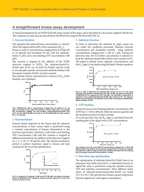
1- Enzyme titration<br />
This step gives the optimal kinase concentration, i.e. that for<br />
which the signal reaches 80% of the maximum (EC 80 ).<br />
Kinase is used at concentrations ranging from 0.10 ng/well<br />
to 10 ng/well, and incubated 30 min with the substratebiotin<br />
(1 µM), and a non-limiting ATP concentration (100<br />
µM).<br />
The reaction is stopped by the addition of the HTRF<br />
detection reagents in EDTA. The substrate-biotin/SA-<br />
XL665 ratio of 8/1 (i.e. 62.2nM SA-XL665) and the ready<br />
to use phospho specific monoclonal antibody labeled with<br />
Europium Cryptate (Eu(K)) are kept constant.<br />
The optimal enzyme concentration is chosen at EC 80 of the<br />
titration curve obtained.<br />
S-B<br />
60000<br />
40000<br />
20000<br />
0<br />
0.0 2.5 5.0 7.5 10.0 12.5<br />
Enzyme ng/well (20 µL final volume)<br />
Fig 2: MAPKAP-K2 used at concentrations ranging from 10 ng/well to 0.1 ng/<br />
well is incubated 30 min with STK substrate 1-biotin (1µM) and a non limiting<br />
ATP concentration (100µM). The STK substrat 1-biotin/streptavidin-XL665 ratio<br />
of 8/1 (i.e 62.2 nM SA-XL665) and ready to use STK Mab-Eu(K) are kept constant<br />
(EC 80 = 0.4 ng/well).<br />
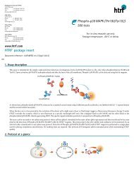
2- Enzyme kinetic<br />
Enzyme kinetic depends on the kinase and the substrate<br />
concentrations. A time course study is performed using<br />
a constant concentration of kinases (determined in the<br />
previous experiment), substrate (1 µM) and a non-limiting<br />
ATP concentration (100 µM).The reaction is stopped at<br />
different end points by the addition of the detection reagents<br />
(at 1, 2, 5, 10, 15, 30 and 60 min). The optimal incubation<br />
period to achieve maximum signal is chosen and kept<br />
constant for the rest of the optimization.<br />
Fig 3: In presence of substrate (1 µM) and ATP (100 µM), the optimal incubation<br />
period for MAPKAP-K2 (0.4 ng/well) to achieve maximum signal and a linear time<br />
course is chosen at 5 min.<br />
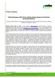
3- Substrate titration<br />
In order to determine the substrate K (app), assays are<br />
M<br />
run under the conditions previously obtained (enzyme<br />
concentration and incubation period) using substrate<br />
concentrations ranging from 1 nM to 2 µM. During the<br />
detection step, the SA-XL665 concentration is adjusted to<br />
keep the substrate/streptavidin-XL665 ratio constant at 8/1.<br />
The signal is plotted versus substrate concentrations, and<br />
the K (app) is calculated using Michaelis-Menten equation.<br />
M<br />
Substrate<br />
Fig 4: In order to determine the substrate KM, assays are run for 5 min using<br />
substrate concentrations ranging from 0.97 nM to 2 µM, and fixed concentrations<br />
of MAPKAP-K2 (0.4 ng/well) and ATP (100 µM). The SA-XL665 concentration is<br />
adjusted to keep substrate/streptavidin-XL665 ratio constant at 8/1. The signal is<br />
plotted versus substrate concentrations and the KM is calculated using Michaelis-<br />
Menten equation: 80 nM.<br />
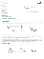
4- ATP titration<br />
Assays are run at a non-limiting substrate concentration with<br />
ATP from 1.7 nM to 300 µM, while the enzyme quantity and<br />
the incubation period are kept constant.<br />
As in the previous step, the K (app) is calculated from the<br />
M<br />
resulting plot of the signal versus ATP concentrations.<br />
ATP K M (app) = 2.30 µM<br />
Fig 5: Assays are run for 5 min with ATP concentrations ranging from 1.7 nM to 300<br />
µM (threefold serial dilution) and using fixed concentrations of kinase (0.4 ng/well)<br />
and substrate (1 µM). As in the previous step, the KM is calculated from the resulting<br />
plot of the signal versus ATP concentrations: 2.30 µM.<br />
5- Detection step optimization<br />
The optimization of substrate-biotin/SA-XL665 ratio is an<br />
important step which may lead to a substantial increase in<br />
signal.The assay is performed using the optimal enzyme,<br />
ATP and substrate concentrations. Three different molar<br />
ratios of substrate-biotin/streptavidin-XL665 are tested<br />
(2/1, 4/1, 8/1). The optimal ratio obtains a good compromise<br />
between signal level and reagent consumption.
![HTRF meeting_130426_villa_pour pdf [Mode de compatibilité]](https://img.yumpu.com/22345646/1/190x135/htrf-meeting-130426-villa-pour-pdf-mode-de-compatibilitac.jpg?quality=85)