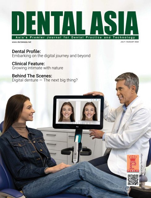
Dental Asia July/August 2022
For more than two decades, Dental Asia is the premium journal in linking dental innovators and manufacturers to its rightful audience. We devote ourselves in showcasing the latest dental technology and share evidence-based clinical philosophies to serve as an educational platform to dental professionals. Our combined portfolio of print and digital media also allows us to reach a wider market and secure our position as the leading dental media in the Asia Pacific region while facilitating global interactions among our readers.
For more than two decades, Dental Asia is the premium journal in linking dental innovators and manufacturers to its rightful audience. We devote ourselves in showcasing the latest dental technology and share evidence-based clinical philosophies to serve as an educational platform to dental professionals. Our combined portfolio of print and digital media also allows us to reach a wider market and secure our position as the leading dental media in the Asia Pacific region while facilitating global interactions among our readers.

Create successful ePaper yourself
Turn your PDF publications into a flip-book with our unique Google optimized e-Paper software.
www.dentalasia.net<br />
JULY / AUGUST <strong>2022</strong><br />
<strong>Dental</strong> Profile:<br />
Embarking on the digital journey and beyond<br />
Clinical Feature:<br />
Growing intimate with nature<br />
Behind The Scenes:<br />
Digital denture — The next big thing?<br />
Photo Courtesy of Align Technology
16<br />
26<br />
34<br />
CONTENTS<br />
TRENDS<br />
16 Connecting patients closer to<br />
their clinics<br />
18 NSK harnesses green power<br />
UNDER THE SPOTLIGHT<br />
19 Integration of guided surgery<br />
into practice<br />
DENTAL PROFILE<br />
20 ZimVie opens doors to<br />
integrated solutions<br />
24 Embarking on the digital<br />
journey and beyond<br />
CLINICAL FEATURE<br />
26 Immediate implant placement<br />
in the aesthetic zone<br />
30 Orthodontic space closure:<br />
Midline diastema related to<br />
mesiodens<br />
34 Growing intimate with nature<br />
42<br />
48<br />
USER REPORT<br />
39 <strong>Dental</strong>-specific solution for 3D<br />
model printing<br />
40 Centripetal layered build-up of<br />
posterior direct composite resin<br />
42 Digital aesthetic smile<br />
reconstruction<br />
46 Same-day visit implant treatment<br />
BEHIND THE SCENES<br />
48 Bringing 3D printing to the next<br />
level for sustainable healing<br />
50 Digital denture – The next big<br />
thing?<br />
52 The bridge between virtual<br />
platform and reality<br />
IN DEPTH WITH<br />
54 Practice transformation with<br />
Invisalign Outcome Simulator Pro<br />
55 Celebrating the anniversary of<br />
Zolid zirconia<br />
SHOW REVIEW<br />
63 GC’s centennial anniversary<br />
celebrations continue at GC<br />
International<br />
SHOW PREVIEW<br />
64 Dentsply Sirona announces<br />
the return off DS World <strong>2022</strong><br />
65 <strong>Dental</strong>Forum APAC <strong>2022</strong>: A<br />
networking hub for regional<br />
dental professionals<br />
REGULARS<br />
4 Editor’s Note<br />
6 <strong>Dental</strong> Updates<br />
56 Product Highlights<br />
66 Giving Back to Society<br />
67 Events Calendar<br />
68 Advertisers’ Index<br />
54<br />
2<br />
DENTAL ASIA JULY / AUGUST <strong>2022</strong>
<strong>Dental</strong> Updates<br />
Whatever tomorrow holds, we’re ready. Our support is continually being<br />
developed and enhanced to ensure that your protection is future-proof.<br />
• A wide range of protection beyond claims<br />
• Advice for any eventuality your career may face<br />
• Fast to respond to unexpected situations<br />
• Support today, tomorrow and yesterday<br />
Always there for you<br />
dentalprotection.org<br />
<strong>Dental</strong> Protection Limited is registered in England (No. 2374160) and is a wholly owned subsidiary of The Medical Protection Society Limited (“MPS”) which is registered in England (No. 00036142).<br />
Both companies use ‘<strong>Dental</strong> Protection’ as a trading name and have their registered office at Level 19, The Shard, 32 London Bridge Street, London, SE1 9SG. <strong>Dental</strong> Protection Limited serves and<br />
supports the dental members of MPS with access to the full range of benefits of membership, which are all discretionary, and set out in MPS’s Memorandum and Articles of Association. MPS is not<br />
an insurance company. <strong>Dental</strong> Protection® is a registered trademark of MPS.<br />
2205233575 06/22<br />
DENTAL ASIA JULY / AUGUST <strong>2022</strong> 3
EDITOR’S NOTE<br />
The world is digital<br />
PABLO SINGAPORE<br />
Publisher<br />
William Pang<br />
williampang@pabloasia.com<br />
Digital technologies have transformed the<br />
way we live and work. Almost everything<br />
can be done within a click at our fingertips,<br />
bringing us so much convenience<br />
and efficiency as we ease through our<br />
everyday.<br />
For the dental industry, the impact of<br />
digitalisation goes way beyond<br />
streamlining treatment procedures – it<br />
elevates patient care and effectively<br />
improves treatment outcomes. As<br />
emphasised by an implantologist, Dr<br />
Jonathan Loa, digital implant planning<br />
makes implant placement more<br />
predictable as it allows greater precision<br />
(pp. 19).<br />
Marie-Laure Pochon, CEO and president<br />
of 3Disc, also highlighted that embracing<br />
digital dentistry can allow practitioners<br />
to improve their treatment process. An<br />
intraoral scanner, for instance, brings more<br />
accuracy in the fabrication of prostheses,<br />
better comfort for the patients and allows<br />
a shorter turnaround time of dental<br />
treatment (pp. 24).<br />
On the laboratory side, 3D printing<br />
continues to benefit the manufacturing of<br />
regenerative implants. Dr Lim Jing, CTO of<br />
Osteopore, shared that this technological<br />
advantage has enabled them to create<br />
a microstructure that is representative<br />
of native bone while meeting the gross<br />
geometrical needs of the reconstruction<br />
area (pp. 48).<br />
However, Dr Naren Rajan, who presented<br />
a digital protocol for ceramic restoration,<br />
stressed: “Simply having technology is<br />
not enough. Using it thoughtfully is how<br />
we realise the true promise of digital<br />
dentistry (pp. 42).”<br />
Hence, as suggested by Maik Walther,<br />
general manager of ZimVie <strong>Dental</strong><br />
<strong>Asia</strong>-Pacific, now is the time for<br />
manufacturers to focus on integrated<br />
solutions to better help dental<br />
professionals use new products and<br />
technologies (pp. 20).<br />
This issue highlights that the dental<br />
landscape is continually growing and<br />
evolving to redefine and innovate<br />
oral healthcare. Tapping into modern<br />
solutions allows practitioners to offer a<br />
wider range of treatments to ultimately<br />
transform their patients’ smiles.<br />
Czarmaine Masigla<br />
Assistant Editor<br />
Publications Director<br />
Senior Editor<br />
Assistant Editor<br />
Graphic Designer<br />
Circulation Manager<br />
PABLO BEIJING<br />
General Manager<br />
PABLO SHANGHAI<br />
Senior Editor<br />
Jamie Tan<br />
jamietan@pabloasia.com<br />
Josephine Tan<br />
josephine@pabloasia.com<br />
Czarmaine Masigla<br />
czarmaine@pabloasia.com<br />
Jolin Tan<br />
jolintan@pabloasia.com<br />
Shu Ai Ling<br />
circulation@pabloasia.com<br />
Ellen Gao<br />
pablobeijing@163.com<br />
Daisy Wang<br />
pabloshanghai@163.net<br />
HEAD OFFICE<br />
PABLO PUBLISHING &<br />
EXHIBITION PTE LTD<br />
3 Ang Mo Kio Street 62 #01-23<br />
Link@AMK, Singapore 569139<br />
Tel: (65) 62665512<br />
Email: info@pabloasia.com<br />
Website: www.dentalasia.net<br />
Company Registration No.: 200001473N<br />
Singapore MICA (P) No. 104/12/2021<br />
Malaysia KDN: PPS1528/07/2013 (022978)<br />
REGIONAL OFFICES<br />
PABLO BEIJING<br />
Tel: +86-10-6509-7728<br />
Email: pablobeijing@163.com<br />
PABLO SHANGHAI<br />
Tel: +86-21-52389737<br />
Email: pabloshanghai@163.net<br />
ADVISORY BOARD<br />
Dr William Cheung<br />
Dr Choo Teck Chuan<br />
Dr Chung Kong Mun<br />
Dr George Freedman<br />
Dr Fay Goldstep<br />
Dr Clarence Tam<br />
Prof Nigel M. King<br />
Dr Anand Narvekar<br />
Dr Kevin Ng<br />
Dr William O’Reilly<br />
A DENTAL ASIA JULY / AUGUST <strong>2022</strong><br />
Dr Wong Li Beng<br />
Dr Adrian U J Yap<br />
Dr Christopher Ho<br />
Dr How Kim Chuan<br />
Dr Derek Mahony<br />
Prof Alex Mersel
DENTAL UPDATES<br />
Envista to acquire Osteogenics Biomedical Business<br />
Envista Holdings Corporation has entered into<br />
a definitive agreement to acquire Osteogenics<br />
Biomedical, Allotech and OBI Biologics<br />
(together “Osteogenics”). The transaction is<br />
expected to close in the third quarter.<br />
regenerative therapies are often a critical<br />
step in implant-based tooth replacements.<br />
By improving bone stability, regenerative<br />
solutions support better clinical outcomes for<br />
more patients.<br />
Osteogenics is a developer of regenerative<br />
solutions for periodontists, oral and<br />
maxillofacial surgeons, and clinicians involved<br />
in implant dentistry throughout the world.<br />
Primarily sold under the Cytoplast brand<br />
name, Osteogenics offers a complete line of<br />
bone grafting products. Bone grafting and<br />
Amir Aghdae, CEO of Envista, said: “Increasing<br />
our capabilities in regenerative solutions<br />
is consistent with our intention to digitise,<br />
personalise, and democratise oral care.<br />
Osteogenics is a recognised pioneer in<br />
membrane technologies used in dental bone<br />
grafting procedures. They are a trusted brand<br />
and have a proven track record of growth. We<br />
are excited to welcome the Osteogenics team<br />
to Envista.” ■<br />
Dentsply Sirona launches DS Core in collaboration with Google Cloud<br />
Dentsply Sirona has unveiled the DS Core, a<br />
new and open platform that integrates the<br />
whole workflow of digital dentistry in a virtual<br />
event, along with other services and solutions.<br />
Developed in collaboration with Google Cloud,<br />
DS Core allows dentists to focus on their<br />
patients and create ways to collaboratively<br />
work with laboratories, partners, and<br />
specialists.<br />
It connects to Dentsply Sirona equipment and<br />
is accessible across multiple devices. Hence,<br />
dentists can maximise the productivity of their<br />
practice by simplifying workflows and easily<br />
adding and integrating new ones.<br />
Furthermore, practitioners can use DS Core<br />
to store different types of patient files and<br />
making them accessible from multiple<br />
locations, while collaborating with partners<br />
and colleagues outside their practice. DS<br />
Core supports GDPR and HIPAA-compliant<br />
file sharing and cloud storage for patient case<br />
files.<br />
Cord Staehler, CTO at Dentsply Sirona, said:<br />
“We are very proud that we are now ready<br />
to take the next step in our mission to make<br />
digital dentistry easy to integrate into dental<br />
offices. In line with our recently launched<br />
collaboration with Google Cloud, this enables<br />
seamless workflows and the highest level of<br />
connectivity with the ultimate goal in mind: the<br />
best treatment outcome for patients.”<br />
Dentsply Sirona has also revealed two new<br />
services: the DS Core Create and DS Core Care.<br />
With the DS Core Create, dental practitioners<br />
can access designs that are tailored for each<br />
patient’s needs across a range of indications<br />
without having to use the software themselves.<br />
It can be integrated with Dentsply Sirona’s<br />
new Primeprint Solution and will grow in the<br />
future. On the other hand, the DS Core Care is<br />
an equipment service and support solution that<br />
helps to increase equipment uptime.<br />
Staehler concluded: “By launching this digital<br />
universe with DS Core at its centre and services<br />
like DS Core Create and DS Core Care, as well<br />
as solutions like Primeprint, we are positioning<br />
Dentsply Sirona at the forefront of digital<br />
dentistry. Most importantly, we help dental<br />
practitioners to unlock the full potential of their<br />
work so that they can focus on what matters<br />
most: treating patients and giving them healthy<br />
smiles.” ■<br />
6 DENTAL ASIA JULY / AUGUST <strong>2022</strong>
DENTAL UPDATES<br />
DENTAL ASIA JULY / AUGUST <strong>2022</strong> 7
DENTAL UPDATES<br />
exocad <strong>Asia</strong> moves to Seoul<br />
exocad has relocated its <strong>Asia</strong> headquarters<br />
to Seoul, South Korea. The company will<br />
coordinate all services and operations<br />
for the <strong>Asia</strong> region from this new office,<br />
located in Seoul’s historic centre.<br />
“We look forward to strengthening our<br />
strategic partnerships in the region and<br />
to benefitting from the robust, high-tech<br />
infrastructure that Seoul offers,” said<br />
Novica Savic, CCO of exocad. “exocad<br />
has been working with a growing number<br />
of South Korean manufacturers for many<br />
years. With the relocation of our <strong>Asia</strong><br />
HQ, we want to be closer to our strategic<br />
partners and users from South Korea.”<br />
South Korea’s digital dentistry market is well<br />
developed and world renowned, making it an<br />
ideal location for exocad’s <strong>Asia</strong>n hub.<br />
“Many global manufacturers and dental<br />
collaborators are already located near exocad’s<br />
new headquarters,” said Kim Gunwoo, exocad’s<br />
sales representative in <strong>Asia</strong>. “The regional<br />
headquarters will allow exocad to deepen its<br />
relationships with these strategic partners<br />
and provide even more comprehensive<br />
services to customers in <strong>Asia</strong>.” ■<br />
Dr Wolff debuts its biomimetic oral care range<br />
The Dr Wolff Group, a family-owned<br />
pharmaceutical and cosmetic company,<br />
has launched its Bioniq Repair Toothpaste<br />
in Singapore. For more than a decade,<br />
the Germany-based company, has led in<br />
the research and development of modern<br />
dental care products to cater to growing<br />
pains of consumers.<br />
Its dental care product, Bioniq Repair-<br />
Toothpaste’s main active ingredient<br />
is the biomimetic tooth enamel called<br />
hydroxyapatite (HAP), which remineralises<br />
the teeth and forms a protective layer<br />
against enamel erosion. This ingredient<br />
alone is known to remineralise the deeper<br />
layers of the teeth effectively, as compared<br />
to other common remineralising agents<br />
used in oral care.<br />
Eduard R. Dörrenberg, managing director, Dr<br />
Wolff, said: “For years, the team at Dr Wolff<br />
has dedicated its research to developing<br />
dental care products with hydroxyapatite as<br />
the main active ingredient, recognising its<br />
benefits for users. We’re excited to finally<br />
bring our product to <strong>Asia</strong>, with Singapore as<br />
the first stop. We hope to raise awareness<br />
on the importance of protecting teeth<br />
enamel with HAP and to plug the gap in the<br />
market with a product that caters to the<br />
dental care needs of Singaporeans, who<br />
predominantly suffer from issues linked to<br />
sensitive teeth and gums. HAP is a mineral<br />
from the calcium phosphate family and is a<br />
proven and effective way of remineralising<br />
our tooth enamel. Good oral health enriches<br />
your quality of life, and oral care can be as<br />
simple as using HAP-based products to keep<br />
oral health issues caused by dental cavities,<br />
sensitive teeth or gingivitis at bay.”<br />
The Bioniq Repair-Toothpaste and Bioniq<br />
Repair-Toothpaste PLUS are the only<br />
toothpastes from Germany to contain 20%<br />
HAP.<br />
Singapore is the first country that the<br />
product has been made available in <strong>Asia</strong>.<br />
Dr Wolff’s Bioniq Repair Toothpaste is<br />
currently available in Germany and Austria,<br />
with over two million units sold last year in<br />
Germany alone. Alongside the latest launch<br />
in Singapore, Dr Wolff has plans to launch<br />
its biomimetic oral care range in Switzerland<br />
and US soon. ■<br />
8 DENTAL ASIA JULY / AUGUST <strong>2022</strong>
DENTAL UPDATES<br />
FDI President addresses the<br />
challenges and opportunities in Africa<br />
for optimal oral health<br />
PERFECTION IN<br />
BONE SURGERY<br />
FDI President, Prof Ihsane Ben Yahya, was invited by the African<br />
Society of Dentistry and Implantology (ASDI) and the African<br />
Journal of Dentistry and Implantology (AJDI) to take part in a<br />
webinar, where she discussed “Optimal Oral Health: challenges<br />
and opportunities for Africa”.<br />
The event took place online and was moderated by Dr Abdellah<br />
Squalli, president of ASDI, and Prof Fethi Maatouk, president<br />
of Conférence des Doyens des Facultés de Médecine Dentaire<br />
d’Afrique.<br />
→ YOUR SURGICAL<br />
APPROACH WILL CHANGE -<br />
THE PIEZOSURGERY® touch<br />
→ best cutting efficiency<br />
→ optimal intraoperative control<br />
→ perfect ergonomics<br />
→ made in Italy<br />
Prof Ben Yahya outlined the epidemiological situation in Africa<br />
and stressed that to this day, millions of people in the region do<br />
not have access to oral healthcare. This is especially concerning<br />
as poor oral health can lead to absenteeism at school and work,<br />
and an overall poor quality of life. She then highlighted some of<br />
the major challenges and the most prevalent pathologies in the<br />
region, which must be tackled to achieve optimal oral health.<br />
Some of the recent positive developments in the oral health<br />
landscape were also shared and she put the spotlight on the<br />
landmark WHO resolution on oral health that was approved<br />
by members states in 2021, as an important indication of the<br />
commitment to the issue at the global policy level.<br />
Prof Ben Yahya also advised that the subsequent WHO oral<br />
health strategy is in line with FDI’s own Vision 2030 report.<br />
She also noted that WHO AFRO’s regional oral health strategy<br />
is currently ongoing and reminded webinar attendees of its<br />
five targets for monitoring and evaluation, emphasising that<br />
strategies to improve oral health must be adopted in regional and<br />
national contexts.<br />
Prof Ben Yahya stressed that while there is still much work to<br />
do, the challenges that confront dentistry can be turned into<br />
opportunities. It was important, now more than ever, to continue<br />
to show resilience and work to effectively prevent and manage<br />
oral diseases so that everyone can enjoy a good oral health, and<br />
thus a good quality of life. ■<br />
→ www.mectron.com<br />
DENTAL ASIA JULY / AUGUST <strong>2022</strong> 9<br />
ad_PStouch_dental_asia_95x250_en_211214.indd 1 14.12.21 15:38
DENTAL UPDATES<br />
Oral-B and Straumann partner on a landmark alliance for scientific education<br />
Oral-B and Straumann have announced a new<br />
global alliance to elevate the importance of<br />
prevention in periodontal and peri-implant<br />
health. The alliance will set new standards<br />
in quality scientific education for dental<br />
professionals and help their patients achieve<br />
better long-term outcomes.<br />
The Oral-B Straumann alliance has a longterm<br />
goal of delivering a holistic programme<br />
of scientific events, professional courses,<br />
webinars and publications, co-created with<br />
and delivered by the experts and thought<br />
leaders in dentistry.<br />
The alliance was launched on 17 Jun <strong>2022</strong><br />
with a Sponsored Scientific Session at<br />
EuroPerio10 Copenhagen – dental congress in<br />
periodontology and implant dentistry – where<br />
both companies also presented their recent<br />
innovations for periodontal and peri-implant<br />
patients.<br />
“As patients around the world invest in dental<br />
implants, they need to recognise that self-care<br />
around implants is just as important as around<br />
their natural teeth. The Oral-B Straumann<br />
Alliance will play an important role in enabling<br />
ongoing dialogue between dental professionals<br />
and their patients so both understand the most<br />
up to date science on prevention of implantrelated<br />
diseases and promotion of periodontal<br />
health,” said J. Leslie Winston, vice-president,<br />
global health care R&D, Procter & Gamble.<br />
Arik Zucker, vice-president, global head of<br />
biomaterials, Straumann Group, added:<br />
“Prevention of tooth or implant loss is for<br />
many years an important topic for us in the<br />
Straumann Group. As one of the world’s<br />
leading dental implant companies, we work<br />
closely with experts from the research and<br />
clinical fields to address the issues caused by<br />
peri-implantitis and periodontitis. The Alliance<br />
with Oral-B complements our efforts and<br />
offers a solid ground for further scientific and<br />
educational activities that will enable more<br />
dental professionals to improve their patients’<br />
lives.” ■<br />
Henry Schein launches NextDent Ortho Flex resin in Singapore in partnership<br />
with expatdental<br />
Medical supplier Henry Schein has launched the<br />
NextDent Ortho Flex in Singapore, a resin that<br />
offers accuracy, break resistance and flexibility<br />
to produce dental appliances in the right<br />
measurements.<br />
Certified by Singapore’s Health Sciences<br />
Authority, the NextDent Ortho Flex resin<br />
smooths key road bump in patients’ dental<br />
journey by providing comfortable dental<br />
appliances in a significantly reduced lead<br />
time in its manufacture for use in restorative<br />
dentistry services.<br />
“At Henry Schein, it is enshrined in our DNA to<br />
stay abreast of the digital advancements and<br />
bring this innovation to dental clinics and labs<br />
around the world. We are thrilled to announce<br />
this partnership with expatdental to enable<br />
in-house manufacturing of splints using 3D<br />
printing technology and recently launched<br />
NextDent Ortho Flex resin. It is also a testimony<br />
of expatdental’s commitment to continuously<br />
advance their clinical procedures and improve<br />
patients’ experience by embracing the latest<br />
available technology,” said Kateryna Iakovenko,<br />
regional sales manager of Henry Schein<br />
Connect<strong>Dental</strong>, SEA.<br />
The NextDent Ortho Flex resin makes its<br />
Singapore debut at expatdental, a premium<br />
proposition from Oracare Group, elevating<br />
its in-house lab capabilities to facilitate<br />
the printing of dental splints for bruxism<br />
treatments. The NextDent Ortho Flex is used in<br />
tandem with the NextDent 5100 3D Printer.<br />
Users can now receive their temporary crowns<br />
and splints in the same week, significantly<br />
faster than the traditional three to four weeks<br />
interval. Offering a broad range of services<br />
including cosmetic dentistry, implant surgery<br />
and bruxism solutions, expatdental’s expertise<br />
lies in providing a seamless, comfortable and<br />
effective experience to all patients through<br />
advanced technologies and dental equipment,<br />
alongside a dedicated, experienced team<br />
focused on dental excellence and quality.<br />
“Our priority at expatdental has always been to<br />
provide efficient, premium and timely services<br />
to all our patients, with their utmost comfort<br />
and convenience in mind. In today’s age of<br />
digital dentistry, the NextDent Ortho Flex resin<br />
presents a new dawn for the future of dentistry,<br />
paving the way for 3D printing innovation, dental<br />
technologies and dental care industries—a<br />
natural acquisition on our end to meet the<br />
unwavering promise of exceptional patient<br />
comfort,” said Dr Shaun Thompson, managing<br />
director of expatdental. ■<br />
10 DENTAL ASIA JULY / AUGUST <strong>2022</strong>
Torq Control®<br />
DENTAL UPDATES<br />
Universal Torque<br />
Wrench<br />
Torq Control® is the Anthogyr universal<br />
torque wrench offering the guarantee of<br />
tightening precision, whatever the type<br />
of implant connection or the difficulties of<br />
access.<br />
Precise tightening is a key factor to secure<br />
implant treatment success. Torq Control®<br />
has been specially designed by Anthogyr<br />
to meet these requirements for all<br />
prosthetic manipulations, in all safety<br />
thanks to automatic declutching.<br />
A must-have, especially for full-arch<br />
restorations.<br />
DENTAL ASIA JULY / AUGUST <strong>2022</strong> 11
DENTAL UPDATES<br />
Medit and Panthera reveal integrated digital workflow for provision of dental<br />
sleep devices<br />
Panthera <strong>Dental</strong>, a provider of CAD/<br />
CAM implant solutions and dental sleep<br />
appliances, has partnered with Medit to<br />
provide patients custom-made sleep apnoea<br />
devices from Panthera <strong>Dental</strong> that are<br />
integrated within the Medit software.<br />
After scanning the patient’s arches and<br />
registering the bite, Medit users can open the<br />
Panthera App from Medit Link, complete the<br />
prescription, and send everything to Panthera<br />
<strong>Dental</strong>. Within eight days, the D-SAD Classic<br />
or X3 will be designed and manufactured by<br />
Panthera <strong>Dental</strong> using its CAD/CAM process.<br />
The appliance will then be shipped to the<br />
dentist via express courier so the patient can<br />
begin treatment in as little as 10 days.<br />
“It is our great pleasure to announce a<br />
successful collaboration with Panthera<br />
<strong>Dental</strong>,” said Inhaeng Cho, CSO of Medit.<br />
“With a company that has long been a<br />
companion of Medit, we have developed<br />
another level of partnership, and we are<br />
eager to provide a seamless workflow to<br />
our users. This integration will provide both<br />
convenience and efficiency for our clinic<br />
users as well as our lab users.”<br />
The scanning process is comfortable and<br />
quick, and the D-SADs are made from<br />
100% medical-grade biocompatible type-12<br />
polyamide nylon, a durable material that is<br />
resistant to bruxism.<br />
A range of plateaus and bands are available,<br />
allowing for over 300 design combinations<br />
to maximise comfort and tongue space,<br />
and minimise side effects, regardless of<br />
the complexity of the patient’s morphology.<br />
D-SADs are also simple for the patient to<br />
adjust and care for at home.<br />
“We are excited to collaborate with a<br />
company that shares our values and vision<br />
for digital dentistry,” said Beatrice Robichaud,<br />
co-founder and vice-president of marketing<br />
and customer experience at Panthera <strong>Dental</strong>.<br />
“Medit has created a workflow that is perfectly<br />
streamlined. There is no need to leave the<br />
Medit app to provide everything we need to<br />
make a great appliance for that customer. It<br />
represents a truly seamless and efficient user<br />
experience that we know customers will love.”<br />
Both Panthera <strong>Dental</strong>’s D-SADs, the Classic<br />
and X3, have FDA 510(k) clearance for snoring<br />
and obstructive sleep apnoea, bear the CE<br />
mark, and comply with the Canadian Medical<br />
Devices Regulations. ■<br />
National <strong>Dental</strong> Centre Singapore held its first international symposium on<br />
oral health research<br />
National <strong>Dental</strong> Centre Singapore (NDCS) has<br />
collaborated with oral health experts from<br />
Sweden, Australia, Hong Kong and Japan<br />
to hold its first International Oral Health<br />
Symposium conducted virtually.<br />
materials research by a young scientist.<br />
Five scientists from each participating<br />
institute presented their novel innovations<br />
and studies that will contribute to the<br />
advancement of oral health.<br />
The two-day symposium comprised of topics<br />
that have impact on the oral health industry<br />
such as regenerative medicine in the area<br />
of tissue engineering, digital transformation<br />
in oral health, oral health policies, precision<br />
medicine and the study of the oral<br />
microbiome.<br />
The symposium, themed “Innovation,<br />
Strategy and Future Perspectives” aimed<br />
to foster stronger collaborations and<br />
exchanges, share research, innovation and<br />
developments in the field of oral health,<br />
and be a learning platform for aspiring<br />
researchers.<br />
The symposium hosted an award<br />
ceremony for the Early Career Investigator<br />
Award, which recognises outstanding,<br />
interdisciplinary scientific work in<br />
“This is an exciting platform for the oral<br />
health research community to gather,<br />
discuss and work together. Beyond<br />
fostering greater collaboration, we<br />
hope it will inspire our researchers and<br />
clinical professionals to continually push<br />
the boundaries for innovative benchto-bedside<br />
solutions,” shared Clinical<br />
Associate Professor Goh Bee Tin, deputy<br />
CEO (Research), NDCS. ■<br />
12 DENTAL ASIA JULY / AUGUST <strong>2022</strong>
DENTAL UPDATES<br />
Ivoclar commits to energy<br />
sustainable future<br />
Ivoclar North America has announced a major investment in solar<br />
energy to power its corporate headquarters located in Amherst,<br />
New York transitioning from fossil fuel dependent to renewable<br />
energy as part of the company’s continued global commitment to a<br />
safer, healthier environment.<br />
PERFECTLY SHAPED.<br />
NATURALLY.<br />
The nearly US$1 million clean energy project started in 2020<br />
and completed in December 2021 is projected to supply the<br />
100,000-square-foot facility with 750MWh of energy per year<br />
with any unused energy returned to the grid for the benefit of the<br />
community. The project included the installation of 1,640 solar<br />
panels by locally owned Buffalo Solar and benefits the environment<br />
by saving the emissions of approximately 532 metric tons CO2<br />
yearly into the air, the equivalent of planting approximately 8,789<br />
trees per year. The company estimates that 65% of its annual<br />
energy requirement will be generated by this project.<br />
As an international company, the Ivoclar Group is a global<br />
manufacturer of dental products, with an ongoing commitment<br />
to supporting people, communities and the environment. The<br />
company aims to sustainably improve the health and quality of<br />
the life of people all over the world. In addition to manufacturing<br />
high-quality products, Ivoclar Group wishes to make an active<br />
contribution to sustainable development as defined by the United<br />
Nations in its Sustainable Development Goals (SDG) and in<br />
accordance with the requirements of the Global Reporting Initiative<br />
(GRI).<br />
The power facility at the headquarters in Schaan, Liechtenstein<br />
which began operating in 2018, supplies the company headquarters<br />
with electricity, warm and cold water, as well as with compressed<br />
air and reduces its consumption of natural gas. Heat recovery<br />
technology is further helping the company's efforts to reduce<br />
its need for natural gas. In the course of the project ‘Global<br />
greenhouse gas balance of the Ivoclar Group", CO2 emissions have<br />
been reduced globally. Moreover, the Ivoclar Group increases its<br />
use of photovoltaics – not only at the NY corporate headquarters in<br />
Amherst, New York.<br />
In addition to the solar panel project, Ivoclar North America has<br />
created an internal task force to explore and evaluate a cleaner and<br />
more sustainable business operation. Some of these initiatives<br />
include new packaging and transportation solutions, utilisation of<br />
hybrid vehicles and reducing plastic water bottle usage.<br />
Now available in<br />
28 VITA SYSTEM 3D-MASTER® shades!<br />
VITAPAN EXCELL®<br />
True to life – in shape and color<br />
• Natural esthetics through layering,<br />
texture and brilliance<br />
• Excellent abrasion durability using MRP<br />
composite material<br />
“We are moving deliberately forward to reduce our carbon emission<br />
footprint in our locality and worldwide as part of Ivoclar’s global<br />
commitment to improving the environment and addressing climate<br />
change,” said Christian Brutzer, president, Ivoclar North America. ■<br />
DENTAL ASIA JULY / AUGUST <strong>2022</strong> 13<br />
3603EN_VITAPAN EXCELL_AZ_95x250.indd 1 15.06.<strong>2022</strong> 16:14:53
DENTAL UPDATES<br />
Septodont releases new version of Biodentine<br />
Since its launch a decade ago, Biodentine,<br />
based on Septodont’s Active Biosilicate<br />
Technology platform, has proven successful in<br />
replacing dentin in the crown and in the root.<br />
Sold in more than 60 countries and with more<br />
than 1,000 worldwide publications, Biodentine<br />
has saved more than five million teeth.<br />
With this success, Septodont has launched<br />
a new version of Biodentine: Biodentine XP,<br />
which includes all the science of Biodentine<br />
embedded in an upgraded system, designed<br />
to provide practitioners an optimal daily<br />
experience from crown to root with nine<br />
indications.<br />
Due to its unique features, Biodentine has<br />
become a gold standard in preserving tooth<br />
structure by offering high bioactivity with<br />
complete dentin bridge formation and sealing<br />
properties. The clinical benefits promote vital<br />
pulp therapy and help save teeth from root<br />
canal treatment and possible extraction.<br />
Biodentine XP ensures a consistent and<br />
perfect mix thanks to its proprietary mixer<br />
and easy delivery directly into the tooth with<br />
the Biodentine applicator gun. The all-in-one<br />
cartridges are available in two formats, XP<br />
200 and XP 500, depending on the procedure<br />
and volume of material needed.<br />
The Biodentine XP Starter Pack contains the<br />
mixer, applicator gun, and packages of XP 200<br />
and XP500 cartridges. Biodentine XP is also<br />
available in boxes of 10 refill cartridges. ■<br />
Formlabs launches <strong>Dental</strong> Academy<br />
Formlabs, a 3D printing company, has launched<br />
Formlabs <strong>Dental</strong> Academy, a new educational<br />
platform dedicated to advancing 3D printing in<br />
dentistry with online and in-person courses, step-bystep<br />
guides, webinars, and more.<br />
The platform includes free and paid courses<br />
designed to enable dental professionals, such as<br />
dental lab technicians, orthodontists, and clinicians<br />
to learn and successfully implement 3D printing<br />
technology in their businesses. <strong>Dental</strong> Academy<br />
courses are available online and in-person.<br />
Implementing 3D printing into a dental treatment<br />
plan benefits practitioners by enabling more<br />
efficient workflows and less downtime while giving<br />
patients access to personalised yet affordable<br />
solutions such as custom dental models, crowns,<br />
dentures, implants, and more.<br />
To address this skills gap, Formlabs created<br />
<strong>Dental</strong> Academy as a free resource for dental<br />
professionals to unlock these benefits with<br />
comprehensive training and educational<br />
content on 3D printing’s uses and benefits,<br />
dental applications, and materials to deliver<br />
the best clinical outcomes, user satisfaction,<br />
and improved performance and print results.<br />
Focus areas will include an introduction to<br />
stereolithography (SLA) 3D printing and its use<br />
for orthodontic and restorative applications,<br />
digital dentistry benefits, training on technology<br />
basics, materials, and implementation strategies<br />
within the clinic and lab, and more. At launch,<br />
courses will be available in English, Spanish,<br />
Swedish and German.<br />
Formlabs <strong>Dental</strong>, a division of Formlabs, has worked<br />
closely with key stakeholders and leaders within<br />
the dental industry to deliver accessible 3D printing<br />
solutions and materials. While Formlabs users have<br />
printed more than 25 million dental parts ranging<br />
from models to surgical guides, Formlabs <strong>Dental</strong><br />
identified a gap in 3D printing education in the field,<br />
slowing further adoption of the technology.<br />
The platform includes a variety of courses with<br />
subject-matter to suit a range of participants’<br />
knowledge, providing both online and inperson<br />
instruction in Formlabs Boston, Berlin,<br />
and Budapest offices as well as trusted private<br />
educational centres. <strong>Dental</strong> Academy will<br />
continue to evolve as new content becomes<br />
available.<br />
“The Swedish organisation for Computer Aided<br />
Digital Dentistry is honoured to be part of the<br />
Formlabs <strong>Dental</strong> Academy. Formlabs has long<br />
been in the forefront of additive manufacturing in<br />
dentistry and now they have a great educational<br />
portal for everyone,” Dr Michael Braian, DDS, CDT,<br />
PhD, founder of SWECADD and Formlabs <strong>Dental</strong><br />
key opinion leader. ■<br />
14 DENTAL ASIA JULY / AUGUST <strong>2022</strong>
DENTAL UPDATES<br />
ZimVie introduces two next-generation dental solutions in the US<br />
ZimVie, a global life sciences company<br />
in the dental and spine markets, has<br />
announced the joint launch of the new,<br />
FDA-cleared T3 PRO Tapered Implant and<br />
Encode Emergence Healing Abutment in<br />
the US.<br />
The T3 PRO is the newest addition to<br />
ZimVie’s family of dental implants and<br />
builds on the proven solutions of the T3<br />
Tapered Implant. The Encode Emergence<br />
Healing Abutment builds upon ZimVie’s<br />
3-in-1 Encode Impression System which<br />
provides clear intraoral scans and<br />
aesthetics, and is designed for patient<br />
comfort and healing.<br />
Both the T3 PRO and the Encode<br />
Emergence reflect significant innovation<br />
to ZimVie’s previous products and promise<br />
an optimised implant experience for both<br />
dentists and patients. ZimVie will begin<br />
commercial rollout of these solutions<br />
in the US, with an intention to expand to<br />
additional countries pending necessary<br />
regulatory approvals.<br />
“We are excited to launch the next<br />
evolution in our T3 family of implants<br />
and Encode impression system, which<br />
has been used in more than two million<br />
implant restoration procedures to date,”<br />
said Indraneel Kanaglekar, senior vicepresident<br />
and president of ZimVie <strong>Dental</strong>.<br />
“ZimVie is committed to providing innovative<br />
treatment options to improve patient care<br />
while partnering with dental professionals<br />
globally to advance dental technology and<br />
end-to-end workflow solutions.” ■<br />
US study analysing tooth survival after root canal reveals the factors impacting<br />
longevity of treated teeth<br />
Teeth survive about 11 years after a root canal,<br />
according to new research from Regenstrief<br />
Institute and Indiana University School of<br />
Dentistry, US. This study is the first to analyse<br />
records from community dental practices,<br />
where most Americans receive dental care.<br />
“The findings of this study give deeper insight<br />
into the longevity of dental procedures<br />
because it provides real-world data on a wider<br />
range of patients, not just those receiving<br />
care in large health systems or those who<br />
are insured,” said first author Thankam<br />
Thyvalikakath, DMD, MDS, PhD, director of<br />
the Regenstrief-IU School of Dentistry dental<br />
informatics programme. “This information<br />
can be used to inform dental practice, and<br />
help patients and dentists make better care<br />
decisions.”<br />
Root canals are an important treatment<br />
to maintain natural teeth affected by<br />
disease. However, over time, the treated<br />
tooth eventually becomes brittle and dies.<br />
Understanding the outcomes of the procedure<br />
is essential to improving dental treatments.<br />
For this study, the research team gathered<br />
deidentified electronic dental records from<br />
the National <strong>Dental</strong> Practice-Based Research<br />
Network, consisting of 99 small group and<br />
solo dentistry practices from around the<br />
country. The data covered more than 46,000<br />
patients who received root canals.<br />
BREAKING DOWN THE ROOT CANAL<br />
DATA<br />
Data analysis revealed that the median<br />
survival time of a tooth after a root canal<br />
is 11.1 years. However, several factors can<br />
impact that, including follow-up treatments.<br />
•Teeth that receive a root canal, and a<br />
subsequent filling and crown last about<br />
20 years<br />
• Teeth that receive either a filling or a<br />
crown after a root canal last around 11<br />
years<br />
• Teeth that receive no restorative work<br />
after a root canal only last about 6.5 years<br />
There were also wide disparities in longevity<br />
among geographic regions.<br />
• Northeast – 20.5 years<br />
• Midwest – 11.2 years<br />
• Southwest – 11.2 years<br />
• South Atlantic – 9.1 years<br />
• South Central – 9.0 years<br />
• Western – 8.7 years<br />
Insurance status also played a significant<br />
role in tooth survival time.<br />
“This data could also inform dental<br />
insurance coverage by demonstrating the<br />
value of crowns and permanent restoration<br />
options,” said Dr Thyvalikakath. “Oral health<br />
is a public health issue that significantly<br />
affects people’s overall health. Leveraging<br />
dental records can help us better<br />
understand ways to improve treatment,<br />
identify causal relationships and maintain<br />
the health of teeth and gums.”<br />
This study provides more representative<br />
data of the overall population than<br />
previous studies. It also demonstrates that<br />
meaningful insights can be gained through<br />
analysis of existing data from routine<br />
dental care. ■<br />
DENTAL ASIA JULY / AUGUST <strong>2022</strong> 15
TRENDS<br />
Connecting patients<br />
closer to their clinics<br />
As a provider of cloud-based mobile and online customer-tech<br />
solutions, Ouch enables the connection between brands and their<br />
consumers. Czarmaine Masigla speaks with Anil Kumar, co-founder<br />
of Ouch, to elaborate on the company’s perspective as a service<br />
provider to the dental industry and beyond.<br />
Technology has become an integral<br />
part of the lives of many around the<br />
world, transforming the way people<br />
live and work. In the healthcare sector,<br />
particularly in dentistry, digitalisation<br />
in this space has raised the bar in<br />
patient care and improved operational<br />
efficiency, thus enhancing the overall<br />
experience for both patients and<br />
medical professionals, Anil Kumar,<br />
co-founder of Ouch, suggested.<br />
There are several automated solutions<br />
available in dentistry, one of which<br />
is automated recalls using a preprogrammed<br />
customer management<br />
system (CMS) and another is the use<br />
of digital scanners. The use of digital<br />
scanners allows dental practitioners<br />
to reduce the need to take physical<br />
impressions, which, in turn, minimises<br />
the need to manually cast and send<br />
models to the labs.<br />
He told <strong>Dental</strong> <strong>Asia</strong>: “During the pandemic,<br />
with borders closed and tight social<br />
distancing regulations, many clinicians<br />
in Singapore found their income streams<br />
from both foreign and domestic patients<br />
had dropped significantly. However, they<br />
need to continue to pay for ongoing<br />
operating costs, among which rental<br />
expenses proved to be significant.<br />
“Telemedicine, which offered an<br />
alternative option for some practices,<br />
unfortunately, was not a viable option for<br />
dentistry.”<br />
THE OUCH APP<br />
Living digitally employs more than<br />
just technologies and tools. This led<br />
Kumar to develop Ouch, a healthcare<br />
app dedicated to providing access to<br />
healthcare services to users. Combining<br />
advanced functions with<br />
an intuitive design<br />
philosophy to<br />
provide the public<br />
with better access<br />
to healthcare, the<br />
Ouch app allows<br />
users to<br />
However, such technologies can be<br />
rather costly and hence the slow<br />
adoption rate, despite the need to<br />
reduce manual courier services<br />
during the COVID-19 pandemic to<br />
help minimise the delivery time of<br />
appliances. That said, Kumar pointed<br />
out that the adoption rate for medicallyrelated<br />
solutions in <strong>Asia</strong>-Pacific has<br />
generally been lukewarm until the<br />
emergence of the pandemic.<br />
16 DENTAL ASIA JULY / AUGUST <strong>2022</strong>
TRENDS<br />
schedule appointments across<br />
multiple specialities such as general<br />
practitioners, dental, veterinary and even<br />
traditional Chinese medicine clinics.<br />
With the Ouch app, several workflows<br />
can be automated. For instance,<br />
appointment registration can be done<br />
via QR code scanning, and payment can<br />
be automatically deducted via the app<br />
after the consultation. The appointment<br />
details – including invoice and medical<br />
certificates – are all consolidated in the<br />
patient’s Ouch app and patients can<br />
conveniently retrieve the information<br />
they need via the app.<br />
Ouch has also planned to introduce two<br />
new modules – the queue management<br />
system (QMS) where walk-in patients<br />
can first request a queue number<br />
virtually before making their way to the<br />
clinic, and group medicine purchase<br />
(GMP) which will enable clinical<br />
practitioners to take advantage of betterpriced<br />
medicine made available when<br />
making bulk purchases by joining other<br />
Ouch clinics.<br />
And in the face of an ageing population,<br />
Ouch has planned to roll out different<br />
languages to the app as well as voice<br />
activation functions in the near future<br />
to help the older generation adapt and<br />
adopt Ouch with ease. Kumar further<br />
stressed the importance for the family<br />
to be able to reach their elderly timely<br />
during emergencies. As such, Ouch<br />
introduced an SOS function, whereby<br />
tapping on the button will trigger a text<br />
message to be sent to the user’s next of<br />
kin, informing them that the user is in an<br />
emergency.<br />
ENDLESS POSSIBILITIES<br />
Looking ahead, Kumar foresees collective<br />
patient empowerment that comes along<br />
with advances in technology noting that<br />
the pandemic has become the catalyst<br />
for the adoption of technology across<br />
different age groups. He explained:<br />
“During the past two years,<br />
awareness, adoption<br />
and proficiency of<br />
technology usage have been greatly<br />
enhanced among the public across<br />
different age groups, from the young to<br />
the old. Healthcare providers should tap<br />
on technology platforms that focus on<br />
the medical care sector to reach out to<br />
more people.”<br />
And where cybersecurity is concerned,<br />
Kumar acknowledged the importance<br />
of data privacy as theft or leakage of<br />
personal information can put individuals<br />
at risk for fraud, identity theft and<br />
cyberattacks. He emphasised that Ouch<br />
has measures in place to protect the<br />
confidentiality of the patients, including<br />
the collection of only information that<br />
is required and storing and using this<br />
information securely and responsibly.<br />
He concluded: “We aim to digitally<br />
transform clinics, offering software-asa-service<br />
(SaaS) solutions for clinic’s<br />
backend system and connecting them<br />
effectively and smartly with their patients<br />
via the Ouch mobile app.<br />
“There are numerous advantages to<br />
adopting SaaS as opposed to traditional<br />
customary business software or<br />
hardware installation models, including<br />
cost-effectiveness, swift configuration<br />
and deployment, seamless updates,<br />
better accessibility and forward<br />
adaptability and scalability. This will be<br />
the starting point and beyond this, the<br />
possibilities are unlimited.” DA<br />
DENTAL ASIA JULY / AUGUST <strong>2022</strong> 17
TRENDS<br />
NSK harnesses green power<br />
Switching the electricity used at their headquarters and A1 Factory in Japan<br />
entirely to renewable energy including photovoltaic power, NSK paves their<br />
path to zero CO2 manufacturing.<br />
Addressing global warming, which is<br />
accompanied by severe climate change, has<br />
become an urgent problem and international<br />
efforts such as the Paris Agreement and the<br />
United Nations Sustainable Development<br />
Goals (SDGs) are becoming more active.<br />
In October 2020, the Japanese government<br />
announced its policy of achieving carbon<br />
neutrality by 2050 and the trend towards<br />
decarbonisation has accelerated in the<br />
country.<br />
Since acquiring the ISO 14001 environmental<br />
management system in 1999, Nakanishi<br />
(NSK), a manufacturer of dental, surgical<br />
and general industrial products, has been<br />
reducing energy consumption by maintaining<br />
environmental conservation at headquarters<br />
and production sites, introducing photovoltaic<br />
power, groundwater recycling air conditioning<br />
systems and automatic air conditioning<br />
control systems.<br />
As a global company, NSK has switched all<br />
energy consumed in domestic production<br />
activities to green power from photovoltaic<br />
power as a further measure to play a more<br />
active role in realising a sustainable society.<br />
NSK has entered into an electric power<br />
purchase contract with EneresPower<br />
Marketing, an electric power retail company,<br />
and has started using its service. Greenhouse<br />
gas (GHG) emissions will be calculated every<br />
physical year and third-party verification<br />
will be conducted to ensure the validity<br />
of emissions while promoting zero CO2<br />
manufacturing.<br />
Looking ahead to acquiring the Environmental<br />
Initiative in the future, NSK is committed to<br />
actively reduce GHG emissions throughout<br />
its business activities in its entire supply<br />
chain and fulfil its social responsibility as a<br />
company in the dental and medical equipment<br />
industry promoting decarbonisation in the<br />
society.<br />
EFFORTS TO REDUCE GHG<br />
EMISSIONS IN ITS SUPPLY CHAIN<br />
Based on GHG Protocol , an international<br />
standard for calculating GHG emissions,<br />
and Basic Guidelines for Calculating GHG<br />
Emissions through the Supply Chain from the<br />
Japanese Ministry of the Environment, NSK will<br />
calculate and reduce emissions from business<br />
activities (Scope 1 and Scope 2) and indirect<br />
emissions outside the scope of its business<br />
activities (Scope 3).<br />
Furthermore, NSK has planned to switch the<br />
energy used in its production activities in Scope 2<br />
to renewable energy-based electricity and reduce<br />
GHG emissions to zero. And under Scope 3, the<br />
company will measure GHG emissions during the<br />
procurement of raw materials and the use and<br />
disposal of products by its customers. Moreover,<br />
NSK will address reducing emissions by<br />
promoting its products that consume less power,<br />
are smaller and lighter, and by reviewing logistics.<br />
NSK believes that the creation of environmentally<br />
conscious products also leads to more<br />
business opportunities, so it intends to<br />
actively work towards the realisation of carbon<br />
neutrality by considering the promotion of its<br />
business strategy and its efforts to tackle the<br />
environmental problem in an integrated manner<br />
with an eye to active participation in climate<br />
protection projects. DA<br />
18 DENTAL ASIA JULY / AUGUST <strong>2022</strong>
UNDER THE SPOTLIGHT<br />
Integration of guided surgery<br />
into practice<br />
Digital workflow combined with usability and performance – there is no better<br />
time than now to embrace digital implant technology as Czarmaine Masigla<br />
writes more.<br />
Breakthrough in dental technology has<br />
empowered dentists to be more confident in<br />
carrying out treatment procedures for their<br />
patients. And patients can ultimately benefit<br />
from more treatment options available and<br />
have procedures performed with higher<br />
accuracy and less discomfort.<br />
In the area of digital implant dentistry,<br />
advancement in treatment planning software,<br />
surgical guides and digital scanning<br />
technology allows for greater precision and<br />
implant reliability. This is particularly true for<br />
Dr Jonathan Loa, a Belgian implantologist,<br />
who believed that implants will more often<br />
be placed in a guided manner and the<br />
developments in implant platforms have<br />
enabled more dentists to integrate dental<br />
implant procedures into their practices.<br />
“Surgical guides are more accessible and<br />
affordable nowadays which is a good thing,”<br />
he noted. “Digital planning makes the implant<br />
placement more predictable and therefore<br />
I expect more general dentists to place<br />
implants.”<br />
For Dr Loa, his first step into digital dentistry<br />
started with the acquisition of an intraoral<br />
scanner in 2020. He was also introduced to<br />
exoplan by a dental technician he works with<br />
who was passionate about digital dentistry.<br />
He shared: “exoplan gives me peace of mind<br />
when placing implants because I know for<br />
sure that the position will be as planned.”<br />
The exoplan from exocad is an implant<br />
planning and surgical guide design software<br />
that allows dental labs, dentists, implant<br />
specialists and surgeons to either plan and<br />
produce surgical guides in-house or outsource<br />
the planning, design and manufacturing.<br />
Besides supporting prosthetic-driven planning,<br />
exoplan offers a comprehensive selection<br />
of validated and tested implant, drill and drill<br />
sleeves libraries.<br />
When using exoplan, Dr Loa valued the wizard<br />
mode which takes the user to the surgical<br />
guide production process, thus making the<br />
software user-friendly. He also favoured the<br />
virtual extraction tool as the cases he performs<br />
are immediate placements after extractions.<br />
He continued: “I like to fact that exoplan is an<br />
open system which, on top of that, is the same<br />
ecosystem my dental technicians work in. This<br />
comes in handy when they need to fabricate<br />
immediate provisional restorations based on<br />
my implant planning.”<br />
STEP AHEAD<br />
Moving forward, Dr Loa remains positive on<br />
the outlook on technological innovations<br />
in the dental industry. One of which is the<br />
advancements in artificial intelligence (AI)<br />
which might leave different degrees of<br />
impact across different fields in dentistry.<br />
He highlighted that AI will not only improve<br />
dentists’ workflows but also enhance the<br />
quality of the work they do.<br />
And in embracing digital dentistry, his advice<br />
to fellow dentists was to keep an eye on the<br />
horizon. “Surround yourself with<br />
passionate people, also<br />
on social media. A<br />
lot of information<br />
can be found on<br />
several social media<br />
platforms, which<br />
can serve as an<br />
inspiration to discover<br />
new techniques or<br />
improve on existing<br />
ones. Of course, you<br />
have to be careful and<br />
critical towards all<br />
this information<br />
but there are a<br />
lot of people<br />
who like to<br />
share their<br />
knowledge,”<br />
he concluded.<br />
DA<br />
DENTAL ASIA JULY / AUGUST <strong>2022</strong> 19
DENTAL PROFILE<br />
ZimVie opens doors to<br />
integrated solutions<br />
Zimmer Biomet reaches a new milestone following the completion<br />
of its spinoff, ZimVie. As the former dental and spine business of<br />
Zimmer Biomet, ZimVie is poised to unleash new opportunities in<br />
the dental industry. Czarmaine Masigla speaks with Maik Walther,<br />
general manager of ZimVie <strong>Dental</strong> <strong>Asia</strong>-Pacific, about this move<br />
and his plans in driving growth in the dental market.<br />
Moving from a dental solutions<br />
provider to now a medical device<br />
company, what are some of the key<br />
takeaways you have brought along<br />
with you and how will they help shape<br />
the strategies you have developed for<br />
ZimVie <strong>Dental</strong>?<br />
Maik Walther: First, I would say that our<br />
customers – the dental professionals<br />
— are looking for the best patient<br />
outcome based on treatment plans,<br />
and this is true across all segments of<br />
the dental industry. So, no matter what<br />
the company’s focus is, one should<br />
keep this in mind. This is the meaning<br />
of being customer-centric from my<br />
point of view, and at ZimVie our vision<br />
is totally in line with it.<br />
The second takeaway for me is that the<br />
dental space, whether it is the industry<br />
or the providers, is currently challenged<br />
by new technologies. Many call it<br />
digital technology, but I would argue<br />
that it is not so much about digital,<br />
but using those new technologies,<br />
like software for design, planning and<br />
guided surgery, 3D printing hardware<br />
and CAM, new materials for digital<br />
technologies, training and education<br />
using digital technologies, advanced<br />
technical service and support to enable<br />
dentists to improve patient outcome<br />
in a quality-assured, repeatable and<br />
integrated way that benefits both their<br />
patients and their practice.<br />
20 DENTAL ASIA JULY / AUGUST <strong>2022</strong>
DENTAL PROFILE<br />
With ZimVie <strong>Dental</strong>, traditionally our<br />
focus is on a very specific application<br />
– implants. But our company as well<br />
as our competitors are shifting from<br />
being a medical parts manufacturer<br />
to an integrated total solutions<br />
provider. My background in a broader<br />
scope of dental products provides<br />
me with a holistic view of customer<br />
needs and enables me to put implant<br />
treatment in the bigger picture of oral<br />
healthcare. With my knowledge and<br />
experience, I am confident in leading<br />
ZimVie <strong>Asia</strong>-Pacific to become the<br />
next leader in integrated solutions.<br />
Can you elaborate more on the debut<br />
of ZimVie, and how can this spinoff<br />
better serve the dental industry?<br />
Walther: With the spinoff, there is the<br />
opportunity for us to combine the<br />
best of two worlds. Number one, with<br />
our background and history where<br />
we are coming from out of Zimmer<br />
Biomet, we have a strong history of<br />
innovation and providing best-in-class<br />
patient outcomes. We have products<br />
which are proven with clinical studies,<br />
clinical outcomes, and more. We<br />
also have built a loyal customer base<br />
and a strong footprint in the implant<br />
industry globally, including <strong>Asia</strong>-<br />
Pacific.<br />
Number two is that with that spinoff,<br />
we are a large part of a smaller<br />
company, similar to a start-up<br />
but with established brands and<br />
products. We will be able to use our<br />
entrepreneurial spirit to advance<br />
what we are doing and be able to<br />
focus on our core business, to create<br />
resources by growing our business<br />
so that we can have innovations to<br />
support our dental professionals, our<br />
customers, and ultimately the patient.<br />
We will also be able to think outside<br />
the box, be curious and authentic,<br />
and eventually be accountable for<br />
our outcome with a strong growth<br />
mindset.<br />
spinoff, we will focus on patient<br />
outcomes and provide solutions<br />
that dental professionals are looking<br />
for. We will be able to tap into that<br />
growth in the area here in <strong>Asia</strong>-Pacific<br />
and deliver what the community is<br />
expecting from us, which are new<br />
products, new technologies, new<br />
integrated workflow solutions for the<br />
best possible clinical outcomes and<br />
of course, patient satisfaction.<br />
I think with a focus on speed,<br />
connectivity and reducing complexity,<br />
we can be more focused on<br />
employees and our customers – two<br />
of our most important assets.<br />
How will digital dentistry transform<br />
the dental landscape, and more<br />
crucially, how can embracing this<br />
concept ultimately benefit both<br />
dental practitioners and their<br />
patients?<br />
Walther: Digital dentistry is<br />
transforming the dental landscape.<br />
This is correct, but I would again see<br />
it more holistically.<br />
The world is already digital. The<br />
key is how can we embed digital<br />
technologies into current workflows<br />
to improve patient outcomes, as well<br />
as the needs and expectations of<br />
dental professionals. Those needs<br />
and expectations can be several:<br />
reduced chair time, fewer patient<br />
visits and reduced margins of<br />
error resulting from the process of<br />
software data capturing to design to<br />
production. The more you can bring<br />
this together, the better.<br />
A smoother workflow not only<br />
improves outcomes and reduces<br />
stress, but also improves staff<br />
retention and satisfaction at the<br />
clinic. To achieve this, the new<br />
technologies need to be validated,<br />
easy to use, integrated and serviced –<br />
ideally out of one provider.<br />
is up to the dental professionals to<br />
decide whether they want to still have<br />
a manual workflow, a fully digital<br />
workflow, or a hybrid of both. And<br />
many parties need to collaborate to<br />
make this happen.<br />
Thus, the better, we as the industry<br />
can help and support the integration<br />
of workflows, new materials and<br />
software with training, education, and<br />
service into the needs of the specific<br />
environment of the healthcare<br />
professionals, the more satisfied our<br />
customers and the patients will be.<br />
This is our purpose, a job we do<br />
because ultimately, we want to enable<br />
our customers to have the benefits<br />
and outcomes they expect. With that,<br />
the dental professionals provide the<br />
patients with what they ultimately<br />
want – a happy and healthy smile.<br />
This is what I believe makes a spinoff<br />
like this exciting and also beneficial<br />
for the dental community. With the<br />
Therefore, I believe that the focus<br />
needs to be on the integration of<br />
automated workflows. Of course, it<br />
DENTAL ASIA JULY / AUGUST <strong>2022</strong> 21
DENTAL PROFILE<br />
In your opinion, how will new<br />
technologies drive growth in the<br />
dentistry market in the <strong>Asia</strong>-Pacific<br />
region?<br />
Walther: The dental landscape in<br />
<strong>Asia</strong>-Pacific is poised for growth not<br />
only based on demographics but also<br />
on global developments which are<br />
changing the world order and shifting<br />
growth, innovation and scale towards<br />
this region. Therefore, the adoption<br />
of new technologies, new business<br />
models for sustainable healthcare<br />
outcomes and healthcare economics<br />
will be accelerated by this vibrant,<br />
fast-paced, solution-oriented, and very<br />
diverse landscape in <strong>Asia</strong>-Pacific.<br />
I see the growth opportunities in a few<br />
areas: restorations solutions based<br />
on integrated digital technologies that<br />
drive patient outcomes and efficiency;<br />
higher adoption rates of biomaterials<br />
in implant surgeries which lead to<br />
predictability and long-term success;<br />
new implant surfaces and macro<br />
design that drive upgrades from older<br />
products; and lastly, more accessible<br />
medical education that increases the<br />
penetration rate of implant treatment,<br />
especially in emerging markets.<br />
What other trends do you see taking<br />
place that will have a sustained impact<br />
on the <strong>Asia</strong>-Pacific dental industry,<br />
and how does ZimVie envision the next<br />
milestone in dentistry?<br />
Walther: We are in the middle of<br />
finishing a milestone a few years from<br />
now. And that is to fully integrate new<br />
technologies to create a dental solution<br />
that improves patient outcomes. There<br />
are new emerging technologies like 3D<br />
printing that will open up possibilities<br />
for new materials and platforms for<br />
producing dental parts.<br />
Our job as a manufacturer is not only<br />
to develop new technologies but also<br />
to do a better job of helping our dental<br />
professionals use new technologies<br />
and new products — this is where<br />
an integrated solution comes in. Our<br />
industry is pursuing solutions with<br />
products that help patients heal faster<br />
by using new surfaces on implants,<br />
combining them with biomaterials,<br />
and then using digital technologies for<br />
a more efficient surgery and healing<br />
process.<br />
Oral health drives the overall health<br />
of the patient, and if you look at<br />
healthcare in a holistic view, dental<br />
health requires a lot of out-of-pocket<br />
money for the patients. But we all<br />
know that oral health will impact your<br />
whole body, whether it is your spine<br />
or whether you will have infections or<br />
not that cause a lot of diseases. So<br />
better outcomes, fewer complications<br />
and shorter chair times at the dental<br />
office will contribute to overall health<br />
economics and also help governments<br />
move forward in addressing the issues<br />
they have with increasing costs for<br />
healthcare.<br />
I believe that like other industries<br />
where new technologies drive<br />
innovation, which then helps our world<br />
to be greener, to overcome the current<br />
threats we have to our environment<br />
and ultimately to mankind, the same<br />
will happen in the dental industry. And<br />
I think <strong>Asia</strong>-Pacific, with the diversity<br />
we have, will drive this trend. DA<br />
22 DENTAL ASIA JULY / AUGUST <strong>2022</strong>
Easy<br />
application<br />
into the<br />
sulcus<br />
THE CORD FROM THE CAPSULE<br />
• Thin cannula with flexible tip – easy and pinpoint<br />
application into the sulcus<br />
• Viscosity Change – paste consistency varies during<br />
application and sulcus widening<br />
• Good visibility – contrasty to the gingiva<br />
• A clean product – quick and easy to spray off<br />
VOCO Retraction<br />
Paste<br />
NEW<br />
VOCO GmbH · Anton-Flettner-Straße 1-3 · 27472 Cuxhaven · Germany · Tel. +49 4721 719-0 · www.voco.dental
DENTAL PROFILE<br />
Embarking on the digital<br />
journey and beyond<br />
From its headquarters in the US and France, 3Disc sets its sight to<br />
pioneer the digital dentistry landscape of tomorrow. Founded in<br />
2010, the company now has R&D divisions located in the US, France<br />
and South Korea to better support clinicians in bringing the highest<br />
quality treatment to their patients, as Czarmaine Masigla finds out<br />
more from Marie-Laure Pochon, CEO and president of 3Disc.<br />
The digital revolution in the dental<br />
industry is unyielding. Not only has it<br />
accelerated equipment innovation, but<br />
embracing new technologies has also<br />
helped streamline dental procedures,<br />
thus enhancing how dentistry is<br />
performed today.<br />
Suggesting digital adoption as the<br />
driving force in the dental market, Marie-<br />
Laure Pochon, CEO and president of<br />
3Disc, explained that embracing digital<br />
dentistry allows dentists to offer a wider<br />
range and more predictable treatments<br />
to their patients. She told <strong>Dental</strong> <strong>Asia</strong>:<br />
“The dentists’ practice relies on very<br />
delicate manual processes with high<br />
technology devices and reliability. This<br />
blend is unique in healthcare and at each<br />
point in time, the question is, therefore,<br />
how to maximise the quality and<br />
reliability of the clinical processes and<br />
the time spent.<br />
“Technology should improve both<br />
the quality of care and the efficiency<br />
of the dentists. This fine balance is<br />
a daily challenge and that is why the<br />
relationship between dentists and their<br />
distributor is so important.”<br />
In Pochon’s perspective, the invention<br />
of the intraoral scanner (IOS) is central<br />
in this transition to a digital dental<br />
landscape as it streamlines dental<br />
practitioners’ workflows. Describing<br />
IOS as the starting point of many<br />
treatments, she explained that IOS<br />
is the first step of the digitalisation<br />
of the dental practices, bringing<br />
more accuracy in the fabrication<br />
of prostheses, better comfort for<br />
the patients and allows a shorter<br />
turnaround time of dental treatment.<br />
“It is interesting to notice that when<br />
a dentist starts to use an IOS, they<br />
would never consider going<br />
back and realise that it has<br />
brought more changes and<br />
advantages than they initially<br />
expected,” she added.<br />
“Today, only a minority of<br />
general or family dentists<br />
are equipped and we see<br />
that the new generation of<br />
IOS, which is more userfriendly<br />
and intuitive,<br />
will help speed up the<br />
adoption. This shift is<br />
happening and doctors<br />
no longer have questions<br />
about precision and returnon-investment<br />
but focus on<br />
how to apply it to all areas<br />
of their daily practice.”<br />
24 DENTAL ASIA JULY / AUGUST <strong>2022</strong>
DENTAL PROFILE<br />
3DISC HERON IOS<br />
In an attempt to bring simplicity into<br />
dental practitioners’ workflows, 3Disc<br />
developed the Heron IOS, an ergonomic<br />
and compact digital 3D imaging solution.<br />
Weighing just 150g, the Heron IOS can<br />
be operated with all CAD/CAM systems<br />
available for clinicians and laboratories.<br />
And when paired with the HeronClinic<br />
software, dental practitioners can<br />
navigate the intuitive interface to meet<br />
their restorative, orthodontic and implant<br />
needs. From case creation, scanning or<br />
case integration, the Heron IOS with a<br />
cloud sharing platform enables dental<br />
practitioners to create and browse<br />
patient cases, scan and share with<br />
labs.<br />
restricted among hyper-specialists and<br />
digital-friendly dentists.<br />
“Our vision is supported by the fact we are<br />
offering an alternative in the marketplace<br />
and continuously developing new features<br />
and open systems that are capable of<br />
driving customised integration with other<br />
systems employed in the dental clinics,”<br />
she added.<br />
THE FUTURE IS DIGITAL<br />
A study by iData Research revealed that<br />
the <strong>Asia</strong>-Pacific digital dentistry market is<br />
expected to reach US$1.1 billion by 2027.<br />
This is fuelled by the emergence of 3D<br />
printers, IOS and CAD/CAM technologies<br />
which will lead to an increase in digital<br />
dentistry procedures moving forward.<br />
She concluded: “We think that the<br />
digitalisation will improve immensely<br />
the way patients are followed by their<br />
dentists in the long run with treatments<br />
and prostheses which will be more<br />
accurate and last longer. The second<br />
benefit will rely on the improved<br />
efficiency of the dentists which will<br />
more than probably allow more patients<br />
to have access to dental care, while<br />
today we know it is less than 40% of the<br />
population who is regularly treated.” DA<br />
The mission of 3Disc has always<br />
been focused on the creation<br />
of an IOS for all dental offices<br />
through a philosophy of<br />
being easy and intuitive,<br />
thereby allowing general<br />
dentists to use it daily,<br />
Pochon emphasised. This<br />
vision, she recalled, was<br />
driven by a realisation<br />
that the first generation<br />
of IOS devices was<br />
complex, hence usage was<br />
Commenting on the <strong>Asia</strong>-Pacific dentistry<br />
market, Pochon described this region as<br />
an “exciting market” because of the high<br />
professionalism of the dentists combined<br />
with their technology-driven mentality<br />
and a strong will to be more efficient,<br />
accelerating the region’s entry into the<br />
digital world. Furthermore, dental<br />
labs have led the transition to<br />
digital over the years and are<br />
now supporting and driving the<br />
adoption of new technologies in dental<br />
clinics to offer new types of prosthetics.<br />
DENTAL ASIA JULY / AUGUST <strong>2022</strong> 25
CLINICAL FEATURE<br />
Immediate implant placement<br />
in the aesthetic zone<br />
In treating a fractured tooth with external root resorption on upper left central<br />
incisor, Dr Eirik Aasland Salvesen and Dr Ida Koll-Frafjord perform a successful<br />
treatment using Straumann’s BLX implant system, XenoFlex and immediate<br />
chairside manufactured provisional crown.<br />
Many factors can be related to apical root<br />
resorption and roundening, ranging from<br />
orthodontic movement to occlusal trauma.<br />
In severe cases, the tooth can even become<br />
mobile.<br />
In normal situations, a fractured tooth<br />
above the gingival margin can be restored<br />
with conventional dentistry with the use<br />
of an intra radicular post and prosthetic<br />
crown. However, in cases where the root<br />
length is no longer optimal to be used as<br />
a retentive element for an intra radicular<br />
post, alternative options must be planned,<br />
which can go from removable partial<br />
prosthesis to a fixed three-unit bridge or<br />
dental implants.<br />
Fig. 1 Fig. 2<br />
Fig. 3<br />
Fig. 4<br />
One of the main benefits of an implant that<br />
is placed and provisionalised immediately<br />
is the potential for aesthetic outcomes<br />
preserving the existing bone morphology<br />
and gingival architecture. To enable this<br />
type of treatment, adequate primary<br />
stability is a prerequisite and the choice<br />
of implant design is directly linked to<br />
the expected outcome. Straumann BLX<br />
implants combine all properties to match<br />
the clinical situations from the surgical to<br />
the restorative perspective.<br />
INITIAL SITUATION<br />
A 53-year-old female patient presented<br />
to the office with a fractured left central<br />
incisor tooth (tooth 21) which had been<br />
repeatedly bonded to a ceramic crown<br />
Fig 1: Initial situation – extraoral view<br />
Fig. 2: Initial situation – intraoral view<br />
Fig. 3: Preoperative periapical radiograph<br />
Fig. 4: Preoperative CBCT scan<br />
(Figs. 1-2). After anamnesis and examination,<br />
we recorded good systemic and oral health<br />
conditions with well-balanced occlusion and<br />
no smoking habits.<br />
Cone beam computed tomography (CBCT)<br />
and periapical x-ray showed external root<br />
resorption with a very small insertion in<br />
the alveolar bone not allowing adequate<br />
conventional intra radicular post (Figs. 3-4).<br />
Considering the fractured tooth was in an<br />
aesthetic zone, the patient requested to have<br />
the tooth restored in the safest and fastest<br />
way possible.<br />
TREATMENT PLANNING<br />
Given the clinical and radiographic<br />
situation with the roots positioned towards<br />
the buccal wall and sufficient apical bone,<br />
extraction followed by immediate implant<br />
placement and immediate restoration was<br />
chosen as the treatment option.<br />
The procedure will be carried out by<br />
placement of a Straumann BLX Roxolid,<br />
SLActive ø4.0x14mm implant with<br />
immediate temporary abutment and<br />
chairside tooth shell pickup technique as<br />
provisional restoration.<br />
26 DENTAL ASIA JULY / AUGUST <strong>2022</strong>
CLINICAL FEATURE<br />
Fig. 5<br />
Fig. 6 Fig. 7<br />
Fig. 8 Fig. 9 Fig. 10<br />
Fig. 11 Fig. 12 Fig. 13<br />
Fig. 14<br />
Fig. 15<br />
Fig. 5: Initial osteotomy with needle drill<br />
Fig. 6: Osteotomy with Ø2.2mm drill<br />
Fig. 7: Tri-dimensional confirmation with Ø2.2<br />
alignment pin<br />
Fig. 8: Osteotomy with Ø3.2mm drill<br />
Fig. 9: Implant pickup<br />
Fig. 10: Implant placement with a surgical handle<br />
Fig. 11: Implant final position assessment<br />
Fig. 12: Implant final position assessment about<br />
HHHgingival margins<br />
Fig. 13: Healing abutment to protect the connection<br />
Figs. 14-15: Straumann Xenoflex application in the<br />
buccal gap<br />
SURGICAL PROCEDURE<br />
Intra sulcular incisions were performed to<br />
release the marginal gingival fibres, allowing<br />
a minimally invasive extraction. The socket<br />
was carefully cleaned to remove ligament<br />
and debris left. Special attention was given<br />
to assessing the integrity of alveolar walls. A<br />
distance of 3mm was observed between the<br />
gingival margin and the buccal wall margin.<br />
Implant bed preparation is considered a<br />
medium density bone workflow starting<br />
with the use of the needle drill aiming at the<br />
palatal wall, creating an entry point for the<br />
next drills with an osteotomy that allows for<br />
a greater area of implant engagement (Fig. 5).<br />
The Ø2.2mm pilot drill was used to the<br />
respective implant length planned followed<br />
by the use of an alignment pin to check<br />
the tri-dimensional orientation (Figs. 6-7).<br />
The drills Ø2.8mm and Ø3.2mm were used<br />
to finalise the osteotomy (Fig. 8). The site<br />
was then probed to assess for possible wall<br />
perforation.<br />
Straumann BLX Ø4.0x14mm implant<br />
was placed starting with the surgical<br />
handle engaging the apical part of the<br />
implant following the same orientation<br />
of the needle drill, correcting the implant<br />
direction as it moves inside the osteotomy<br />
(Figs. 9-10).<br />
A 45Ncm primary stability was achieved<br />
at the final position. After placement, the<br />
implant position was verified horizontally<br />
with an occlusal mirror, and vertically with<br />
the use of surgical probe. At this moment,<br />
the abutment gingival height was also<br />
selected (Figs. 11-12).<br />
A healing abutment was placed onto the<br />
implant (Fig. 13) to protect the connection.<br />
With this, a granular bone substitute<br />
(Xenoflex) could be applied in the gap<br />
between the implant and buccal wall,<br />
which was gently compacted in with the<br />
2.8mm side of the surgical probe (Figs.<br />
14-16). Collacone was used to hold the bone<br />
DENTAL ASIA JULY / AUGUST <strong>2022</strong> 27
CLINICAL FEATURE<br />
granules distant from the gingival margins,<br />
avoiding the possibility of soft tissue<br />
fenestration and protecting them from<br />
fibroblastic infiltration (Figs. 17-18).<br />
PROSTHETIC PROCEDURE<br />
A Ø4mm with 2.5mm gingival height<br />
temporary abutment was placed onto the<br />
implant and it showed no crestal bone<br />
interference allowing for the appropriate<br />
creation of emergence profile (Fig. 19). The<br />
tooth shell that was designed and milled in<br />
PMMA before the procedure (Figs. 20-21)<br />
was tested to make sure the contours and<br />
retention wings were appropriate without<br />
interference (Figs. 22-23).<br />
The palatal side including a small portion<br />
of the incisal edge to allow screw channel<br />
access was opened (Figs. 24-25). The<br />
lateral wings of the tooth shell allow the<br />
provisional temporary crown to stay<br />
immobile during the pickup procedure,<br />
which is a common problem clinicians face<br />
when performing the immediate pickup<br />
technique.<br />
Fig. 16 Fig. 17<br />
Fig. 18<br />
Fig. 20<br />
Fig. 19<br />
The tooth shell was bonded to the<br />
temporary abutment with the use of<br />
flowable light-curing composite and a slim<br />
emergence profile was created for the<br />
subgingival portion using the same material<br />
(Fig. 26). At this moment the retention<br />
wings have been removed.<br />
With no compression to the soft tissue,<br />
the provisional crown was seated and the<br />
incisal edge adjusted to make sure the<br />
antagonist teeth were not touching it in<br />
excursive movements. The provisional<br />
crown was torqued to 25Ncm and the screw<br />
access was properly closed and polished<br />
(Fig. 27). The patient was again seen seven<br />
days after for post-operative and periapical<br />
radiographic control (Figs. 28-29).<br />
The final prosthesis was designed using<br />
Straumann CARES Visual with digital<br />
workflow. During the designing step, it<br />
was possible to see that the screw access<br />
would stay completely to the palatal side<br />
with no interference in the aesthetics of the<br />
restoration.<br />
Fig. 21<br />
Fig. 22<br />
Fig. 23 Fig. 24<br />
Fig. 16: Granules condensation with the surgical probe<br />
Fig. 17: Application of Straumann collacone<br />
Fig. 18: Biomaterials application finalised<br />
Fig. 19: Periapical radiograph to confirm temporary abutment gingival height<br />
Fig. 20: Temporary crown designed in CoDiagnotiX<br />
Fig. 21: Temporary crown milled in PMMA<br />
Figs. 22-23: Fitting assessment of provisional tooth shell<br />
Fig. 24: Screw access open<br />
28 DENTAL ASIA JULY / AUGUST <strong>2022</strong>
CLINICAL FEATURE<br />
Fig. 25 Fig. 26<br />
A monolithic ceramic (Zirconiam Prettau)<br />
crown with a facial cutback and layering<br />
technique was milled and cemented<br />
extraorally to RB/WB Variobase with<br />
1,5mm gingival height corresponding to<br />
the remodelling of the bone architecture<br />
as it heals (Fig. 30). The restoration was<br />
seated and screwed to a torque of 25Ncm<br />
after occlusion, contact points and<br />
excursive movements have been checked<br />
(Figs. 31-33).<br />
Fig. 27 Fig. 28<br />
Fig. 30<br />
TREATMENT OUTCOME<br />
The patient works as a dental nurse<br />
herself in a dental practice and has<br />
direct interaction with many patients<br />
during her working shifts. For her, it was<br />
extremely gratifying that she was able<br />
to leave the dental chair knowing the<br />
compromised tooth had been extracted<br />
and the implant placed and restored<br />
immediately. The final crown matched<br />
the texture and shade of the adjacent<br />
teeth bringing harmony to the smile.<br />
The patient refers the final crown as “the<br />
cherry on the cake”. DA<br />
ABOUT THE AUTHORS<br />
Fig. 29<br />
Fig. 32<br />
Fig. 25: Temporary provisional tooth shell in<br />
position for pick up<br />
Fig. 26: Temporary provisional tooth shell after<br />
pick up and emergence profile<br />
Fig. 27: Temporary crown seated and polished<br />
Fig. 28: Temporary crown one-week post<br />
operative<br />
Fig. 29: Periapical radiograph one-week post<br />
operative<br />
Fig. 30: Monolithic ceramic crown cemented to<br />
RB/WB Variobase abutment<br />
Fig. 31: Final result smile<br />
Fig. 32: Final result close up<br />
Fig. 33: Final result radiograph<br />
Fig. 31<br />
Fig. 33<br />
Dr Eirik Aasland<br />
Salvesen<br />
graduated as a<br />
Doctor in <strong>Dental</strong><br />
Surgery from<br />
the University<br />
of Bergen and<br />
specialised in Periodontology. He is<br />
the co-founder of Oris <strong>Dental</strong>, a dental<br />
service organisation in Stavanger,<br />
Norway, and the executive director of<br />
Oris Academy. He is a long time ITI<br />
fellow and a proponent of immediate<br />
treatment protocols.<br />
Dr Ida Koll-Frafjord is<br />
a general practitioner<br />
at Oris <strong>Dental</strong>.<br />
DENTAL ASIA JULY / AUGUST <strong>2022</strong> 29
CLINICAL FEATURE<br />
Orthodontic space closure: Midline<br />
diastema related to mesiodens<br />
A comprehensive treatment approach to address severe midline diastema<br />
associated with mesiodens and malocclusion by Dr Emil Angelo Santos.<br />
A 10-year-old male patient was<br />
referred to our private office (Figs.<br />
1a-c). His chief complaint was the<br />
presence of a large midline gap<br />
accompanied by an unidentified<br />
structure in between the upper<br />
central incisors. His medical history<br />
was uneventful.<br />
Fig. 1a Fig. 1b Fig. 1c<br />
Previous dental history revealed<br />
that the patient has never visited a<br />
dentist before due to poor socioeconomic<br />
status.<br />
Intraoral examination, in the<br />
correlation of his panoramic<br />
radiograph, showed that the patient<br />
was in the mixed dentition stage<br />
with the permanent maxillary central<br />
incisors and first molars erupted<br />
(Fig. 2). A 6mm midline diastema due<br />
to an erupted mesiodens along with<br />
anterior crossbite associated with<br />
crowding was noted. The molars<br />
on the right side were in Class I<br />
relationship, whereas on the left side<br />
were in Class II (Figs. 3a-c).<br />
Fig. 2<br />
The panoramic radiograph also<br />
revealed that there is an impacted<br />
mesiodens in addition to the one<br />
that is clinically visible.<br />
Fig. 3a Fig. 3b Fig. 3c<br />
The postero-anterior radiograph<br />
was unremarkable (Fig. 4), but the<br />
cephalometric radiograph indicated<br />
that the patient has an orthognathic<br />
maxilla and a retrognathic mandible,<br />
leading to a Class II malocclusion<br />
(Fig. 5).<br />
Fig. 4<br />
Fig. 5<br />
30 DENTAL ASIA JULY / AUGUST <strong>2022</strong>
CLINICAL FEATURE<br />
Fig. 6a<br />
Fig. 6b<br />
Fig. 6c<br />
Fig. 7a<br />
Fig. 7b<br />
Fig. 7c<br />
Fig. 8a<br />
Fig. 8b<br />
Fig. 8c<br />
Treatment options were<br />
outlined including removal of<br />
the infected primary teeth,<br />
surgery of the mesiodens,<br />
and finally the orthodontic<br />
treatment to address the spaces<br />
and locked lateral incisors.<br />
Aesthetic restorative composite<br />
treatment was also suggested to<br />
close the diastema, but further<br />
on decided that orthodontic<br />
treatment would be a better<br />
choice to address the patient’s<br />
malocclusion at the same time.<br />
TREATMENT<br />
The first step in interceptive<br />
treatment was to inform the<br />
patient and his parents of the<br />
need for extraction of the<br />
infected primary tooth and<br />
the surgical removal of the<br />
erupted and impacted mesiodens<br />
in between the upper central<br />
incisors to reestablish the normal<br />
dental occlusion and tooth<br />
position.<br />
Surgical removal of the mesiodens<br />
was then performed and comanaged<br />
with the oral surgeon,<br />
and after two weeks, orthodontic<br />
treatment was introduced to<br />
the patient through the use of<br />
standard edgewise brackets (Figs.<br />
6a-c).<br />
The first stage of orthodontic<br />
treatment was done through<br />
an initial 2x2 appliance bracket<br />
system in the upper arch and a 2x4<br />
fixed appliance in the lower with<br />
both having 0.016 stainless steel<br />
wires as the initial wires.<br />
A month after the orthodontic<br />
treatment was started, 1mm<br />
spontaneous closure on the<br />
diastema was noted. Central<br />
incisors observed a more levelled<br />
position than the initial position.<br />
At this stage, space closure was<br />
initiated on the space left by the<br />
mesiodens, before starting to<br />
align the upper lateral incisors.<br />
A 0.016x0.016 square stainlesssteel<br />
wires were placed on both<br />
upper and lower arches, followed<br />
by initial space closure of upper<br />
diastema (Figs. 7a-c).<br />
Total space closure was almost<br />
2mm during this stage. After<br />
three months of treatment,<br />
space closure was almost done,<br />
therefore allowing space to be<br />
DENTAL ASIA JULY / AUGUST <strong>2022</strong> 31
CLINICAL FEATURE<br />
Fig. 9a<br />
Fig. 9b<br />
Fig. 9c<br />
Fig. 10a<br />
Fig. 10b<br />
Fig. 10c<br />
Fig. 11a<br />
Fig. 11b<br />
Fig. 11c<br />
regained in the area of the upper<br />
lateral incisors (Figs. 8a-c).<br />
Four months of treatment led<br />
to complete space closure of<br />
the upper incisors, and brackets<br />
are now placed on teeth 12 and<br />
22 to initiate their levelling and<br />
alignment. At this time, both the<br />
upper and lower arches were now<br />
in a 2x4 fixed appliance set-up<br />
as a part of the first phase of<br />
orthodontic treatment (Figs. 9a-d).<br />
Overlay 0.014 nickel-titanium<br />
wires were used to align the lateral<br />
incisors. Replacement of the 0.014<br />
thermal nickel-titanium wires<br />
for the piggyback technique was<br />
done after a month with an 0.016<br />
thermal nickel-titanium wire.<br />
An additional two months were<br />
spent bringing teeth 12 and 22 into<br />
the arch alignment. The final wire<br />
that was used was a 0.017x0.025<br />
stainless steel wire where bends<br />
were incorporated for detailing<br />
the occlusion for the first phase of<br />
orthodontic treatment (Figs. 10a-c).<br />
The analysis of the final<br />
photographs showed the<br />
reestablishment of normal dental<br />
development, highlighting the<br />
efficiency and effectiveness<br />
of the treatment (Figs. 11a-c).<br />
Furthermore, due to the aesthetic<br />
improvement of the smile, the<br />
patient reported a higher level of<br />
self-confidence and was generally<br />
happier than in the original<br />
situation.<br />
A re-evaluation of the case will<br />
be done once all the permanent<br />
teeth have erupted to determine<br />
the need for a complete fixed<br />
appliance in the future.<br />
DISCUSSION<br />
In the course of dental<br />
development, the interaction<br />
of different factors can cause<br />
malocclusion. This case involved<br />
the interaction of two major<br />
factors: two mesiodens, one visible<br />
clinically and the other needs<br />
surgical removal. This has led to a<br />
6mm midline diastema and a lack<br />
of space for the lateral incisors to<br />
erupt properly.<br />
Prompt detection of large midline<br />
diastemas and erupted mesiodens<br />
32 DENTAL ASIA JULY / AUGUST <strong>2022</strong>
CLINICAL FEATURE<br />
is more common because they are<br />
located in the maxillary anterior<br />
region and are easily noticed by<br />
parents or general practitioners or<br />
even the patient himself.<br />
This case, however, does not ratify<br />
this idea because the patient was<br />
only referred to the orthodontist at<br />
the age of 10 years and two months,<br />
although his parents reported that<br />
they noticed it when the patient<br />
was still seven years old.<br />
Before<br />
Along with this problem, the<br />
maxillary lateral incisors were<br />
misaligned and did not erupt at<br />
the expected age, which is seven<br />
to eight years old. This is due to<br />
the presence of the mesiodens and<br />
diastema, blocking the space for<br />
the lateral eruption.<br />
A simple space regaining could<br />
encourage the spontaneous<br />
eruption of lateral incisors, which<br />
is a more logical and conservative<br />
approach similar to the case<br />
presented.<br />
Older literature also supports<br />
the cause-and-effect connection<br />
between the mesiodens and the<br />
midline diastema as described in<br />
this clinical case. Therefore, there<br />
was a call for closing the diastema<br />
related to the mesiodens present.<br />
For the patient’s chief complaint,<br />
the clinical option was the surgical<br />
removal of the mesiodens followed<br />
by the orthodontic space closure of<br />
the remaining space in the midline.<br />
A month after the mesiodens<br />
removal, there was a 1.5mm<br />
reduction in the diastema due<br />
to the immediate availability<br />
of the space which allowed<br />
the teeth to move with a fixed<br />
appliance. Although there is a<br />
biological tendency to initiate<br />
the unconstrained closure of the<br />
After<br />
midline diastema, this case showed<br />
that an orthodontic intervention<br />
was necessary to facilitate the<br />
alignment of the maxillary lateral<br />
incisors.<br />
With a straightforward and rapid<br />
method of using an orthodontic<br />
fixed appliance and 2x4 appliance,<br />
the treatment objective was<br />
successfully achieved in six months,<br />
showing the effectiveness and<br />
coherence of the procedure. DA<br />
REFERENCES<br />
1.<br />
Edwards JG. Soft tissue surgery to<br />
alleviate orthodontic relapse. Dent Clin<br />
North Am. 1993 Apr;37(2):205–225.<br />
2.<br />
Beckar A. The median diastema. Dent<br />
Clin Nirth Am. 1978 Oct;22(4):685–710.<br />
3.<br />
Bishara SE. Management of diastema<br />
in orthodontics. Am J Orthod. 1972<br />
Jan;61(1):55–63.<br />
4.<br />
Steigman S, Weissberg Y. Spaced<br />
dentition. An Epidemiologic Study.<br />
Angle Orthod.1985;55(2):167–176.<br />
doi:10.1043/0003-<br />
3219(1985)0552.0.CO;2<br />
5.<br />
Andrews LF. The six keys to normal<br />
occlusion. Am J Orthod. 1972;62(3):296–<br />
309.<br />
ABOUT THE AUTHOR<br />
Dr Emil Angelo<br />
Santos graduated<br />
with a degree of<br />
Doctor of <strong>Dental</strong><br />
Medicine in 2015<br />
and Masters<br />
of Science in<br />
Clinical Dentistry<br />
major in Orthodontics in 2018 from<br />
the University of the East, College<br />
and Graduate School of Dentistry,<br />
respectively. He is a lecturer at De<br />
Ocampo Memorial College, a clinical<br />
consultant at the Graduate School of<br />
Orthodontics, Manila Central University,<br />
and a core trainer at UY <strong>Dental</strong> Clinic<br />
Group. Dr Santos is also the owner and<br />
head dentist of the Dayanghirang <strong>Dental</strong><br />
Clinic Manila branch.<br />
DENTAL ASIA JULY / AUGUST <strong>2022</strong> 33
CLINICAL FEATURE<br />
Growing intimate with nature<br />
An increase in social media applications and online video conferencing has<br />
created a surge in patient demand for smile enhancements. With their high<br />
expectations, Dr Anand Narvekar discusses a treatment technique that enables<br />
a clinician to achieve the right balance of aesthetics, function and longevity to<br />
satisfy their patients.<br />
As dentists, we often play the role of an<br />
oral architect as we constantly strive to<br />
reproduce and replicate natural aesthetics<br />
and function while meeting our patient’s<br />
needs. We have the responsibility to not<br />
only meet the patient’s ever-growing<br />
demands but to also adopt a minimally<br />
invasive treatment approach that could<br />
deliver predictable outcomes with long-term<br />
treatment success.<br />
Another area of dentistry that has gained<br />
popularity among young adults is cosmetic<br />
orthodontic treatment with the quest to<br />
enhance smiles. I have been seeing more<br />
patients with relapse after orthodontic<br />
treatment in my practice recently mainly due<br />
to poor treatment planning, cosmetic-driven<br />
orthodontic treatment without the use of<br />
post ortho retainers, and poor oral hygiene<br />
maintenance leading to caries.<br />
The clinical case showcased below is a classic<br />
case of post orthodontic relapse where the<br />
spacing between the upper front teeth had<br />
occurred affecting the patient’s smile.<br />
CASE STUDY<br />
A 36-year-old female patient visited the<br />
dental office with a chief complaint of<br />
spacing between her upper front teeth.<br />
The patient presented with a history of<br />
orthodontic treatment a couple of years<br />
ago to close the gap between her teeth.<br />
This appeared to be a typical case of relapse<br />
possibly caused by infrequent wearing or<br />
complete lack of retention plates in the posttreatment<br />
maintenance phase.<br />
Initial photographic documentation revealed<br />
prominent diastemas between the central<br />
incisors and minor spacings in the laterals.<br />
In terms of morphology, the teeth have no<br />
prominent line angles, hence appearing flat<br />
and squarish. Both centrals were different<br />
in size and anatomical form, with the distal<br />
part of tooth 21 appearing straighter than<br />
tooth 11. There were no signs of traumatic<br />
occlusion such as abfraction or loss of canine<br />
tips and no history of parafunctional habits.<br />
The smile line was average with a thick and<br />
healthy gingival tissue type (Figs. 1-2).<br />
Upon further observation and detailed<br />
communication with the ceramist, we<br />
decided to proceed with indirect lithium<br />
disilicate ceramic laminates (Shofu Vintage<br />
LD) for teeth 12, 11, 21 and 22. The canines<br />
were not included as the teeth showed<br />
healthy morphology with no signs of<br />
structural loss. The treatment plan was<br />
completely briefed to the patient and was<br />
finalised.<br />
INDIRECT AESTHETIC SIMULATION<br />
The essential photographic and clinical<br />
findings were noted down and all the<br />
information was sent to the laboratory<br />
for the ceramist to proceed with the wax<br />
mockup. The mockup was done on the cast<br />
without any teeth preparation in line with<br />
the minimally invasive treatment approach<br />
adopted to preserve as much enamel as<br />
possible. A very thin layer of wax was layered,<br />
taking perfect care of the morphology, and<br />
few textures and contours were created on<br />
the labial surface (Figs. 3a-b).<br />
Fig. 1<br />
Fig. 2<br />
Fig. 1: Patient smile before treatment<br />
Fig. 2: Intraoral view of the patient smile before<br />
treatment<br />
SIMULATION IN THE PATIENT’S MOUTH<br />
The first impression from the wax mockup<br />
was made with putty to create an index, which<br />
aided in transferring the idealised aesthetic<br />
and functional outcome intraorally without any<br />
tooth preparation. The same index was also<br />
used to fabricate the temporary restorations,<br />
which were placed in the patient’s mouth to<br />
simulate the final form and function of the<br />
restorations. The index was cut back following<br />
the garland-shaped gingival line to ensure easy<br />
excess removal.<br />
PREPARATION CONTROL<br />
Two impressions were made with putty, the<br />
34 DENTAL ASIA JULY / AUGUST <strong>2022</strong>
CLINICAL FEATURE<br />
first one was for the incisal edge and the<br />
second one for maintaining the labial<br />
surface in three planes (cervical, middle and<br />
incisal).<br />
MOCKUP TRANSFER AND<br />
TEMPORISATION<br />
Luxatemp (DMG) was dispensed into the<br />
first putty index labially and was placed over<br />
the upper arch with mild to firm pressure<br />
both labially and incisally. The excess was<br />
removed in the semi-hard phase along the<br />
gingival margin (approximately after two<br />
minutes), guided by the garland-shaped<br />
cut-back. The wax-up was then transferred<br />
to the aesthetic zone from teeth 12 to 22.<br />
Fig. 3a Fig. 3b<br />
Fig. 4<br />
Fig. 4a<br />
Fig. 5<br />
This procedure was carried out without<br />
any local anaesthesia and lip controls were<br />
checked with the thickness of the material.<br />
The Aesthetic Pre-Evaluative Temporary<br />
(APT), a term coined by author Gürel in<br />
2003, helps the patient to experience<br />
the outcome in terms of tooth form,<br />
function, and phonetics without any tooth<br />
preparation. This technique provides the<br />
clinician with the opportunity to make<br />
minor adjustments and address any patient<br />
concerns before tooth preparation. In<br />
this case, we decided to follow the more<br />
feminine tooth anatomy where the line<br />
angles and incisal edges were slightly<br />
rounded (Fig. 4).<br />
Fig. 6 Fig. 7<br />
After the patient approved the APT, we<br />
proceeded with minimal preparation using<br />
a 0.5mm depth cutting diamond bur (Shofu).<br />
The diamond bur was moved in horizontal<br />
directions in sections cervical, middle and<br />
incisal third. These horizontal depths were<br />
marked by pencil. The residual mockup was<br />
removed with a scaler and probe (Figs. 5-6).<br />
Surfaces that needed to be reduced during<br />
the tooth preparation remained within the<br />
imprint of the pencil mark.<br />
For the incisal edge, a 1mm depth cutting<br />
diamond bur was used to create the butt<br />
joint (Fig. 7). As planned, we increased<br />
the length of the incisor by 0.75mm to<br />
compensate width to the height ratio.<br />
The butt joint on the palatal surface was<br />
Fig. 8<br />
Figs. 3a-b: Non-prep wax mockup created by the ceramist with surface texture and contours<br />
Fig. 4: Temporisation using the putty index to simulate the outcome<br />
Fig. 5: Minimal tooth preparation using 0.5mm depth cutting diamond bur (Shofu)<br />
Fig. 6: The horizontal depths were marked, and residual temporary material was removed with a scaler<br />
and probe<br />
Fig. 7: 1mm depth cutting diamond bur was used to create the butt joint at the incisal edge<br />
Fig. 8: The labial and incisal edge preparation was checked with the putty index<br />
selected instead of the step design for the<br />
flush and smooth interface of ceramic and<br />
the natural tooth. In case of a step, a very<br />
thin layer of ceramic is created that may<br />
lead to chipping over time.<br />
A selective preparation was performed<br />
on the labial surface ensuring sufficient<br />
thickness for the all-ceramic restoration<br />
which will provide harmony in the alignment.<br />
The labial and incisal edge preparation were<br />
checked with the putty index (Fig. 8).<br />
This was followed by creating a smooth<br />
chamfer on the enamel surface to establish<br />
a reliable bond. The cervical preparation was<br />
then extended into the interproximal areas<br />
keeping all enamel intact. The prepared<br />
DENTAL ASIA JULY / AUGUST <strong>2022</strong> 35
CLINICAL FEATURE<br />
tooth surfaces of all four teeth were<br />
smoothened with a fine diamond bur and a<br />
Super-Snap Purple polishing disk (Shofu).<br />
Retraction cord #000 was placed gently<br />
inside the sulcus and impressions were<br />
made with a one-step technique using heavy<br />
and light body impression material (Fig. 9).<br />
Temporary restorations were placed with<br />
the same putty index using the spot etching<br />
technique.<br />
Fig. 9<br />
Fig. 10<br />
Finishing of the temporary restorations was<br />
performed with a finely tapered diamond<br />
bur and Super-Snap X-Treme polishing disks<br />
without touching the soft tissues. To achieve<br />
a smooth and shiny surface, a coat of glazing<br />
agent was applied to the temporaries to<br />
avoid plaque accumulation.<br />
Fig. 11 Fig. 12<br />
INDIRECT LAMINATE VENEER<br />
FABRICATION PROCESS<br />
Lithium disilicate laminates with Shofu<br />
Vintage LD pressable system were created<br />
by Ceramist Madan Soman. Below is an<br />
explanation of the fabrication steps followed<br />
to create the life-like restorations.<br />
• No prep mockups were created paying<br />
attention to the incisal plane and diastema<br />
to facilitate temporisation using the APT<br />
technique.<br />
• The mockup was then transferred to<br />
the patient’s mouth and was thoroughly<br />
checked and examined before tooth<br />
preparation.<br />
• The final impressions were made and the<br />
master casts were retrieved using die<br />
stone.<br />
• Dies were made and additional casts were<br />
poured for checking the fit in all possible<br />
angles.<br />
• Wax-up was done followed by investing<br />
and pressing of the Shofu Vintage LD<br />
lithium disilicate ingots under the<br />
recommended parameters.<br />
• Devesting was done followed by fitting<br />
and finishing, further cutback was carried<br />
out using Shofu Dura green DIA WH6 and<br />
TC4 abrasives.<br />
• The incisal third build-up was done using<br />
Shofu Vintage LD opal 58 porcelain and<br />
fired as per the recommended firing<br />
schedule.<br />
Fig. 13 Fig. 14<br />
Fig. 9: Placement of retraction cords #000<br />
Fig. 10: Indirect lithium disilicate laminates with Vintage LD Press system for try-in<br />
Fig. 11: Final indirect lithium disilicate laminate veneers with Vintage LD Press system ready for placement<br />
Fig. 12: Preparation of teeth with split rubber dam technique and adjacent teeth protected from etching<br />
and air abrasion procedure using Teflon tape<br />
Fig. 13: Final adjustments in the margins using the Compomaster diamond impregnated polishers<br />
Fig. 14: Recall visit after three to four months showing good tissue response<br />
• Upon successful firing, the laminates were<br />
sent to the clinic for try-in.<br />
• After try-in and confirmation by the<br />
clinician the laminates were sent back<br />
to the lab for a final check and surface<br />
detailing.<br />
• Final glazing and staining were carried<br />
out using Vintage ART LF stains AS and<br />
GP and firing were done under the<br />
recommended protocols.<br />
• Upon satisfactory final firing, the<br />
laminates were delivered to the clinic.<br />
The indirect laminates created by the<br />
ceramist using pressable lithium disilicate<br />
Shofu Vintage LD were received from the<br />
laboratory for try-in. Upon confirmation,<br />
the glazed laminate veneers were endorsed<br />
for final delivery and cementation (Figs.<br />
10-11).<br />
CEMENTATION PROTOCOL<br />
The veneers were tried-in using glycerine<br />
gel to simulate the outcome. When the fit<br />
and shade results were satisfactory for the<br />
clinician and the patient, the restorations<br />
were sent back to the lab for final glazing.<br />
Upon receipt of the final laminate veneer<br />
restorations from the lab, we proceeded<br />
with the veneer cementation.<br />
The veneers were cleaned using<br />
ultrasonic cleaner and later etched with<br />
5% hydrofluoric acid for 60 seconds,<br />
which was then cleaned and checked for<br />
frosty appearance. They were further<br />
conditioned for 20 seconds with 37%<br />
phosphoric acid. After a thorough<br />
cleaning, a silane coupling agent was<br />
applied and the veneers were covered<br />
under the lid.<br />
36 DENTAL ASIA JULY / AUGUST <strong>2022</strong>
CLINICAL FEATURE<br />
DENTAL ASIA JULY / AUGUST <strong>2022</strong> 37
CLINICAL FEATURE<br />
instructed to wear twice weekly during the<br />
night. The patient was recalled after three<br />
to four months to check tissue response and<br />
hygiene maintenance (Figs. 14-17).<br />
Fig. 15 Fig. 16<br />
CONCLUSION<br />
When the treatment plan involves an<br />
indirect restoration, working closely with the<br />
ceramist and identifying the most suitable<br />
type of restoration to meet the patient’s<br />
expectations while preserving sound tooth<br />
structure is crucial. Today, lithium disilicate<br />
laminate veneers are considered one of the<br />
most widely accepted indirect restorations<br />
that are precise and reliable for transforming<br />
smiles with laminate veneers.<br />
Fig. 17<br />
For the preparation of teeth before bonding<br />
veneers, the split rubber dam technique<br />
was used. Adjacent teeth (canines) were<br />
protected from etching and air abrasion<br />
procedures using Teflon tape (Fig. 12).<br />
Air abrasion was done on all the prepared<br />
teeth using 50-micron aluminium oxide<br />
powder for 5-10 seconds each, which<br />
removed all temporary residues and biofilm.<br />
After this step, all the prepared teeth were<br />
cleaned thoroughly with water and dried and<br />
37% phosphoric acid was applied entirely on<br />
the labial surface and the incisal edges.<br />
After 15 seconds, the teeth were cleaned with<br />
water for approximately 10 seconds. The<br />
prepared teeth were air dried with highvacuum<br />
suction before uniformly applying<br />
the sixth-generation bonding agent (Shofu<br />
FL Bond II) to all the prepared teeth. The<br />
excess was removed using a dry applicator<br />
tip, ensuring an equal amount of bonding<br />
agent on the entire surface. For luting,<br />
Beautifil Injectable X (Inc shade) was selected.<br />
Figs. 15-16: Before and after images of the<br />
patient smile<br />
Fig. 17: Close-up view of the final restorations<br />
showcasing natural like-like aesthetics<br />
This shade does not interfere with the final<br />
colour of the restorations after bonding. In<br />
addition, its high compressive strength can<br />
contribute to the longevity of the restoration<br />
inside the patient’s mouth.<br />
The veneers were manually placed all<br />
together, gently adding pressure with the<br />
thumb and forefinger, from the incisal to<br />
the cervical surface. Once the restorations<br />
were finally seated, we light-cured each<br />
veneer from all sides for only two seconds<br />
(tack-cure). Removal of the excess cured<br />
gelatinous luting cement was done using a<br />
periodontal sickle or a curve probe. The final<br />
cure was performed on each veneer for 40<br />
seconds from each side. Any further excess<br />
material was removed using a no. 12 blade.<br />
Final adjustments in the marginal areas were<br />
carried out using the Shofu Compomaster<br />
diamond impregnated polishers (Fig. 13).<br />
Bite adjustment was then checked using a<br />
40-micron and 8-micron foil followed by a<br />
negative fremitus test. Finally, the pathway<br />
motion (dynamic occlusion) was checked<br />
using 200-micron horseshoe paper. A soft<br />
night guard was given to the patient for the<br />
initial phase till she got acquainted with her<br />
new restorations. An impression was made<br />
for an ortho clear retainer which she was<br />
Shofu Vintage LD press system offers<br />
natural aesthetics and predictability for<br />
long-lasting restorations as showcased in<br />
this clinical case. It is equally important to<br />
pay attention to detail when bonding the<br />
indirect restoration as following the bonding<br />
protocols plays an essential role in the<br />
longevity of the restoration. DA<br />
REFERENCES:<br />
• Minimally invasive veneers. Galip Gürel<br />
• Gürel G (2003) Predictable, precise, and<br />
repeatable tooth preparation for porcelain laminate<br />
veneers. Pract Proced Aesthet Dent 15:17-24<br />
• Gürel G (2007) Porcelain laminate veneers:<br />
Minimal tooth preparation by design. Dent Clin<br />
North Am 51: 419-431<br />
• Piemjai M, Arksornnukit M (2007) Compressive<br />
fracture resistance of porcelain laminates bonded<br />
to enamel or dentin with four adhesive systems. J<br />
Prosthodont 16: 457-464<br />
• Magne P, Belser UC (2004) Novel porcelain<br />
laminate preparation approach driven by diagnostic<br />
mockup. J Esthet Restor Dent 16: 7-16<br />
ABOUT THE AUTHOR<br />
Dr Anand Narvekar<br />
is an adjunct<br />
faculty at various<br />
private dental<br />
institutes and is a<br />
clinical trainer for<br />
Minimally Invasive Cosmetic Dentistry<br />
(MiCD). He is a key opinion leader and<br />
advisor for many global manufacturers<br />
and routinely conducts courses on<br />
various aesthetic and occlusion-related<br />
techniques in India and internationally.<br />
38 DENTAL ASIA JULY / AUGUST <strong>2022</strong>
USER REPORT<br />
<strong>Dental</strong>-specific<br />
solution for 3D model<br />
printing<br />
Consistency and accuracy – an example of how<br />
Prof Shen Yung-Kang embraces technology<br />
advancements by deploying Renfert’s Simplex 3D<br />
filament printer to streamline model production.<br />
Integrating modern technology into<br />
everyday dental practice enables dental<br />
clinicians to provide more precise and,<br />
perhaps, more economical, treatment<br />
options to their patients. In the planning<br />
stage of treatment, for example, the<br />
process can be simulated and optimised in<br />
the virtual world made possible by today’s<br />
advances in digital dentistry.<br />
The preparation required prior to<br />
treatment can be broken into several<br />
aspects and, depending on the treatment<br />
plan, might involve digital model<br />
production. To better support the<br />
practitioners in fabricating dental models,<br />
Renfert developed Simplex, a dentalspecific<br />
all-in-one 3D filament printer<br />
system.<br />
A user of Simplex is Prof Shen Yung-Kang,<br />
a professor at the School of <strong>Dental</strong><br />
Technology at Taipei Medical University<br />
(TMU) and a member of the TMU Research<br />
Centre of Biomedical Devices, who has<br />
deployed the system for the production<br />
of oral guide plates. Highlighting speed<br />
as one of the features he favoured about<br />
Simplex, Prof Shen elaborated on other<br />
aspects a 3D printer should be equipped<br />
with, one of which is printing orientation<br />
as it impacts the quality, accuracy and<br />
surface finish of the final product.<br />
“It is crucial to have reproducible and<br />
reliable results in using a 3D printer to<br />
ensure consistent product quality every<br />
time. For clinics, 3D printers can help<br />
technicians deepen their knowledge about<br />
this industry,” he shared.<br />
Renfert’s Simplex consists of a modified<br />
filament printer, slicer software with<br />
parameters that are already stored, and<br />
high-quality special filaments. Designed<br />
with a plug-and-play concept, Simplex<br />
offers a spectrum of orthodontic model<br />
fabrication and pre-installed pre-sets<br />
for various models. Once the print bed<br />
has been loaded virtually, the optimised<br />
slicer software will select the appropriate<br />
parameters for the user to carry out 3D<br />
filament printing of diagnostic, working or<br />
aligner models.<br />
Particularly, Simplex is designed with four<br />
filaments with their high layer and print bed<br />
adhesion, as well as constant dimensional<br />
accuracy with improved mechanical and<br />
physical printing properties. The four<br />
filaments are the Simplex study model,<br />
a white bio-filament with a high level of<br />
detail reproduction suitable for producing<br />
planning and diagnostic models; Simplex<br />
working model, a viridian green bio-filament<br />
for accurate fitting working models; Simplex<br />
aligner model, a white special filament<br />
that is designed for the special needs of<br />
aligner production and thermoforming<br />
technology and Simplex multi-use<br />
model, a white bio-filament with a<br />
high hard gypsum content for a natural<br />
surface effect.<br />
Featuring a dimensional accuracy of<br />
≥50µm, Simplex boost a low-noise<br />
production process of 47-48dB and is<br />
packed with the Filament Monitoring<br />
System function that allows the user to<br />
receive a precise, reproducible result.<br />
It also features intuitive touchscreen<br />
navigation and a closed<br />
building chamber with<br />
a lockable door and<br />
removable cover<br />
with a fan integrated,<br />
making the unit a<br />
“convenient and safe<br />
companion” for daily<br />
model production. DA<br />
DENTAL ASIA JULY / AUGUST <strong>2022</strong> 39
USER REPORT<br />
Centripetal layered build-up of<br />
posterior direct composite resin<br />
To achieve better marginal adaptation to the gingival floor and long-term<br />
aesthetic of posterior composite restoration, Dr Christopher Ho presents a novel<br />
technique which replaces the lost tooth structure by layering from the periphery<br />
towards the centre of the cavity.<br />
There has been the emergence of nanohybrid<br />
composite resins that enable<br />
dentists to provide strong, life-like aesthetic<br />
restorations. They promise better polish,<br />
the longevity of lustre, better handling and<br />
high strength. All composite resins when<br />
polymerised shrink from 1.6-5% which<br />
can lead to marginal openings, internal<br />
debonding, cuspal flexure and microfracture.<br />
This polymerisation shrinkage can further<br />
lead to microleakage and marginal staining,<br />
caries, post-operative sensitivity, and pulpal<br />
problems.<br />
The nanohybrid-filled composites, such<br />
as Harmonise, contain much smaller filler<br />
particles of 0.02 microns compared to 0.4-1<br />
microns with the hybrids. With the smaller<br />
particle size, there is a subsequent higher<br />
filler loading, which enables considerably less<br />
shrinkage and also allows better polish and<br />
gloss retention.<br />
In considering the restoration of teeth,<br />
layering of composite resin is carried out to:<br />
• Minimise polymerisation stresses,<br />
• Increase polymerisation depth,<br />
• Achieve good anatomic contour, and<br />
• Obtain the best aesthetic result.<br />
Implementation of different incremental<br />
layering techniques is used to achieve the<br />
best aesthetic result but also to minimise<br />
the stresses that are encountered with a<br />
direct composite restoration. There are<br />
many different layering techniques used<br />
for posterior teeth from the horizontal and<br />
vertical build-up to oblique layering and<br />
successive cusp build-up techniques.<br />
No clear advantage for any technique has been<br />
evidenced from the literature, but a thorough<br />
understanding of composite shrinkage and<br />
how to combat the stresses induced to a tooth<br />
are the responsibility of every clinician that<br />
places direct resin restorations.<br />
RESTORATIVE SEQUENCE<br />
Shade Selection (Fig. 1): Selection should be<br />
made at the start of the appointment and<br />
before rubber dam placement to prevent<br />
incorrect matching due to dehydration and<br />
the subsequent increase in value.<br />
Rubber dam placement (Fig. 2): After the<br />
administration of local anaesthesia, the teeth<br />
were isolated with a rubber dam to achieve<br />
adequate isolation. This allows protection<br />
from contaminants like blood, saliva and<br />
crevicular fluid.<br />
Preparation (Fig. 3): Removal of amalgam and<br />
caries were done using a rotary instrument,<br />
which included carious dentin and residual<br />
amalgam staining. The preparation only<br />
involved affected tooth structure, and the<br />
adhesive preparation allowed maximum<br />
preservation of tooth structure.<br />
Etching (Fig. 4): The Selective Etch technique<br />
was performed with 37% phosphoric acid<br />
on enamel only. The tooth was then washed<br />
thoroughly with water spray. Optibond<br />
Versa (Kerr) was applied with a disposable<br />
Fig. 1<br />
Fig. 2<br />
Fig. 3<br />
Fig. 4<br />
40 DENTAL ASIA JULY / AUGUST <strong>2022</strong>
USER REPORT<br />
applicator for 20 seconds. This was done with<br />
a scrubbing motion and left the dentin glossy,<br />
demonstrating good resin impregnation. The<br />
bond was light cured.<br />
Internal adaptation (Fig. 5): A layer of<br />
flowable composite resin was used as the<br />
first increment. Due to its lower viscosity,<br />
it is self-adapting and enables an intimate<br />
contact with the dentin. This was first placed<br />
at the gingival margin and cured followed by<br />
another very thin layer at the pulpal floor.<br />
Flowable composite resin has also been<br />
discussed to have a stress-absorbing property<br />
with it having a high modulus of elasticity,<br />
which allows it to bend and stretch more, and<br />
absorb stress from polymerisation shrinkage.<br />
Fig. 5<br />
Fig. 6<br />
Incremental layering: Layering the<br />
restoration reduces polymerisation<br />
shrinkage and enhances the aesthetics of the<br />
restoration.<br />
Fig. 7 Fig. 8<br />
Building the proximal contact (Fig. 6): A<br />
sectional matrix was used with a Lucidwedge<br />
(Hawe-Neos). The proximal contact was<br />
then built up with Harmonise composite to<br />
simulate the enamel in this area. This cavity<br />
form has become a simple Class I restoration.<br />
Fig. 9 Fig. 10<br />
Building the artificial dentin (Fig. 7): The<br />
artificial dentin layer was built up with an<br />
incremental technique. Each layer was built<br />
up in diagonal increments of less than 2mm.<br />
Opposing walls were not contacted by the<br />
same increment to minimise wall-to-wall<br />
shrinkage and cuspal deflection. The shade<br />
used was an A3 body shade of composite. This<br />
layer of artificial dentin was stopped short of<br />
where the anatomic layer of enamel begins<br />
and was approximately 1mm short of the final<br />
anatomic contour.<br />
Building the artificial enamel layer (Fig. 8): It<br />
has been stated by Muia that the colour of a<br />
tooth comes from the dentin with the enamel<br />
acting like a fibreoptic rod transmitting the<br />
light. Therefore, the final enamel layer was<br />
translucent. Any warming effects like yellow<br />
tints or opacities like white spots can be<br />
added before this final layer of translucent<br />
goes on. These tints are built up internally<br />
giving them a real depth to them. A layer<br />
of enamel composite was built up and the<br />
surface was invaginated with an instrument<br />
while still soft. This layer is contoured with<br />
its associated fissures and cusps and light<br />
cured.<br />
Addition of occlusal staining (Fig. 9):<br />
This can be achieved with a number 10<br />
endodontic file or a probe with some brown<br />
or ochre tint (Kerr Kolor Plus). The tint was<br />
run into the fissures and the excess was<br />
removed with a disposable micro brush.<br />
This final layer was cured.<br />
Finishing and polishing (Fig. 10): This was<br />
performed to reproduce the shape, contour<br />
and lustre of the natural dentition. Initial<br />
contouring was done with multi-fluted<br />
finishing burs and finishing discs for the<br />
proximal contours. All areas of the<br />
restoration were polished and checked for<br />
occlusal interferences. Polishing was carried<br />
out with rubber silicone points and the<br />
Optishine (Hawe-Neos), which maintained<br />
the surface texture and anatomy. DA<br />
ABOUT THE AUTHOR<br />
Dr Christopher<br />
Ho is a specialist<br />
prosthodontist<br />
who completed<br />
postgraduate<br />
studies in Masters of<br />
Clinical Dentistry in<br />
Prosthodontics from<br />
the University of London, and a Doctorate in<br />
Clinical Dentistry in Prosthodontics from the<br />
University of Sydney. He is a fellow of the<br />
Pierre Fauchard Academy, American College<br />
of Dentists, and a special field member of<br />
the Royal Australasian College of <strong>Dental</strong><br />
Surgeons in Prosthodontics.<br />
DENTAL ASIA JULY / AUGUST <strong>2022</strong> 41
USER REPORT<br />
Digital aesthetic smile<br />
reconstruction<br />
A digital protocol was adopted for an eight-unit ceramic restoration<br />
utilising 3Shape clinical and laboratory workflows as Dr Naren Rajan<br />
elaborates more.<br />
The patient was a 53-year-old<br />
female in good overall health<br />
with a non-contributory medical<br />
history. She wanted to have<br />
a natural-looking upgrade to<br />
her teeth without the result of<br />
looking too white and was not<br />
interested in treating the lower<br />
teeth.<br />
Fig. 1 Fig. 2<br />
Upon clinical examination, the<br />
patient presented with an edgeto-edge<br />
bite with asymmetric<br />
wear on the upper anterior as<br />
well as two existing laminates<br />
on the upper right cuspid and<br />
lateral.<br />
The treatment plan included<br />
a referral for pre-prosthetic<br />
orthodontic therapy followed by<br />
restoration of the upper eight<br />
anterior teeth with layered<br />
ceramic restorations.<br />
The intended outcome was to<br />
restore symmetry, form, function<br />
and aesthetics to the patient’s<br />
smile without significantly<br />
altering her overall appearance.<br />
TREATMENT<br />
The patient was first referred to<br />
the orthodontist to address her<br />
edge-to-edge occlusion prior to<br />
any restorative treatment. The<br />
goals of orthodontic treatment<br />
Fig. 3<br />
Figs. 1-2: Post ortho presentation<br />
Fig. 3: TRIOS Smile Design – proposed outcome<br />
Fig. 4: <strong>Dental</strong> System digital waxup<br />
were to level the gingival<br />
margins, align the teeth and to<br />
re-establish correct overjet and<br />
overbite.<br />
We utilised TRIOS Smile Design at<br />
the time of orthodontic referral<br />
to communicate our treatment<br />
goals to the orthodontist as<br />
well as share our vision of the<br />
potential results with the patient.<br />
When orthodontic treatment was<br />
completed, the patient returned<br />
for updated records that included<br />
new photography and postorthodontic<br />
TRIOS scans (Visit 1).<br />
Fig. 4<br />
The TRIOS scans were brought<br />
into <strong>Dental</strong> System and a digital<br />
waxup was created using the Temp<br />
on Prepared Model workflow. The<br />
digital waxup was output as a<br />
stereolithography (STL) model and<br />
printed using a desktop 3D printer.<br />
A <strong>Dental</strong> Desktop order was created<br />
for crowns on teeth 14, 13, 12, 22, 23<br />
and 24, and veneers on teeth 11 and<br />
21. The Pre-Preparation scan was<br />
enabled.<br />
On the day of preparation (Visit<br />
2), a putty matrix was made and<br />
used to create a biscaryl intraoral<br />
42 DENTAL ASIA JULY / AUGUST <strong>2022</strong>
USER REPORT<br />
mockup prior to beginning tooth<br />
preparations. While waiting for<br />
anaesthesia, we scanned the<br />
upper arch with a mockup applied<br />
as a Pre-Preparation, the lower<br />
opposing arch and two-bite<br />
registrations. After marking the<br />
teeth as directed, we carefully<br />
trimmed away the clinical crowns<br />
of the teeth being treated to<br />
maintain as many attached tissues<br />
to allow for easily scannable areas<br />
while capturing the preparations.<br />
Fig. 5<br />
The initial depth cuts were made<br />
directly through the bisacryl<br />
mockup to ensure conservative<br />
tooth reduction in additive design<br />
areas. After the preparations<br />
were completed and refined, we<br />
placed dual retraction cords and<br />
allowed them to remain in place<br />
for 10-15 minutes. The top cord<br />
was then removed and all eight<br />
preparations were scanned to<br />
complete the necessary records.<br />
The case was post-processed and<br />
evaluated to ensure all areas were<br />
adequately recorded.<br />
Fig. 6a Fig. 6b Fig. 6c<br />
Fig. 7a<br />
Fig. 7b<br />
Provisional restorations were<br />
fabricated with a bisacryl shrink<br />
wrap technique using the same<br />
putty matrix from the mockup.<br />
The provisionals were trimmed<br />
and polished, and the patient was<br />
dismissed.<br />
Fig. 7c<br />
Fig. 5: Bisacryl mockup applied before preparation<br />
Figs. 6a-6c: Preparation<br />
Figs. 7a-d: Final outcome<br />
Fig. 7d<br />
We saw the patient after a week<br />
from post-op (Visit 3) at which<br />
time we evaluated the<br />
provisionals and made minor<br />
occlusal adjustments in<br />
excursions. Once both the patient<br />
and clinician were satisfied,<br />
we obtained a TRIOS scan of<br />
the provisionals. It was only at<br />
this time that we instructed<br />
the laboratory to proceed<br />
with manufacturing the final<br />
restorations copied as close<br />
as possible to the approved<br />
provisionals.<br />
Approximately three weeks later,<br />
the patient returned for her delivery<br />
visit (Visit 4). The provisionals were<br />
removed and the restorations were<br />
tried in. The restorations required<br />
only one minor contact adjustment<br />
between teeth 22 and 23 where we<br />
felt the contact was slightly tight.<br />
Otherwise, the units fit impeccably<br />
well and satisfied the patient’s initial<br />
treatment goals.<br />
The units were isolated and bonded<br />
with resin cement and the patient<br />
was seen another week later for a<br />
post insertion visit (Visit 5) where<br />
we checked the occlusion and took<br />
postoperative photographs. A posttreatment<br />
TRIOS scan was also taken<br />
in centric relation (CR) to be used for<br />
the fabrication of an occlusal guard.<br />
DIGITAL ECOSYSTEM<br />
The long-term benefit of using<br />
3Shape digital workflows is being<br />
able to implement and grow<br />
digital skills over time at your<br />
own pace. Being a TRIOS user<br />
DENTAL ASIA JULY / AUGUST <strong>2022</strong> 43
USER REPORT<br />
since 2015, I began by scanning<br />
and outsourcing design and<br />
manufacturing. I started with<br />
routine cases and slowly moved<br />
into more complex cases as my<br />
skills developed and I gained<br />
confidence with my laboratory<br />
partners.<br />
Fig. 8<br />
As we mastered scanning<br />
techniques, we added more<br />
components to our digital<br />
ecosystem. We began with the<br />
3Shape excitement apps built into<br />
<strong>Dental</strong> Desktop then added 3D<br />
printing and more recently 3Shape<br />
<strong>Dental</strong> System lab software.<br />
Fig. 9 Fig. 10<br />
In this case, we combined TRIOS,<br />
TRIOS Smile Design and TRIOS<br />
Patient Monitoring as well as<br />
<strong>Dental</strong> System to complete the<br />
treatment. The initial smile design<br />
not only engaged the patient about<br />
the potential result, but it helped<br />
to guide the orthodontist as to<br />
our restorative end goals. The<br />
smile design was then also used<br />
in <strong>Dental</strong> System in the RealView<br />
engine to help interpret the design<br />
into a 3D waxup model.<br />
After the provisionals were made<br />
and TRIOS scans taken at critical<br />
milestones, we were able to align<br />
the preoperative, mockup and<br />
provisional scans in TRIOS Patient<br />
Monitoring to ensure that the<br />
original design was respected and<br />
transferred to the patient before<br />
having the final restorations<br />
fabricated. The TRIOS scan of the<br />
final ceramics was also aligned<br />
to evaluate if the results were<br />
accomplished in harmony with the<br />
initial design.<br />
The benefits to the patient were<br />
being able to have substantive<br />
engagement in the entire process<br />
from inception to execution.<br />
She was able to contribute her<br />
wishes and envision the scope<br />
of treatment and was also able<br />
to have the treatment completed<br />
without a single physical<br />
impression taken in her mouth.<br />
As a clinician, utilising the entire<br />
3Shape digital ecosystem allows<br />
me to offer precision with more<br />
predictability and less stress<br />
than ever before in my 18 years of<br />
practice. Being able to accomplish<br />
the digital waxup myself ensures<br />
the case is designed according<br />
to my vision and my lab partners<br />
simply need to copy my<br />
provisionals. Using the ecosystem<br />
in this manner has essentially<br />
helped me to undertake more<br />
comprehensive treatments with<br />
confidence in my practice that I<br />
was previously hesitant to treat.<br />
Simply having technology is not<br />
enough. Using it thoughtfully is<br />
how we realise the true promise of<br />
digital dentistry. DA<br />
Fig. 11<br />
Fig. 8: TRIOS Patient Monitoring of planned digital waxup vs. executed provisionals<br />
Figs. 9-10: Comparison of pre-op – mockup – provisionals<br />
Fig. 11: TRIOS Patient Monitoring of final restorations vs provisionals<br />
ABOUT THE AUTHOR<br />
Dr Naren Rajan<br />
has over 18 years<br />
of experience<br />
as a full-time<br />
general practice<br />
in Mendham,<br />
New Jersey,<br />
and focuses on<br />
restorative, implant and aesthetic<br />
dentistry. He graduated from Rutgers<br />
School of <strong>Dental</strong> Medicine with<br />
honours, followed by a general<br />
practice residency at Jersey Shore<br />
University Medical Centre. Dr Rajan<br />
was inducted into OKU, the national<br />
dental honour society and received<br />
his fellowship in the AGD. He is<br />
actively involved in dental education<br />
serving as assistant director of<br />
digital dentistry at the Touro College<br />
of <strong>Dental</strong> Medicine and a founding<br />
member at CADPro Academy in<br />
Woodbury, New York.<br />
44 DENTAL ASIA JULY / AUGUST <strong>2022</strong>
The perfect duo<br />
USER REPORT<br />
BRILLIANT EverGlow ® &<br />
ONE COAT 7 UNIVERSAL<br />
BRILLIANT EverGlow is a universal submicron hybrid<br />
composite of the latest generation and a true all-round<br />
material. It has been developed with special focus on high,<br />
long-lasting gloss and shade aesthetics and handling<br />
convenience. Meeting highest requirements for “brilliant”<br />
anterior and posterior restorations, it is the ideal modern<br />
filling material.<br />
ONE COAT 7 UNIVERSAL is a light-cured,<br />
one component bonding agent used<br />
effortlessly, for all adhesive restoration<br />
techniques. The outstanding shear bond<br />
strength ensures excellent adhesion to<br />
enamel and dentine.<br />
More information about<br />
BRILLIANT EverGlow<br />
More information about<br />
ONE COAT 7 UNIVERSAL<br />
COLTENE.<strong>Asia</strong>Pacific<br />
007439 05.22<br />
DENTAL ASIA JULY / AUGUST <strong>2022</strong> 45
USER REPORT<br />
Same-day visit implant treatment<br />
What does an immediate implant procedure entail? As a user of the Anthogyr<br />
Axiom PX implant system, Dr Sebrina Abdul Malik expounds and highlights its<br />
advantages over delayed loading.<br />
A 42-year-old anxious healthy<br />
male patient presented with a<br />
fractured grossly decayed tooth<br />
on his upper left lateral incisor<br />
(Tooth 22). Clinically, he had<br />
completely lost his crown due to<br />
untreated caries, leaving no ferrule<br />
for restoration, therefore it had<br />
to be extracted. He is an irregular<br />
attender to the dental office, very<br />
anxious with a dentist, a nonsmoker<br />
who has a moderate oral<br />
hygiene.<br />
Pre-operative panoramic radiograph Fractured tooth 22<br />
There was insufficient ferrule to<br />
restore the tooth as it would need<br />
crown lengthening, a root canal<br />
with a post and core, therefore the<br />
prognosis was guarded. Treatment<br />
options were explained to the<br />
patient and he asked for the tooth<br />
extraction and the immediate<br />
implant placement. He was more<br />
comfortable with this treatment<br />
option which he considered more<br />
suitable for him. The panoramic<br />
radiograph showed a generalised<br />
healthy periodontium.<br />
Treatment options for the implant<br />
procedure and technique were<br />
discussed in detail including<br />
whether a delayed or an immediate<br />
approach has to be done. In this<br />
case, both immediate or delayed<br />
implant placement approaches<br />
are equally successful. Since the<br />
patient is asymptomatic, with no<br />
existing acute periapical lesion or<br />
Socket of extracted tooth 22 Extracted tooth 22<br />
infection, he is considered suitable and pain as well as reduces the need<br />
for an immediate implant procedure. for suturing. Most patients would be<br />
inclined towards this technique due<br />
What does an immediate implant to the above advantages.<br />
procedure require? This procedure<br />
first involves a flapless extraction TREATMENT<br />
of the tooth followed by an implant A flapless atraumatic extraction of<br />
insertion into the socket during the broken upper left lateral incisor<br />
the same surgery. Since there is was carried out with coupland<br />
no surgical incision nor buccal flap elevators and upper incisor forceps.<br />
raised, this results in less surgical The empty socket was irrigated<br />
trauma, shorter surgical time,<br />
with a saline solution and curetted,<br />
reduction in post-operative swelling leaving only a healthy surrounding<br />
46 DENTAL ASIA JULY / AUGUST <strong>2022</strong>
USER REPORT<br />
alveolar bone. Then, an Axiom<br />
PX (Anthogyr) 4.0x14.0mm was<br />
optimally positioned, and a great<br />
primary stability of 40Ncm is<br />
obtained.<br />
Since the ‘jumping gap’ is bigger<br />
than 2.0mm, a Xenogeneic bone<br />
graft – Bio Oss 0.25gram was<br />
packed and compressed into the<br />
socket. A healing abutment was<br />
used to maintain the gingival<br />
emergence profile and favour the<br />
healing of the soft tissue for the<br />
future prosthetic crown. Implant<br />
positioning was controlled by<br />
a post operative-panoramic<br />
radiograph.<br />
One-stage surgery with healing screw<br />
Post-operative x-ray<br />
Four months later, a closed tray<br />
impression technique was carried<br />
out and a cement retained zirconia<br />
crown was successfully placed.<br />
Three months review<br />
Final restoration<br />
CONCLUSION<br />
This case has changed the patient<br />
perception of a dentist and that<br />
gave him the motivation to care<br />
for his dental health. He has<br />
other carious teeth as well which<br />
are undergoing treatment with<br />
dental restorations and he is now<br />
much more motivated to care for<br />
his dentition after the implant<br />
placement. He has now become<br />
more comfortable at the dental<br />
office and no longer anxious to<br />
attend for treatments.<br />
A high-quality dental implant<br />
and crown treatment have<br />
been devered in only one visit<br />
with minimal surgical trauma, a<br />
reduced post-operative swelling<br />
and pain. The Axiom PX (Anthogyr)<br />
dental implant is designed to<br />
engage well in the socket for<br />
immediate implantation with great<br />
stability. Studies have shown the<br />
success rate of immediate implant<br />
technique is nearly as successful<br />
to delayed approach if done the<br />
correct way. DA<br />
Closed tray techniques<br />
Crown received from the lab<br />
ACKNOWLEDGEMENT<br />
The author would like to extend her<br />
appreciation to Patrick Lab for their<br />
prosthetic work.<br />
Radiograph to verify fully seated restoration<br />
ABOUT THE AUTHOR<br />
Dr Sebrina Abdul Malik<br />
graduated from the<br />
Trinity College Dublin,<br />
University of Ireland,<br />
with a Bachelor of<br />
<strong>Dental</strong> Science in<br />
2009 and has been<br />
practising general dentistry since then.<br />
With an interest in dental implants and<br />
dentoalveolar surgery, she co-founded<br />
Azure <strong>Dental</strong> in Singapore where she<br />
currently works. A certified implant dentist,<br />
she is also a fellow of the International<br />
Congress of Oral Implantologist and<br />
an active member of the International<br />
Team of Implantology and Academy of<br />
Osseointegration.<br />
DENTAL ASIA JULY / AUGUST <strong>2022</strong> 47
BEHIND THE SCENES<br />
Bringing 3D printing to the<br />
next level for sustainable healing<br />
What can 3D printing bring forth to the manufacturing of regenerative implants?<br />
Dr Lim Jing, CTO of Osteopore, elaborates more.<br />
Three dimensional or 3D printing is an<br />
additive manufacturing process that has<br />
been around for a while now – but is<br />
still guaranteed to attract attention. It<br />
produces a physical object from a digital<br />
design and has been creating a buzz in the<br />
healthcare industry since the 1990s when<br />
dental implants and custom prosthetics<br />
took off.<br />
While there are many different types of<br />
3D printing available, Osteopore has been<br />
embracing the technological advantage<br />
that 3D printing has over traditional<br />
manufacturing techniques. This has<br />
enabled us to create a microstructure<br />
that is representative of native bone while<br />
meeting the gross geometrical needs of<br />
the reconstruction area and facilitating<br />
technology differentiation from other<br />
medical implant providers.<br />
It involves harnessing the body’s<br />
regenerative capacity to rebuild lost<br />
tissues and the bioresorbable materials<br />
in Osteopore’s implants leverage<br />
the combined technologies of tissue<br />
engineering, regenerative medicine and 3D<br />
printing techniques.<br />
3D printing technology allows us to<br />
make regenerative implants. Osteopore’s<br />
bioresorbable implant is the first of its<br />
kind to be successfully developed and<br />
commercialised for surgical use, and this<br />
technology paves the way for the future<br />
of healthcare. When used appropriately,<br />
the solutions created with 3D printing<br />
regularly outperform traditional implant<br />
methods in terms of design and associated<br />
long-term healthcare costs. 3D printing<br />
allows the creation of complex geometries<br />
that copy the shape and function of natural<br />
bone and allows efficient productisation,<br />
particularly in customised implants.<br />
Given the complex nature of bone<br />
microarchitecture, 3D printing enables<br />
production at a cost-effective scale.<br />
Pairing advancements in technology<br />
and automation enables the production<br />
of implants around the clock and even<br />
remotely, bringing forth a compelling<br />
commercial industrial business case for<br />
the technology alongside the medical<br />
rationale of what is possible.<br />
But most importantly, 3D printing is<br />
reshaping what implants can do, and how<br />
patients can be treated – often patient<br />
comfort and experience during recovery<br />
are improved.<br />
Additive manufacturing’s role in the medical<br />
field continues to develop and mature,<br />
and while in some medical specialities the<br />
hype of 3D printing has gone down – the<br />
value that 3D printing can provide to this<br />
field will be recognised once the industry<br />
understands and accepts the technology<br />
and its benefits.<br />
The compelling argument for the technology<br />
is that it has the combination of being able<br />
to produce something as specific as the<br />
biomimetic architecture at Osteopore,<br />
as well as its scalability at the same time.<br />
Besides being consistent and reproducible,<br />
3D printing allows Osteopore to<br />
manufacture products at scale and produce<br />
them in a way that meets quality standards.<br />
48 DENTAL ASIA JULY / AUGUST <strong>2022</strong>
BEHIND THE SCENES<br />
What is the largest implant Osteopore has<br />
printed?<br />
The largest implant Osteopore has<br />
produced is 36cm in length and was<br />
implanted in an Australian patient in<br />
Queensland for a shin bone reconstruction<br />
surgery. He has since recovered well and<br />
can ambulate without crutches.<br />
Osteopore honeycomb microstructure<br />
BREAKDOWN OF 3D PRINTING<br />
Choice of 3D Printing<br />
Osteopore utilises Fused Deposition<br />
Modelling, or Fused Filament Fabrication<br />
as it has been more recently called. This<br />
method of 3D printing is so far one of the<br />
most reliable and reproducible among other<br />
techniques and hence is suited to producing<br />
parts and design.<br />
In addition, the microstructure that is<br />
incorporated into Osteopore products<br />
can be consistently reproduced in good<br />
quality using this method. Also, the physical<br />
properties of parts produced by this method<br />
of 3D printing fit well with the needs of the<br />
application area.<br />
What are the available products?<br />
Osteopore’s product portfolio includes<br />
Osteomesh, Osteoplug and Osteoplug-C<br />
in various sizes. These are available offthe-shelf<br />
to allow surgeons quick access<br />
to products so that they can treat patients<br />
as soon as possible. In addition, Osteopore<br />
provides customised implants designed based<br />
on the patient’s and surgeon’s treatment plan.<br />
What are bone graft substitutes?<br />
Traditionally, bone graft substitutes are<br />
derived from animal, cadaveric, or synthetic<br />
sources.<br />
Osteomesh<br />
Guided Bone<br />
Regeneration<br />
They are used to fill bone voids in the<br />
skeletal system. Although their particulate<br />
nature allows them to fill irregularly sized<br />
bone voids easily, they do not provide<br />
structural support to the skeletal system.<br />
As a result, they are mostly used in smaller<br />
areas of bone loss, or areas of lower stress<br />
activation.<br />
Why is porosity necessary?<br />
Porosity is a basic requirement for bone<br />
ingrowth and blood vessel ingrowth. It is<br />
also representative of the natural structure<br />
of bone: a highly porous and interconnected<br />
pore system. Without an interconnected<br />
pore system, bone tissue may consequently<br />
grow around the implant rather than<br />
through the implant – this may not lead<br />
to the intended outcomes of bridging and<br />
providing support to the skeletal system.<br />
Are bone graft substitutes porous?<br />
They are in a way, but there is a substantial<br />
amount of variability depending on how<br />
they are packed into the bone void. If they<br />
are packed too tightly, the pores become<br />
compressed or are isolated, making them<br />
unamenable to the ingrowth of tissue. If<br />
they are packed too loosely, it may not be<br />
ideal for bone formation.<br />
How does 3D printing create consistent<br />
porosity?<br />
3D printing with Osteopore’s microstructure<br />
allows us to define the pore spaces and<br />
reproduce them consistently. In this<br />
way, the consistency of providing open<br />
channels for bone and vessel ingrowth is<br />
ensured.<br />
How does 3D printing enable the implant<br />
to work?<br />
The pillars of tissue engineering include<br />
scaffolds, cells and growth factors.<br />
Osteopore’s technology provides a scaffold<br />
that is bioresorbable and designed to allow<br />
cells and growth factors to be incorporated<br />
at the point of surgery. In our off-the-shelf<br />
products, cells and growth factors can grow<br />
into the scaffold structure due to the open<br />
pore system.<br />
How does the Osteopore design facilitate<br />
bio-stimulation?<br />
The combination of our choice of material,<br />
3D printing technique, and pore design<br />
allows the scaffold to withstand some level<br />
of compression forces. This is important as<br />
it allows these forces to be transmitted to<br />
cells for them to experience appropriate<br />
levels of mechanical stimulation to aid in<br />
bone repair and growth. DA<br />
ABOUT THE AUTHOR<br />
Dr Lim Jing holds a<br />
PhD from Nanyang<br />
Technological University,<br />
Singapore. Prior to<br />
joining Osteopore,<br />
Dr Lim researched<br />
biomaterials for tissue<br />
engineering and regenerative medicine and<br />
developed material fabrication platforms.<br />
He published 14 articles in internationally<br />
peer-reviewed journals during that time.<br />
Dr Lim joined Osteopore in December<br />
2014 and has led Osteopore to important<br />
regulatory and quality milestones,<br />
expanded product and therapy portfolio<br />
and contributed to the improvement in<br />
manufacturing efficiency.<br />
DENTAL ASIA JULY / AUGUST <strong>2022</strong> 49
BEHIND THE SCENES<br />
Digital denture –<br />
The next big thing?<br />
An insight into his everyday practice, Michael Wiernek, CEO and<br />
founder of Tandprotetikeren Aarhus Laboratory, shares his personal<br />
yet holistic view of digital dentures.<br />
There is hardly an area of life that<br />
has not yet been affected by the<br />
far-reaching technical change in<br />
recent years: Digitalisation is on<br />
everybody’s mind and, of course,<br />
dentists and dental technicians and<br />
their day-to-day practice have not<br />
been left untouched.<br />
This also holds for digital dentures,<br />
according to Michael Wiernek, CEO<br />
and founder of Tandprotetikeren<br />
Aarhus Laboratory. From his base<br />
in Denmark, Wiernek has over 22<br />
years of work experience and is<br />
also a denturist himself. Here, he<br />
elaborates on the opportunities<br />
and challenges in the area of digital<br />
dentures.<br />
hours. In the beginning, we couldn't<br />
believe it ourselves, but it is true.<br />
Did you go through a big learning<br />
process for digital manufacturing<br />
or was it easy and intuitive for<br />
you?<br />
Wiernek: For me, going digital has<br />
been a unique yet tough journey,<br />
which I started in March 2018.<br />
The following six months involved<br />
truly hard work because no one in<br />
Denmark knew how to do it. After<br />
the summer of 2018, I acquired a<br />
lot of know-how, but still, I could<br />
not do everything I wanted to. So,<br />
I went outside Denmark to the US,<br />
Holland and Germany to network and<br />
exchange knowledge with experts in<br />
the field. It took me many evenings<br />
and nights trying to figure things<br />
out at home, but after a year I fully<br />
committed to it and it paid off. I<br />
know almost everything about digital<br />
dentures now and even when I do not,<br />
I always find a way to make it work.<br />
What convinced you to manufacture<br />
your dentures with Ivotion, and what<br />
difference do you see compared with<br />
Do you only work digitally or do<br />
you also manufacture conventional<br />
dentures?<br />
Michael Wiernek: We work only<br />
digitally; never with conventional<br />
dentures. I consider us to be an<br />
almost completely digital laboratory.<br />
My colleagues and I were one of<br />
the first dental technician teams in<br />
Scandinavia to use digital dentures.<br />
As a result, we have created a<br />
laboratory that almost eliminates<br />
the need for gypsum for producing<br />
dentures. With the digital denture<br />
method, we have reduced 10 hours<br />
of lab work to approximately 1.5<br />
Michael Wiernek, CEO and founder of Tandprotetikeren Aarhus Laboratory<br />
50 DENTAL ASIA JULY / AUGUST <strong>2022</strong>
BEHIND THE SCENES<br />
Upper and lower jaw dentures milled in<br />
a PrograMill PM7<br />
Wiernek designing an Ivotion denture in the 3Shape <strong>Dental</strong> System<br />
how conventional dentures are<br />
fabricated?<br />
Wiernek: For me, the most<br />
convincing argument was the fact<br />
that with Ivotion, you have a full<br />
denture plate in one, which no<br />
one else can provide. In addition,<br />
speed is an enormous advantage,<br />
since you do not have to glue the<br />
denture base and teeth together.<br />
The biggest difference between<br />
a conventional denture and<br />
a digital denture is that I can<br />
significantly reduce patient<br />
visits by working with the<br />
Ivotion Denture System. We<br />
can cut them down from five to<br />
six visits to two to three visits<br />
per denture, and we are fully<br />
done with satisfied and smiling<br />
patients.<br />
What was the feedback you have<br />
received from your patients?<br />
Wiernek: The feedback is<br />
great because patients only<br />
have to come for one or two<br />
appointments before the denture<br />
fits. The patients are much more<br />
satisfied than in the past. They<br />
have also become much more<br />
relaxed since I can assure them<br />
that the result will fit them<br />
perfectly.<br />
With Ivotion, they no longer have<br />
the same pain level as with the<br />
conventional method, since it is<br />
not only much better and faster<br />
but also fits them perfectly. In<br />
addition, the reproducibility of<br />
the created prosthesis is simple.<br />
Digital dentures are an absolute<br />
win from the patient's point of<br />
view.<br />
Did you invest directly in the<br />
entire system or did you do it<br />
in stages? You also purchased<br />
a PrograMill PM7. How did you<br />
feel when you made the decision<br />
to purchase it and how would<br />
you evaluate the situation<br />
today?<br />
Wiernek: In the beginning, I<br />
started by using the scanner and<br />
the 3Shape dental system. At a<br />
very early stage, I also decided<br />
to only implement and follow the<br />
respective Ivoclar Digital Denture<br />
workflow and take it step by step.<br />
Ivotion Denture System helps Wiernek<br />
deliver ideal results for his patients<br />
Seven months later I made the next<br />
move and got the lab scanner and<br />
then the 3D printer.<br />
About the PrograMill PM7, I thought<br />
about it for two years, because of the<br />
high investment amount. However,<br />
I knew I could not resist because<br />
the PM7 is a very capacious mill for<br />
high productivity. If I am investing<br />
in something new, I am going to<br />
go with the best. I knew that in the<br />
long run, I would invest in the PM7<br />
anyway, knowing that if I wanted<br />
to hire more denturists or increase<br />
my production, the PM7 would be<br />
the right one. I have never regretted<br />
it, as for me it is the best available<br />
machine. And it has paid off. DA<br />
DENTAL ASIA JULY / AUGUST <strong>2022</strong> 51
BEHIND THE SCENES<br />
The bridge between virtual<br />
platform and reality<br />
With Dicom to Print (D2P) and Geomagic Freeform software,<br />
practitioners can work interactively with patient-specific virtual models<br />
to simulate surgical procedures and improve the planning process.<br />
As a reseller of Oqton software<br />
solutions, Chennai-headquartered<br />
Graft3D Healthcare Solutions<br />
provides design and metal 3D<br />
printing of patient-specific<br />
implants (PSIs). With its team of<br />
PSI design engineers and medical<br />
professionals including surgeons,<br />
Graft3D can create individualised<br />
implants for complex surgical<br />
procedures using 3D solutions.<br />
Rebuilding any anatomical defect<br />
is a difficult skill that involves<br />
surgeons manually shaping<br />
modeling, and placing bone<br />
cement, bone grafts, or titanium<br />
meshes. Surgeons can use PSIs<br />
to correct complex, symmetrical<br />
anatomical defects and develop<br />
healthcare treatment for greater<br />
accuracy and shorter rehabilitation<br />
times. <strong>Dental</strong> work is a prime<br />
candidate for PSIs.<br />
For example, a 50-year-old female<br />
patient was having difficulty<br />
chewing due to the loss of her<br />
posterior teeth. Subperiosteal<br />
implants are ideal for cases such<br />
as atrophic maxilla and mandible<br />
ridges. Using Oqton’s D2P software<br />
and Geomagic Freeform organic<br />
3D design software, as well as<br />
3D printing for the anatomical<br />
model and implants, Graft3D<br />
restored her edentulous jaw<br />
Fig. 1: Conversion of DICOM to STL file in D2P software<br />
with a subperiosteal implant<br />
Image processing with D2P<br />
and minimally-invasive surgery Dr Preetha referred the patient for<br />
while improving results. Surgical a cone beam computed tomography<br />
planning facilitated the precise<br />
(CBCT) scan of both maxilla and<br />
design of the implant and screw mandible with less than 0.6mm<br />
position, which reduced repetitive slicing thickness. She rendered<br />
drills during surgery.<br />
the image in digital imaging and<br />
communication in medicine (DICOM)<br />
SURGICAL PLANNING<br />
and converted it into an STL file<br />
Graft3D CEO V. Iraimudi, BE,<br />
using D2P software from Oqton,<br />
MS (by research) designed and<br />
which provides information including<br />
analysed the patient-specific<br />
bone density and soft tissues. The<br />
implant. <strong>Dental</strong> surgeon Dr C.<br />
D2P software has received a US FDA<br />
Viha Preetha, BDS, processed the 510(k) device clearance, European<br />
images and planned the surgery Economic Area (EEA) CE mark and<br />
virtually with the maxillofacial<br />
Israeli AMAR for medical use. Using<br />
surgeon, Dr B. Karthick, MDS.<br />
D2P, she extracted the exact amount<br />
52 DENTAL ASIA JULY / AUGUST <strong>2022</strong>
BEHIND THE SCENES<br />
of bone exposed during CBCT<br />
radiation to perform an accurate and<br />
minimally invasive osteotomy (Fig. 1).<br />
Plan and design with Geomagic<br />
Freeform<br />
Next, Iraimudi virtually planned and<br />
designed customised subperiosteal<br />
implants for every step of the<br />
procedure using Oqton’s Geomagic<br />
Freeform software and a Touch haptic<br />
device.<br />
Fig. 2 Fig. 3<br />
He imported the converted STL file<br />
into Geomagic Freeform as an exact<br />
virtual replica of the patient’s jaw.<br />
He then used Geomagic Freeform<br />
and a Touch haptic device to design<br />
the implant framework for location,<br />
length, depth, and angle using the<br />
interocclusal distance to position the<br />
abutment length and angulation for<br />
the prosthetic replacement<br />
(Fig. 2).<br />
By superimposing the pre-operative<br />
impression over the mandibular<br />
STL file in Geomagic Freeform, he<br />
designed a custom implant and saved<br />
time by assessing the procedure<br />
50–60% faster, avoiding clinical<br />
obstacles, and eliminating multiple<br />
post-operative impressions and<br />
patient appointments.<br />
3D printing anatomical models and<br />
implants with plastic and metal AM<br />
Next, Iraimudi used the mandibular<br />
STL file to 3D print an anatomical<br />
model of the patient’s jaw and<br />
customised implants for the surgical<br />
procedure with a combination<br />
of plastic and metal additive<br />
manufacturing (AM) technologies<br />
(Fig. 3).<br />
He had the anatomical model of the<br />
patient’s jaw 3D printed on a plastic<br />
3D printer with fused deposition<br />
modelling (FDM) technology using<br />
PLA. He had the customised implants<br />
3D printed on a metal 3D printer<br />
using medical-grade titanium<br />
Fig. 4 Fig. 5<br />
Fig. 2: Customised implants and anatomical model designed in Geomagic Freeform<br />
Fig. 3: 3D-printed plastic anatomical model of patient’s jaw with customised metal implants<br />
Fig. 4: Modified Ginwalla incision and elevated buccal flap<br />
Fig. 5: Post-operative orthopantomogram<br />
Ti6Al4V (Ti-64), which is compatible<br />
with human anatomy.<br />
SURGICAL PROCEDURE<br />
The prosthetic replacement operation<br />
was comprised of three steps that took<br />
place over one week. The surgeon<br />
informed the patient of each step<br />
before surgery.<br />
Dr Karthick diagnosed the patient<br />
with a posterior edentulous mandible<br />
and administered local anaesthesia<br />
(inferior alveolar nerve block) to each<br />
side of the mandible. Then, an alveolar<br />
incision was performed in the middle<br />
third of the premolar region (modified<br />
Ginwalla incision) and the buccal flap<br />
was elevated, relieving the underlying<br />
periosteum (Fig. 4).<br />
A customised subperiosteal implant<br />
was placed on the edentulous ridge, on<br />
each side of the mandible. Dr Karthick<br />
also placed the screws and adjusted<br />
the implants accordingly with minimal<br />
bone scraping and closed the buccal<br />
flap with simple suturing. Finally, a<br />
post-operative radiograph was taken<br />
(Fig. 5).<br />
OUTCOME<br />
Periodontal disease is the main cause<br />
of edentulous conditions. Bone loss<br />
occurs in infectious conditions, which<br />
can lead to standard dental implant<br />
failure. Clinicians can avoid such<br />
outcomes by customising minimallyinvasive,<br />
patient-specific subperiosteal<br />
implants through surgical planning.<br />
Using Oqton’s D2P and Geomagic<br />
Freeform software, clinicians can work<br />
interactively with patient-specific<br />
virtual models to simulate surgical<br />
procedures and improve the planning<br />
process. 3D-printed patient-specific<br />
implants are the bridge between a<br />
virtual platform and reality. DA<br />
DENTAL ASIA JULY / AUGUST <strong>2022</strong> 53
IN DEPTH WITH<br />
Practice transformation with<br />
Invisalign Outcome Simulator Pro<br />
Align Technology introduces the next generation of its advanced patient<br />
communication tool.<br />
Invisalign Outcome Simulator Pro enables<br />
doctors to show patients their potential new<br />
smile after Invisalign treatment, using in-face<br />
visualisation or 3D dentition view, all done<br />
chairside in minutes.<br />
It is available on iTero Element Plus Series<br />
scanners and imaging systems, and expands<br />
Align’s existing Invisalign Outcome Simulator<br />
technology. It adds the benefits of the<br />
ClinCheck ‘In-Face’ Visualisation tool that<br />
combines a photo of a patient’s face with<br />
their 3D treatment simulation, creating a<br />
personalised view of how their new smile<br />
could look.<br />
“The Invisalign Outcome Simulator Pro<br />
with in-face visualisation has completely<br />
transformed my consultation process. It’s<br />
been a real game changer in driving case<br />
acceptance as the emotional impact from<br />
a patient seeing their smile transformation<br />
in their own face is incredibly powerful,”<br />
said Dr Johnathan Fitzpatrick, an aesthetic<br />
and restorative dentist based in Glasgow,<br />
Scotland, who participated in the limited<br />
market release of the Invisalign Outcome<br />
Simulator Pro. “I recently had a patient who<br />
came in for a hygiene appointment and<br />
ended up accepting an Invisalign treatment<br />
after seeing the in-face visualisation of their<br />
potential future smile.”<br />
Invisalign Outcome Simulator Pro supports<br />
a more streamlined workflow and overall<br />
consultation, making the most of patient<br />
chair time. Patient photos are easily captured<br />
on a mobile device using the Invisalign<br />
Practice App and the scan is taken on<br />
the iTero Element Plus series scanner.<br />
With those digital patient images, the<br />
Invisalign Outcome Simulator Pro starts<br />
automatically in the background, enabling<br />
doctors to continue the consultation<br />
using other iTero tools in their chairside<br />
discussions with their patients while<br />
the Invisalign treatment simulation is<br />
generated.<br />
“The new Invisalign Outcome Simulator<br />
Pro tool is a demonstration of our<br />
continued commitment to bringing<br />
greater efficiency and more value<br />
to doctors through innovation and<br />
simplified workflows via the iTero<br />
Element portfolio,” said Yuval Shaked,<br />
executive vice-president, iTero scanners<br />
and services business. “It is an exciting<br />
new development that furthers Align’s<br />
vision of an integrated platform of digital<br />
products and services, providing a<br />
seamless solution for doctors to efficiently<br />
and effectively treat their patients with<br />
Invisalign clear aligners and to build their<br />
practices through a holistic, end-to-end<br />
digital treatment experience.”<br />
“Invisalign Outcome Simulator Pro is<br />
another example of our drive to empower<br />
doctors with the latest advances in dental<br />
science and technology via the Align<br />
Digital platform to help doctors transform<br />
smiles and change lives with Invisalign<br />
treatment,” said Raj Pudipeddi, chief<br />
product and marketing officer, executive<br />
vice-president and managing director,<br />
<strong>Asia</strong>-Pacific.<br />
Invisalign Outcome Simulator Pro is<br />
currently in limited market release and<br />
will be available on all iTero Element Plus<br />
Series scanners and imaging systems<br />
starting in H2 <strong>2022</strong>. DA<br />
54 DENTAL ASIA JULY / AUGUST <strong>2022</strong>
IN DEPTH WITH<br />
Celebrating the anniversary of<br />
Zolid zirconia<br />
This year, Amann Girrbach celebrates two anniversaries at once: 15 years of<br />
zirconia production at the Austrian site and 10 years of Zolid.<br />
Amann Girrbach, a manufacturer in<br />
the field of zirconia, is celebrating two<br />
anniversaries this year: 15 years ago, the<br />
company produced zirconia blocks for<br />
the fabrication of dentures for the first<br />
time, and exactly 10 years ago, the Zolid<br />
brand was launched. Customers will<br />
therefore receive a 10-year warranty<br />
on all Zolid materials as of immediate.<br />
At the same time, Amann Girrbach will<br />
be hosting various webinars on zirconia<br />
throughout the year under the hashtag<br />
#nosurprisezirconia.<br />
Amann Girrbach was one of the first<br />
companies to start manufacturing<br />
zirconia for dentures 15 years ago. Due<br />
to the rising demand for increasingly<br />
aesthetic zirconias and the desire<br />
for increased cost-effectiveness and<br />
process reliability, the manufacturer<br />
introduced the Zolid brand five<br />
years later. With Zolid, computeraided<br />
design (CAD)/computer-aided<br />
manufacturing (CAM) blanks that<br />
achieved high aesthetics without<br />
requiring complex veneering processes<br />
were available to users.<br />
Since then, the company has<br />
been continuously developing the<br />
corresponding materials and processing<br />
methods. Meanwhile, blanks such as<br />
the Zolid Gen-X allow for life-like<br />
restorations with integrated colour<br />
gradients in 16 Vita shades. Amann<br />
Girrbach reported that in the context<br />
of quality assurance, only the best<br />
raw materials are processed using the<br />
strictest test methods and certified<br />
processes. To give an example, it is<br />
one of the first dental companies to be<br />
certified by the new Medical Device<br />
Regulation (MDR), thus supporting users<br />
in complying with MDR obligations.<br />
“This year we are celebrating two<br />
special anniversaries that once again<br />
demonstrate that we at Amann Girrbach<br />
are constantly driving the boundaries<br />
of technical possibilities forwards.<br />
At the same time, we are taking this<br />
opportunity to underline our quality<br />
promise with a 10-year warranty<br />
on all Zolid materials,” said Melanie<br />
Röschmann, director of global business<br />
unit — consumables at Amann Girrbach.<br />
“We view ourselves as a holistic system<br />
provider and support our customers<br />
along the entire customer journey. With<br />
this in mind, we will also be focusing<br />
more on training and education in the<br />
coming years about zirconia. This year,<br />
we will be kicking off with a series of<br />
webinars on the subject of working with<br />
zirconia.” DA<br />
DENTAL ASIA JULY / AUGUST <strong>2022</strong> 55
PRODUCT HIGHLIGHTS<br />
Mectron<br />
Next-generation ultrasonics<br />
Mectron has developed Micropiezo,<br />
a piezoelectric scaler designed to<br />
simplify day-to-day clinical practice.<br />
Micropiezo helps to minimise<br />
patients’ discomfort when<br />
undergoing a treatment, which, in<br />
turn, increases the quality of care for<br />
both the patient and the clinician.<br />
The compact, ergonomic and<br />
minimalistic MICROPIEZO device is<br />
the ideal solution for meeting the<br />
needs of hygienists and their patients.<br />
Supporting the needs of hygienists<br />
and their patients, the compact and<br />
ergonomic Micropiezo is packed<br />
with a ultra-slim ultrasonic handpiece<br />
with circular LED light for optimal<br />
illumination surrounding the treatment<br />
area at all times and a multifunctional<br />
touch keyboard with colour coding<br />
according to the clinical indication<br />
selected.<br />
Micropiezo features three functions<br />
with 12 dedicated power settings:<br />
scaling and perio, endodontic and<br />
prosthetics.<br />
The Soft Mode ensures ultra-gentle<br />
scaling for sensitive patients. This<br />
function avoids excessive ultrasound<br />
oscillation, allowing a gentle and<br />
efficient insert movement. ■<br />
Kerr <strong>Dental</strong><br />
Power obturation with no strings attached<br />
Obturation is a critical step in root canal<br />
procedure as it helps to prevent leakage,<br />
entomb remaining bacteria and reduce the<br />
chance of reinfection.<br />
The elements IC obturation system from<br />
Kerr <strong>Dental</strong> is designed to be used with<br />
the Continuous Wave warm vertical<br />
condensation technique. The new extended<br />
battery life and inductive charging base<br />
makes the elements IC the latest technology<br />
for 3D endodontic obturation without cords<br />
or wires getting in the way.<br />
With elements IC, users can get excellent,<br />
predictable results faster and easier, and<br />
rely on its longer lasting battery, inductive<br />
charging, and a comfortable, ergonomic<br />
design.<br />
The downpack device provides fast heating<br />
of the heat plugger with precisely controlled<br />
temperature and timing, making it suitable<br />
for single-motion downpack obturation of the<br />
apical portion of the root canal. It features an<br />
adjustable heating range from 140-400°C and<br />
heats to 200°C in less than 0.5 seconds.<br />
The ergonomic backfill device features<br />
a motorised extruder system for precise<br />
temperature and speed control for a 3D<br />
obturation of the root canal system, utilising<br />
single-use gutta percha cartridges.<br />
“I really liked elements IC. Thanks to the<br />
developments, it is faster and more efficient<br />
to work with, than elements Free. I like that<br />
I can check the gutta percha level on the<br />
backfill unit. Easier to keep it clean thanks<br />
to the new induction charger. My assistant<br />
loves it. Thanks to the ring button on the<br />
backfill unit more comfortable to use under<br />
microscope,” said Dr Janos Zoltan from Nagy,<br />
Hungary. ■<br />
56 DENTAL ASIA JULY / AUGUST <strong>2022</strong>
PRODUCT HIGHLIGHTS<br />
DENTAL ASIA MAY / JUNE <strong>2022</strong> 57
PRODUCT HIGHLIGHTS<br />
VITA<br />
The system solution for zirconia reconstructions<br />
Vita YZ Solutions makes it possible to<br />
have a precise and accurate shade<br />
production of fully or partially veneered<br />
and monolithic restorations.<br />
It is a coordinated material system made<br />
from ZrO2 blanks and accompanying<br />
system components, such as liquids and<br />
stains, and available as non-coloured,<br />
monochromatic and multichromatic<br />
versions in up to four translucency levels<br />
(T/HT/ST/XT).<br />
Vita YZ Solutions reproduces tooth shades<br />
usinga material system with ideally matched<br />
components and create highly accurate<br />
restorations with material blanks with edge<br />
stability, homogeneous microstructures and<br />
tested sintering properties.<br />
Users are able to achieve efficient, solid and<br />
aesthetic results thanks to tooth-shaded<br />
pre-coloured (multi)colour blanks with good<br />
shades matching to the Vita classical A1–D4<br />
shade guide. ■<br />
Ultradent<br />
LED curing light suited for all photocurable dental materials<br />
Equipped with a lens that is 50% larger<br />
than other lights, the VALO Grand Curing<br />
Light has all the features of the VALO<br />
Curing Light family. The 12mm lens is<br />
designed to easily adapt to 10mm size<br />
molars for fast and effective curing. The<br />
second operation button at the bottom<br />
allows for more intuitive operation.<br />
VALO Grand Lights use custom multiwavelength<br />
LEDs required to produce<br />
high-intensity light from 385-515nm<br />
to polymerise all photocurable dental<br />
materials. With this strength, it can<br />
penetrate even ceramic materials and<br />
harden the base resin cement, similar<br />
to high-quality halogen light.<br />
All VALO Grand Hardened Light<br />
products start from solid rods of highgrade<br />
aerospace-reinforced aluminium<br />
that are precision-machined in<br />
Ultradent, Utah, US. The one-piece<br />
construction ensures durability<br />
and provides improved<br />
heat dissipation.<br />
With an ergonomic and streamlined<br />
design, the VALO Grand Light can easily<br />
reach all treatment areas. Aluminium<br />
rods, machined components and<br />
tempered glass lenses create the most<br />
durable hardened light.<br />
VALO Grand Wired Curing Lights<br />
have a medical grade, internationally<br />
approved power supply and are suitable<br />
for 100-240V electrical outlets. These<br />
handpieces can be fitted into standard<br />
dental unit brackets or custom mounted<br />
using the brackets included in the kit.<br />
The VALO Grand Wireless Curing<br />
Light is powered by two rechargeable<br />
lithium iron phosphate batteries that<br />
consistently provide the high-energy<br />
power required by the VALO light. ■<br />
58 DENTAL ASIA JULY / AUGUST <strong>2022</strong>
PRODUCT HIGHLIGHTS<br />
GC <strong>Asia</strong> <strong>Dental</strong><br />
Force-absorbing hybrid ceramic CAD/CAM block<br />
GC <strong>Dental</strong>’s CERASMART270 is developed<br />
using the full-coverage silane coating<br />
(FSC) technology, a filler treatment<br />
method for homogeneous filler dispersion,<br />
having a great impact on physical<br />
properties.<br />
A stronger bond between the filler and<br />
matrix is achieved, together with an<br />
increase in filler load, resulting in a<br />
stronger material with improved aesthetic<br />
properties.<br />
With its improved strength and wear<br />
resistance, the surface of CERASMART270<br />
remains smooth and glossy over time,<br />
which is not only beneficial for the<br />
aesthetic appearance, but also limits the<br />
wear of the opposing dentition.<br />
The improved breaking energy<br />
and flexibility of the material are<br />
contributing to buffer the masticatory<br />
forces. This makes the material suitable<br />
for treatment of the worn dentition.<br />
The milling of CERASMART270 is fast<br />
and efficient, making it possible to<br />
create restorations with precise, fine<br />
margins and an optimal fit in a very<br />
short time.<br />
Additionally, it can easily be polished,<br />
saving time and money. When higher<br />
aesthetics outcome is desired, the<br />
material can easily be glazed and<br />
characterised using the wear resistant<br />
coating OPTIGLAZE colour.<br />
The flexibility of CERASMART270 also<br />
lies in its ability to be either sandblasted<br />
or etched before luting, so that it adapts<br />
to every practice. ■<br />
DENTAL ASIA JULY / AUGUST <strong>2022</strong> 59
PRODUCT HIGHLIGHTS<br />
Neoss<br />
Scan with precision<br />
As part of Neoss’ milestone celebration,<br />
delegates at the Neoss Integrate <strong>2022</strong><br />
congress in Gothenburg, Sweden were<br />
given the first access to the NeoScan<br />
1000 intraoral scanner which is set for full<br />
commercial launch in September <strong>2022</strong>.<br />
The NeoScan 1000 is an intraoral scanner<br />
that supports a flexible workflow with<br />
open and compatible output.<br />
“The NeoScan 1000 is a superfast,<br />
lightweight, and easy-to-use scanner. I<br />
had the pleasure of being part of early<br />
testing and have used the scanner for<br />
several digital impression indications at<br />
my clinic with excellent results. Digital<br />
dentistry is in need of more cost-efficient<br />
solutions so that clinicians can use it to<br />
its full potential. The NeoScan 1000 has<br />
the potential to do just this,” said Dr<br />
Marcus Dagnelid, DDS, board-certified<br />
prosthodontist.<br />
With a simplified cable connection,<br />
full-touch screen support, and without<br />
the need for powder or pre-heating, the<br />
NeoScan 1000 delivers the practice’s<br />
need at a competitive price. Its motioncapture<br />
feature and buttons on the<br />
handpiece allows users to complete the<br />
entire process of scanning, optimising,<br />
and exporting without touching the<br />
computer.<br />
Moreover, thanks to high-resolution<br />
and true-colour images, it allows users<br />
to distinguish tooth structure and soft<br />
tissues, so that margin lines and undercut<br />
areas can be easily identified.<br />
“I am excited to introduce the NeoScan 1000<br />
into our range of intuitive dental solutions.<br />
The performance of the scanner is beyond<br />
my expectations with clear competitive<br />
advantages. The scanner will allow Neoss’ to<br />
significantly expand its proprietary digital<br />
dental offering,” said Dr Robert Gottlander,<br />
CEO and president of Neoss Group. ■<br />
W&H<br />
Innovative tools for excellent preparation results<br />
The Synea dental turbine line from W&H<br />
comprises two key handpieces - Synea<br />
Fusion and Synea Vision - that can be<br />
adopted for daily clinical needs.<br />
Synea Vision is the instrument line in<br />
the Synea series. It features two head<br />
sizes and delivers a quiet operation due<br />
to ceramic ball bearings and the gearing<br />
technology, WK-99/WK-93.<br />
Synea Vision has a comprehensive<br />
protection and long-term value<br />
retention with the new surface coating.<br />
Grip and ergonomics are enhanced with<br />
optimised shank profile and monobloc<br />
design.<br />
The compact glass rod allows for<br />
illumination of the treatment site paired<br />
with durable instrument construction<br />
with stainless-steel external parts that<br />
can be disinfected with thermo washer<br />
and is sterilisable. It also has an integrated<br />
maintenance-free spray filter for top water<br />
quality.<br />
On the other hand, Synea Fusion assures<br />
clinicians with proven Synea quality at an<br />
outstanding price. It has long lifespan and<br />
quiet running with ceramic ball bearings<br />
for model WG-99. It has optimum access,<br />
offering best view due to small head-size.<br />
Synea Fusion features safe cooling with<br />
Quattro Spray (four port spray) for model<br />
WG-99 and monobloc design for optimum<br />
hygiene and ergonomics. ■<br />
60 DENTAL ASIA JULY / AUGUST <strong>2022</strong>
PRODUCT HIGHLIGHTS<br />
DÜrr <strong>Dental</strong><br />
Compressed air meeting the<br />
highest demands<br />
→ DISCOVER PERFECTION<br />
IN PROPHYLAXIS -<br />
THE NEW COMBI touch<br />
As a member of the Dürr <strong>Dental</strong> portfolio, the Tornado<br />
series of compressors are said to consume less energy<br />
while boosting increased efficiency to help users<br />
reduce their practice’s operating costs and protect the<br />
environment at the same time.<br />
→ ultra-gentle prophylaxis<br />
→ ergonomic handling<br />
→ made in Italy<br />
→ 40 years experience<br />
The Tornado series of compressors is equipped with a<br />
membrane drying unit that ensures the power of the<br />
compressor is always available. Furthermore, it also<br />
enables a continuous supply of dry compressed air even<br />
in continuous operation.<br />
In terms of audibility, the compressor unit ensures quiet<br />
operation for both the practitioner and the patient at<br />
just 56dbA made possible via the aerodynamic, soundabsorbing<br />
housing.<br />
All compressor components are designed to operate for<br />
10,000 hours, which is equivalent to about 20 years of<br />
service life, Dürr <strong>Dental</strong> claimed. However, the filters are<br />
recommended to be changed regularly.<br />
The Tornado range comes in three designs: one-cylinder<br />
unit and 20-litre tank for one user, two-cylinder and<br />
20-litre tank for two users, and four-cylinder compressor<br />
with 50-litre tank to cater four users in the operatory. ■<br />
→ www.mectron.com<br />
DENTAL ASIA JULY / AUGUST <strong>2022</strong> 61
PRODUCT HIGHLIGHTS<br />
BUSCH<br />
Filigree all-ceramic cutters<br />
From Busch is a new range of Kera cutters of<br />
ceramic materials for rotary instruments. The<br />
ceramic material is 25% up to 60% lighter,<br />
according to Busch, offering a different<br />
cutting experience and milling behaviour for<br />
both patient and user. These ceramic tools<br />
can also be fully disinfected and sterilised.<br />
Application advantages are evident in terms<br />
of processing acrylics and zirconia blanks. Its<br />
ceramic white surface brings sophistication,<br />
making it ideal for chairside use when<br />
processing splints or plastic prostheses in<br />
the treatment room.<br />
The medium (X) and fine X-cut (FX) are<br />
used for shaping and precision milling, the<br />
effective GQSR cut guarantees excellent<br />
results when processing non-hardening<br />
acrylics. ■<br />
All-ceramic cutters, one per cut type and<br />
application<br />
Carestream <strong>Dental</strong><br />
Making CBCT even more intuitive<br />
Carestream <strong>Dental</strong> has launched the Neo Edition<br />
of the CS 8200 3D family. The Neo Edition is<br />
an evolution of the CS 8200 3D, and optional<br />
Scan Ceph module, that improves ease of use,<br />
contributes to a better clinical experience, and<br />
leads to more successful outcomes for patients.<br />
As with the original CS 8200 3D family, the<br />
Neo Edition includes 2D panoramic imaging,<br />
CBCT imaging, 3D object scanning and optional<br />
cephalometric imaging. Up to nine selectable<br />
fields of view (FOV), from 4x4cm to 12x10cm,<br />
cover both daily clinical applications and more<br />
advanced treatments.<br />
To reassure doctors that they are seeing the full<br />
picture, and to reduce dose for full arch scans<br />
on smaller patients, the Neo Edition features<br />
additional FOV including 10x5cm and 10x10cm.<br />
For endodontists focusing on even the smallest<br />
details of root and canal morphology, the<br />
EndoHD mode delivers high-resolution scans<br />
(75µm); the system can even capture full arch<br />
images at the highest resolution for endodontic<br />
scans, making it ideal for cases involving multiple<br />
quadrants and teeth.<br />
Doctors who introduce CBCT to their practices<br />
are often looking to expand their treatment<br />
capabilities, like placing implants or designing<br />
surgical guides. So, the new 5x8cm FOV of<br />
the CS 8200 3D enables doctors to easily<br />
capture opposing teeth to better manage<br />
occlusion for implant cases. Optional add-on<br />
software, and modules like Prosthetic-Driven<br />
Implant Planning and Swissmeda, integrate<br />
with the CS Imaging software suite to help<br />
doctors create more comprehensive implant<br />
treatment plans, or design and order surgical<br />
guides.<br />
To facilitate proper patient positioning, a<br />
low-dose scout image lets users control<br />
the imaging area prior the examination to<br />
reduce the need for retakes. Follow-up exams<br />
are also faster and easier to compare over<br />
time since the unit memorises the scanning<br />
parameters used for each.<br />
Plus, the system’s new user interface makes<br />
it easier, faster and more intuitive to set up<br />
the unit for exams—meaning fewer clicks and<br />
more confidence. And since panoramic image<br />
remains the foundation of most treatment<br />
plans, the system comes with a new low dose<br />
panoramic programme, reducing dose to 50%.<br />
Once CBCT is adopted into their practice,<br />
doctors will want to ensure they are gaining<br />
the most from their investment. The optional<br />
CS UpStream, an advanced equipment<br />
monitoring service, leads to faster, more<br />
responsive service while reducing equipment<br />
downtime through automated diagnostic<br />
feedback to Carestream <strong>Dental</strong>. CS UpStream<br />
is available as an add-on to the CS Advantage<br />
programme. ■<br />
62 DENTAL ASIA JULY / AUGUST <strong>2022</strong>
SHOW REVIEW<br />
GC’s centennial anniversary<br />
celebrations continue at<br />
GC International<br />
Having unveiled a new company<br />
motto “100 years of Quality in <strong>Dental</strong>”<br />
last February, GC hosted numerous<br />
celebratory events globally to mark<br />
its centennial anniversary. The latest<br />
celebration, hosted by GC International,<br />
took place at the Grand Casino Luzern<br />
in Switzerland on 25 Apr <strong>2022</strong>.<br />
Over 250 attendees were at the event<br />
where they had a glimpse into GC’s<br />
plans for the next decade. Among<br />
the guests were Prof Ihsane Ben<br />
Yahya, president of the FDI World<br />
<strong>Dental</strong> Federation; Kojiro Shiraishi,<br />
ambassador of Japan to Switzerland;<br />
and Prof Dr Christian Schwarzenegger,<br />
vice-president of the University of<br />
Zurich.<br />
The opening remarks were delivered<br />
by Dr Kiyotaka Nakao, president<br />
and CEO of GC Corporation and GC<br />
International. He expressed gratitude<br />
to all stakeholders and highlighted the<br />
advancements in dentistry as well as<br />
the growth and evolution of GC going<br />
forward.<br />
GC International was established in<br />
the Swiss city in 2013 to facilitate<br />
GC’s internationalisation, promote<br />
new product development on a global<br />
scale and manage other multinational<br />
business operations. As GC’s global<br />
headquarters, GC International leads the<br />
organisation towards its Vision 2021<br />
strategy, which is to become the leading<br />
dental company committed to realising<br />
a healthy and long-living society.<br />
Prior to the celebration in Luzern, GC<br />
hosted the fifth International <strong>Dental</strong><br />
Symposium at the Tokyo International<br />
Forum from 16-17 Apr <strong>2022</strong>. This<br />
educational event gathered over 100<br />
lecturers across the globe, united around<br />
the symposium’s theme of “Smile for<br />
the World – Beyond the Century”.<br />
The symposium held a total of 28<br />
educational sessions where attendees<br />
gained insights into dentistry and had a<br />
hands-on experience with the latest GC<br />
products and equipment. DA<br />
GC Chairman Makoto Nakao<br />
DENTAL ASIA JULY / AUGUST <strong>2022</strong> 63
SHOW PREVIEW<br />
Dentsply Sirona<br />
announces the return off<br />
DS World <strong>2022</strong><br />
Dentsply Sirona World <strong>2022</strong> will once<br />
again bring together the ultimate<br />
experience in digital dentistry with<br />
a line-up of clinicians, industry<br />
professionals and speakers as well as<br />
multiple networking opportunities.<br />
Taking place from 15-17 Sep <strong>2022</strong><br />
at the Caesars Forum in Las Vegas,<br />
DS World <strong>2022</strong> will be hosted by<br />
Dr Sameer Puri, vice-president of<br />
education at CDOCS, who said: “This<br />
event offers an opportunity to engage<br />
with dental experts, gain access<br />
to the latest education course and<br />
innovation in dentistry and earn CE<br />
credits.<br />
“It allows dental professionals to<br />
be the first to experience the latest<br />
systems and equipment, get hands-on<br />
with the most cutting-edge technology<br />
in the dental industry and discover<br />
how to apply these learnings to grow<br />
their practice.”<br />
One highlight at DS World <strong>2022</strong> will<br />
be the live surgery component where<br />
Dr Farhad Boltchi, a periodontist and<br />
CDOCS resident faculty member, will<br />
perform simulcasted dental surgery<br />
live. Attendees will be able to follow<br />
the entire implant surgery process<br />
and witness how digital technology<br />
can help achieve desired outcomes.<br />
Full practices are encouraged<br />
to attend with a new Bring your<br />
Team offer to enable all dental<br />
professionals to meet and learn<br />
from each other.<br />
Besides educational activities, DS<br />
World <strong>2022</strong> will also provide<br />
entertainment with performances<br />
from rock band Journey and<br />
comedian David Spade.<br />
And as part of Dentsply Sirona’s<br />
commitment to transforming<br />
dentistry to improve oral health and<br />
engaging dental professionals all over<br />
the world, the company has extended<br />
this experience in digital dentistry<br />
beyond the US. Local DS World events<br />
will also be held in Madrid, Spain, from<br />
16-17 Sep <strong>2022</strong> , and in Fethiye, Turkey,<br />
from 27-29 Oct <strong>2022</strong>. DA<br />
Dr Sameer Puri, vice-president of<br />
education at CDOCS, will be the host for<br />
DS World <strong>2022</strong>, which is scheduled to<br />
take place from 15-17 Sep <strong>2022</strong> at the<br />
Caesars Forum in Las Vegas<br />
64 DENTAL ASIA JULY / AUGUST <strong>2022</strong>
SHOW PREVIEW<br />
<strong>Dental</strong>Forum APAC <strong>2022</strong>:<br />
A networking hub for regional<br />
dental professionals<br />
The <strong>Asia</strong>-Pacific dental industry<br />
will be coming together to discuss<br />
topics such as artificial intelligence<br />
(AI) in dentistry, procurement trends<br />
and the effects of the <strong>Dental</strong> Service<br />
Organisation (DSO) model on the<br />
sector at <strong>Dental</strong>Forum APAC <strong>2022</strong>.<br />
Industry professionals will be joined<br />
by a range of international suppliers<br />
and solutions providers to explore the<br />
future of dentistry in <strong>Asia</strong>, Australia<br />
and New Zealand.<br />
Several senior dentists, practice<br />
owners and clinical directors from<br />
Singapore, Malaysia, Indonesia,<br />
Vietnam and Australia have confirmed<br />
their attendance, which will not only<br />
see presentations from industry<br />
innovators but also the return of<br />
<strong>Dental</strong>Forum’s one-on-one meetings<br />
between dental groups and suppliers.<br />
The keynote speech will be delivered<br />
by Dr Vimal Arora, CCO of Clove <strong>Dental</strong>,<br />
who will examine the DSO model’s<br />
effect on the industry landscape.<br />
<strong>Dental</strong>Monitoring will be investigating<br />
the future of AI in dentistry, and there<br />
will be a presentation of the findings of<br />
Group Dentistry Now’s global survey on<br />
procurement.<br />
With such rapid changes in the<br />
industry, <strong>Dental</strong>Forum APAC <strong>2022</strong><br />
offers an opportunity for industry<br />
peers to meet, reflect and share best<br />
practices. Attendees will also be able<br />
to network, develop and grow their<br />
practice.<br />
Organised by OpenRoom Events,<br />
this will be the second iteration of<br />
<strong>Dental</strong>Forum APAC and will mark<br />
a return to the region after a threeyear<br />
hiatus due to the pandemic.<br />
It adds to OpenRoom’s portfolio<br />
of <strong>Dental</strong>Forum events, which are<br />
already crucial fixtures in the US, UK<br />
and European dental industry. Next<br />
year, <strong>Dental</strong>Forum will also expand<br />
into the Middle East and North Africa<br />
(MENA) region.<br />
<strong>Dental</strong>Forum APAC <strong>2022</strong> will take<br />
place from 15-16 Aug <strong>2022</strong> at the<br />
Shangri-La Hotel in Singapore. DA<br />
DENTAL ASIA JULY / AUGUST <strong>2022</strong> 65
GIVING BACK TO SOCIETY<br />
FDI and Toothy Tigers team up<br />
Led by Prof Peter Mossey, FDI’s Oral Health<br />
in Comprehensive Cleft Care Task Team<br />
has teamed up with the Dundee University<br />
<strong>Dental</strong> School students, called “Toothy<br />
Tigers”, to produce age-specific leaflets<br />
for people born with cleft as well as their<br />
families and caregivers.<br />
A total of five leaflets have been developed<br />
for age groups ranging from 0 to above<br />
18 that provide tips on preventing oral<br />
diseases such as reducing the frequency<br />
and amount of sugar consumed and visiting<br />
the dentist regularly. They also offer advice<br />
on cultivating good oral hygiene habits from<br />
a young age such as brushing teeth twice<br />
daily, using a toothpaste with fluoride, and<br />
flossing everyday.<br />
Recently Toothy Tigers, whose aim is to<br />
educate children about the value of their<br />
oral health, were selected to present<br />
posters showcasing their contribution to<br />
the cleft care leaflets during two events,<br />
including at the National Education for<br />
Scotland virtual conference in April <strong>2022</strong>.<br />
They also provided children with guidance<br />
on how to maintain good oral hygiene as<br />
well as dietary tips, during educational oral<br />
health workshops.<br />
TWO ORGANISATIONS WITH A<br />
COMBINED GOAL<br />
FDI and Toothy Tigers believe it is crucial<br />
to educate, guide and encourage children<br />
to adopt positive habits from their<br />
youngest years to reduce dental fear and<br />
set in motion good oral health practices for<br />
life. FDI encourages everyone to use the<br />
leaflets to either learn about cleft care or to<br />
educate patients with cleft, their families and<br />
caregivers about the importance of taking care<br />
of their mouth to live with a healthy smile.■<br />
exocad supports National Association of <strong>Dental</strong> Laboratories Disaster<br />
Relief Fund for US lab industry<br />
exocad has donated €10,000 to the USbased<br />
National Association of <strong>Dental</strong><br />
Laboratories (NADL) Disaster Relief Fund,<br />
a portion of which included proceeds<br />
made from a charitable t-shirt campaign at<br />
International <strong>Dental</strong> Show (IDS).<br />
“On behalf of the National Association of<br />
<strong>Dental</strong> Laboratories we are grateful for<br />
the significant contribution by exocad to<br />
NADL’s Disaster Relief Fund,” said Tad<br />
Friess, president of NADL. “The Relief Fund<br />
was established by NADL in 2005 after<br />
Hurricane Katrina. Since the fund was<br />
launched, the Relief Fund has provided<br />
emergency assistance to hundreds of<br />
dental laboratory professionals who have<br />
been personally or professionally impacted<br />
due to damage stemming from hurricanes,<br />
tornadoes, wildfires, floods and other<br />
natural disasters. exocad’s contribution will<br />
ensure NADL is able to continue to provide<br />
emergency assistance to those impacted.”<br />
exocad contributes this effort to provide<br />
financial assistance to dental laboratory<br />
professionals affected by natural disasters.<br />
“The impact of natural disasters touches<br />
everyone,” said Tillmann Steinbrecher, CEO<br />
of exocad. “We are glad we can help support<br />
people in the dental lab industry who are<br />
facing hardships.”<br />
NADL is a trade organisation representing<br />
nearly 1,000 commercial laboratories in<br />
the US. ■<br />
66<br />
DENTAL ASIA JULY / AUGUST <strong>2022</strong>
EVENTS CALENDAR<br />
AUGUST<br />
4 – 7 Malaysia International <strong>Dental</strong> Exhibition and<br />
Conference <strong>2022</strong><br />
Kuala Lumpur, Malaysia<br />
Venue: Kuala Lumpur Convention Centre<br />
https://web.mda.org.my/midec<strong>2022</strong><br />
11 – 13 The International Vietnam <strong>Dental</strong> Equipment<br />
and Materials Exhibition<br />
Ho Chi Minh City, Vietnam<br />
Venue: SECC – 799 Nguyen Van Linh Str., Dist 7<br />
http://vietnamdental.com.vn<br />
15 – 16 <strong>Dental</strong>Forum APAC <strong>2022</strong><br />
Singapore<br />
Venue: Shangri-La, Singapore<br />
https://openroomevents.com/dentalforum-<br />
apac-<strong>2022</strong>.php<br />
18 – 20 NZDA Conference and Expo <strong>2022</strong><br />
Christchurch, New Zealand<br />
Venue: Te Pae<br />
https://www.nzdaconference.org.nz<br />
20 – 21 Interdental Indonesia <strong>2022</strong><br />
Jakarta, Indonesia<br />
Venue: Pullman Hotel Jakarta Central Park<br />
https://www.asa-exhibitions.com/interdental<br />
24 – 26 The 5th Medical Philippines <strong>2022</strong><br />
Manila, Philippines<br />
Venue: SMX Exhibition and Convention Center<br />
http://www.philmedical.com<br />
25 – 27 Indo Healthcare Expo<br />
Jakarta, Indonesia<br />
Venue: Jakarta International Expo<br />
https://indohealthcareexpo.com<br />
27 – 28 Famdent Hyperabad<br />
Hyperabad, India<br />
Venue: Hitex Exhbition Centre<br />
https://www.famdent.com/famdent-hyderabad<br />
SEPTEMBER<br />
13 – 16 China <strong>Dental</strong> Show <strong>2022</strong><br />
Shanghai, China<br />
Venue: National Exhibition and Convention Center<br />
http://www.chinadentalshow.com/en<br />
15 – 17 Dentsply Sirona World <strong>2022</strong><br />
Las Vegas, NV, US<br />
Venue: Caesars Forum<br />
https://www.dentsplysirona.com/en-us/ds-world.html<br />
26 – 29 <strong>Dental</strong> Expo <strong>2022</strong> – The 52nd Moscow<br />
International dental forum and exhibition<br />
OCTOBER<br />
Moscow, Russia<br />
Venue: Crocus Expo Fairgrounds<br />
https://en.dental-expo.com/dental-expo-en<br />
7 – 9 International <strong>Dental</strong> Exhibition and<br />
Meeting <strong>2022</strong><br />
Singapore<br />
Venue: Marina Bay Sands, Singapore<br />
https://www.idem-singapore.com<br />
13 – 15 Central <strong>Asia</strong> <strong>Dental</strong> Expo <strong>2022</strong> (CADEX) -<br />
6th International <strong>Dental</strong> Exhibition<br />
Almaty, Kazakhstan<br />
Venue: ATAKENT Expo 11th Pavilion Timiryazev Str<br />
https://cadex.kz/en<br />
13 – 15 <strong>Dental</strong> World <strong>2022</strong><br />
Budapest, Hungary<br />
Venue: HUNGEXPO Budapest Fair Center<br />
https://dentalworld.hu/dental-world-<strong>2022</strong>-en<br />
With the evolving COVID-19 situation, kindly check with<br />
organisers for updates on the related events. Please refer to the<br />
local airports’ websites for the latest travel advisories too.<br />
DENTAL ASIA JULY / AUGUST <strong>2022</strong> 67
ADVERTISERS’ INDEX<br />
COMPANY<br />
PAGE<br />
3Shape A/S<br />
Inside Front Cover<br />
3Shape A/S 1<br />
Align Technology 5<br />
Anthogyr France 11<br />
Carestream <strong>Dental</strong> 7<br />
Coltene 45<br />
Dentsply Sirona<br />
Outside Back Cover<br />
exocad GmbH<br />
Inside Back Cover<br />
IDEM Singapore 57<br />
Mectron 61<br />
Mectron 9<br />
Medical Protection Society 3<br />
Rolence 59<br />
For information, visit us at www.dentalasia.net or<br />
contact us at sales@pabloasia.com<br />
Shofu <strong>Dental</strong> <strong>Asia</strong>-Pacific 37<br />
VITA 13<br />
@dentalasia<br />
VOCO 23<br />
Company Registration No: 200001473N<br />
All rights reserved. Views of writers do not necessarily reflect the views of the Publisher. No part of this publication may be reproduced in any form or by any means, without<br />
prior permission in writing from the Publisher and copyright owner. Whilst every care is taken to ensure accuracy of the information in this publication, the Publisher accepts no<br />
liability for damages caused by misinterpretation of information, expressed or implied, within the pages of the magazine. All advertisements are accepted on the understanding<br />
that the Advertiser is authorised to publish the contents of the advertisements, and in this respect, the Advertiser shall indemnify the Publisher against all claims or suits for<br />
libel, violation of right of privacy and copyright infringements. <strong>Dental</strong> <strong>Asia</strong> is a controlled-circulation bi-monthly magazine. It is mailed free-of-charge to readers who meet a set<br />
of criteria. Paid subscription is available to those who do not fit our terms of control. Please refer to subscription form provided in the publication for more details.<br />
Printed by Times Printers Pte Ltd<br />
68<br />
DENTAL ASIA JULY / AUGUST <strong>2022</strong>
LEARN.<br />
CONNECT.<br />
ENJOY.<br />
October 3–4<br />
SECURE<br />
YOUR<br />
TICKET<br />
Secure your ticket to Insights <strong>2022</strong>!<br />
Join exocad on the sun-soaked island of Mallorca for this year’s premier digital dentistry<br />
event. Enjoy our exciting program of software demos, partner lectures and international<br />
guest speakers. Then unwind to live music at our fabulous welcome party.<br />
Don’t miss out. Book today!<br />
exocad.com/insights<strong>2022</strong>
Only<br />
makes it happen<br />
Digital solutions for<br />
Chairside Dentistry<br />
Learn more about<br />
www.dentsplysirona.com/CEREC<br />
CEREC SpeedFire<br />
CEREC Primemill<br />
CEREC Primescan