Aberrant Blood Grouping Results in a Patient with Splenomegaly ...
Aberrant Blood Grouping Results in a Patient with Splenomegaly ...
Aberrant Blood Grouping Results in a Patient with Splenomegaly ...

Create successful ePaper yourself
Turn your PDF publications into a flip-book with our unique Google optimized e-Paper software.
538<br />
case study [blood bank<strong>in</strong>g/transfusion medic<strong>in</strong>e]<br />
<strong>Aberrant</strong> <strong>Blood</strong> <strong>Group<strong>in</strong>g</strong><br />
<strong>Results</strong> <strong>in</strong> a <strong>Patient</strong><br />
<strong>with</strong> <strong>Splenomegaly</strong><br />
and Thrombocytopenia<br />
Nancy L. Sapanara, MD, 1 Vanlila Swami, MD, 1<br />
Emmanuel C. Besa, MD 2<br />
Departments of 1 Pathology and Laboratory Medic<strong>in</strong>e, and<br />
2 Medic<strong>in</strong>e, Medical College of Pennsylvania Hospital and<br />
Drexel University College of Medic<strong>in</strong>e, Philadelphia, PA<br />
DOI: 10.1309/1637L2KFT6F728ME<br />
<strong>Patient</strong><br />
43-year-old male.<br />
Chief Compla<strong>in</strong>t<br />
The patient presented to our outpatient laboratory for<br />
pre-operative blood work prior to splenectomy. He had<br />
persistent thrombocytopenia that was felt to be secondary<br />
to hypersplenism. An appropriately labeled blood<br />
sample was sent and received <strong>in</strong> the blood bank for type<br />
and cross.<br />
History of Present Illness<br />
The patient had presented 8 months earlier <strong>with</strong><br />
anemia, thrombocytopenia, and splenomegaly. He was<br />
diagnosed <strong>with</strong> kappa light cha<strong>in</strong>-restricted chronic<br />
lymphocytic leukemia (CLL) <strong>with</strong> high CD38 expression,<br />
Rai stage IV. Complete remission was achieved<br />
after treatment <strong>with</strong> cyclophosphamide, fludarab<strong>in</strong>e,<br />
and rituximab.<br />
Past Medical History<br />
No significant history other than as above.<br />
Physical Exam<strong>in</strong>ation<br />
The pert<strong>in</strong>ent positive f<strong>in</strong>d<strong>in</strong>g on physical exam was an<br />
enlarged spleen extend<strong>in</strong>g to the iliac crest. The patient<br />
was afebrile and <strong>with</strong>out palpable lymphadenopathy.<br />
Pr<strong>in</strong>cipal Laboratory F<strong>in</strong>d<strong>in</strong>gs<br />
[T1] and [T2]<br />
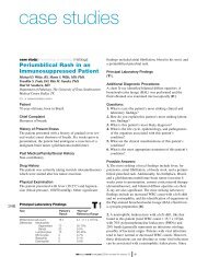
<strong>Patient</strong>’s Forward and Reverse <strong>Blood</strong> <strong>Group<strong>in</strong>g</strong> and Rh Test<strong>in</strong>g <strong>Results</strong><br />
laboratorymedic<strong>in</strong>e> september 2004> number 9> volume 35<br />
Questions:<br />
1. What is (are) this patient’s most strik<strong>in</strong>g laboratory<br />
result(s)?<br />
2. How do you expla<strong>in</strong> this patient’s most strik<strong>in</strong>g laboratory<br />
result(s)?<br />
3. What is this patient’s blood group and Rh type?<br />
4. What other tests can be done to confirm this patient’s<br />
blood group and the presence of hypogammaglobul<strong>in</strong>emia?<br />
5. What are other possible causes of ABO discrepancies?<br />
Possible Answers:<br />
1. The discrepancy between forward (group O) and reverse<br />
(group AB) ABO blood typ<strong>in</strong>g results [T1];<br />
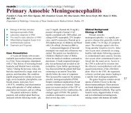
decreased globul<strong>in</strong>s; mildly <strong>in</strong>creased WBC count;<br />
markedly decreased platelet count; and modestly<br />
<strong>in</strong>creased blood urea nitrogen (BUN) concentration<br />
[T2]. The failure of expected agglut<strong>in</strong>ation reactions to<br />
occur dur<strong>in</strong>g forward and reverse typ<strong>in</strong>g is called an<br />
Forward <strong>Group<strong>in</strong>g</strong> Rh <strong>Group<strong>in</strong>g</strong> Reverse <strong>Group<strong>in</strong>g</strong><br />
Reaction Conditions Anti-A Anti-B Anti-D D u Rh control A 1 Cells B Cells<br />
Immediate sp<strong>in</strong> 0 0 0 0 0 0 0<br />
Incubation 5 m<strong>in</strong>, RT 0 0<br />
Incubation 10 m<strong>in</strong>, 3°C 0 0<br />
Incubation 60 m<strong>in</strong>, RT vw+ vw+<br />
RT, room temperature; 0, no reaction; vw+, very weakly positive<br />
Pr<strong>in</strong>cipal Laboratory F<strong>in</strong>d<strong>in</strong>gs<br />
Test <strong>Patient</strong>’s Result “Normal”<br />
Reference Range<br />
Hematology<br />
WBC count 12.1 4.0-10.8 x10 3 /µL<br />
Hemoglob<strong>in</strong> 14.3 14.0-18.0 g/dL<br />
Hematocrit 42.9 42.0-52.0%<br />
MCV 94.8 80-94 fL<br />
Platelet count 33 150-450 x10 3 /µL<br />
Differential<br />
Neutrophils 69 42-75%<br />
Bands 30 0-5%<br />
Monocytes 1 5-10%<br />
Chemistry<br />
Sodium 139 133-145 mEq/L<br />
Potassium 4.2 3.3-5.1 mEq/L<br />
Chloride 102 96-108 mEq/L<br />
Bicarbonate 23 22-29 mmol/L<br />
BUN 20 6-19 mg/dL<br />
Creat<strong>in</strong><strong>in</strong>e 0.9 0.5-1.2 mg/dL<br />
Glucose 90 70-110 mg/dL<br />
Total prote<strong>in</strong> 6.1 5.9-8.4 g/dL<br />
Album<strong>in</strong> 4.7 3.2-5.2 g/dL<br />
Globul<strong>in</strong>s * 1.4 2.5-3.5 g/dL<br />
<strong>Blood</strong> Bank<br />
Antibody screen Neg Neg<br />
Direct coombs test Neg Neg<br />
* Calculated from total prote<strong>in</strong> concentration – album<strong>in</strong> concentration<br />
WBC, white blood cell; MCV, mean cell volume; BUN, blood urea nitrogen<br />
©<br />
T2<br />
T1
“ABO discrepancy.” Discrepant forward and reverse ABO<br />
group<strong>in</strong>g results should be brought to the attention of the<br />
blood bank director and reviewed <strong>in</strong> tandem <strong>with</strong> the patient’s<br />
cl<strong>in</strong>ical history, which <strong>in</strong> this case, <strong>in</strong>dicated the<br />
diagnosis, chronic lymphocytic leukemia (CLL). To enhance<br />
the reverse group reaction, the tubes were <strong>in</strong>cubated<br />
at room temperature for 5 m<strong>in</strong>utes, and at 3ºC for 10 m<strong>in</strong>utes.<br />
The results <strong>in</strong> all 4 tubes were still negative [T1]. Repeat<br />
test<strong>in</strong>g provided the same results. While not standard<br />
procedure, the reverse group typ<strong>in</strong>g was repeated after a<br />
60 m<strong>in</strong>ute <strong>in</strong>cubation at room temperature, and the results<br />
were very weakly positive for both A 1 and B reagent<br />
RBCs [T1].<br />
2. Hypogammaglobul<strong>in</strong>emia is known to occur <strong>in</strong> patients<br />
<strong>with</strong> CLL as a result of both leukemia-related immune<br />
dysfunction and therapy-related immunosuppression. The<br />
deficiency <strong>in</strong> humoral immunity is largely responsible for<br />
CLL patients’ <strong>in</strong>creased risk of <strong>in</strong>fection-related morbidity<br />
and mortality, especially by encapsulated microorganisms. 1<br />
The overall decrease <strong>in</strong> antibody production that occurs<br />
dur<strong>in</strong>g immunosuppression also causes a decrease <strong>in</strong> ABO<br />
antibody levels. Weak, or miss<strong>in</strong>g, ABO antibodies due to<br />
CLL-related hypogammaglobul<strong>in</strong>emia are the most logical<br />
explanation for the failure of the patient’s serum to react<br />
aga<strong>in</strong>st the A 1 and B cells at immediate sp<strong>in</strong>. Moreover,<br />
hypogammaglobul<strong>in</strong>emia follow<strong>in</strong>g rituximab therapy has<br />
been reported previously. 2-4 In each of these reports, however,<br />
the disease be<strong>in</strong>g treated was post-transplant lymphoproliferative<br />
disorder not CLL. Nevertheless, it is<br />
possible that rituximab therapy contributed to the development<br />
of hypogammaglobul<strong>in</strong>emia <strong>in</strong> the CLL patient. The<br />
decreased globul<strong>in</strong> value may also be a manifestation of<br />
the patient’s hypogammaglobul<strong>in</strong>emia. An elevated WBC<br />
count may be related to a variety of conditions, <strong>in</strong>clud<strong>in</strong>g<br />
<strong>in</strong>fection, stress (physical or emotional), and hematologic<br />
disorders. Thrombocytopenia may result from decreased<br />
production or <strong>in</strong>creased consumption of platelets caused<br />
by immune- and nonimmune-mediated mechanisms. In<br />
this patient <strong>with</strong> marked splenomegaly, <strong>in</strong>creased platelet<br />
sequestration by the spleen is the most likely cause of<br />
thrombocytopenia. Changes <strong>in</strong> <strong>in</strong>travascular volume due<br />
to hypogammaglobul<strong>in</strong>emia is the most likely cause of<br />
this patient’s slightly <strong>in</strong>creased blood urea nitrogen (BUN)<br />
concentration.<br />
3. Group O, Rh-negative.<br />
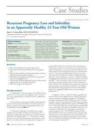
4. To confirm the patient’s blood type, additional cell typ<strong>in</strong>g<br />
can be done to rule out the presence of an <strong>in</strong>hibitory<br />
substance <strong>in</strong> the patient’s blood that could cause all reactions<br />
to appear negative. When we tested the patient’s<br />
RBCs us<strong>in</strong>g anti-C, anti-E, anti-c, and anti-e antisera, his<br />
RBCs were found to have both c and e antigens, thus rul<strong>in</strong>g<br />
out the presence of a global <strong>in</strong>hibitor <strong>in</strong> his blood<br />
[T3]. The patient’s Rh phenotype is rr, the most common<br />
phenotype found <strong>in</strong> Rh-negative white (Caucasian) blood<br />
©<br />
donors. 5 To confirm and characterize this patient’s<br />
hypogammaglobul<strong>in</strong>emia, serum prote<strong>in</strong> electrophoresis<br />
and/or quantification of serum levels of the 3 pr<strong>in</strong>cipal<br />
serum immunoglobul<strong>in</strong>s, IgA, IgG, and IgM, could be<br />
performed. Quantification of these immunoglobul<strong>in</strong>s <strong>in</strong><br />
our patient’s serum <strong>in</strong>dicated a marked decrease <strong>in</strong> IgA<br />
[ number 9> volume 35<br />
C E c e<br />
<strong>Patient</strong>’s RBCs 0 0 4+ 3+<br />
Positive control 3+ 2+ 4+ 2+<br />
Negative control 0 0 0 0<br />
0, no reaction<br />
T3<br />
539
540<br />
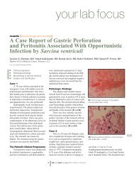
Possible Causes of <strong>Aberrant</strong> <strong>Blood</strong> Group Typ<strong>in</strong>g Reactions*<br />
Factors Affect<strong>in</strong>g Typ<strong>in</strong>g Reaction<br />
Type of Forward Reverse<br />
<strong>Aberrant</strong><br />
Result<br />
Positive Acquired B antigen <strong>in</strong> patients <strong>with</strong>:<br />
gastric or colon cancers<br />
<strong>in</strong>test<strong>in</strong>al obstructions<br />
other conditions (eg, <strong>in</strong>flammatory bowel disease; UTI <strong>with</strong><br />
E. coli O86; pancreatic cancer, primary and metastatic)<br />
Rouleaux formation due to:<br />
elevated globul<strong>in</strong><br />
elevated fibr<strong>in</strong>ogen<br />
Wharton’s jelly<br />
dextran/plasma expanders<br />
Polyagglut<strong>in</strong>ation:<br />
microbial-associated (ie, T polyagglut<strong>in</strong>ation)<br />
nonmicrobial-associated (ie, Tn activation)<br />
Autoagglut<strong>in</strong>ation due to:<br />
potent cold autoantibodies<br />
Antibody-coated RBCs due to:<br />
warm autoantibodies<br />
transfusion reaction<br />
Acriflav<strong>in</strong> antibodies<br />
Negative Weak A or B subgroups caus<strong>in</strong>g weak or mixed field<br />
agglut<strong>in</strong>ation reactions or no reaction<br />
Excess blood group-specific soluble substances <strong>in</strong> blood<br />
samples from patients’ <strong>with</strong>:<br />
gastric cancer<br />
pancreatic cancer<br />
muc<strong>in</strong>ous ovarian cysts<br />
Antigen depression due to disease<br />
*Exclud<strong>in</strong>g technical and human error. UTI, ur<strong>in</strong>ary tract <strong>in</strong>fection<br />
repeat<strong>in</strong>g the test on the same sample suspended <strong>in</strong> sal<strong>in</strong>e<br />
may resolve the discrepancy by wash<strong>in</strong>g away the excess<br />
BGSS. 7 Excess BGSS has been associated <strong>with</strong> carc<strong>in</strong>oma<br />
of the stomach and pancreas. 7 There is, however, at least 1<br />
case where excess BGSS was identified <strong>in</strong> the serum, cyst<br />
fluid, and ascites of a woman <strong>with</strong> muc<strong>in</strong>ous cystadenoma<br />
of the ovary. 8 After surgical resection of the cyst, the<br />
serum BGSS returned to a normal level. Weak antigens<br />
are less common than weak or miss<strong>in</strong>g antibodies, but<br />
when encountered, they too may cause aberrant typ<strong>in</strong>g<br />
reactions. Weakly react<strong>in</strong>g antigens that may be present<br />
<strong>in</strong>clude subgroups of A and/or B. Group A is subdivided<br />
<strong>in</strong>to A 1 and A 2 phenotypes, which together represent approximately<br />
99% of group A <strong>in</strong>dividuals, and several rare<br />
A phenotypes, A 3 , A x , A end , A m , A y , and A el , which comprise<br />
the rema<strong>in</strong><strong>in</strong>g 1%. The rare A subgroups may yield<br />
negative, weak, or mixed-field agglut<strong>in</strong>ation reactions<br />
upon test<strong>in</strong>g <strong>with</strong> anti-A, putt<strong>in</strong>g the patient at risk of<br />
be<strong>in</strong>g mistyped as group O. Similarly, B subgroups B 3 ,<br />
B x , B m , and B el demonstrate variable reactivity to anti-B.<br />
The presence of unexpected antibodies may also produce<br />
confus<strong>in</strong>g results. For example, anti-A 1 may be present <strong>in</strong><br />
laboratorymedic<strong>in</strong>e> september 2004> number 9> volume 35<br />
Unexpected antibody present <strong>in</strong> blood group<strong>in</strong>g reagent that<br />
may react <strong>with</strong> an unknown, unexpected patient antigen<br />
Genetic chimerism (cis-AB) (rare)<br />
Unexpected ABO antibody <strong>in</strong> patient’s serum such as:<br />
anti-A1 <strong>in</strong> A subgroups (<strong>in</strong>clud<strong>in</strong>g A2B)<br />
anti-B <strong>in</strong> cis-AB <strong>in</strong>dividuals<br />
anti-H <strong>in</strong> A1 and A1B <strong>in</strong>dividuals<br />
Rouleaux formation due to:<br />
elevated globul<strong>in</strong><br />
elevated fibr<strong>in</strong>ogen<br />
Wharton’s jelly<br />
dextran/plasma expanders<br />
Unexpected alloantibody <strong>in</strong> the patient’s serum that may<br />
react <strong>with</strong> an unknown, unexpected reagent RBC<br />
antigen<br />
Cold reactive allo- or auto-antibodies that may agglut<strong>in</strong>ate<br />
reagent red blood cells<br />
Weak or miss<strong>in</strong>g antibodies <strong>in</strong> patients <strong>with</strong>:<br />
hypogammaglobul<strong>in</strong>emia<br />
newborn or elderly<br />
immunodeficiency<br />
immunosuppression<br />
congenital deficiency (agammaglobul<strong>in</strong>emia)<br />
Genetic chimerism (cis-AB) (rare)<br />
up to 8% of blood group A 2 <strong>in</strong>dividuals and <strong>in</strong> 22% to<br />
35% of blood group A 2 B <strong>in</strong>dividuals, 6 caus<strong>in</strong>g a positive<br />
reaction to A 1 cells dur<strong>in</strong>g reverse typ<strong>in</strong>g. This discrepancy<br />
may be resolved by repeat<strong>in</strong>g the forward typ<strong>in</strong>g<br />
us<strong>in</strong>g anti-A 1 lect<strong>in</strong> reagent <strong>in</strong>stead of the usual anti-A<br />
reagent. Group A 1 cells will react <strong>with</strong> both anti-A 1 lect<strong>in</strong><br />
and anti-A, but group A 2 cells will only react <strong>with</strong> anti-A.<br />
A negative reaction <strong>with</strong> anti-A 1 will confirm the suspicion<br />
that one is most likely work<strong>in</strong>g <strong>with</strong> group A 2 cells.<br />
In addition to blood group A 2 , some of the rare A<br />
subgroups can also produce anti-A 1 . If a positive reaction<br />
to A 1 cells is observed on reverse typ<strong>in</strong>g and this f<strong>in</strong>d<strong>in</strong>g<br />
is coupled <strong>with</strong> an absent or weak reaction to anti-A on<br />
forward typ<strong>in</strong>g, a patient will erroneously appear to be<br />
group O. F<strong>in</strong>ally, an unusual case has been reported <strong>in</strong><br />
which a transfusion-naïve blood group A 1 <strong>in</strong>dividual produced<br />
anti-A 1 autoantibodies. 9 Acquired B antigens are<br />
another important, although uncommon, cause of ABO<br />
discrepancies. The B antigen is acquired as a result of<br />
bacterial enzymatic activity that modifies N-acetylgalactosam<strong>in</strong>e,<br />
the immunodom<strong>in</strong>ant sugar on A red blood<br />
cells, to D-galactosam<strong>in</strong>e. D-galactosam<strong>in</strong>e is similar<br />
©<br />
T4
enough to D-galactose, the immunodom<strong>in</strong>ant sugar of B<br />
red blood cells, to cause reactivity <strong>with</strong> most anti-B<br />
reagents, but not <strong>with</strong> autologous anti-B. Acquired B antigens<br />
are most commonly seen <strong>in</strong> conditions where the<br />
breakdown of normal bowel mucosa allows bacterial enzymes<br />
to enter the systemic circulation. The underly<strong>in</strong>g<br />
disease state is often gastric cancer, 10 colon cancer, 11 or<br />
<strong>in</strong>test<strong>in</strong>al obstruction, 7 but the acquired B phenomenon<br />
has been reported <strong>in</strong> patients <strong>with</strong> <strong>in</strong>flammatory bowel<br />
disease, 12 ur<strong>in</strong>ary tract <strong>in</strong>fection <strong>with</strong> E. coli O86, 13 and<br />
pancreatic cancer 14 as well. ABO typ<strong>in</strong>g reactions may be<br />
mis<strong>in</strong>terpreted as positive <strong>in</strong> the presence of rouleaux formation<br />
or polyagglut<strong>in</strong>ation. Rouleaux may be attributable<br />
to conditions <strong>in</strong> which there are elevated levels of<br />
globul<strong>in</strong>, such as multiple myeloma, elevated levels of fibr<strong>in</strong>ogen,<br />
or <strong>in</strong> the presence of dextran, other plasma expanders,<br />
or Wharton’s jelly. 7 In case of the latter,<br />
repeat<strong>in</strong>g the tests on the same sample after wash<strong>in</strong>g the<br />
patient’s RBCs <strong>with</strong> sal<strong>in</strong>e may resolve the discrepancy by<br />
remov<strong>in</strong>g the Wharton’s jelly. Polyagglut<strong>in</strong>ation is<br />
caused by an alteration <strong>in</strong> the RBC membrane that enables<br />
agglut<strong>in</strong>ation to occur <strong>in</strong> the presence of most ABO-compatible<br />
sera. Alteration of the RBC membrane may occur<br />
by microbial- or nonmicrobial-associated methods. In either<br />
case, normal carbohydrate residues are removed from<br />
the RBC membrane caus<strong>in</strong>g exposure of what are called<br />
hidden, or “cryptantigens.” Cryptantigens then react to<br />
naturally occurr<strong>in</strong>g IgM antibodies <strong>in</strong> the patients’ serum<br />
and cause agglut<strong>in</strong>ation. 15 Cold autoantibodies may be<br />
potent enough to cause agglut<strong>in</strong>ation of the patient’s<br />
RBCs used <strong>in</strong> forward group<strong>in</strong>g, as well as the reagent<br />
cells used <strong>in</strong> reverse group<strong>in</strong>g. To resolve the forward<br />
group<strong>in</strong>g problem, the patient’s RBCs could be <strong>in</strong>cubated<br />
at 37ºC, then washed <strong>with</strong> sal<strong>in</strong>e that has also been<br />
warmed to 37ºC. If warm<strong>in</strong>g methods are unsuccessful,<br />
dithiothreitol (DTT) can be used to remove IgM-related<br />
agglut<strong>in</strong>ation. 6 For reverse-group<strong>in</strong>g discrepancies, the<br />
reagent RBCs may need to be warmed to 37ºC before<br />
mix<strong>in</strong>g <strong>with</strong> the patient’s serum. If IgM-related agglut<strong>in</strong>ation<br />
still occurs, adsorption may be required to remove<br />
IgM antibody from the serum. Warm autoantibodies may<br />
also cause ABO discrepancies; however, they typically<br />
yield weaker reactions than those observed <strong>with</strong> cold antibodies.<br />
When warm autoantibodies are suspected to be the<br />
cause of false-positive results <strong>in</strong> forward group<strong>in</strong>g, elution<br />
may be required to remove bound immunoglobul<strong>in</strong> from<br />
the RBCs. Last, RBCs may become coated <strong>with</strong> warm antibodies<br />
after a transfusion reaction, secondary to the use<br />
of certa<strong>in</strong> drugs such as alpha-methyldopa, or <strong>in</strong> patients<br />
<strong>with</strong> warm autoimmune hemolytic anemia. 7<br />
©<br />
<strong>Patient</strong> Outcome<br />
Given our patient’s profound hypogammaglobul<strong>in</strong>emia,<br />
splenectomy was postponed so that he could first receive<br />
replacement doses of <strong>in</strong>travenous immunoglobul<strong>in</strong> (IvIg).<br />
Later, he underwent splenectomy <strong>with</strong> removal of a 1,550gram<br />
spleen, <strong>with</strong>out complication. It showed no<br />
evidence of CLL by histologic exam<strong>in</strong>ation or flow<br />
cytometry. The patient’s platelet count recovered;<br />
however, he rema<strong>in</strong>ed hypogammaglobul<strong>in</strong>emic.<br />
Keywords: ABO discrepancy, hypogammaglobul<strong>in</strong>emia,<br />
chronic lymphocytic leukemia, forward group<strong>in</strong>g, reverse<br />
group<strong>in</strong>g<br />
1. Tsiodras S, Samonis G, Keat<strong>in</strong>g MJ, et al. Infection and immunity <strong>in</strong><br />
chronic lymphocytic leukemia. Mayo Cl<strong>in</strong>ic Proceed<strong>in</strong>gs. 2000;75:1039-<br />
1054.<br />
2. Imashuku S, Teramura T, Morimoto A, et al. Prolonged<br />
hypogammaglobul<strong>in</strong>emia follow<strong>in</strong>g rituximab treatment for post transplant<br />
Epste<strong>in</strong>-Barr virus-associated lymphoproliferative disease. Bone Marrow<br />
Transplantation. 2004;33:129-130.<br />
3. Castagnola E, Dallorso S, Faraci M, et al. Long-last<strong>in</strong>g<br />
hypogammaglobul<strong>in</strong>emia follow<strong>in</strong>g rituximab adm<strong>in</strong>istration for Epste<strong>in</strong>-<br />
Barr virus-related post-transplant lymphoproliferative disease preemptive<br />
therapy. J Hematotherapy & Stem Cell Research. 2003;12:9-10.<br />
4. Verschuuren EA, Stevens SJ, van Imhoff GW, et al. Treatment of<br />
posttransplant lymphoproliferative disease <strong>with</strong> rituximab: The remission,<br />
the relapse, and the complication. Transplantation. 2002;73:100-104.<br />
5. Beadl<strong>in</strong>g WV, Cool<strong>in</strong>g L, Henry JB. Immunohematology. In: Henry JB.<br />
Cl<strong>in</strong>ical Diagnosis and Management by Laboratory Methods. Philadelphia:<br />
W.B. Saunders; 2001:660-717.<br />
6. ABO, H, and Lewis blood groups and structurally related antigens. In:<br />
Brecher ME, Ed. AABB Technical Manual. 14 th ed. 2002;271-293.<br />
7. Harmen<strong>in</strong>g DM, Firestone D. The ABO blood group system. In: Harmen<strong>in</strong>g<br />
DM. Modern <strong>Blood</strong> Bank<strong>in</strong>g and Transfusion Practices. Philadelphia: F.A.<br />
Davis; 1999:90-125.<br />
8. Kom<strong>in</strong>ato Y, Fujikura T, Takizawa H, et al. Investigation of blood group A<br />
substance <strong>in</strong> the circulation of a patient <strong>with</strong> ovarian cyst. Experimental &<br />
Cl<strong>in</strong>ical Immunogenetics. 1991;8:24-28.<br />
9. Rogers VB, Reid ME, Ellisor SS, et al. Auto-anti-A1: Another cause of<br />
ABO discrepancy. Transfusion. 1981;21:92-95.<br />
10. Beadl<strong>in</strong>g WV, Cool<strong>in</strong>g L, Henry JB. Immunohematology. In: Henry JB.<br />
Cl<strong>in</strong>ical Diagnosis and Management by Laboratory Methods. Philadelphia:<br />
W.B. Saunders; 2001:660-717.<br />
11. Northoff H, Wolpl A, Bewersdorf H, et al. An ABO-blood group<br />
abnormality lead<strong>in</strong>g to the detection of a colon-carc<strong>in</strong>oma. Blut.<br />
1983;46:161-164.<br />
12. Roth S, Todd CE, Shaw D. Transient acquired blood group B antigen<br />
associated <strong>with</strong> diverticular bowel disease. Acta Haematologica.<br />
1987;77:188-190.<br />
13. Vojvodic S. Acquired B antigen <strong>in</strong> a pregnant woman belong<strong>in</strong>g to the A1<br />
blood group: Case report. Medic<strong>in</strong>ski Pregled. 2001;54:490-492.<br />
14. Itzkowitz SH, Yuan M, Ferrell LD, et al. Cancer-associated alterations of<br />
blood group antigen expression <strong>in</strong> the human pancreas. J Nat’l Cancer Inst.<br />
1987;79:425-434.<br />
15. Walker PS. Polyagglut<strong>in</strong>ation. In: Harmen<strong>in</strong>g DM. Modern <strong>Blood</strong> Bank<strong>in</strong>g<br />
and Transfusion Practices. Philadelphia: F.A. Davis; 1999:474-488.<br />
laboratorymedic<strong>in</strong>e> september 2004> number 9> volume 35<br />
541