Henry Schein MENA AEEDC Brochure 2023

You also want an ePaper? Increase the reach of your titles
YUMPU automatically turns print PDFs into web optimized ePapers that Google loves.
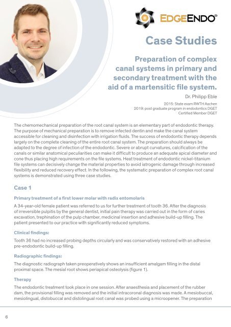
Case Studies<br />
Preparation of complex<br />
canal systems in primary and<br />
secondary treatment with the<br />
aid of a martensitic file system.<br />
Dr. Philipp Eble<br />
2015: State exam RWTH Aachen<br />
2019: post graduate program in endodontics DGET<br />
Certified Member DGET<br />
of the primary access cavity for better accessibility of the canals was carried out with long-neck carbide<br />
round bur. Based on the preoperative diagnostic X-ray, the length of the root canals could be preliminarily<br />
approximated. The canals were continuously rinsed with 6% NaOCl during the further course of therapy.<br />
After preparation of the access cavity, coronal expansion of the root canals followed using EdgeEndo X7<br />
files size 17.06. Electrometric determination of the canal length using a Morita Root ZX Mini Apex Locator<br />
was performed with C-Pilots size 8-10. After the working length was determined, the glide path was<br />
rotationally extended with EdgeFile X7 size 17.04 and 25.04 and finally prepared to 30.04 (Figure 2).<br />
The preparation was followed by a rinse with 17% EDTA for 60 seconds per canal, followed by the final<br />
sound-activated rinse with 6% NaOCl for 60 seconds per canal. The preparation and the fit of<br />
the congruent EdgeEndo X7 gutta-percha tips were confirmed with the help of a master point image<br />
(Figure 4). After drying the canals and access cavity with microsuction and paper tips, the obturation<br />
of the canal system followed using the warm vertical compaction technique. A heat-resistant bioceramic<br />
sealer was used for this purpose (figure 3). The subsequent closure was done with a bulk fill flow<br />
composite (figure 5).<br />
The chemomechanical preparation of the root canal system is an elementary part of endodontic therapy.<br />
The purpose of mechanical preparation is to remove infected dentin and make the canal system<br />
accessible for cleaning and disinfection with irrigation fluids. The success of endodontic therapy depends<br />
largely on the complete cleaning of the entire root canal system. The preparation should always be<br />
adapted to the degree of infection of the endodontic. Severe or abrupt curvatures, calcification of the<br />
canals or similar anatomical peculiarities can make it difficult to produce an adequate apical diameter and<br />
cone thus placing high requirements on the file systems. Heat treatment of endodontic nickel-titanium<br />
file systems can decisively change the material properties to avoid iatrogenic damage through increased<br />
flexibility and reduced recovery effect. In the following, the systematic preparation of complex root canal<br />
systems is demonstrated using three case studies.<br />
Fig 1<br />
Fig 2<br />
Fig 3<br />
Case 1<br />
Primary treatment of a first lower molar with radix entomolaris<br />
A 34-year-old female patient was referred to us for further treatment of tooth 36. After the diagnosis<br />
of irreversible pulpitis by the general dentist, initial pain therapy was carried out in the form of caries<br />
excavation, trephination of the pulp chamber, medicinal insertion and adhesive build-up filling. The<br />
patient presented to our practice with significantly reduced symptoms.<br />
Clinical findings:<br />
Tooth 36 had no increased probing depths circularly and was conservatively restored with an adhesive<br />
pre-endodontic build-up filling.<br />
Radiographic findings:<br />
Fig 4<br />
Fig 5<br />
Fig 1: Preoperative diagnostic image<br />
Fig 2: View of the mesial canal system after preparation<br />
Fig 3: View after obturation<br />
Fig 4: Masterpoint image<br />
Fig 5: After root filling and adhesive closure<br />
The diagnostic radiograph taken preoperatively shows an insufficient amalgam filling in the distal<br />
proximal space. The mesial root shows periapical osteolysis (figure 1).<br />
Therapy<br />
The endodontic treatment took place in one session. After anaesthesia and placement of the rubber<br />
dam, the provisional filling was removed and the initial intracoronal diagnosis was made. A mesiobuccal,<br />
mesiolingual, distobuccal and distolingual root canal was probed using a microopener. The preparation<br />
6 7