Mikrobiologische Reinigung und Desinfektion Wirksamkeit einer dreistufigen Ultraschallbearbeitung Protokoll für CAD-CAM-Implantataufbauten
- No tags were found...

You also want an ePaper? Increase the reach of your titles
YUMPU automatically turns print PDFs into web optimized ePapers that Google loves.
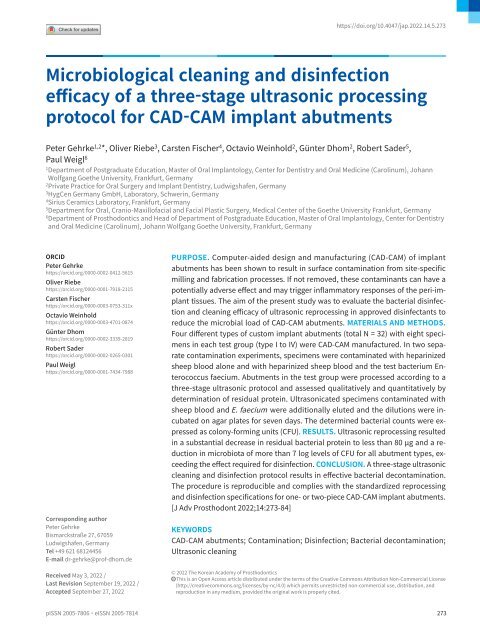
https://doi.org/10.4047/jap.2022.14.5.273<br />
Microbiological cleaning and disinfection<br />
efficacy of a three-stage ultrasonic processing<br />
protocol for <strong>CAD</strong>-<strong>CAM</strong> implant abutments<br />
Peter Gehrke 1,2 *, Oliver Riebe 3 , Carsten Fischer 4 , Octavio Weinhold 2 , Günter Dhom 2 , Robert Sader 5 ,<br />
Paul Weigl 6<br />
1<br />
Department of Postgraduate Education, Master of Oral Implantology, Center for Dentistry and Oral Medicine (Carolinum), Johann<br />
Wolfgang Goethe University, Frankfurt, Germany<br />
2<br />
Private Practice for Oral Surgery and Implant Dentistry, Ludwigshafen, Germany<br />
3<br />
HygCen Germany GmbH, Laboratory, Schwerin, Germany<br />
4<br />
Sirius Ceramics Laboratory, Frankfurt, Germany<br />
5<br />
Department for Oral, Cranio-Maxillofacial and Facial Plastic Surgery, Medical Center of the Goethe University Frankfurt, Germany<br />
6<br />
Department of Prosthodontics and Head of Department of Postgraduate Education, Master of Oral Implantology, Center for Dentistry<br />
and Oral Medicine (Carolinum), Johann Wolfgang Goethe University, Frankfurt, Germany<br />
ORCID<br />
Peter Gehrke<br />
https://orcid.org/0000-0002-0412-5615<br />
Oliver Riebe<br />
https://orcid.org/0000-0001-7918-2115<br />
Carsten Fischer<br />
https://orcid.org/0000-0003-0753-311x<br />
Octavio Weinhold<br />
https://orcid.org/0000-0003-4701-0874<br />
Günter Dhom<br />
https://orcid.org/0000-0002-3339-2819<br />
Robert Sader<br />
https://orcid.org/0000-0002-0265-0301<br />
Paul Weigl<br />
https://orcid.org/0000-0001-7434-7988<br />
Corresponding author<br />
Peter Gehrke<br />
Bismarckstraße 27, 67059<br />
Ludwigshafen, Germany<br />
Tel +49 621 68124456<br />
E-mail dr-gehrke@prof-dhom.de<br />
Received May 3, 2022 /<br />
Last Revision September 19, 2022 /<br />
Accepted September 27, 2022<br />
PURPOSE. Computer-aided design and manufacturing (<strong>CAD</strong>-<strong>CAM</strong>) of implant<br />
abutments has been shown to result in surface contamination from site-specific<br />
milling and fabrication processes. If not removed, these contaminants can have a<br />
potentially adverse effect and may trigger inflammatory responses of the peri-implant<br />
tissues. The aim of the present study was to evaluate the bacterial disinfection<br />
and cleaning efficacy of ultrasonic reprocessing in approved disinfectants to<br />
reduce the microbial load of <strong>CAD</strong>-<strong>CAM</strong> abutments. MATERIALS AND METHODS.<br />
Four different types of custom implant abutments (total N = 32) with eight specimens<br />
in each test group (type I to IV) were <strong>CAD</strong>-<strong>CAM</strong> manufactured. In two separate<br />
contamination experiments, specimens were contaminated with heparinized<br />
sheep blood alone and with heparinized sheep blood and the test bacterium Enterococcus<br />
faecium. Abutments in the test group were processed according to a<br />
three-stage ultrasonic protocol and assessed qualitatively and quantitatively by<br />
determination of residual protein. Ultrasonicated specimens contaminated with<br />
sheep blood and E. faecium were additionally eluted and the dilutions were incubated<br />
on agar plates for seven days. The determined bacterial counts were expressed<br />
as colony-forming units (CFU). RESULTS. Ultrasonic reprocessing resulted<br />
in a substantial decrease in residual bacterial protein to less than 80 µg and a reduction<br />
in microbiota of more than 7 log levels of CFU for all abutment types, exceeding<br />
the effect required for disinfection. CONCLUSION. A three-stage ultrasonic<br />
cleaning and disinfection protocol results in effective bacterial decontamination.<br />
The procedure is reproducible and complies with the standardized reprocessing<br />
and disinfection specifications for one- or two-piece <strong>CAD</strong>-<strong>CAM</strong> implant abutments.<br />
[J Adv Prosthodont 2022;14:273-84]<br />
KEYWORDS<br />
<strong>CAD</strong>-<strong>CAM</strong> abutments; Contamination; Disinfection; Bacterial decontamination;<br />
Ultrasonic cleaning<br />
© 2022 The Korean Academy of Prosthodontics<br />
cc This is an Open Access article distributed <strong>und</strong>er the terms of the Creative Commons Attribution Non-Commercial License<br />
(http://creativecommons.org/licenses/by-nc/4.0) which permits unrestricted non-commercial use, distribution, and<br />
reproduction in any medium, provided the original work is properly cited.<br />
pISSN 2005-7806 · eISSN 2005-7814<br />
273
The Journal of Advanced Prosthodontics<br />
INTRODUCTION<br />
Dental implant abutments are part of the prosthetic<br />
superstructure with direct contact with the surro<strong>und</strong>ing<br />
oral tissues. In addition to prefabricated<br />
stock abutments, computer-aided design and manufacturing<br />
(<strong>CAD</strong>-<strong>CAM</strong>) enables the fabrication of customized<br />
abutments. 1 They facilitate the compensation<br />
of axial divergences between the implant and the<br />
corresponding crown as well as the individualization<br />
of the abutment shoulder with an anatomical emergence<br />
profile for implant-supported single-tooth restorations.<br />
2,3 The peri-implant mucosa is commonly<br />
known as hypovascular and hypocellular scar tissue.<br />
Immunologically, it is inferior to the periodontal tissue<br />
aro<strong>und</strong> teeth, as it exhibits lower resistance to<br />
bacterial induced infections. 4 Surface properties of<br />
the abutment such as topography, roughness, hydrophilicity,<br />
surface energy, contaminants, and macromolecular<br />
conditioning affect the biological response<br />
at the hard- and soft tissue interface in multiple<br />
ways. 5 Hence, recurrent hazards with potential negative<br />
impact on the attachment of the peri-implant<br />
tissues should be prevented. 6,7 <strong>CAD</strong>-<strong>CAM</strong> production<br />
for custom abutments have been proven to result in<br />
surface contamination from microwear particulates,<br />
cooling lubricants, and general laboratory debris. 8,9<br />
Analysis revealed contamination on their outer and<br />
inner surfaces following laboratory procedures. 10,11<br />
The presence of these micro-residues at the critical<br />
abutment-tissue junction may provoke inflammatory<br />
responses of the peri-implant tissues and mechanically<br />
compromise the stability of the implant-abutment<br />
junction. 12 Cleaning and subsequent disinfection<br />
of the abutment surface is therefore mandatory.<br />
European health regulations and the guidelines of the<br />
American Dental Association (ADA) both approved<br />
cleaning and disinfection regimens for semi-critical<br />
medical devices, including <strong>CAD</strong>-<strong>CAM</strong> implant abutments.<br />
13,14 This refers to either dry heat sterilization,<br />
steam treatment of the components <strong>und</strong>er pressure<br />
at 134°C (autoclaving) or ultrasonic cleaning by<br />
means of approved disinfectants. While heat-stable<br />
metallic implant abutments (e.g. titanium or titanium<br />
nitride) can be safely autoclaved without compromising<br />
their material properties, 15 sterilization of monohttps://doi.org/10.4047/jap.2022.14.5.273<br />
type ceramic or hybrid abutments <strong>und</strong>er moist heat<br />
and pressure may lead to permanent damage to the<br />
crystal-ceramic framework (degradation) or to the<br />
adhesive bond of hybrid abutments and is therefore<br />
controversially discussed. 16-19 In addition, it should<br />
be noted that the physical process of sterilization by<br />
autoclaving using a combination of appropriate heat<br />
and pressure is able to kill all viable forms of microbiota,<br />
but it cannot effectively remove particulate debris<br />
from <strong>CAD</strong>-<strong>CAM</strong> abutments. The reprocessing of<br />
implant abutments by steam cleaning in the laboratory<br />
(vaporization), although frequently employed,<br />
is an ineffective method and fails to achieve the normatively<br />
required efficacy of disinfection (DIN EN<br />
14885:2018). 20,21 In addition to cleanliness, the surface<br />
topography within the submucosal region of the<br />
abutment structure should be considered. Roughness,<br />
wettability and surface energy are important<br />
properties in this context. These parameters exhibit<br />
some correlation, as roughness can strongly influence<br />
wetting behavior. 22 It can be assumed that there is a<br />
critical threshold for surface roughness, where the<br />
accumulation of bacteria and plaques is low, at the<br />
same time best supporting the attachment of fibroblasts<br />
and the adaptation of the peri-implant mucosa.<br />
23 One of the novel cleaning techniques is plasma<br />
pretreatment of implant abutments, which has been<br />
the subject of promising pilot studies. 24,25 However, it<br />
should be considered that this is not a validated cleaning<br />
method for laboratory-fabricated implant prosthetic<br />
components. 14,26 Due to the lack of legal validation<br />
and the fact that plasma devices are not widely<br />
used in dental laboratories or practices, 21 the primary<br />
focus should be on cleaning methods with a realistic<br />
relationship between the technical efforts and costs of<br />
the devices and a reliable cleaning efficacy.<br />
Alternatively, recent investigations have demonstrated<br />
that a validated three-step ultrasonic cleaning<br />
procedure significantly reduces surface contamination<br />
of monotype and hybrid <strong>CAD</strong>-<strong>CAM</strong> abutments without<br />
adversely affecting their tensile bond strength. 9,19 The<br />
abutments are cleaned in three successive ultrasonic<br />
baths, one with an antibacterial cleaning solution, the<br />
other with 80% ethyl alcohol and finally with medically<br />
pure water (Cleaning System; Bredent GmbH<br />
& Co. KG, Senden, Germany) at 30°C for 5 min each,<br />
274 https://jap.or.kr
Microbiological cleaning and disinfection efficacy of a three-stage ultrasonic<br />
processing protocol for <strong>CAD</strong>-<strong>CAM</strong> implant abutments<br />
J Adv Prosthodont 2022;14:273-84<br />
yielding a reduction in deposit particles and organic<br />
and inorganic contaminants. 10 While qualitative and<br />
semi-quantitative effectiveness in removing these<br />
contaminants from <strong>CAD</strong>-<strong>CAM</strong>-fabricated surfaces has<br />
been demonstrated by microscopic and chemical<br />
analyses, 9,10 data on the disinfection efficacy of the<br />
three-step ultrasonic procedure are lacking. Therefore,<br />
the aim of the present study was to evaluate its<br />
potential for ultrasonic disinfection to reduce the microbial<br />
load of <strong>CAD</strong>-<strong>CAM</strong> implant abutments. The null<br />
hypothesis tested was that the three-step ultrasonic<br />
cleaning protocol results in a normatively required reduction<br />
of residual protein ≤ 80 µg per sample 27 and<br />
a decrease of microbiota by at least 5 decadal logarithms<br />
(lg) of colony-forming units (CFU) relative to<br />
the positive control and is therefore suitable for disinfection<br />
of implant abutments.<br />
MATERIALS AND METHODS<br />
The laboratory procedures for sample production<br />
have been described in detail in a previous publication.<br />
10 Briefly summarized, a total of 32 <strong>CAD</strong>-<strong>CAM</strong><br />
implant abutments were virtually constructed (Implant<br />
Studio; 3Shape, Copenhagen, Denmark) and<br />
fabricated (<strong>CAD</strong>Abut F and <strong>CAD</strong>Abut D; BEGO Implant<br />
Systems GmbH & Co. KG, Bremen, Germany). The<br />
master cast of a clinical case involving the replacement<br />
of the right maxillary central incisor with an<br />
implant (Semados SCX D 4.1/L 11.5; BEGO Implant<br />
Systems GmbH & Co. KG, Bremen, Germany) served<br />
as the basis for the digital abutment design. The geometry<br />
of the virtual design had uniform dimensions<br />
of 10.5 mm height and 6.5 mm shoulder width for all<br />
abutment specimens. According to AAMI TIR30:2011/<br />
R2016, 3 test samples per test setup are required. Due<br />
to the validation of the reprocessing efficacy of the<br />
process according to EN ISO 17664, it was decided<br />
to test representative abutments of 4 different types<br />
with a sample size of 3 per setup. The 32 <strong>CAD</strong>-<strong>CAM</strong><br />
abutments were divided into four groups (n = 8 each)<br />
depending on the material used and <strong>CAD</strong>-<strong>CAM</strong> manufacturing<br />
process (Fig. 1): monotype abutments (onepiece)<br />
and hybrid abutments (two-piece). Lab-pro-<br />
<strong>CAD</strong>-<strong>CAM</strong> Implant Abutment N = 32<br />
One-piece zirconia<br />
abutments (type I)<br />
n = 8<br />
Two-piece lithium-disilcate<br />
ti-base abutments (type II)<br />
n = 8<br />
Two-piece zirconia ti-base<br />
abutments (type III)<br />
n = 8<br />
One-piece titanium<br />
abutments (type IV)<br />
n = 8<br />
Assessment of cleaning efficacy<br />
Assessment of disinfection efficacy<br />
Soiling of 4 test specimens of each abutment type I-IV<br />
with hep. sheep blood n = 16<br />
Soiling of 4 test specimens of each abutment type I-IV<br />
with hep. sheep blood & test bacteria (E. faecium) n = 16<br />
Test group I-V: Finevo ultrasonic<br />
cleaning for each test group n = 3<br />
Positive control:<br />
Uncleaned n = 1<br />
Test group I-V: Finevo ultrasonic<br />
cleaning for each test group n = 3<br />
Positive control:<br />
Uncleaned n = 1<br />
Qualitative analysis of cleaning:<br />
Visual inspection for each test group at 400x n = 3<br />
Quantitative analysis of residual protein:<br />
OPA-assay for each test group n = 3 and positive control n = 1<br />
Cultivation & elution of test bacteria:<br />
Census of colony-forming-units (CFU) for each test group<br />
n = 3 and positive control n = 1<br />
Soiling test 2<br />
Soiling test 1<br />
Fig. 1. Study design.<br />
https://jap.or.kr 275
The Journal of Advanced Prosthodontics<br />
https://doi.org/10.4047/jap.2022.14.5.273<br />
cessed materials for monotype abutments included<br />
zirconia and titanium, while hybrid abutments were<br />
comprised of zirconia or lithium disilicate <strong>CAD</strong>-<strong>CAM</strong><br />
copings that were bonded to prefabricated titanium<br />
bases (Ti-Base D 4.1 mm) (Semados SCX; BEGO Implant<br />
Systems GmbH & Co. KG, Bremen, Germany).<br />
Accordingly, the following four material groups were<br />
employed for testing: one-piece zirconia abutments<br />
(type I), two-piece lithium disilicate (LDS) meso-abutments<br />
bonded to titanium bases (type II), two-piece<br />
zirconia meso-abutments bonded to titanium bases<br />
(type III), and one-piece titanium abutments (type IV)<br />
(Fig. 2). A list of materials and manufacturers can be<br />
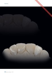
Fig. 2. Samples of the investigated <strong>CAD</strong>-<strong>CAM</strong> abutment<br />
types I-IV from left to right. Type I: zirconia monotype<br />
abutment (<strong>CAD</strong>Abut F, BeCe <strong>CAD</strong> Zirconia XH), type II:<br />
lithium disilicate hybrid abutment (<strong>CAD</strong>Abut D, IPS e.max<br />
<strong>CAD</strong> LT on Ti-Base), type III: zirconia hybrid abutment<br />
(<strong>CAD</strong>Abut D, Zirconia LT <strong>CAD</strong> on Ti-Base), type IV: titanium<br />
monotype abutment (<strong>CAD</strong>Abut F, <strong>CAD</strong> Titanium).<br />
fo<strong>und</strong> in Table 1. The bonding surfaces of the titanium<br />
inserts and the ceramic copings of the two-piece<br />
hybrid abutments were grit-blasted (aluminum oxide<br />
particles 50 μm; 2 bar/0.25 MPa; 10 s; distance 10 mm)<br />
and cleaned with ethanol. Afterwards, the titanium<br />
inserts were moistened with a metal primer (GC Metal<br />
Primer II; GC EUROPE N.V, Leuven, Belgium), while<br />
a bonding agent (Monobond Plus; Ivoclar Vivadent,<br />
Schaan, Liechtenstein) was applied to the basal portion<br />
of the ceramic copings. Each hybrid abutment<br />
was luted with a dimethacrylate/hydroxyethyl methacrylate<br />
(DMA/HEMA)-based cement (Multilink Implant;<br />
Ivoclar Vivadent, Schaan, Liechtenstein) as recommended<br />
by the manufacturer. The excess cement<br />
was removed and the adhesive joint was polished<br />
with silicone polishers and polishing paste according<br />
to a previously documented protocol. 28 Upon completion<br />
of a defined 3-step ultrasonic cleaning protocol,<br />
all test abutment types I-IV were evaluated in terms of<br />
both cleaning and disinfection effectiveness.<br />
To test cleanability, in a first trial a test soil was prepared<br />
from heparinized sheep blood (Fiebig Xebios<br />
Diagnostics GmbH, Düsseldorf, Germany, lot number<br />
31406500/01) diluted 1:5 with 0.85% NaCl and reactivated<br />
with protamine and 1% mucin according to DIN<br />
EN ISO 15883-5 29 and AAMI TIR 30:2011. 30 For contamination,<br />
four test specimens of each abutment type<br />
(type I-IV, total n = 16) were immersed in the test soil<br />
and subsequently dried for one hour at 22 ± 2°C <strong>und</strong>er<br />
laminar room air.<br />
After soiling, three test specimens per abutment<br />
type were subjected to a three-stage ultrasonic cleaning<br />
protocol using ultra-high frequency waves in combination<br />
with disinfecting agents (Finevo Cleaning<br />
Table 1. Type, material, manufacturer, and sample size of tested <strong>CAD</strong>-<strong>CAM</strong> abutments<br />
Abutment type Abutment design Material Product & Manufacturer No. of samples<br />
Type I Monotype Abutment Y-TZP Zirconia<br />
Type II<br />
Type II<br />
Hybrid Abutment<br />
Hybrid Abutment<br />
Lithium Disilicate Coping/<br />
Ti-Base<br />
Y-TZP Zirconia Coping/<br />
Ti-Base<br />
Type IV Monotype Abutment Titanium Grade 5<br />
<strong>CAD</strong>Abut F, BeCe <strong>CAD</strong> Zirconia XH,<br />
Semados SCX, BEGO Implant Systems<br />
<strong>CAD</strong>Abut D, IPS e.max <strong>CAD</strong> LT/ Ivoclar<br />
Vivadent AG on Ti-Base, Semados SCX,<br />
BEGO Implant Systems<br />
<strong>CAD</strong>Abut D, Zirconia LT <strong>CAD</strong> on Ti-Base,<br />
Semados SCX, BEGO Implant Systems<br />
<strong>CAD</strong>Abut F, <strong>CAD</strong> Titanium, Semados<br />
SCX, BEGO Implant Systems<br />
8<br />
8<br />
8<br />
8<br />
276 https://jap.or.kr
Microbiological cleaning and disinfection efficacy of a three-stage ultrasonic<br />
processing protocol for <strong>CAD</strong>-<strong>CAM</strong> implant abutments<br />
J Adv Prosthodont 2022;14:273-84<br />
Fig. 3. Set-up of type IV titanium monotype abutment for<br />
visual inspection after ultrasonic reprocessing.<br />
System; Bredent GmbH & Co. KG, Senden, Germany)<br />
(Fig. 3). According to this protocol, the samples were<br />
cleansed three times in an ultrasonic device (Finevoclean;<br />
Sirius Ceramics, Frankfurt, Germany) in separate<br />
glass beakers at 30°C for 5 min each. The first<br />
bath contained an antibacterial cleaning solution (FI-<br />
NEVO 01, serial number 841117230. BEGO Implant<br />
Systems GmbH & Co. KG, Bremen, Germany), the second<br />
bath contained 80% ethyl alcohol, and the third<br />
bath contained medically purified water (Aqua Destilata,<br />
Sanismart GmbH, Waltrop, Germany). The total<br />
cleaning time amounted to 15 min. Declared by the<br />
manufacturer, the cleaning device solution contains<br />
1.5 g of chlorhexidine gluconate and 15 g of cetrimide<br />
per 100 g as active ingredients. The fourth test specimen<br />
of each abutment type remained uncleaned as a<br />
positive control.<br />
The cleanliness of this first group of test specimens<br />
was qualitatively assessed after ultrasonic reprocessing<br />
by visual inspection with a magnifying glass at 400<br />
× magnification (Fig. 3). Visible cleanliness served as<br />
the acceptance criterion in accordance with the requirements<br />
for reprocessed instruments (Guideline<br />
German Society of Hospital Hygiene). 31 Specimens visually<br />
free of contamination were subsequently subjected<br />
to quantitative analysis for protein residues<br />
using a modified o-phthalaldehyde (OPA) spectrophotometric<br />
method.<br />
Prior to protein analysis, the reprocessed test abutments<br />
were subjected to elution according to DIN EN<br />
ISO 15883-5:2021-11. 29 For this purpose, they were<br />
eluted in 5 ml elution solution (1% sodium dodecyl<br />
sulfate, SDS, pH 11) in a 15 ml tube on a rotary shaker<br />
for 10 min at 300 rpm. Glass beads were added for<br />
better retrieval of the test soil. Screening of protein<br />
in the SDS eluate was based on the modified OPA<br />
method. The principle of the OPA assay is the chemical<br />
conversion of o-phthaldialdehyde and free amino<br />
groups in the presence of a thiol component to<br />
form fluorescent isoindole compo<strong>und</strong>s (absorbance<br />
340 nm; emission 450 nm). Since the thiol reagent<br />
mercaptoethanol is not suitable for general use, 32 it<br />
is replaced by N,N-dimethyl-2-mercaptoethylammonium<br />
chloride in the modified OPA method. Advantageous<br />
is a more stable extinction behavior compared<br />
to mercaptoethanol. The quantity of residual protein<br />
was evaluated according to the requirements for reprocessed<br />
instruments (Guideline German Society of<br />
Hospital Hygiene, 27 DIN EN ISO 15883-5, 29 and AAMI<br />
TIR 30:2011 30 ). They define a residual protein quantity<br />
of more than 150 µg per sample as threshold value,<br />
while a concentration of > 80 to ≤ 150 µg is classified<br />
as critical value, and ≤ 80 µg per sample as reference<br />
value. The cleaning guidelines based on international<br />
standards suggest a residual protein content of < 6.4<br />
µg protein/cm² of the product. The extent of depletion<br />
was calculated in percentage (%) based on the<br />
original degree of contamination (positive controls).<br />
An additional four test specimens of each abutment<br />
type (Type I-IV, total n = 16) were soiled in a second<br />
soiling test according to DIN EN ISO 15883-5 29 to test<br />
the disinfection efficacy. This particular test soiling<br />
consisted of heparinized sheep blood (Fiebig Xebios<br />
Diagnostics GmbH, Düsseldorf, Germany, charge<br />
31406500/01)) diluted 1:5 with 0.85% NaCl and reactivated<br />
with protamine and 1% mucin as well as with<br />
the test germ E. faecium. The second passage of the<br />
test bacterium cultured on brain-heart infusion agar<br />
(BHI) was adjusted to 1.5-5.0 × 10 9 CFU/ml (colony<br />
forming units) in a dilution solution of 0.85% NaCl<br />
and 0.1% tryptone. After centrifugation, the dilution<br />
solution was decanted and the volume was supplemented<br />
with heparinized sheep blood. Thereafter,<br />
the suspension was carefully homogenized with glass<br />
beads. For contamination, the test specimens were<br />
immersed in the test soiling and then dried for 1 h at<br />
https://jap.or.kr 277
The Journal of Advanced Prosthodontics<br />
https://doi.org/10.4047/jap.2022.14.5.273<br />
22 ± 2°C in a laminar air flow box. Three test abutments<br />
per group were ultrasonically cleaned and disinfected<br />
according to the procedure described above.<br />
One abutment per product type remained unprocessed<br />
and served as a positive control.<br />
The ultrasonically processed test abutments were<br />
eluted into tryptic soy broth (TSB). It consists of 3.0%<br />
polysorbate 80, 3.0% saponine, 0.1% histidine, and<br />
0.1% cysteine. The elution volume was 10 ml. The test<br />
samples were eluted in 10 ml of elution solution in a<br />
15 ml tube on a rotary shaker for 10 min at 300 rpm.<br />
Glass beads were added for better recovery of the test<br />
soils.<br />
Dilutions were prepared from the positive controls<br />
using a dilution solution of 0.1% tryptone and 0.85%<br />
sodium chloride and spread to tryptic soy agar (TSA).<br />
From each of the processed samples, 1.0 and 0.1 ml<br />
of the eluate were plated onto TSA. All samples were<br />
incubated at 36 ± 1°C for seven days. The grown colonies<br />
were counted visually and the determined germ<br />
counts were expressed as the decadal logarithm (lg)<br />
of the colony-forming units (CFU). The remaining elution<br />
volume was incubated for 7 days at 37°C to allow<br />
growth of pre-damaged test bacteria that were not<br />
completely killed (enrichment). Over the course of<br />
the 7 days, this was visible as turbidity of the sample<br />
and assessed with a detected colony count of < 10 (resulting<br />
in a logarithmic value of < 1), in case no residual<br />
germs could be detected on the culture medium.<br />
To evaluate the disinfection efficacy of the three-step<br />
cleaning protocol, the microbial load of the test abutments<br />
was subtracted from the microbial load of the<br />
positive controls, i.e., the reduction factor (RF) was<br />
calculated according to the following formula: RF = lg<br />
CFU/positive control - lg CFU/test specimens. The criterion<br />
for acceptance was a reduction of the test organism<br />
(bacteria) by ≥ 5 lg steps.<br />
RESULTS<br />
All test abutments (type I-IV) passed the qualitative-visual<br />
inspection with a magnifying glass at 400<br />
× magnification for cleanliness after being subjected<br />
to the three-stage cleaning procedure. The prerequisite<br />
for determining the amount of residual protein<br />
was thus met for all test specimens.<br />
For the uncleaned one-piece zirconia abutment<br />
(positive control type I), a protein concentration of<br />
670.55 µg bovine serum albumine (BSA) was detected<br />
in the 1:1.25 diluted eluate by modified OPA analysis,<br />
whereas after ultrasonic cleaning, protein was not detectable<br />
in the <strong>und</strong>iluted eluate for any of the three<br />
test type I specimens examined, and the depletion<br />
was ≥99.99% (Table 2). The one-piece titanium test<br />
abutments (type IV) exhibited a similar high cleaning<br />
efficacy based on the initial BSA of 1492.15 µg for the<br />
positive control (Table 2). In both two-piece hybrid<br />
abutments (type II, type III), minimal traces of BSA<br />
were present in the serum after cleaning. The protein<br />
concentration was a maximum of 1.83 µg BSA for lithium<br />
disilicate hybrid abutments (type II) (Table 2) and<br />
67.74 µg BSA for zirconia hybrid abutments (type III)<br />
(Table 2). However, the extent of depletion was over<br />
95% for type III abutments and over 99% for type<br />
II abutments. As a result, the test requirements for<br />
cleanliness for all investigated one-piece- and twopiece<br />
abutments were successfully met (reference<br />
value: ≤ 80 µg residual protein quantity per sample).<br />
31 The limit of detection was 3.9 µg/ml, resulting<br />
in a limit of quantification of 19.5 µg/sample.<br />
DISCUSSION<br />
Recent in vitro studies have detected contaminants<br />
on the surfaces of titanium and zirconia <strong>CAD</strong>-<strong>CAM</strong> customized<br />
implant abutments from various manufacturers.<br />
8-10 All tested specimens exhibited process-related<br />
roughening, micro wear deposits, and organic<br />
and inorganic debris upon delivery. These contaminants<br />
can be caused by machining residues, coolant<br />
or chemical washing protocols after industrial milling<br />
as a remnant of surface processing in centralized<br />
production. The fabrication of <strong>CAD</strong>-<strong>CAM</strong> abutments<br />
in the dental laboratory is basically subject to the<br />
same sources of contamination. In addition, the risk<br />
of remaining blasting media, excess adhesive, hand<br />
grease, polishing agents and rubber residues must<br />
be taken into account. 9,33 Soft tissues surro<strong>und</strong>ing<br />
implants are hypovascular and hypocellular scar tissues<br />
with a considerably lower immunologic capacity<br />
than periodontal tissues aro<strong>und</strong> teeth. 12,34 If not<br />
removed, these particulate contaminants can have<br />
278 https://jap.or.kr
Microbiological cleaning and disinfection efficacy of a three-stage ultrasonic<br />
processing protocol for <strong>CAD</strong>-<strong>CAM</strong> implant abutments<br />
J Adv Prosthodont 2022;14:273-84<br />
Table 2. Results of quantitative protein analysis based on modified o-phthalaldehyde spectrophotometric method (OPA)<br />
for: type I abutments (one-piece zirconia abutments); type II abutments (two-piece lithium disilicate abutments); type III<br />
abutments (two-piece zirconia abutments); type IV abutments (one-piece titanium abutments)<br />
Type I sample<br />
Dilution factor<br />
Extinction<br />
at 340 nm (OPA)<br />
Protein concentration<br />
[µg BSA/TS]<br />
Extend of<br />
depletion [%]<br />
Criterion<br />
[≤ 80 µg residual<br />
protein TS]<br />
PC-I 1:1.25 0.293 670.55 - -<br />
Test I-1 1.0 0.000 b.d. ≥ 99.99 Pass<br />
Test I-2 1.0 -0.001 b.d. ≥ 99.99 Pass<br />
Test I-3 1.0 0.000 b.d. ≥ 99.99 Pass<br />
Type II sample<br />
PC-II 1:1.25 0.344 787.27 - -<br />
Test II-1 1.0 0.001 1.83 ≥ 99.73 Pass<br />
Test II-2 1.0 0.001 1.83 ≥ 99.73 Pass<br />
Test II-3 1.0 -0.001 b.d. ≥ 99.99 Pass<br />
Type III sample<br />
PC-III 1:1.25 0.652 1492.15 - -<br />
Test III-1 1.0 0.037 67.74 95.46 Pass<br />
Test III-2 1.0 -0.001 b.d. ≥ 99.99 Pass<br />
Test III-3 1.0 0.034 62.25 95.83 Pass<br />
Type IV sample<br />
PC-IV 1:1.25 0.625 1492.15 - -<br />
Test IV-1 1.0 -0.002 b.d. ≥ 99.99 Pass<br />
Test IV-2 1.0 -0.003 b.d. ≥ 99.99 Pass<br />
Test IV-3 1.0 0.000 b.d. ≥ 99.99 Pass<br />
Three test specimens (Test X-1,2,3) were subjected to ultrasonic cleaning once. The fourth test specimen remained uncleaned as positive control.<br />
BSA = Bovine Serum Albumin, b.d. = below detection, PC = positive control, TS = test sample.<br />
a potentially adverse effect and may trigger inflammatory<br />
responses of the peri-implant tissues. 24,28,35<br />
Titanium particles have been shown to trigger acute<br />
inflammation via increased interleukin (IL)-1 β secretion<br />
and IL-1-associated signaling by promoting the<br />
NALP3 inflammasome, which is a temporal enzyme<br />
of PGE2 synthesis, and RANL/RANKL that differentiates<br />
osteoclasts. 36,37 In an effort of surface cleaning,<br />
the impact of different cleaning, disinfection and<br />
sterilization methods for implant abutments is therefore<br />
comprehensively discussed. 25,26,38,39 However, a<br />
solid clinical relationship between abutment cleanliness<br />
and peri-implant bone preservation has not<br />
yet been demonstrated. 40 It is worth noting that both<br />
European and North American health regulations<br />
have approved only sterilization or high-level ultraso<strong>und</strong><br />
disinfection procedures for cleaning semi-critical<br />
medical devices such as <strong>CAD</strong>-<strong>CAM</strong> implant abutments.<br />
13,14 Despite the existence of these regulatory<br />
guidelines for adequate cleaning or sterilization,<br />
there does not appear to be a consistent application.<br />
21 Sterilization by autoclaving involves a physical<br />
process that uses a combination of appropriate heating<br />
and pressure to remove or destroy all viable forms<br />
of microorganisms, including bacterial spores. Some<br />
concerns have been raised about the use of resin cements<br />
with regard to their hydrothermal aging resistance<br />
during autoclavation, as a detrimental effect on<br />
bond strength (degradation) has been reported. 41 A<br />
recent systematic review on the bond strength of resin<br />
based cements, however, showed that thermo-artificial<br />
aging has minimal effect if the adhesive surface<br />
was pretreated, such as sandblasted and/or coated<br />
with acid monomers. 42 More recent in vitro results on<br />
https://jap.or.kr 279
The Journal of Advanced Prosthodontics<br />
https://doi.org/10.4047/jap.2022.14.5.273<br />
Table 3. Results of disinfection test for: type I abutments (one-piece zirconia abutments); type II abutments (two-piece<br />
lithium disilicate abutments); type III abutments (two-piece zirconia abutments); type IV abutments (one-piece titanium<br />
abutments)<br />
Type I sample<br />
Holding time CFU/TS Enrichment* log/TS RF Criterion<br />
PC-I - 1.45 × 10 8 - 8.19 - -<br />
Test I-1<br />
< 10 + < 1.00 > 7.19 Pass<br />
Test I-2 5 min.<br />
< 10 + < 1.00 > 7.19 Pass<br />
Test I-3 0 - 0.00 ≥ 8.19 Pass<br />
Type II sample<br />
PC-II - 4.4 × 10 8 - 8.64 - -<br />
Test II-1 5 min. 0 - 0.00 ≥ 8.64 Pass<br />
Test II-2 0 - 0.00 ≥ 8.64 Pass<br />
Test II-3 0 - 0.00 ≥ 8.64 Pass<br />
Type III sample<br />
PC-III - 1.47 × 10 8 - 8.17 - -<br />
Test III-1 5 min. 0 - 0.00 ≥ 8.17 Pass<br />
Test III-2 0 - 0.00 ≥ 8.17 Pass<br />
Test III-3 0 - 0.00 ≥ 8.17 Pass<br />
Type IV sample<br />
PC-IV - 1.33 × 10 8 - 8.12 - -<br />
Test IV-1 5 min. 0 - 0.00 ≥ 8.12 Pass<br />
Test IV-2 0 - 0.00 ≥ 8.12 Pass<br />
Test IV-3 0 - 0.00 ≥ 8.12 Pass<br />
Three test specimens (Test X-1,2,3) were subjected to ultrasonic cleaning once. The fourth test specimen remained uncleaned as positive control. CFU = colony<br />
forming units, log = logarithm [of colony forming units], TS = test sample, RF = reduction factor = (initial contamination [lg] - residual contamination [lg],<br />
PC = positive control, + = Turbidity due to bacteria growth in sub cultivation (enrichment), - = lack of turbidity due to lack of bacterial growth, * = Incubation<br />
of the eluate 7d at 36 ± 1°C.<br />
the structural integrity and bond strength between<br />
zirconia frameworks and titanium-based abutments<br />
after autoclave sterilization have also shown no negative<br />
influence on bond strength. 18,43<br />
While sterilization has the ability to effectively eliminate<br />
microbial surface contamination to achieve<br />
aseptic and sterile settings, particulate debris on <strong>CAD</strong>-<br />
<strong>CAM</strong> abutments cannot be removed by this process<br />
alone. However, ultrasonic cleaning with high-frequency<br />
waves in approved disinfectants is validated<br />
and effective in the mechanical removal of foreign<br />
bodies and microbiota from the surface of abutments.<br />
10,39 In vitro results employing a three-stage ultrasonic<br />
cleaning procedure confirm generally good<br />
cell viability on titanium specimens and improved<br />
cell attachment and reduced inflammatory response<br />
of human gingival fibroblasts (HGF) on zirconia surfaces.<br />
28,44 Ultrasonic devices are widely available and<br />
employed in clinics and dental laboratories and can<br />
be conveniently used for abutment hygiene. 45,46<br />
While the effectiveness of the three-step ultrasonic<br />
cleaning protocol for removing particulate contamination<br />
from <strong>CAD</strong>-<strong>CAM</strong>-fabricated surfaces has<br />
already been conclusively demonstrated, 9,10 the<br />
present study aimed to evaluate the microbiological<br />
disinfection efficacy of this regimen for <strong>CAD</strong>-<strong>CAM</strong><br />
implant abutments. The present data show that an<br />
antibacterial cleaning solution with chlorhexidine<br />
gluconate and cetrimide as active ingredients, followed<br />
by ethyl alcohol and medically pure water in<br />
sonication treatment, resulted in effective bacterial<br />
decontamination. It revealed a decrease in residual<br />
protein of less than 80 μg and a reduction in microbiota<br />
of more than 7 lg levels of colony-forming units<br />
280 https://jap.or.kr
Microbiological cleaning and disinfection efficacy of a three-stage ultrasonic<br />
processing protocol for <strong>CAD</strong>-<strong>CAM</strong> implant abutments<br />
J Adv Prosthodont 2022;14:273-84<br />
(CFU) for all abutment types, exceeding the effect required<br />
for cleaning and disinfection efficacy (Table 3).<br />
OPA assays failed to detect residual protein quantity<br />
for the ultrasonically processed zirconia (type I) and<br />
titanium (type IV) monotype test abutments. Only<br />
minimal traces of protein residues were measured in<br />
the eluates of the two-piece hybrid abutments (type<br />
II, type III) after ultrasonic cleaning and disinfection,<br />
amounting to 1.83 µg BSA equivalent for lithium disilicate<br />
hybrid abutments (type II) and 67.74 µg BSA<br />
for zirconia hybrid abutments (type III) (Table 2). The<br />
degree of disinfection, as measured by the residual<br />
protein concentration, was thus dependent on the<br />
manufacturing process of the respective <strong>CAD</strong>-<strong>CAM</strong><br />
abutment. Nevertheless, all specimens successfully<br />
met the standard reference value of ≤ 80 μg residual<br />
protein per sample. 31 Since the titanium and ceramic<br />
abutments tested had non-porous surfaces, it can<br />
be hypothesized that the materials employed did not<br />
have a significant influence on the disinfection effect.<br />
It can only be assumed that macro- and micro-design<br />
of the abutments such as geometry, cavities, surface<br />
roughness, micro texturing and polishing had a potential<br />
influence on the efficacy of high-frequency<br />
microwaves and the disinfection regime. A possible<br />
explanation for the marginally inferior cleaning results<br />
of the tested hybrid abutments after sonication<br />
processing could be the fact that their manufacturing<br />
process is predominantly manual and thus minimal<br />
residues of polishing agents and hand grease may not<br />
be completely removed by ultraso<strong>und</strong> and cleaning<br />
and disinfection. The residual levels of protein fo<strong>und</strong><br />
on hybrid abutments in the current study are consistent<br />
with the results of previous investigations. An in<br />
vitro study using scanning electron microscopy (SEM),<br />
X-ray dispersion analysis (EDX) and computer-assisted<br />
planimetric analysis (CAPM) to evaluate the quality<br />
and quantity of process-related surface contamination<br />
of various customized implant abutments<br />
also concluded that hybrid abutments, in contrast to<br />
monotype abutments, exhibited minimal amounts of<br />
debris after ultrasonic cleaning. 10 Whether the adhesive<br />
gap and bonding material of hybrid abutments<br />
have an influence on the removal of protein contamination<br />
remains to be investigated. The specific influence<br />
of the abutment material and the post-processing<br />
measures on the cleanliness of the abutment<br />
before and after ultrasonic cleaning has yet to be determined<br />
in this contest. For the detection of residual<br />
protein, the modified OPA assay was chosen because<br />
it is considered a suitable method for the quantitative<br />
determination of free and terminal amino groups<br />
(sensitivity: 0.03 - 1 μg/ml). 47 In practice, this method<br />
was proven to be easy to adopt, relatively simple to<br />
use, and readily implementable <strong>und</strong>er the given conditions.<br />
The normative acceptance criterion for confirming<br />
disinfection efficacy with a reduction of test<br />
germs by at least 5 lg levels compared to the positive<br />
control was clearly exceeded in the present study.<br />
Consequently, the null hypothesis can be considered<br />
accepted, as the ultrasonic protocol successfully met<br />
the test requirements for disinfection of all investigated<br />
one- and two-piece implant abutments.<br />
The test soiling of blood and Enterococcus faecium<br />
proved to have good reproducibility in the evaluation<br />
of medical devices. 48 In the present study, the<br />
addition of bacterial cultures and blood as test soil<br />
allowed the validation of the disinfection efficacy on<br />
the tested <strong>CAD</strong>-<strong>CAM</strong> implant abutments. Disinfection<br />
alone is not able to achieve the necessary reduction<br />
of microorganisms, and therefore efficient cleaning,<br />
e.g. by ultraso<strong>und</strong>, is also advised. Since customized<br />
abutments were sequentially immersed in three different<br />
solutions with ultraso<strong>und</strong>, the physical effects<br />
of ultrasonic cleaning and the effect of antimicrobial<br />
solutions were not separately evaluated in the limitation<br />
of this study. Thus, further studies will be necessary<br />
to assess each effect of antimicrobial solutions<br />
and ultrasonic procedure separately. Although the reproducibility<br />
of the in vitro investigation was ensured<br />
by the use of a defined contamination and ultrasonic<br />
cleaning protocol, the employment of an artificial soil<br />
could be considered a drawback of the present study.<br />
Another limitation concerns the relatively small number<br />
of samples per subgroup and their limited variety<br />
of material and size, which should be expanded in<br />
future studies with a larger number and selection of<br />
samples.<br />
CONCLUSION<br />
Based on the present findings, the following conclu-<br />
https://jap.or.kr 281
The Journal of Advanced Prosthodontics<br />
https://doi.org/10.4047/jap.2022.14.5.273<br />
sions were obtained:<br />
A three-step ultrasonic cleaning and disinfection<br />
process is reproducible and complies with the standardized<br />
reprocessing and disinfection specifications<br />
for custom one- or two-piece <strong>CAD</strong>-<strong>CAM</strong> implant<br />
abutments made of titanium or ceramic. The cleaning<br />
solution with the active ingredients chlorhexidine<br />
gluconate and cetrimide, followed by ethyl alcohol<br />
and medically pure water in the ultrasonic treatment,<br />
leads to effective bacterial decontamination. Ultrasonic<br />
reprocessing resulted in a substantial decrease<br />
of residual protein of less than 80 µg and a reduction<br />
in microbiota of more than 7 lg levels of colony-forming<br />
units for all abutment types, exceeding the effect<br />
required for disinfection.<br />
ACKNOWLEDGEMENTS<br />
The authors gratefully acknowledge Jochen Schlenker<br />
for his support and valuable advise as well as Patrick<br />
Mühlbeyer for his excellent technical assistance.<br />
The authors thank BEGO Medical GmbH for providing<br />
the test samples for the experimental investigation.<br />
REFERENCES<br />
1. Kapos T, Evans C. <strong>CAD</strong>/<strong>CAM</strong> technology for implant<br />
abutments, crowns, and superstructures. Int J Oral<br />
Maxillofac Implants 2014;29:117-36.<br />
2. Zarauz C, Pitta J, Pjetursson B, Zwahlen M, Pradies G,<br />
Sailer I. Esthetic outcomes of implant-supported single<br />
crowns related to abutment type and material: a<br />
systematic review. Int J Prosthodont 2021;34:229-49.<br />
3. Gehrke P, Bleuel K, Fischer C, Sader R. Influence of<br />
margin location and luting material on the amount of<br />
<strong>und</strong>etected cement excess on <strong>CAD</strong>/<strong>CAM</strong> implant abutments<br />
and cement-retained zirconia crowns: an in-vitro<br />
study. BMC Oral Health 2019;19:111.<br />
4. Ivanovski S, Lee R. Comparison of peri-implant and<br />
periodontal marginal soft tissues in health and disease.<br />
Periodontol 2000 2018;76:116-30.<br />
5. Rupp F, Gittens RA, Scheideler L, Marmur A, Boyan BD,<br />
Schwartz Z, Geis-Gerstorfer J. A review on the wettability<br />
of dental implant surfaces I: theoretical and experimental<br />
aspects. Acta Biomater 2014;10:2894-906.<br />
6. Canullo L, Tallarico M, Radovanovic S, Delibasic B,<br />
Covani U, Rakic M. Distinguishing predictive profiles<br />
for patient-based risk assessment and diagnostics<br />
of plaque induced, surgically and prosthetically triggered<br />
peri-implantitis. Clin Oral Implants Res 2016;27:<br />
1243-50.<br />
7. Welander M, Abrahamsson I, Bergl<strong>und</strong>h T. The mucosal<br />
barrier at implant abutments of different materials.<br />
Clin Oral Implants Res 2008;19:635-41.<br />
8. Canullo L, Micarelli C, Lembo-Fazio L, Iannello G,<br />
Clementini M. Microscopical and microbiologic characterization<br />
of customized titanium abutments after<br />
different cleaning procedures. Clin Oral Implants Res<br />
2014;25:328-36.<br />
9. Gehrke P, Tabellion A, Fischer C. Microscopical and<br />
chemical surface characterization of <strong>CAD</strong>/<strong>CAM</strong> zircona<br />
abutments after different cleaning procedures. A<br />
qualitative analysis. J Adv Prosthodont 2015;7:151-9.<br />
10. Gehrke P, Abazari C, Schlichter K, Fischer C, Duddeck<br />
D, Romanos GE, Weigl P. Qualitative and semi-quantitative<br />
assessment of processing-related surface contamination<br />
of one- and two-piece <strong>CAD</strong>/<strong>CAM</strong> abutments<br />
before and after ultrasonic cleaning. Materials<br />
(Basel) 2020;13:3225.<br />
11. Canullo L, Micarelli C, Iannello G. Microscopical and<br />
chemical surface characterization of the gingival portion<br />
and connection of an internal hexagon abutment<br />
before and after different technical stages of preparation.<br />
Clin Oral Implants Res 2013;24:606-11.<br />
12. Mishra PK, Wu W, Rozo C, Hallab NJ, Benevenia J,<br />
Gause WC. Micrometer-sized titanium particles can induce<br />
potent Th2-type responses through TLR4-independent<br />
pathways. J Immunol 2011;187:6491-8.<br />
13. Infection control recommendations for the dental office<br />
and the dental laboratory. ADA Council on Scientific<br />
Affairs and ADA Council on Dental Practice. J Am<br />
Dent Assoc 1996;127:672-80.<br />
14. ISO 17664-1. Processing of health care products - Information<br />
to be provided by the medical device manufacturer<br />
for the processing of medical devices - Part<br />
1: Critical and semi-critical medical devices. International<br />
Standard Organization (ISO); Geneva; Switzerland,<br />
2021.<br />
15. Park JH, Olivares-Navarrete R, Baier RE, Meyer AE,<br />
Tannenbaum R, Boyan BD, Schwartz Z. Effect of<br />
cleaning and sterilization on titanium implant surface<br />
properties and cellular response. Acta Biomater 2012;<br />
282 https://jap.or.kr
Microbiological cleaning and disinfection efficacy of a three-stage ultrasonic<br />
processing protocol for <strong>CAD</strong>-<strong>CAM</strong> implant abutments<br />
J Adv Prosthodont 2022;14:273-84<br />
8:1966-75.<br />
16. Lang R, Hiller KA, Kienböck L, Friedl K, Friedl KH. Influence<br />
of autoclave sterilization on bond strength between<br />
zirconia frameworks and Ti-base abutments<br />
using different resin cements. J Prosthet Dent 2022;<br />
127:617.e1-617.e6.<br />
17. Gehrke P, Sing T, Fischer C, Spintzyk S, Geis-Gerstorfer<br />
J. Marginal and internal adaptation of hybrid abutment<br />
assemblies after central and local manufacturing,<br />
respectively. Int J Oral Maxillofac Implants 2018;<br />
33:808-14.<br />
18. Pils D, Baeppler RJ, Junker R, Kielbassa AM, Nothdurft<br />
FP. Application of a standard autoclaving protocol<br />
does not harm structural integrity of two-piece zirconia<br />
abutments <strong>und</strong>er detachment force testing. Clin<br />
Oral Investig 2019;23:3133-7.<br />
19. Wiedenmann F, Liebermann A, Spintzyk S, Eichberger<br />
M, Stawarczyk B. Influence of different cleaning procedures<br />
on tensile bond strength between zirconia<br />
abutment and titanium base. Int J Oral Maxillofac Implants<br />
2019;34:1318-27.<br />
20. DIN EN 14885:2018. Chemical disinfectants and antiseptics<br />
- Application of European Standards for chemical<br />
disinfectants and antiseptics. 2018.<br />
21. Canullo L, Tallarico M, Chu S, Peñarrocha D, Özcan M,<br />
Pesce P. Cleaning, disinfection, and sterilization protocols<br />
employed for customized implant abutments:<br />
an international survey of 100 universities worldwide.<br />
Int J Oral Maxillofac Implants 2017;32:774-8.<br />
22. Rupp F, Liang L, Geis-Gerstorfer J, Scheideler L, Hüttig<br />
F. Surface characteristics of dental implants: A review.<br />
Dent Mater 2018;34:40-57.<br />
23. Bollen CM, Papaioanno W, Van Eldere J, Schepers<br />
E, Quirynen M, van Steenberghe D. The influence of<br />
abutment surface roughness on plaque accumulation<br />
and peri-implant mucositis. Clin Oral Implants Res<br />
1996;7:201-11.<br />
24. Canullo L, Penarrocha-Oltra D, Marchionni S, Bagan L,<br />
Penarrocha-Diago MA, Micarelli C. Soft tissue cell adhesion<br />
to titanium abutments after different cleaning<br />
procedures: Preliminary results of a randomized clinical<br />
trial. Med Oral Patol Oral Cir Bucal 2014;19:e177-<br />
83.<br />
25. Canullo L, Tallarico M, Peñarrocha-Oltra D, Monje A,<br />
Wang HL, Peñarrocha-Diago M. Implant abutment<br />
cleaning by plasma of argon: 5-year follow-up of a<br />
randomized controlled trial. J Periodontol 2016;87:<br />
434-42.<br />
26. Kern M. On the scientific evidence that the sterilization<br />
of customized implant abutments is required.<br />
Eur J Oral Implantol 2015;8:219.<br />
27. German guideline for the validation of manual cleaning<br />
and manual chemical disinfection of medical devices<br />
compiled by DGKH-German Society for Hospital<br />
Hygiene. DGSV-German Society for Sterile Supply,<br />
AKI-working group instrument reprocessing in cooperation<br />
with the VAH-association for applied hygiene.<br />
5th ed., 2017. Supplement; 5-14.<br />
28. Gehrke P, Smeets R, Gosau M, Friedrich RE, Madani<br />
E, Duddeck D, Fischer C, Tebbel F, Sader R, Hartjen P.<br />
The influence of an ultrasonic cleaning protocol for<br />
cad/cam abutment surfaces on cell viability and inflammatory<br />
response in vitro. In Vivo 2019;33:689-98.<br />
29. DIN EN ISO 15883-5. Washer-disinfectors - Part 5: Performance<br />
requirements and test method criteria for<br />
demonstrating cleaning efficacy. International Standard<br />
Organization (ISO); Geneva; Switzerland, 2021.<br />
30. Technical information report AMI TIR30:2011 (R2016).<br />
A compendium of processes, materials, test methods,<br />
and acceptance criteria for cleaning reusable medical<br />
devices. Textbook, Association for the Advancement<br />
of Medical Instrumentation, 2011, p. 9-17.<br />
31. Guideline compiled by German Society of Hospital<br />
Hygiene (DGKH, DGSV and AK). Validation and routine<br />
monitoring of automated cleaning and thermal disinfection<br />
processes for medical devices. 5th ed., 2017. p.<br />
1-4.<br />
32. Kampf G, Bloss R, Martiny H. Surface fixation of dried<br />
blood by glutaraldehyde and peracetic acid. J Hosp<br />
Infect 2004;57:139-43.<br />
33. Happe A, Röling N, Schäfer A, Rothamel D. Effects of<br />
different polishing protocols on the surface roughness<br />
of Y-TZP surfaces used for custom-made implant<br />
abutments: a controlled morphologic SEM and profilometric<br />
pilot study. J Prosthet Dent 2015;113:440-7.<br />
34. Piattelli A, Pontes AE, Degidi M, Iezzi G. Histologic<br />
studies on osseointegration: soft tissues response to<br />
implant surfaces and components. A review. Dent Mater<br />
2011;27:53-60.<br />
35. Nakajima K, Odatsu T, Shinohara A, Baba K, Shibata<br />
Y, Sawase T. Effects of cleaning methods for custom<br />
abutment surfaces on gene expression of human gin-<br />
https://jap.or.kr 283
The Journal of Advanced Prosthodontics<br />
https://doi.org/10.4047/jap.2022.14.5.273<br />
gival fibroblasts. J Oral Sci 2017;59:533-9.<br />
36. Geng D, Mao H, Wang J, Zhu X, Huang C, Chen L, Yang<br />
H, Xu Y. Protective effects of COX-2 inhibitor on titanium-particle-induced<br />
inflammatory osteolysis via<br />
the down-regulation of RANK/RANKL. Acta Biomater<br />
2011;7:3216-21.<br />
37. Yang SY, Zhang K, Bai L, Song Z, Yu H, McQueen DA,<br />
Wooley PH. Polymethylmethacrylate and titanium alloy<br />
particles activate peripheral monocytes during<br />
periprosthetic inflammation and osteolysis. J Orthop<br />
Res 2011;29:781-6.<br />
38. Canullo L, Penarrocha D, Micarelli C, Massidda O, Bazzoli<br />
M. Hard tissue response to argon plasma cleaning/sterilisation<br />
of customised titanium abutments<br />
versus 5-second steam cleaning: results of a 2-year<br />
post-loading follow-up from an explanatory randomised<br />
controlled trial in periodontally healthy patients.<br />
Eur J Oral Implantol 2013;6:251-60.<br />
39. Kim S, Choi C, Cha Y, Chang JS. The efficacy of convenient<br />
cleaning methods applicable for customized<br />
abutments: an in vitro study. BMC Oral Health 2021;<br />
21:78.<br />
40. Mehl C, Kern M, Zimmermann A, Harder S, Huth S,<br />
Selhuber-Unkel C. Impact of cleaning procedures on<br />
adhesion of living cells to three abutment materials.<br />
Int J Oral Maxillofac Implants 2017;32:976-84.<br />
41. Özcan M, Bernasconi M. Adhesion to zirconia used<br />
for dental restorations: a systematic review and meta-analysis.<br />
J Adhes Dent 2015;17:7-26.<br />
42. Inokoshi M, De Munck J, Minakuchi S, Van Meerbeek B.<br />
Meta-analysis of bonding effectiveness to zirconia ceramics.<br />
J Dent Res 2014;93:329-34.<br />
43. Bergamo E, Zahoui A, Luri Amorin Ikejiri L, Marun M,<br />
Peixoto da Silva K, G Coelho P, Soares S, A Bonfante<br />
E. Retention of zirconia crowns to Ti-base abutments:<br />
effect of luting protocol, abutment treatment and autoclave<br />
sterilization. J Prosthodont Res 2021;65:171-<br />
5.<br />
44. Yan S, Li M, Komasa S, Agariguchi A, Yang Y, Zeng Y,<br />
Takao S, Zhang H, Tashiro Y, Kusumoto T, Kobayashi Y,<br />
Chen L, Kashiwagi K, Matsumoto N, Okazaki J, Kawazoe<br />
T. Decontamination of titanium surface using different<br />
methods: an in vitro study. Materials (Basel)<br />
2020;13:2287.<br />
45. Jatzwauk L, Schöne H, Pietsch H. How to improve instrument<br />
disinfection by ultraso<strong>und</strong>. J Hosp Infect<br />
2001;48:S80-3.<br />
46. Bentley EM. The value of ultrasonic cleaners in dental<br />
practice. Br Dent J 1994;177:53-6.<br />
47. Guan A, Wang Y, Phillips KS. An extraction free modified<br />
o-phthalaldehyde assay for quantifying residual<br />
protein and microbial biofilms on surfaces. Biofouling<br />
2018;34:925-34.<br />
48. Zuhlsdorf B, Martiny H. Intralaboratory reproducibility<br />
of the German test method of prEN ISO 15883-1 for<br />
determination of the cleaning efficacy of washer-disinfectors<br />
for flexible endoscopes. J Hosp Infect 2005;<br />
59:286-91.<br />
284 https://jap.or.kr