Sucrose (JrSUT1) and hexose (JrHT1 and JrHT2 ... - Tree Physiology
Sucrose (JrSUT1) and hexose (JrHT1 and JrHT2 ... - Tree Physiology
Sucrose (JrSUT1) and hexose (JrHT1 and JrHT2 ... - Tree Physiology

You also want an ePaper? Increase the reach of your titles
YUMPU automatically turns print PDFs into web optimized ePapers that Google loves.
220 DECOURTEIX ET AL.<br />
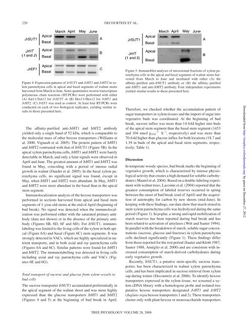
Figure 4. Expression patterns of <strong>JrSUT1</strong> <strong>and</strong> <strong>JrHT1</strong> <strong>and</strong> <strong>JrHT2</strong> in xylem<br />
parenchyma cells in apical <strong>and</strong> basal segments of walnut stems<br />
harvested from March to June. Semi-quantitative reverse transcriptase<br />
polymerase chain reactions (RT-PCRs) were performed with either<br />
(A) Sut11/Sut12 for <strong>JrSUT1</strong> or (B) Hex11/Hex12 for <strong>JrHT1</strong> <strong>and</strong><br />
<strong>JrHT2</strong>. (C) JrEF1 was used as control. At least four RT-PCRs were<br />
conducted on each of two biological replicates, yielding similar results<br />
to those presented here.<br />
The affinity-purified anti-<strong>JrHT1</strong> <strong>and</strong> <strong>JrHT2</strong> antibody<br />
yielded only a single b<strong>and</strong> of 52 kDa, which is comparable to<br />
the molecular mass of other <strong>hexose</strong> transporters (Williams et<br />
al. 2000, Vignault et al. 2005). The protein pattern of <strong>JrHT1</strong><br />
<strong>and</strong> <strong>JrHT2</strong> contrasted with that of <strong>JrSUT1</strong> (Figure 5B). In the<br />
apical xylem parenchyma cells, <strong>JrHT1</strong> <strong>and</strong> <strong>JrHT2</strong> were barely<br />
detectable in March, <strong>and</strong> only a faint signals were observed in<br />
April <strong>and</strong> June. The greatest amount of <strong>JrHT1</strong> <strong>and</strong> <strong>JrHT2</strong> was<br />
found in May, coinciding with a period of intense radial<br />
growth in walnut (Daudet et al. 2005). In the basal xylem parenchyma<br />
cells, no significant signal was found, except in<br />
May, when <strong>JrHT1</strong> <strong>and</strong> <strong>JrHT2</strong> were abundant. In May, <strong>JrHT1</strong><br />
<strong>and</strong> <strong>JrHT2</strong> were more abundant in the basal than in the apical<br />
stem segment.<br />
Immunolocalization analysis of the <strong>hexose</strong> transporters was<br />
performed in sections harvested from apical <strong>and</strong> basal stem<br />
segments of 1-year-old stems at the end of April (beginning of<br />
bud break). No signal was detected when the immunolocalization<br />
was performed either with the saturated primary antibody<br />
(data not shown) or in the absence of the primary antibody<br />
(Figures 6B, 6D, 6F <strong>and</strong> 6H). For <strong>JrSUT1</strong>, immunolabeling<br />
was limited to the living cells of the xylem in both apical<br />
(Figure 6A) <strong>and</strong> basal (Figure 6C) stem segments. It was<br />
strongly detected in VACs, which are highly specialized in nutrient<br />
transports, <strong>and</strong> in both axial <strong>and</strong> ray parenchyma cells<br />
(Figures 6A <strong>and</strong> 6C). Similar patterns were found for <strong>JrHT1</strong><br />
<strong>and</strong> <strong>JrHT2</strong>. The immunolabelling was detected in living cells<br />
including axial <strong>and</strong> ray parenchyma cells <strong>and</strong> VACs (Figures<br />
6E <strong>and</strong> 6G).<br />
Total transport of sucrose <strong>and</strong> glucose from xylem vessels to<br />
bud cells<br />
The sucrose transporter <strong>JrSUT1</strong> accumulated preferentially in<br />
the apical segment of the walnut shoot <strong>and</strong> was more highly<br />
expressed than the glucose transporters <strong>JrHT1</strong> <strong>and</strong> <strong>JrHT2</strong><br />
(Figures 4 <strong>and</strong> 5) at the beginning of bud break in April.<br />
TREE PHYSIOLOGY VOLUME 28, 2008<br />
Figure 5. Immunoblot analyses of microsomal fractions of xylem parenchyma<br />
cells in the apical <strong>and</strong> basal segments of walnut stems harvested<br />
from March to June <strong>and</strong> incubated with either (A) the<br />
affinity-purified anti-<strong>JrSUT1</strong> antibody or (B) the affinity–purified<br />
anti-<strong>JrHT1</strong> <strong>and</strong> anti-<strong>JrHT2</strong> antibody. Four independent experiments<br />
yielded similar results to those presented here.<br />
Therefore, we checked whether the accumulation pattern of<br />
sugar transporters in xylem tissues <strong>and</strong> the import of sugar into<br />
vegetative buds was coordinated. At the beginning of bud<br />
break, sucrose influx was more than 14-fold higher into buds<br />
of the apical stem segment than the basal stem segment (1453<br />
<strong>and</strong> 104 nmol g DM –1 h –1 , respectively) <strong>and</strong> was more than<br />
70-fold higher than glucose influx for both locations (18.7 <strong>and</strong><br />
1.39 in buds of the apical <strong>and</strong> basal stem segments, respectively;<br />
Table 1).<br />
Discussion<br />
In temperate woody species, bud break marks the beginning of<br />
vegetative growth, which is characterized by intense physiological<br />
activity that creates a high dem<strong>and</strong> for soluble carbohydrates<br />
(Maurel et al. 2004). Based on a double-labeling experiment<br />
with walnut trees, Lacointe et al. (2004) reported that the<br />
greatest consumption of labeled reserves occurred in spring<br />
between the onset of bud break (end of April) <strong>and</strong> the acquisition<br />
of autotrophy for carbon by new shoots (mid-June). In<br />
keeping with these findings, our data show that starch stored in<br />
stem xylem parenchyma cells was hydrolyzed during the same<br />
period (Figure 1). In poplar, a strong <strong>and</strong> rapid mobilization of<br />
starch reserves has been reported during bud break <strong>and</strong> has<br />
been related to activation of amylases (Witt <strong>and</strong> Sauter 1994).<br />
In parallel with the breakdown of starch, soluble sugar concentrations<br />
(sucrose, glucose <strong>and</strong> fructose) in xylem parenchyma<br />
cells declined significantly (Figure 1). These findings differ<br />
from those reported for the rest period (Sauter <strong>and</strong> Kloth 1987,<br />
Sauter 1988, Améglio et al. 2000) <strong>and</strong> are consistent with increased<br />
consumption of starch-derived carbohydrates during<br />
early vegetative growth.<br />
Recently, <strong>JrSUT1</strong>, a putative stem-specific sucrose transporter,<br />
has been characterized in walnut xylem parenchyma<br />
cells, <strong>and</strong> has been implicated in sucrose retrieval from xylem<br />
sap during winter (Decourteix et al. 2006). To identify <strong>hexose</strong><br />
transporters expressed in the xylem tissue, we screened a xylem<br />
cDNA library with a homologous probe <strong>and</strong> isolated two<br />
putative <strong>hexose</strong> transporters designated <strong>JrHT1</strong> <strong>and</strong> <strong>JrHT2</strong><br />
(Juglans regia <strong>hexose</strong> transporters 1 <strong>and</strong> 2). These transporters<br />
cluster only with plant <strong>hexose</strong> or monosaccharide transporters<br />
Downloaded from<br />
http://treephys.oxfordjournals.org/ by guest on December 27, 2012