Single-Molecule RNA Science - Xiaowei Zhuang - Harvard University
Single-Molecule RNA Science - Xiaowei Zhuang - Harvard University
Single-Molecule RNA Science - Xiaowei Zhuang - Harvard University

You also want an ePaper? Increase the reach of your titles
YUMPU automatically turns print PDFs into web optimized ePapers that Google loves.
Annu. Rev. Biophys. Biomol. Struct. 0.0:${article.fPage}-${article.lPage}. Downloaded from arjournals.annualreviews.org<br />
by HARVARD COLLEGE on 04/05/05. For personal use only.<br />
10 Feb 2005 13:0 AR AR243-BB34-17.tex XMLPublish SM (2004/02/24) P1: JRX<br />
AR REVIEWS IN ADVANCE10.1146/annurev.biophys.34.040204.144641<br />
(Some corrections may occur before final publication online and in print)<br />
I N<br />
R E V I E W S<br />
A D V A N C E<br />
SINGLE-MOLECULE <strong>RNA</strong> SCIENCE<br />
Annu. Rev. Biophys. Biomol. Struct. 2005. 34:399–414<br />
doi: 10.1146/annurev.biophys.34.040204.144641<br />
Copyright c○ 2005 by Annual Reviews. All rights reserved<br />
First published online as a Review in Advance on March 3, 2005<br />
<strong>Xiaowei</strong> <strong>Zhuang</strong><br />
Department of Chemistry and Chemical Biology and Department of Physics, <strong>Harvard</strong><br />
<strong>University</strong>, Cambridge, Massachusetts 02138; email: zhuang@chemistry.harvard.edu<br />
KeyWords single-molecule experiments, <strong>RNA</strong> folding, <strong>RNA</strong> catalysis,<br />
fluorescence resonance energy transfer, optical tweezers<br />
■ Abstract The development of single-molecule detection and manipulation has<br />
allowed us to monitor the behavior of individual biological molecules and molecular<br />
complexes in real time. This approach significantly expands our capability to characterize<br />
complex dynamics of biological processes, allowing transient intermediate<br />
states and parallel kinetic pathways to be directly observed. Exploring this capability<br />
to elucidate complex dynamics, recent single-molecule experiments on <strong>RNA</strong> folding<br />
and catalysis have improved our understanding of the folding energy landscape of <strong>RNA</strong><br />
and allowed us to better dissect complex <strong>RNA</strong> catalytic reactions, including translation<br />
by the ribosome.<br />
CONTENTS<br />
INTRODUCTION .....................................................399<br />
EXPERIMENTAL TECHNIQUES ........................................401<br />
<strong>Single</strong>-<strong>Molecule</strong> FRET ................................................401<br />
Optical Tweezers.....................................................403<br />
SINGLE-MOLECULE FRET STUDIES OF <strong>RNA</strong> FOLDING ..................403<br />
Folding of Large, Multidomain <strong>RNA</strong> Enzymes .............................403<br />
Folding of Small <strong>RNA</strong> Enzymes ........................................405<br />
<strong>Single</strong>-<strong>Molecule</strong> �-Value Analysis ......................................406<br />
SINGLE-MOLECULE FRET STUDIES OF <strong>RNA</strong> CATALYSIS .................407<br />
Catalysis by a Small <strong>RNA</strong> Enzyme ......................................407<br />
Translation by the Ribosome ...........................................407<br />
SINGLE-MOLECULE FORCE STUDIES OF <strong>RNA</strong> FOLDING<br />
AND UNFOLDING ...................................................408<br />
Folding and Unfolding of Simple Secondary or Tertiary Structures .............408<br />
Unfolding of a Large, Multidomain <strong>RNA</strong> Enzymes .........................409<br />
CONCLUSIONS AND FUTURE DIRECTIONS .............................409<br />
INTRODUCTION<br />
This is not the first time that we have witnessed <strong>RNA</strong> taking center stage. Some<br />
20 years ago, two independent, groundbreaking discoveries changed our view<br />
about this biomacromolecule: Found to function as catalysts, <strong>RNA</strong> molecules could<br />
1056-8700/05/0609-0399$20.00 399
Annu. Rev. Biophys. Biomol. Struct. 0.0:${article.fPage}-${article.lPage}. Downloaded from arjournals.annualreviews.org<br />
by HARVARD COLLEGE on 04/05/05. For personal use only.<br />
10 Feb 2005 13:0 AR AR243-BB34-17.tex XMLPublish SM (2004/02/24) P1: JRX<br />
AR REVIEWS IN ADVANCE10.1146/annurev.biophys.34.040204.144641<br />
400 ZHUANG<br />
no longer be viewed as merely messengers that pass information from the DNA<br />
to proteins (19, 28). Since then, a growing number of essential reactions in cells<br />
are have been found to be catalyzed by <strong>RNA</strong> enzymes. Two examples exist at the<br />
heart of the central dogma of biology: The ribosome, the giant ribonucleoprotein<br />
assembly responsible for protein synthesis in cells, uses its <strong>RNA</strong> components to<br />
perform major catalytic functions (12, 37, 38). The spliceosome also likely uses<br />
its <strong>RNA</strong> constituent to catalyze the splicing reaction of the precursor <strong>RNA</strong> (70,<br />
71), while the 100 or so protein splicing factors regulate the reaction. In addition<br />
to these essential catalytic functions, we now witness a second revolution in the<br />
world of <strong>RNA</strong>: its remarkable regulatory role. In the past decade or so, hundreds<br />
of small noncoding <strong>RNA</strong>s, micro<strong>RNA</strong>s, and endogenous small-interfering <strong>RNA</strong>s<br />
were discovered that regulate gene expression either by inducing messenger <strong>RNA</strong><br />
degradation or by suppressing translation in both prokaryotic and eukaryotic cells<br />
(3, 11, 17). Aside from these posttranscriptional regulations, gene-regulation by<br />
noncoding <strong>RNA</strong> at the transcription level has also been observed recently (17, 74).<br />
In keeping with this expanding list of fundamental biological roles, <strong>RNA</strong> also finds<br />
exciting applications in modern biotechnology and medicine, as gene regulatory<br />
tools, as therapeutic agents, and in drug discovery (9, 14, 60, 64).<br />
Although <strong>RNA</strong> science is attracting a tremendous amount of attention, singlemolecule<br />
studies of <strong>RNA</strong> are also gaining momentum. There are good reasons for<br />
this to happen. With a growing appreciation of the importance of <strong>RNA</strong>, a mechanistic<br />
understanding of how <strong>RNA</strong> molecules adopt specific structures that mediate<br />
their functions is becoming more desirable than ever. Research on this <strong>RNA</strong> folding<br />
problem suggests that the free energy landscape of <strong>RNA</strong> is often highly rugged (8,<br />
24, 41–43, 45, 46, 50, 52–54, 56, 66, 67, 69, 78–82). As a result, an <strong>RNA</strong> molecule<br />
can traverse a multitude of kinetic paths and intermediate conformational states<br />
before attaining its native structure. For example, oligonucleotide hybridization,<br />
hydroxyl radical footprinting, circular dichroism, and UV absorption assays have<br />
led to the discovery that <strong>RNA</strong> folding occurs through a series of intermediate<br />
states (46, 56, 69, 79, 80). <strong>RNA</strong> enzymes were also seen to fold along multiple<br />
pathways by native gel electrophoresis and enzymatic activity assays (41–43,<br />
52). However, these methods that measure the average property of an ensemble of<br />
molecules can only probe accumulative intermediate states and significantly populated<br />
pathways; they cannot detect nonaccumulative, transient intermediate states.<br />
The existence of a large number of folding pathways will also make the characterization<br />
of each individual pathway difficult by ensemble assays. In these cases,<br />
single-molecule approaches come to the rescue owing to their intrinsic capability<br />
to detect nonaccumulative transient intermediate states and heterogeneity in the<br />
system.<br />
Similarly, the reactions catalyzed by <strong>RNA</strong> enzymes, especially those by large<br />
ribonucleoprotein complexes such as the ribosome, tend to be complex, involving<br />
many reaction steps and reaction intermediates. These steps are typically not<br />
synchronized for an ensemble of molecules, making the reactions difficult to dissect.<br />
Although stalling the reaction complexes at specific intermediate states has
Annu. Rev. Biophys. Biomol. Struct. 0.0:${article.fPage}-${article.lPage}. Downloaded from arjournals.annualreviews.org<br />
by HARVARD COLLEGE on 04/05/05. For personal use only.<br />
10 Feb 2005 13:0 AR AR243-BB34-17.tex XMLPublish SM (2004/02/24) P1: JRX<br />
AR REVIEWS IN ADVANCE10.1146/annurev.biophys.34.040204.144641<br />
SINGLE-MOLECULE <strong>RNA</strong> SCIENCE 401<br />
provided us with a lot of molecular insights into these reactions, this kind of<br />
manipulation is not necessarily possible for every intermediate state. Monitoring<br />
the reaction of a single molecule or molecular complex naturally overcomes this<br />
synchronization problem, offering us an opportunity to resolve each microscopic<br />
step of a complex reaction.<br />
The power of single-molecule techniques to elucidate complex dynamics does<br />
not come without sacrifices. Because of the limited amount of signal that a single<br />
molecule can produce, the time and spatial resolutions of single-molecule experiments<br />
are relatively limited compared with ensemble experiments. Luckily, <strong>RNA</strong><br />
folding and catalytic reactions are not so fast, typically occurring on the timescales<br />
of milliseconds to minutes. In addition, these reactions often involve large conformational<br />
changes. These properties allow current, state-of-the-art single-molecule<br />
experiments to explore the <strong>RNA</strong> world. Indeed, these experiments have already<br />
made significant contributions to our understanding of the structural biology and<br />
enzymology of <strong>RNA</strong>. In this article, I review some of the recent developments in<br />
<strong>RNA</strong> science made possible by single-molecule techniques, focusing on those by<br />
fluorescence resonance energy transfer (FRET) and optical tweezers.<br />
EXPERIMENTAL TECHNIQUES<br />
<strong>Single</strong>-<strong>Molecule</strong> FRET<br />
FRET is a powerful assay to measure the intra- and intermolecular motions of<br />
biomolecules in real time (57, 63, 75). In this assay, a pair of fluorescent donor and<br />
acceptor molecules is attached to the host biomolecule(s) of interest. The dipoledipole<br />
interaction between the donor and acceptor causes energy transfer between<br />
them, leading to a decrease in the donor emission and an increase in the acceptor<br />
emission. The energy transfer efficiency E is given by E = 1/(1 + (R/R0) 6 ),<br />
where R is the distance between the donor and the acceptor. The Förster radius R0<br />
is typically 3 to 8 nm, making FRET sensitive to changes of a few nanometers in<br />
the donor-acceptor distance, which can reflect either the conformation changes of<br />
the host molecule or relative motions between two molecules. The dependence of<br />
energy transfer efficiency on the orientation of the donor and acceptor fluorophores,<br />
however, introduces uncertainty into FRET-based distance determinations. This<br />
problem can be mitigated if the dyes are attached to the host molecule with flexible<br />
linkers to ensure their free rotation. As this condition is difficult to satisfy rigorously<br />
in practice, researchers should be cautious when translating FRET efficiencies into<br />
absolute distances quantitatively.<br />
The development of single-molecule fluorescence spectroscopy (33, 36, 77)<br />
has allowed FRET to be measured at the single-molecule level (21, 55). Here,<br />
I briefly discuss a few aspects of single-molecule detection that are critical for<br />
unveiling the dynamic information of biomolecules. Like all single-molecule<br />
fluorescence techniques, efficient reduction of the background signal is critical
Annu. Rev. Biophys. Biomol. Struct. 0.0:${article.fPage}-${article.lPage}. Downloaded from arjournals.annualreviews.org<br />
by HARVARD COLLEGE on 04/05/05. For personal use only.<br />
10 Feb 2005 13:0 AR AR243-BB34-17.tex XMLPublish SM (2004/02/24) P1: JRX<br />
AR REVIEWS IN ADVANCE10.1146/annurev.biophys.34.040204.144641<br />
402 ZHUANG<br />
for single-molecule FRET. This is often achieved by confocal detection or by<br />
excitation with an evanescent wave generated by total internal reflection. The total<br />
internal reflection microscope allows a whole field of molecules, typically several<br />
hundred of them, to be detected simultaneously. Such a parallel detection<br />
greatly facilitates the building of statistics, especially for irreversible processes.<br />
However, wide-field detection requires the use of cameras, which limit the data<br />
acquisition rate to 1 kilocycle per second or slower using state-of-the-art charge<br />
couple device (CCD) cameras with on-chip amplification. Higher data acquisition<br />
rates, up to tens of megacycles per second, can be achieved with confocoal<br />
microscopes using sensitive point detectors, such as avalanche photodiodes or<br />
photomultipliers.<br />
Besides physical instrumentation, special manipulation of biomolecules is often<br />
required for real-time characterizations. With a confocal microscope, it is possible<br />
to detect freely diffusing molecules in solution (13, 36). Although this detection<br />
geometry avoids any potentially adverse effect arising from the confinement of<br />
molecules, the range of timescales accessible by this method is severely limited<br />
by the dwell time of a molecule in the confocal detection volume, typically in the<br />
submilliseconds governed by diffusion. Therefore, to expand the dynamic range<br />
of the single-molecule experiments, various strategies were developed to confine<br />
the biomolecules of interest within the detection volume of a microscope.<br />
The two most commonly used strategies are confinement by matrices, such as<br />
agarose or polyacrylamide gels (32), and immobilization to surfaces with specific<br />
interactions, such as via the biotin-streptavidin bridge (22). The latter method is<br />
compatible with rapid buffer exchange and therefore is better suited for characterizing<br />
reactions. Although surface immobilization may perturb the biomolecules,<br />
this perturbation has been rather insignificant for most of the <strong>RNA</strong> systems studied<br />
to date, at neutral or basic pH values. For systems that involve proteins, which<br />
tend to interact strongly with surfaces through hydrophobic interactions, surface<br />
passivation is often required to retain the integrity of the system. Polyethylene<br />
glyco (PEG) is a commonly used passivating reagent (34). Another method is to<br />
entrap the biomolecule in a lipid vesicle attached to a surface (48). This technique<br />
potentially offers a close-to-physiological environment for the biomolecule;<br />
however, a method for rapidly changing the buffer in the vesicle needs to be<br />
developed before this method can be broadly used for characterizing biological<br />
processes.<br />
With the molecules confined in the detection volume, dynamics can be probed<br />
at the single-molecule level over a wide range of timescales, ranging from millisecond<br />
to minutes or even hours. The lower limit is imposed by the photon<br />
emission rate from a single fluorophore under conditions in which photoblinking<br />
or photobleaching is not severe enough to render the experiment impractical.<br />
The recent development of CCD cameras with on-chip amplification allows this<br />
limit to be achieved even in the wide-field-detection geometry. This limit can be<br />
further improved by using fluorescence correlation spectroscopy if the process<br />
under investigation is reversible and if the forward and backward reaction occurs
Annu. Rev. Biophys. Biomol. Struct. 0.0:${article.fPage}-${article.lPage}. Downloaded from arjournals.annualreviews.org<br />
by HARVARD COLLEGE on 04/05/05. For personal use only.<br />
10 Feb 2005 13:0 AR AR243-BB34-17.tex XMLPublish SM (2004/02/24) P1: JRX<br />
AR REVIEWS IN ADVANCE10.1146/annurev.biophys.34.040204.144641<br />
SINGLE-MOLECULE <strong>RNA</strong> SCIENCE 403<br />
spontaneously (25, 27, 76). In this case, the time resolution is limited by the response<br />
time of the detectors, which is of the order of tens of nanoseconds for<br />
avalanche photodiodes or photomultipliers.<br />
<strong>Single</strong>-molecule FRET has been used to study a variety of biological systems.<br />
In this review, I only cover its application to <strong>RNA</strong> systems (4–7, 22, 35, 47, 51,<br />
54, 67, 78, 81, 82) but refer interested readers to a number of recent review articles<br />
for other applications (20, 75, 83).<br />
Optical Tweezers<br />
Optical tweezers rely on a tightly focused laser beam(s) to trap a particle in three<br />
dimensions and, through redirection of the beam, manipulate the particles (1).<br />
Focused light exerts two forces on the particle, the gradient force and the scattering<br />
force. When the refractive index of the particle is larger than that of the surrounding<br />
medium, the gradient force draws the particle toward the focus of the beam, where<br />
the light field is the strongest. The scattering force arises from the radiation pressure<br />
exerted on the particle by photons that are either absorbed or scattered. When<br />
balanced, these two forces hold the particle just slightly downstream of the light<br />
focus.<br />
Optical tweezers allow the manipulation and detection of single biomolecules.<br />
As biological molecules are often too small to be manipulated directly by optical<br />
tweezers, a micron-sized dielectric sphere is often attached to the molecule of<br />
interest to serve as a handle. This allows researchers to manipulate the position of<br />
the attached biomolecule and to exert a well-defined force. Both parameters can<br />
be determined with exquisite accuracy: The position of the microsphere can be<br />
measured to within 0.1 nm, and the force to 0.1 pN (39, 58). Forces well above<br />
100 pN can be achieved with a dual-beam optical tweezers (10).<br />
Since the invention of optical tweezers, this technique has been adopted to<br />
study a variety of biological systems. This review focuses on the studies on <strong>RNA</strong><br />
systems (23, 30, 31, 40). Other single-molecule force spectroscopy techniques,<br />
such as those using atomic force microscopes and magnetic tweezers, have also<br />
provided a wealth of information on biological molecules (61, 62, 83). These<br />
single-molecule techniques are fully expected to make significant contributions to<br />
our understanding of <strong>RNA</strong> systems as well.<br />
SINGLE-MOLECULE FRET STUDIES OF <strong>RNA</strong> FOLDING<br />
Folding of Large, Multidomain <strong>RNA</strong> Enzymes<br />
The capability of single-molecule FRET to follow conformational changes of<br />
<strong>RNA</strong> was clear early on. In one of the first applications of single-molecule FRET<br />
to biological systems, Ha et al. (22) showed that the ligand-induced conformational<br />
changes of an <strong>RNA</strong> three-way junction could be monitored in real time at<br />
the single-molecule level. <strong>Zhuang</strong> et al. (81) performed the first single-molecule
Annu. Rev. Biophys. Biomol. Struct. 0.0:${article.fPage}-${article.lPage}. Downloaded from arjournals.annualreviews.org<br />
by HARVARD COLLEGE on 04/05/05. For personal use only.<br />
10 Feb 2005 13:0 AR AR243-BB34-17.tex XMLPublish SM (2004/02/24) P1: JRX<br />
AR REVIEWS IN ADVANCE10.1146/annurev.biophys.34.040204.144641<br />
404 ZHUANG<br />
<strong>RNA</strong> folding study by using a large, multidomain <strong>RNA</strong> enzyme, the Tetrahymena<br />
ribozyme (Figure 1a, see color insert), as a model system. The power of singlemolecule<br />
FRET was already apparent in this early work. A repertoire of<br />
dynamic properties that are difficult to analyze with ensemble experiments, including<br />
nonaccumulative folding intermediate states, parallel folding pathways,<br />
and equilibrium conformational fluctuations, were directly observed in singlemolecule<br />
FRET trajectories of the Tetrahymena ribozyme (81).<br />
FRET trajectories of individual ribozyme molecules directly reveal folding<br />
intermediates (Figure 1b): The addition of Mg 2+ to trigger folding causes the<br />
FRET signal to increase from the unfolded state value of 0.1 to an intermediate<br />
value (0.3) before attaining the native value (0.9). Statistical analysis of the<br />
folding times of each molecule indicated that the Tetrahymena ribozyme folds<br />
along at least three distinct pathways, with folding rate constant being 1 s, 1 min,<br />
and 0.01 hr, respectively. Kinetic traps exist on these pathways. While the two<br />
slower pathways have been observed previously in ensemble measurements, the<br />
most rapid pathway was first observed in this single-molecule FRET experiment<br />
(81).<br />
In a follow-up study designed to gain a greater understanding of the various<br />
pathways (54), folding of the Tetrahymena ribozyme was started from distinct regions<br />
on the energy landscape by varying the monovalent salt concentration of the<br />
prefolding solution. Different monovalent salt concentrations led to dramatically<br />
different folding behaviors, each with a distinct folding rate constant and traversing<br />
a different set of intermediate states. These results were interpreted as the<br />
molecules folding along several discrete channels separated by large energy barriers.<br />
Site-specific mutation analysis suggests that these different folding behaviors<br />
are caused by the different secondary structures formed under various salt concentrations<br />
(54), consistent with previous ensemble experiments (43, 44). Recent<br />
results from a hydroxyl radical footprinting assay suggest that high concentrations<br />
of monovalent salt induce an extensive amount of tertiary structures in addition<br />
to secondary structure changes (66). These preformed tertiary structures may also<br />
be partly responsible for the observed changes in the folding kinetics induced by<br />
preincubation with monovalent salt.<br />
More recently, FRET has been applied to another large <strong>RNA</strong> molecule, the<br />
catalytic domain of Bacillus subtilis RNase P <strong>RNA</strong> (78). Ensemble characterizations<br />
of this enzyme indicate the existence of three conformational states: the<br />
unfolded state, the intermediate state, and the native state. However, analysis of<br />
the molecules at equilibrium using single-molecule FRET suggest that at least<br />
seven different conformations exist at intermediate Mg 2+ concentrations, as distinguished<br />
by their corresponding FRET levels and connectivity between different<br />
states. The equilibrium structural dynamics of individual RNase P molecules also<br />
shows a high level of heterogeneity.<br />
These single-molecule results indicate that large <strong>RNA</strong> molecules fold across<br />
a highly rugged energy landscape, along a multitude of folding pathways, and<br />
through many intermediate folding states. These findings not only corroborate
Annu. Rev. Biophys. Biomol. Struct. 0.0:${article.fPage}-${article.lPage}. Downloaded from arjournals.annualreviews.org<br />
by HARVARD COLLEGE on 04/05/05. For personal use only.<br />
10 Feb 2005 13:0 AR AR243-BB34-17.tex XMLPublish SM (2004/02/24) P1: JRX<br />
AR REVIEWS IN ADVANCE10.1146/annurev.biophys.34.040204.144641<br />
SINGLE-MOLECULE <strong>RNA</strong> SCIENCE 405<br />
previous and concurrent ensemble characterizations using various footing-printing<br />
methods, enzymatic activity assays, gel-mobility measurements, and optical scattering<br />
or spectroscopic techniques (8, 24, 41–43, 45, 46, 50, 52, 53, 56, 66, 69,<br />
79, 80), but also reveal new folding intermediates and folding pathways difficult<br />
to access by ensemble assays, thereby significantly improving our understanding<br />
of <strong>RNA</strong> folding.<br />
Folding of Small <strong>RNA</strong> Enzymes<br />
To date, single-molecule studies of small <strong>RNA</strong> enzymes have concentrated on one<br />
type of ribozyme, the hairpin ribozyme (Figure 2a, see color insert), although other<br />
systems are beginning to be explored. The hairpin ribozyme has been suggested<br />
to assume either an extended, undocked conformation or a docked conformation<br />
in which the two loop domains, A and B, make tertiary contacts (73). Two forms<br />
of hairpin ribozymes have been studied at the single-molecule level: the two-way<br />
junction ribozyme, a minimal hairpin ribozyme with high enzymatic activity, and<br />
the wild-type ribozyme, which has a four-way junction connecting helix 2 and<br />
helix 3 (15, 73) (Figure 2a). The first single-molecule experiment was performed<br />
on the two-way junction ribozyme (82). Strikingly, the FRET time traces of single<br />
molecules show highly heterogeneous undocking kinetics with several different<br />
rate constants (Figure 2b,c). Furthermore, the molecules retain their particular<br />
undocking rates through many docking-undocking cycles, and conversion between<br />
different undocking behaviors requires at least several hours (Figure 2b). These<br />
results suggest the existence of rather large numbers of conformational states that<br />
are all relatively stable under functional conditions, which is remarkable for such<br />
a small <strong>RNA</strong> enzyme.<br />
The heterogeneous undocking kinetics appear to be ubiquitous to all hairpin<br />
ribozymes. For the two-way junction ribozyme, both the wild type and variants with<br />
significantly different docking and undocking rate constants show heterogeneous<br />
undocking behavior (7, 51, 82). In addition, heterogeneous docking kinetics are<br />
seen in a variant with a modification at the domain junction (51). A study on<br />
the four-way junction hairpin ribozyme, a construct that more closely resembles<br />
the natural form of the ribozyme, also shows pronounced heterogeneity in both<br />
docking and undocking kinetics (67).<br />
The comparison between the single-molecule studies of the two- and fourway<br />
junction ribozymes reveals an interesting phenomenon: The latter form folds<br />
(docks) two to three orders of magnitude faster (67). Evidence suggests that the<br />
dramatic enhancement in the folding rate is a result of the intrinsic structural<br />
dynamics of the four-way junction: The ribozyme appears to fold into its native<br />
form via an intermediate state in which the four-way junction juxtaposes the loop<br />
domains, A and B, into proximity but without substantial loop-loop interactions<br />
(67). This result is further supported by a more recent single-molecule study of<br />
hairpin ribozymes freely diffusing in solution (47). There, even when the two loops<br />
are replaced with perfectly base-paired helices, these helices are still juxtaposed
Annu. Rev. Biophys. Biomol. Struct. 0.0:${article.fPage}-${article.lPage}. Downloaded from arjournals.annualreviews.org<br />
by HARVARD COLLEGE on 04/05/05. For personal use only.<br />
10 Feb 2005 13:0 AR AR243-BB34-17.tex XMLPublish SM (2004/02/24) P1: JRX<br />
AR REVIEWS IN ADVANCE10.1146/annurev.biophys.34.040204.144641<br />
406 ZHUANG<br />
into proximity by the four-way junction, whereas both two-way and three-way<br />
junctions lack this capability. Undocking kinetics does not seem to be accelerated<br />
by the junction dynamics.<br />
These results suggest that even small <strong>RNA</strong> enzymes can have highly complex<br />
structural dynamics, featuring a rugged energy landscape in the conformational<br />
space. Unlike in the case of large, multidomain <strong>RNA</strong> molecules, the level of complexity<br />
in the structural dynamics of small ribozymes had not been observed previously<br />
in ensemble experiments. These single-molecule studies make us wonder<br />
whether rugged energy landscapes and complex structural dynamics might very<br />
well be general properties of all <strong>RNA</strong>s, not just reserved for the large, complex<br />
<strong>RNA</strong> enzymes.<br />
<strong>Single</strong>-<strong>Molecule</strong> �-Value Analysis<br />
Characterization of the transition state ensemble is critical for a mechanistic understanding<br />
of the macromolecule folding problem. While transition-state analysis<br />
has been performed routinely for protein folding, its implementation in <strong>RNA</strong> folding<br />
has been challenging owing to the rugged energy landscape of <strong>RNA</strong>, which<br />
often leads to the coexistence of distinct structural transitions. The power of singlemolecule<br />
measurements to isolate individual transitions makes them ideally suited<br />
for transition-state analysis of <strong>RNA</strong> folding, A single-molecule �-value analysis<br />
based on FRET was developed to characterize the degree to which specific intramolecular<br />
interactions are formed in the transition states of <strong>RNA</strong> folding (4,<br />
7), following the �-value analysis introduced by Fersht (16) for characterizing<br />
protein folding.<br />
Using FRET to monitor the folding and unfolding transitions of the hairpin ribozyme,<br />
in conjunction with metal-ion titrations and tertiary-contact-destabilizing<br />
mutations, Bokinsky et al. (7) showed that the transition state is compact with the<br />
Mg 2+ -mediated interactions between the two domains of the hairpin ribozyme<br />
formed at a strength similar to that in the folded state, whereas the native tertiary<br />
contacts are not substantially formed. The conformational entropy loss and the<br />
breaking of secondary structures may be the primary mechanisms limiting the<br />
folding rate of the ribozyme. The folding transition states of some proteins also<br />
feature native-like topology without substantial native tertiary contacts (29).<br />
This single-molecule �-value analysis is not only applicable to an elementary<br />
folding reaction of a small <strong>RNA</strong> enzyme, it can also be used to characterize the<br />
transition state of a local folding step of a large <strong>RNA</strong> enzyme (4, 81). After extensive<br />
effort to disrupt every single tertiary contact between the P1 duplex and catalytic<br />
core of the Tetrahymena ribozyme, Bartley et al. (4) have shown that not one of<br />
these tertiary contacts is formed in the transition state of P1-docking, which is<br />
believed to be the last folding step of the ribozyme. Their results suggest that P1docking<br />
is rate-limited by kinetic traps. This work demonstrated the potential of<br />
this single-molecule �-value analysis to characterize each individual folding step<br />
of a complex, multistep, multipath folding reaction.
Annu. Rev. Biophys. Biomol. Struct. 0.0:${article.fPage}-${article.lPage}. Downloaded from arjournals.annualreviews.org<br />
by HARVARD COLLEGE on 04/05/05. For personal use only.<br />
10 Feb 2005 13:0 AR AR243-BB34-17.tex XMLPublish SM (2004/02/24) P1: JRX<br />
AR REVIEWS IN ADVANCE10.1146/annurev.biophys.34.040204.144641<br />
SINGLE-MOLECULE <strong>RNA</strong> SCIENCE 407<br />
SINGLE-MOLECULE FRET STUDIES OF <strong>RNA</strong> CATALYSIS<br />
<strong>Single</strong>-molecule methods are particularly well suited for detecting transient intermediate<br />
states and thereby resolving the microscopic steps of a complex reaction.<br />
This is demonstrated in the following two examples, which are at the two extremes<br />
of the ribozyme world, a small <strong>RNA</strong> enzyme and the ribosome.<br />
Catalysis by a Small <strong>RNA</strong> Enzyme<br />
Following the reaction of single hairpin ribozyme molecules using FRET, <strong>Zhuang</strong><br />
et al. (82) have shown that this ribozyme catalyzes the cleavage reaction of its<br />
substrate in several distinct steps (Figure 2c): (a) substrate binding; (b) folding<br />
of the ribozyme-substrate complex into a catalytically active state (docking);<br />
(c) cleavage of the substrate; (d) the unfolding (undocking) of the ribozyme-product<br />
complex; and (e) release of the products. These results confirmed a previously<br />
proposed reaction pathway based on ensemble measurements (15, 72, 73). The<br />
determination of the rate constants of each step indicates that the overall cleavage<br />
reaction of the two-way junction ribozyme is primarily rate-limited by the two<br />
structural transition steps, (b) and (d), and by the internal equilibrium constants<br />
of the reversible cleavage step, (c) (82). The heterogeneous undocking kinetics<br />
mentioned above result in heterogeneous reaction kinetics.<br />
The rate-limiting mechanism of the four-way junction ribozyme is different.<br />
Cleavage appears to dramatically accelerate undocking in the case of the four-way<br />
junction ribozyme. As a result the undocking rate is faster than the ligation rate,<br />
and the overall cleavage reaction rate is primarily limited by the internal cleavage<br />
reaction (35). In contrast, the undocking rate constant of the two-way junction<br />
ribozyme bound to its natural cleavage products is much slower than the ligation<br />
rate constant (51; G. Bokinsky, N. Walter & X. <strong>Zhuang</strong>, unpublished data). This<br />
discrepancy between the two-way and four-way ribozymes indicates an interesting<br />
allostery effect that the junction has on the docked structure of the loop domains.<br />
Translation by the Ribosome<br />
Early works by Hochstrasser and coworkers (26, 65) have shown that surfaceimmobilized<br />
ribosome complexes retain their peptidyl transferase activity and can<br />
be detected at the single-complex level. These experiments gave researchers hope<br />
that single-molecule experiments might someday provide exciting new insights<br />
into the molecular mechanisms of protein synthesis in cells.<br />
This promise has been realized in a recent, tour de force experiment by<br />
Blanchard et al. (5, 6). To set the stage for new discoveries, these authors first<br />
demonstrated that surface-immobilized ribosomes are highly active in t<strong>RNA</strong><br />
accommodation and translocation and in peptide-bond formation (6). This is nontrivial<br />
considering the complexity of this vast ribonucleoprotein. Indeed specific<br />
surface immobilization and extensive surface passivation are necessary.
Annu. Rev. Biophys. Biomol. Struct. 0.0:${article.fPage}-${article.lPage}. Downloaded from arjournals.annualreviews.org<br />
by HARVARD COLLEGE on 04/05/05. For personal use only.<br />
10 Feb 2005 13:0 AR AR243-BB34-17.tex XMLPublish SM (2004/02/24) P1: JRX<br />
AR REVIEWS IN ADVANCE10.1146/annurev.biophys.34.040204.144641<br />
408 ZHUANG<br />
Next, using single-molecule FRET to follow the accommodation of the t<strong>RNA</strong>elongation<br />
factor Tu-GTP ternary complex in real time, Blanchard et al. (5) resolved<br />
a multistep movement of the t<strong>RNA</strong> into the ribosome and identified the<br />
intermediate states in the t<strong>RNA</strong>-delivery pathway using antibiotics and nonhydrolyzable<br />
GTP analogs. Previously unknown intermediate states were discovered<br />
both in the initial codon-recognition steps and in the kinetic proofreading steps<br />
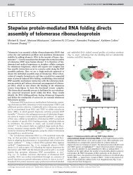
following GTP hydrolysis (Figure 3, see color insert). These results, complementing<br />
previous kinetic characterizations of translation (18, 49), have provided new<br />
insights into the structural basis for the initial cognate t<strong>RNA</strong> selection, kinetic<br />
proofreading, and thus for the fidelity of translation by the ribosome. Interesting<br />
dynamic t<strong>RNA</strong> fluctuations between the classical state and hybrid states have also<br />
been observed (6). The translocation of t<strong>RNA</strong>s from the classical to the hybrid<br />
state after peptide-bond formation has been suggested before; however, it is surprising<br />
that t<strong>RNA</strong> fluctuates spontaneously between these two states both before<br />
and after peptide-bond formation. This observation again highlights the capability<br />
of single-molecule experiments to resolve dynamic behaviors.<br />
SINGLE-MOLECULE FORCE STUDIES OF <strong>RNA</strong><br />
FOLDING AND UNFOLDING<br />
Folding and Unfolding of Simple Secondary<br />
or Tertiary Structures<br />
The use of mechanical forces to induce folding and unfolding provides a few unique<br />
advantages in characterizing macromolecule folding: (a)Itallows the folding and<br />
unfolding reaction to occur along a well-defined reaction coordinate, the molecular<br />
end-to-end distance; and (b) the folding free energy can be readily measured (68).<br />
Liphardt et al. (31) first characterized unfolding and refolding of small <strong>RNA</strong><br />
molecules induced by mechanical forces using optical tweezers. They have shown<br />
that, with a slow-enough force-loading rate, the folding and unfolding of secondary<br />
structures, such as an <strong>RNA</strong> hairpin or a three-helix junction, can occur in<br />
equilibrium. In contrast, for a molecule with tertiary contacts, such as the P5abc<br />
domain of the Tetrahymena ribozyme in the presence of Mg2+ , folding and unfolding<br />
are nonequilibrium processes even at the slowest accessible force-loading rate.<br />
Interestingly, the transition state for folding for secondary structure formation is<br />
dramatically different from that for tertiary structure formation. In the former case,<br />
the transition state is relatively far from the folded state on the reaction coordinate,<br />
i.e., the molecular end-to-end distance, indicating a relatively soft transition.<br />
In contrast the transition state for tertiary structure folding is close to the folded<br />
state, suggesting that tertiary structures are much more brittle than secondary<br />
structures.<br />
As promised, the free energy difference between the folded and unfolded<br />
states is readily calculated from the force-extension curves of molecules being<br />
folded or unfolded at equilibrium, and compare well with theoretical predictions.
Annu. Rev. Biophys. Biomol. Struct. 0.0:${article.fPage}-${article.lPage}. Downloaded from arjournals.annualreviews.org<br />
by HARVARD COLLEGE on 04/05/05. For personal use only.<br />
10 Feb 2005 13:0 AR AR243-BB34-17.tex XMLPublish SM (2004/02/24) P1: JRX<br />
AR REVIEWS IN ADVANCE10.1146/annurev.biophys.34.040204.144641<br />
SINGLE-MOLECULE <strong>RNA</strong> SCIENCE 409<br />
Remarkably, the folding free energy can even be derived from force-extension<br />
curves of nonequilibrium (un)folding processes using a recently developed nonequilibrium<br />
statistical mechanics theorem, the Jarzynski’s equality (30).<br />
Unfolding of a Large, Multidomain <strong>RNA</strong> Enzymes<br />
More recently, Onoa et al. (40) extended mechanical unfolding to a large <strong>RNA</strong><br />
enzyme, the Tetrahymena ribozyme (Figure 4a, see color insert). The forceextension<br />
curves of individual <strong>RNA</strong> molecules show several discrete unfolding<br />
steps (Figure 4b), indicating the existence of multiple unfolding intermediate states.<br />
The molecular interactions disrupted at each of these unfolding steps have been<br />
identified by studying progressively larger pieces of the Tetrahymena ribozyme,<br />
determining the number of nucleotides released at each step, and using mutations<br />
or antisense oligonucleotides to perturb specific interactions in the <strong>RNA</strong>. Interestingly,<br />
most of these unfolding barriers are imposed by tertiary interactions, even<br />
though the overall thermodynamic stability of the molecule largely arises from the<br />
secondary structures, namely the base-paired helices. Unlike thermal or solution<br />
unfolding, in which tertiary structures are dissolved before secondary structures,<br />
the partially unfolded intermediates observed in this mechanical unfolding experiment<br />
contain both secondary structures and tertiary contacts.<br />
Mechanical unfolding experiments have been used to characterize the secondary<br />
structure formation of <strong>RNA</strong> as large as the 16S ribosome <strong>RNA</strong>. Even in this case,<br />
well-defined unfolding intermediates are observed (23).<br />
CONCLUSIONS AND FUTURE DIRECTIONS<br />
A distinct advantage of single-molecule techniques is their capability to detect<br />
transient, nonaccumulative states and heterogeneous behavior. Such capabilities<br />
make these techniques well suited to investigate the structural dynamics and catalytic<br />
reactions of <strong>RNA</strong> molecules. Indeed, single-molecule experiments have<br />
already yielded important insights into <strong>RNA</strong> folding by detecting new folding intermediate<br />
states and pathways, and by revealing a daunting level of complexity<br />
in the conformational dynamics of both large and small <strong>RNA</strong>. <strong>Single</strong>-molecule<br />
experiments have also allowed us to better dissect the complex catalytic reactions<br />
of <strong>RNA</strong> enzymes by resolving individual reaction steps and discovering<br />
new reaction intermediates that are critical for understanding the reaction<br />
mechanisms.<br />
What we have witnessed in the past few years is likely just the tip of the iceberg.<br />
Future single-molecule studies will certainly make more significant contributions<br />
to <strong>RNA</strong> science. As a growing number of essential reactions in cells have now<br />
been catalyzed by ribonucleoprotein enzymes, a bright future direction for singlemolecule<br />
studies would be to characterize the folding, assembly, and enzymatic<br />
reactions of these important cellular ribonucleoproteins. The recent work on the
Annu. Rev. Biophys. Biomol. Struct. 0.0:${article.fPage}-${article.lPage}. Downloaded from arjournals.annualreviews.org<br />
by HARVARD COLLEGE on 04/05/05. For personal use only.<br />
10 Feb 2005 13:0 AR AR243-BB34-17.tex XMLPublish SM (2004/02/24) P1: JRX<br />
AR REVIEWS IN ADVANCE10.1146/annurev.biophys.34.040204.144641<br />
410 ZHUANG<br />
delivery of t<strong>RNA</strong> to ribosome gives us confidence that these complex ribonucleoproteins<br />
are amendable to single-molecule studies.<br />
Another exciting direction is to apply these single-molecule studies to live cells.<br />
After all, the cellular environment is different from that in solution. A complete<br />
understanding of <strong>RNA</strong> folding, catalysis, and regulation requires investigation in<br />
live cells. Recent studies show that the trafficking of individual messenger <strong>RNA</strong>s<br />
and viral <strong>RNA</strong> genes can be monitored in living cells (2, 59). Although these studies<br />
have provided important insights into the trafficking properties of <strong>RNA</strong>-protein<br />
complexes, the investigations relied on the attachment of many fluorophores to each<br />
complex to compete with the cell autofluorescence background, a strategy that is<br />
not necessarily applicable to other <strong>RNA</strong> studies. Information that can be extracted<br />
from a single fluorophore in living cells is rather limited with the current stateof-the-art<br />
technology. However, intensive effort is being invested into developing<br />
brighter fluorescent reporters for single-molecule studies in live cells. Indeed,<br />
several research centers have recently been elected to tackle this problem. I am<br />
optimistic that these efforts will make it possible to investigate <strong>RNA</strong> folding and<br />
catalysis and the regulatory roles of <strong>RNA</strong> at the single-molecule level in living<br />
cells.<br />
ACKNOWLEDGMENTS<br />
This work is supported in part by the ONR, NSF, and a Packard <strong>Science</strong> and<br />
Engineering Fellowship. I thank S.C. Blanchard and S. Dumont for providing<br />
Figures 3 and 4, respectively.<br />
The Annual Review of Biophysics and Biomolecular Structure is online at<br />
http://biophys.annualreviews.org<br />
LITERATURE CITED<br />
1. Ashkin A, Dziedzic JM, Bjorkholm JE,<br />
Chu S. 1986. Observation of a single-beam<br />
gradient force optical trap for dielectric particles.<br />
Opt. Lett. 11:288–90<br />
2. Babcock HP, Chen C, <strong>Zhuang</strong> X. 2004.<br />
Using single-particle tracking to study nuclear<br />
trafficking of viral genes. Biophys. J.<br />
87:2749–58<br />
3. Bartel DP. 2004. Micro<strong>RNA</strong>s: genomics,<br />
biogenesis, mechanism, and function. Cell<br />
116:281–97<br />
4. Bartley LE, <strong>Zhuang</strong> X, Das R, Chu S, Herschlag<br />
D. 2003. Exploration of the transition<br />
state for tertiary structure formation<br />
between <strong>RNA</strong> helix and a large structured<br />
<strong>RNA</strong>. J. Mol. Biol. 328:1011–26<br />
5. Blanchard SC, Gonzalez RLJ, Kim HD,<br />
Chu S, Puglisi JD. 2004. t<strong>RNA</strong> selection<br />
and kinetic proofreading in translation. Nat.<br />
Struct. Mol. Biol. 11:1008–14<br />
6. Blanchard SC, Kim HD, Gonzalez RLJ,<br />
Puglisi JD, Chu S. 2004. t<strong>RNA</strong> dynamics<br />
on the ribozome during translation.<br />
Proc. Natl. Acad. Sci. USA 101:12893–<br />
98<br />
7. Bokinsky G, Rueda D, Misra VK, Gordus<br />
A, Rhodes MM, et al. 2003. <strong>Single</strong>molecule<br />
transition-state analysis of <strong>RNA</strong><br />
folding. Proc. Natl. Acad. Sci. USA 100:<br />
9302–7<br />
8. Buchmueller KL, Webb AE, Richardson<br />
DA, Weeks KM. 2000. A collapsed
Annu. Rev. Biophys. Biomol. Struct. 0.0:${article.fPage}-${article.lPage}. Downloaded from arjournals.annualreviews.org<br />
by HARVARD COLLEGE on 04/05/05. For personal use only.<br />
10 Feb 2005 13:0 AR AR243-BB34-17.tex XMLPublish SM (2004/02/24) P1: JRX<br />
AR REVIEWS IN ADVANCE10.1146/annurev.biophys.34.040204.144641<br />
non-native <strong>RNA</strong> folding state. Nat. Struct.<br />
Biol. 7:362–66<br />
9. Burgstaller P, Jenne A, Blind M. 2002. Aptamers<br />
and aptazymes: accelerating small<br />
molecule drug discovery. Curr. Opin. Drug<br />
Discov. Dev. 5:690–700<br />
10. Bustamante C, Bryant Z, Smith SB. 2003.<br />
Ten years of tension: single-molecule DNA<br />
mechanics. Nature 421:423–27<br />
11. Carrington JC, Ambros V. 2003. Role of<br />
micro<strong>RNA</strong>s in plant and animal development.<br />
<strong>Science</strong> 301:336–38<br />
12. Carter AP, Clemons WM, Brodersen DE,<br />
Morgan-Warren RJ, Wimberly BT, Ramakrishnan<br />
V. 2000. Functional insights<br />
from the structure of the 30S ribosomal<br />
subunit and its interactions with antibiotics.<br />
Nature 407:340–48<br />
13. Deniz AA, Dahan M, Grunwell JR, Ha<br />
T, Faulhaber AE, et al. 1999. <strong>Single</strong>pair<br />
fluorescence resonance energy transfer<br />
on freely diffusing molecules: observation<br />
of Forster distance dependence and<br />
subpopulations. Proc. Natl. Acad. Sci. USA<br />
96:3670–75<br />
14. Famulok M, Verma S. 2002. In vivoapplied<br />
functional <strong>RNA</strong>s as tools in proteomics<br />
and genomics research. Trends<br />
Biotechnol. 20:462–66<br />
15. Fedor MJ. 2000. Structure and function of<br />
the hairpin ribozyme. J. Mol. Biol. 297:<br />
269–91<br />
16. Fersht A. 1999. Structure and Mechanism<br />
in Protein <strong>Science</strong>. New York: Freeman<br />
17. Gottesman S. 2004. The small <strong>RNA</strong> regulators<br />
of Escherichia coli: roles and<br />
mechanisms. Annu. Rev. Microbiol. 58:<br />
303–28<br />
18. Gromadski KB, Rodnina MV. 2004. Kinetic<br />
determinants of high-fidelity t<strong>RNA</strong><br />
discrimination on the ribosome. Mol. Cell<br />
13:191–200<br />
19. Guerrier-Takada C, Gardinier K, Pace T,<br />
Altman S. 1983. The <strong>RNA</strong> moiety of ribonuclease<br />
P is the catalytic subunit of the<br />
enzyme. Cell 35:849–57<br />
20. Ha T. 2004. Structural dynamics and processing<br />
of nucleic acids revealed by sin-<br />
SINGLE-MOLECULE <strong>RNA</strong> SCIENCE 411<br />
gle molecule spectroscopy. Biochemistry<br />
43:4055–63<br />
21. Ha T, Enderle T, Ogletree DF, Chemla<br />
DS, Selvin PR, Weiss S. 1996. Probing the<br />
interaction between two single molecules:<br />
fluorescence resonance energy transfer between<br />
a single donor and a single acceptor.<br />
Proc. Natl. Acad. Sci. USA 93:6264–68<br />
22. Ha T, <strong>Zhuang</strong> X, Kim H, Orr JW,<br />
Williamson JR, Chu S. 1999. Ligandinduced<br />
conformational changes observed<br />
in single <strong>RNA</strong> molecules. Proc. Natl. Acad.<br />
Sci. USA 96:9077–82<br />
23. Harlepp S, Marchal T, Robert J, Leger JF,<br />
Xayaphoummine A, et al. 2003. Probing<br />
complex <strong>RNA</strong> structures by mechanical<br />
force. Eur. Phys. J. E 12:605–15<br />
24. Heilman-Miller SL, Pan J, Thirumalai D,<br />
Woodson SA. 2001. Role of counterion<br />
condensation in folding of the Tetrahymena<br />
ribozyme II. Counterion-dependence<br />
of folding kinetics. J. Mol. Biol. 309:57–<br />
68<br />
25. Hess ST, Huang SH, Heikal AA, Webb<br />
WW. 2002. Biological and chemical applications<br />
of fluorescence correlation spectroscopy:<br />
a review. Biochemistry 41:697–<br />
705<br />
26. Jia YW, Sytnik A, Li LQ, Vladimirov S,<br />
Cooperman BS, Hochstrasser RM. 1997.<br />
Nonexponential kinetics of a single<br />
t<strong>RNA</strong>(Phe) molecule under physiological<br />
conditions. Proc. Natl. Acad. Sci. USA<br />
94:7932–36<br />
27. Kim HD, Nienhaus GU, Ha T, Orr<br />
JW, Williamson JR, Chu S. 2002.<br />
Mg 2+ -dependent conformational change of<br />
<strong>RNA</strong> studied by fluorescence correlation<br />
and FRET on immobilized single<br />
molecules. Proc. Natl. Acad. Sci. USA 99:<br />
4284–89<br />
28. Kruger K, Grabowski PJ, Zaug AJ, Sands<br />
J, Gottschling DE, Cech TR. 1982. Selfsplicing<br />
<strong>RNA</strong>: auto-excision and autocyclization<br />
of the ribosomal-<strong>RNA</strong> intervening<br />
sequence of Tetrahymena. Cell<br />
31:147–57<br />
29. Lindorff-Larsen K, Vendruscolo M, Paci
Annu. Rev. Biophys. Biomol. Struct. 0.0:${article.fPage}-${article.lPage}. Downloaded from arjournals.annualreviews.org<br />
by HARVARD COLLEGE on 04/05/05. For personal use only.<br />
10 Feb 2005 13:0 AR AR243-BB34-17.tex XMLPublish SM (2004/02/24) P1: JRX<br />
AR REVIEWS IN ADVANCE10.1146/annurev.biophys.34.040204.144641<br />
412 ZHUANG<br />
E, Dobson CM. 2004. Transition states for<br />
protein folding have native topologies despite<br />
high structural variability. Nat. Struct.<br />
Mol. Biol. 11:443–49<br />
30. Liphardt J, Dumont S, Smith SB, Tinoco I,<br />
Bustamante C. 2002. Equilibrium information<br />
from nonequilibrium measurements in<br />
an experimental test of Jarzynski’s equality.<br />
<strong>Science</strong> 296:1832–35<br />
31. Liphardt J, Onoa B, Smith SB, Tinoco I,<br />
Bustamante C. 2001. Reversible unfolding<br />
of single <strong>RNA</strong> molecules by mechanical<br />
force. <strong>Science</strong> 292:733–37<br />
32. Lu HP, Xun LY, Xie XS. 1998. <strong>Single</strong>molecule<br />
enzymatic dynamics. <strong>Science</strong><br />
282:1877–82<br />
33. Moerner WE, Orrit M. 1999. Illuminating<br />
single molecules in condensed matter. <strong>Science</strong><br />
283:1670–76<br />
34. Mrksich M, Whitesides GM. 1997. Using<br />
self-assembled monolayers that present<br />
oligo(ethylene glycol) groups to control the<br />
interactions of proteins with surfaces. ACS<br />
Symp. Ser. 680:361–73<br />
35. Nahas MK, Wilson TJ, Hohng SC, Jarvie<br />
K, Lilley DMJ, Ha T. 2004. Observation of<br />
internal cleavage and ligation reactions of a<br />
ribozyme. Nat. Struct. Mol. Biol. 11:1107–<br />
13<br />
36. Nie SM, Zare RN. 1997. Optical detection<br />
of single molecules. Annu. Rev. Biophys.<br />
Biomol. Struct. 26:567–96<br />
37. Nissen P, Hansen J, Ban N, Moore PB,<br />
Steitz TA. 2000. The structural basis of ribosome<br />
activity in peptide bond synthesis.<br />
<strong>Science</strong> 289:920–30<br />
38. Noller HF, Hoffarth V, Zimniak L. 1992.<br />
Unusual resistance of peptidyl transferase<br />
to protein extraction procedure. <strong>Science</strong><br />
265:1709–12<br />
39. Nugent-Glandorf L, Perkins TT. 2004.<br />
Measuring 0.1-nm motion in 1 ms in an<br />
optical microscope with differential backfocal-plane<br />
detection. Opt. Lett. 29:2611–<br />
13<br />
40. Onoa B, Dumont S, Liphardt J, Smith SB,<br />
Tinoco I, Bustamante C. 2003. Identifying<br />
kinetic barriers to mechanical unfold-<br />
ing of the T. thermophila ribozyme. <strong>Science</strong><br />
299:1892–95<br />
41. Pan J, Deras ML, Woodson SA. 2000. Fast<br />
folding of a ribozyme by stabilizing core<br />
interactions: evidence for multiple folding<br />
pathways in <strong>RNA</strong>. J. Mol. Biol. 296:133–<br />
44<br />
42. Pan J, Thirumalai D, Woodson SA. 1997.<br />
Folding of <strong>RNA</strong> involves parallel pathways.<br />
J. Mol. Biol. 273:7–13<br />
43. Pan J, Woodson S. 1998. Folding intermediates<br />
of a self-splicing <strong>RNA</strong>: mis-pairing<br />
of the catalytic core. J. Mol. Biol. 280:597–<br />
609<br />
44. Pan J, Woodson SA. 1999. The effect of<br />
long-range loop-loop interactions on folding<br />
of the Tetrahymena self-splicing <strong>RNA</strong>.<br />
J. Mol. Biol. 294:955–65<br />
45. Pan T, Fang X, Sosnick TR. 1999. Pathway<br />
modulation, circular permutation and rapid<br />
folding under kinetic control. J. Mol. Biol.<br />
286:721–31<br />
46. Pan T, Sosnick TR. 1997. Intermediates<br />
and kinetics traps in the folding of a large<br />
ribozyme revealed by circular dichroism<br />
and UV absorbance spectroscopies and catalytic<br />
activity. Nat. Struct. Biol. 4:931–<br />
38<br />
47. Pljevaljcic G, Millar DP, Deniz AA. 2004.<br />
Freely diffusing single hairpin ribozymes<br />
provide insights into the role of secondary<br />
structure and partially folded states in <strong>RNA</strong><br />
folding. Biophys. J. 87:457–67<br />
48. Rhoades E, Gussakovsky E, Haran G. 2003.<br />
Watching protein folding one molecule at a<br />
time. Proc. Natl. Acad. Sci. USA 100:3197–<br />
203<br />
49. Rodnina MV, Wintermeyer W. 2001. Fidelity<br />
of aminoacyl-t<strong>RNA</strong> selection on the<br />
ribosome: kinetic and structural mechanisms.<br />
Annu. Rev. Biochem. 70:415–35<br />
50. Rook MS, Treiber DK, Williamson JR.<br />
1998. Fast folding mutants of the Tetrahymena<br />
group I ribozyme reveal a rugged<br />
folding energy landscape. J. Mol. Biol.<br />
281:609–20<br />
51. Rueda D, Bokinsky G, Rhodes MM,<br />
Rust MJ, <strong>Zhuang</strong> X, Walter NG. 2004.
Annu. Rev. Biophys. Biomol. Struct. 0.0:${article.fPage}-${article.lPage}. Downloaded from arjournals.annualreviews.org<br />
by HARVARD COLLEGE on 04/05/05. For personal use only.<br />
10 Feb 2005 13:0 AR AR243-BB34-17.tex XMLPublish SM (2004/02/24) P1: JRX<br />
AR REVIEWS IN ADVANCE10.1146/annurev.biophys.34.040204.144641<br />
<strong>Single</strong>-molecule enzymology of <strong>RNA</strong>: essential<br />
functional groups impact catalysis<br />
from a distance. Proc. Natl. Acad. Sci. USA<br />
101:10066–71<br />
52. Russell R, Herschlag D. 1999. New pathways<br />
in folding of the Tetrahymena group<br />
I<strong>RNA</strong> enzyme. J. Mol. Biol. 291:1155–67<br />
53. Russell R, Millett IS, Tate MW, Kwok LW,<br />
Nakatani B, et al. 2002. Rapid compaction<br />
during <strong>RNA</strong> folding. Proc. Natl. Acad. Sci.<br />
USA 99:4266–71<br />
54. Russell R, <strong>Zhuang</strong> X, Babcock H, Millett<br />
IS, Doniach S, et al. 2002. Exploring<br />
the folding landscape of a structured <strong>RNA</strong>.<br />
Proc. Natl. Acad. Sci. USA 99:155–60<br />
55. Schutz GJ, Trabesinger W, Schmidt T.<br />
1998. Direct observation of ligand colocalization<br />
on individual receptor molecules.<br />
Biophys. J. 74:2223–26<br />
56. Sclavi B, Sullivan M, Chance MR,<br />
Brenowitz M, Woodson SA. 1998. <strong>RNA</strong><br />
folding at millisecond intervals by synchrotron<br />
hydroxyl radical footprinting. <strong>Science</strong><br />
279:1940–43<br />
57. Selvin PR. 1995. Fluorescence resonance<br />
energy transfer. Methods Enzymol.<br />
246:6264–68<br />
58. Shaevitz JW, Abbondanzieri EA, Landick<br />
R, Block SM. 2003. Backtracking by single<br />
<strong>RNA</strong> polymerase molecules observed at<br />
near-base-pair resolution. Nature 426:684–<br />
87<br />
59. Shav-Tal Y, Darzacq X, Shenoy SM, Fusco<br />
D, Janicki SM, et al. 2004. Dynamics of single<br />
mRNPs in nuclei of living cells. <strong>Science</strong><br />
304:1797–800<br />
60. Silverman SK. 2003. Rube Goldberg<br />
goes (ribo)nuclear? Molecular switches<br />
and sensors made from <strong>RNA</strong>. <strong>RNA</strong> 9:377–<br />
83<br />
61. Strick TR, Croquette V, Bensimon D. 2000.<br />
<strong>Single</strong>-molecule analysis of DNA uncoiling<br />
by a type II topoisomerase. Nature<br />
404:901–4<br />
62. Strick TR, Kawaguchi T, Hirano T. 2004.<br />
Real-time detection of single-molecule<br />
DNA compaction by condensin I. Curr.<br />
Biol. 14:874–80<br />
SINGLE-MOLECULE <strong>RNA</strong> SCIENCE 413<br />
63. Stryer L, Haugland RP. 1967. Energy transfer:<br />
a spectroscopic ruler. Proc. Natl. Acad.<br />
Sci. USA 58:719–26<br />
64. Sullenger BA, Gilboa E. 2002. Emerging<br />
clinical applications of <strong>RNA</strong>. Nature<br />
418:252–58<br />
65. Sytnik A, Vladimirov S, Jia YW, Li LQ,<br />
Cooperman BS, Hochstrasser RM. 1999.<br />
Peptidyl transferase center activity observed<br />
in single ribosomes. J. Mol. Biol.<br />
285:49–54<br />
66. Takamoto K, He Q, Morris S, Chance<br />
MR, Brenowitz M. 2002. Monovalent<br />
cations mediate formation of native tertiary<br />
structures of the Tetrahymena thermophila<br />
ribozyme. Nat. Struct. Biol. 9:<br />
928–33<br />
67. Tan E, Wilson TJ, Nahas MK, Clegg RM,<br />
Lilley DM, HA T. 2003. A four-way junction<br />
accelerates hairpin ribozyme folding<br />
via a discrete intermediate. Proc. Natl.<br />
Acad. Sci. USA 100:9308–13<br />
68. Tinoco I. 2004. Force as a useful variable in<br />
reaction: unfolding <strong>RNA</strong>. Annu. Rev. Biophys.<br />
Biomol. Struct. 33:363–85<br />
69. Treiber DK, Rook MS, Zarrinkar PP,<br />
Williamson JR. 1998. Kinetic intermediates<br />
trapped by native interactions in <strong>RNA</strong><br />
folding. <strong>Science</strong> 279:1943–46<br />
70. Valadkhan S, Manley JL. 2002. Intrinsic<br />
metal binding by a spliceosomal <strong>RNA</strong>. Nat.<br />
Struct. Biol. 9:498–99<br />
71. Villa T, Pleiss JA, Guthrie C. 2002. Spliceosomal<br />
sn<strong>RNA</strong>s: Mg2 + -dependent chemistry<br />
at the catalytic core? Cell 109:149–52<br />
72. Walter NG, Burke JM. 1997. Real-time<br />
monitoring of hairpin ribozyme kinetics<br />
through base-specific quenching of<br />
fluorescein-labeled substrates. <strong>RNA</strong> 3:392–<br />
404<br />
73. Walter NG, Burke JM. 1998. The hairpin<br />
ribozyme: structure, assembly and catalysis.<br />
Curr. Opin. Chem. Biol. 2:24–30<br />
74. Wassarman KM. 2004. <strong>RNA</strong> regulators of<br />
transcription. Nat. Struct. Mol. Biol 11:<br />
803–4<br />
75. Weiss S. 2000. Measuring conformational<br />
dynamics of biomolecules by single
Annu. Rev. Biophys. Biomol. Struct. 0.0:${article.fPage}-${article.lPage}. Downloaded from arjournals.annualreviews.org<br />
by HARVARD COLLEGE on 04/05/05. For personal use only.<br />
10 Feb 2005 13:0 AR AR243-BB34-17.tex XMLPublish SM (2004/02/24) P1: JRX<br />
AR REVIEWS IN ADVANCE10.1146/annurev.biophys.34.040204.144641<br />
414 ZHUANG<br />
molecule fluorescence spectroscopy. Nat.<br />
Struct. Biol. 7:724–29<br />
76. Widengren J, Rigler R. 1998. Fluorescence<br />
correlation spectroscopy as a tool to<br />
investigate chemical reactions in solutions<br />
and on cell surfaces. Cell. Mol. Biol.<br />
44:857–79<br />
77. Xie XS, Trautman JK. 1998. Optical studies<br />
of single molecules at room temperature.<br />
Annu. Rev. Phys. Chem. 49:441–80<br />
78. Xie Z, Srividya N, Sosnick TR, Pan T,<br />
Scherer NF. 2004. <strong>Single</strong>-molecule studies<br />
highlight conformational heterogeneity in<br />
the early folding steps of a large ribozyme.<br />
Proc. Natl. Acad. Sci. USA 101:534–<br />
39<br />
79. Zarrinkar PP, Wang J, Williamson JR.<br />
1996. Slow folding kinetic of RNase P<br />
<strong>RNA</strong>. <strong>RNA</strong> 2:564–73<br />
80. Zarrinkar PP, Williamson JR. 1994. Kinetic<br />
intermediates in <strong>RNA</strong> folding. <strong>Science</strong><br />
265:918–24<br />
81. <strong>Zhuang</strong> X, Bartley L, Babcock H, Russell<br />
R, Ha T, et al. 2000. A single<br />
molecule study of <strong>RNA</strong> catalysis<br />
and folding. <strong>Science</strong> 288:2048–51<br />
82. <strong>Zhuang</strong> X, Kim H, Pereira M, Babcock<br />
H, Walter N, Chu S. 2002. Correlating<br />
of structural dynamics and function in<br />
single ribozyme molecules. <strong>Science</strong> 296:<br />
1473–77<br />
83. <strong>Zhuang</strong> X, Rief M. 2003. <strong>Single</strong>-molecule<br />
folding. Curr. Opin. Struct. Biol. 13:<br />
88–97
Annu. Rev. Biophys. Biomol. Struct. 0.0:${article.fPage}-${article.lPage}. Downloaded from arjournals.annualreviews.org<br />
by HARVARD COLLEGE on 04/05/05. For personal use only.<br />
<strong>Zhuang</strong>.qxd 2/10/05 1:02 PM Page 1<br />
SINGLE-MOLECULE <strong>RNA</strong> SCIENCE C-1<br />
Figure 1 A single-molecule FRET study of the Tetrahymena ribozyme. (a) The<br />
Tetrahymena ribozyme. The Cy3 and Cy5 dyes serve as the fluorescent donor and<br />
acceptor. The ribozyme was immobilized to surface via a biotin-streptavidin interaction.<br />
(b) Folding time trajectories showing the unfolding and folding of a single<br />
ribozyme molecule. Red trace: the acceptor signal; green trace: the donor signal;<br />
blue trace: the FRET value, defined as the acceptor signal divided by the sum of the<br />
donor and acceptor signals. The unfolding and refolding were triggered by removing<br />
and adding Mg 2+ at 3 s and 30 s, respectively. Figure adopted from Reference 81.
Annu. Rev. Biophys. Biomol. Struct. 0.0:${article.fPage}-${article.lPage}. Downloaded from arjournals.annualreviews.org<br />
by HARVARD COLLEGE on 04/05/05. For personal use only.<br />
<strong>Zhuang</strong>.qxd 2/10/05 1:02 PM Page 2<br />
C-2 ZHUANG<br />
Figure 2 A single-molecule FRET study of the hairpin ribozyme. (a) The two-way junction<br />
hairpin ribozyme. The Cy3 and Cy5 dyes serve as the fluorescent donor and acceptor.<br />
(b) The FRET time traces of individual ribozyme molecules show heterogeneous undocking<br />
kinetics (upper three panels). The FRET levels at 0.8 and 0.2 correspond to the docked<br />
and undocked states, respectively. The distributions of dwell times of undocked and docked<br />
states, obtained from hundreds of time traces, indicate that the two-way junction ribozyme<br />
docks with a single rate constant but undocks with at least four different rate constants<br />
given in (c). Conversion between different undocking behaviors requires at least several<br />
hours (lower panel). (c) The reaction pathway of the hairpin ribozyme. Rz, ribozyme; S,<br />
substrate; 3�P, 3� cleavage product; 5�P, 5� cleavage product. The rate constants were<br />
obtained from single-molecule FRET experiments in conjunction with ensemble cleavage<br />
and ligation assays. Figure adopted from Reference 82.
Annu. Rev. Biophys. Biomol. Struct. 0.0:${article.fPage}-${article.lPage}. Downloaded from arjournals.annualreviews.org<br />
by HARVARD COLLEGE on 04/05/05. For personal use only.<br />
<strong>Zhuang</strong>.qxd 2/10/05 1:02 PM Page 3<br />
SINGLE-MOLECULE <strong>RNA</strong> SCIENCE C-3<br />
Figure 3 A model of t<strong>RNA</strong> accommodation into the A site of the ribozyme inferred from a recent single-molecule FRET experiment and<br />
previous kinetic characterizations. The ribozyme is shown in yellow. Exit (E), peptidyl (P), and aminoacyl (A) sites are depicted as yellow<br />
rectangles. t<strong>RNA</strong>s are shown in blue. The elongation factor Tu (EF-Tu) is shown in three colors (light blue, red, and purple), each corresponding<br />
to a different state. Step 1: Initial binding of the EF-Tu(GTP)t<strong>RNA</strong> ternary complex with the ribosome. Step 2: Contact is made<br />
between the anticodon of t<strong>RNA</strong> and the messenger <strong>RNA</strong> codon. Step 3: Productive codon recognition triggers the t<strong>RNA</strong> to move closer to<br />
the P site. Step 4: GTP hydrolysis. Step 5: EF-Tu changes from the GTP- to the GDP-bound form. Step 6: Release of t<strong>RNA</strong> from EF-Tu<br />
to accommodate at the peptidyl transferase center. Step 7: Peptide-bond formation. The value of FRET between the two t<strong>RNA</strong>s is indicated.<br />
Figure adopted from Reference 5.
Annu. Rev. Biophys. Biomol. Struct. 0.0:${article.fPage}-${article.lPage}. Downloaded from arjournals.annualreviews.org<br />
by HARVARD COLLEGE on 04/05/05. For personal use only.<br />
<strong>Zhuang</strong>.qxd 2/10/05 1:02 PM Page 4<br />
C-4 ZHUANG<br />
Figure 4 A single-molecule force study of the Tetrahymena ribozyme. (a) Secondary<br />
structure of the Tetrahymena ribozyme. Gray lines label sequences targeted by complementary<br />
DNA oligonucleotides, and “M” labels are site-directed mutations, both of which<br />
were used for identifying the molecular interactions disrupted at each unfolding step (kinetic<br />
barriers for mechanical unfolding). The letters a through h indicate the proposed<br />
positions of the kinetic barriers on the ribozyme. (b) Representative unfolding (black)<br />
and refolding (pink) force-extension curves of the ribozyme displaying several unfolding<br />
steps. The letters a through h correspond to the kinetic barriers. Figure adopted from<br />
Reference 40.