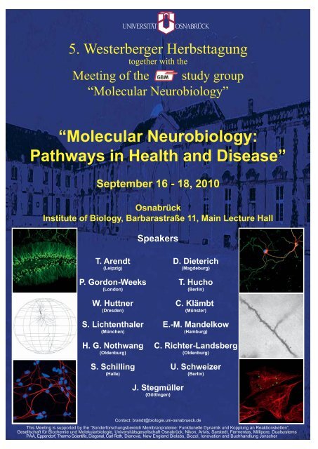
“Molecular Neurobiology: Pathways in Health and Disease”
“Molecular Neurobiology: Pathways in Health and Disease”
“Molecular Neurobiology: Pathways in Health and Disease”

You also want an ePaper? Increase the reach of your titles
YUMPU automatically turns print PDFs into web optimized ePapers that Google loves.
5. Westerberger Herbsttagung<br />
together with the<br />
Meet<strong>in</strong>g of the study group<br />
<strong>“Molecular</strong> <strong>Neurobiology</strong>”<br />
<strong>“Molecular</strong> <strong>Neurobiology</strong>:<br />
<strong>Pathways</strong> <strong>in</strong> <strong>Health</strong> <strong>and</strong> <strong>Disease”</strong><br />
September 16 - 18, 2010<br />
Osnabrück<br />
Institute of Biology, Barbarastraße 11, Ma<strong>in</strong> Lecture Hall<br />
T. Arendt<br />
(Leipzig)<br />
P. Gordon-Weeks<br />
(London)<br />
W. Huttner<br />
(Dresden)<br />
S. Lichtenthaler<br />
(München)<br />
Speakers<br />
D. Dieterich<br />
(Magdeburg)<br />
T. Hucho<br />
(Berl<strong>in</strong>)<br />
C. Klämbt<br />
(Münster)<br />
E.-M. M<strong>and</strong>elkow<br />
(Hamburg)<br />
H. G. Nothwang C. Richter-L<strong>and</strong>sberg<br />
(Oldenburg) (Oldenburg)<br />
S. Schill<strong>in</strong>g<br />
(Halle)<br />
J. Stegmüller<br />
(Gött<strong>in</strong>gen)<br />
U. Schweizer<br />
(Berl<strong>in</strong>)<br />
Contact: br<strong>and</strong>t@biologie.uni-osnabrueck.de<br />
This Meet<strong>in</strong>g is supported by the “Sonderforschungsbereich Membranprote<strong>in</strong>e: Funktionelle Dynamik und Kopplung an Reaktionsketten”,<br />
Gesellschaft für Biochemie und Molekularbiologie, Universitätsgesellschaft Osnabrück, Nikon, Arivis, Sarstedt, Fermentas, Millipore, Dualsystems<br />
PAA, Eppendorf, Thermo Scientific, Diagonal, Carl Roth, Dianova, New Engl<strong>and</strong> Biolabs, Biozol, Ionovation <strong>and</strong> Buchh<strong>and</strong>lung Jonscher
Dear Collegues,<br />
it is our pleasure to welcome you to the 5 th Westerberg Meet<strong>in</strong>g<br />
(“Westerberger Herbsttagung zu den Perspektiven der molekularen<br />
Neurobiologie”) on the Science Campus of the University of Osnabrück. This<br />
year, the Westerberg Meet<strong>in</strong>g is organized together with the Study Group<br />
<strong>“Molecular</strong> <strong>Neurobiology</strong>” of the GBM (Gesellschaft für Biochemie und<br />
Molekularbiologie). The Study Group was founded <strong>in</strong> 1977 as one of the first<br />
<strong>and</strong> def<strong>in</strong>es itself as a population of scientists with the aim to approach the<br />
function of the nervous system on molecular level. The topics are discussed <strong>in</strong><br />
a frame of biyearly Meet<strong>in</strong>gs, which take place at the location of the Speaker<br />
of the Study Group.<br />
The Meet<strong>in</strong>g is entitled "Molecular <strong>Neurobiology</strong>: <strong>Pathways</strong> <strong>in</strong> <strong>Health</strong> <strong>and</strong><br />
Disease" <strong>and</strong> the aim for the next two days is to present new results as<br />
lectures, progress talks or posters, to facilitate contacts between established<br />
<strong>and</strong> young researchers, <strong>and</strong> to exchange <strong>in</strong>formation <strong>and</strong> material on all<br />
levels. A special focus of the Study Group as well as of the biyearly<br />
Westerberg Meet<strong>in</strong>gs is to promote young researchers. We hope to achieve<br />
this by <strong>in</strong>vit<strong>in</strong>g both, established researchers <strong>and</strong> young scientists, from all<br />
over Germany <strong>and</strong> neighbor<strong>in</strong>g countries. In addition, we are putt<strong>in</strong>g a special<br />
emphasis on Poster presentations <strong>in</strong>clud<strong>in</strong>g award<strong>in</strong>g with attractive Poster<br />
prizes. In the tradition of our Westerberg Meet<strong>in</strong>gs we try our best to provide a<br />
familiar <strong>and</strong> <strong>in</strong>formal atmosphere, which can be also witnessed from the<br />
booklets of the previous Meet<strong>in</strong>gs. (available at http://www.biologie.uniosnabrueck.de/Neurobiologie/<br />
neurobiol/General_<strong>in</strong>fo.html).<br />
In the City of Peace, Osnabrück, the history is apparent on every corner, with<br />
the rema<strong>in</strong>s of the old city wall <strong>and</strong> its watchtowers, the castle dat<strong>in</strong>g from the<br />
17th century <strong>and</strong> the town hall where the Peace of Westphalia was declared<br />
<strong>in</strong> 1648. The young University of Osnabrück, which was founded <strong>in</strong> 1973,<br />
has about 10,000 students with more than 1000 students on the Science<br />
Campus, which is located <strong>in</strong> the "Westerberg" area.<br />
We are delighted to welcome all<br />
of you <strong>in</strong> Osnabrück. We thank<br />
you for your contribution <strong>and</strong><br />
participation <strong>and</strong> look forward to<br />
an excit<strong>in</strong>g Meet<strong>in</strong>g.<br />
Rol<strong>and</strong> Br<strong>and</strong>t<br />
(on behalf of the organiz<strong>in</strong>g<br />
committee)<br />
1
Program: “5. Westerberger Herbsttagung” together with the<br />
“Meet<strong>in</strong>g of the GBM Study group ‘Molecular <strong>Neurobiology</strong>’”<br />
<strong>“Molecular</strong> <strong>Neurobiology</strong>: <strong>Pathways</strong> <strong>in</strong> <strong>Health</strong> <strong>and</strong> <strong>Disease”</strong><br />
Thursday, September 16, 2010<br />
From 16:00 Registration<br />
18:30 Buffet<br />
19:30 Open<strong>in</strong>g of the Meet<strong>in</strong>g <strong>and</strong> welcome addresses (Rol<strong>and</strong> Br<strong>and</strong>t,<br />
Department of <strong>Neurobiology</strong>, <strong>and</strong> May-Britt Kallenrode, Vice<br />
president for Research, University of Osnabrück)<br />
20:00 Keynote lecture: Wiel<strong>and</strong> Huttner (Max-Planck-Institute of<br />
Molecular Cell Biology <strong>and</strong> Genetics, Dresden): “Cell biology of<br />
neural stem <strong>and</strong> progenitor cells”<br />
21:00 Get together with dr<strong>in</strong>ks <strong>and</strong> live music (“Sturm und Klang”)<br />
Friday, September 17, 2010<br />
Session I: Neuronal development (Chair: Thomas Arendt)<br />
09:00 - 09:30 Lecture 1: Phillip Gordon-Weeks (K<strong>in</strong>g's College, London, GB):<br />
“Microtubule-Act<strong>in</strong> Filament Coupl<strong>in</strong>g <strong>in</strong> Axon Guidance”<br />
09:30 - 10:00 Lecture 2: Daniela Dieterich (Leibniz Institute for <strong>Neurobiology</strong>,<br />
Magdeburg): “Profil<strong>in</strong>g neuron-glia communication with a 'click'”<br />
10:00 - 10:15 Progress talk 1: Anna-Lena Hillje (Center for Molecular Biology<br />
of Inflammation, Münster): “Inhibition of TRIM32 <strong>in</strong>duced neural<br />
stem cell differentiation by k<strong>in</strong>ase <strong>in</strong>dependent function of<br />
atypical prote<strong>in</strong> k<strong>in</strong>ase C”<br />
10:15 - 10:45 Coffee break<br />
10:45 - 11:15 Lecture 3: Christian Klämbt (University of Münster): <strong>“Molecular</strong><br />
control of glial migration <strong>in</strong> the Drosophila PNS”<br />
11:15 - 11:45 Lecture 4: Judith Stegmüller (Max-Planck-Institute of<br />
Experimental Medic<strong>in</strong>e, Gött<strong>in</strong>gen): “Novel roles for cell cycle<br />
regulators <strong>in</strong> neuronal morphogenesis”<br />
11:45 - 12:00 Progress talk 2: Eva Bunk (Center for Molecular Biology of<br />
Inflammation, Münster): “Cellular organization of adult<br />
neurogenesis <strong>in</strong> the common marmoset”<br />
12:00 - 15:00 Lunch buffet <strong>and</strong> Poster view<strong>in</strong>g (Poster presentation: 13-14 for<br />
even numbers; 14-15 for odd numbers)<br />
Session II: Ma<strong>in</strong>tenance <strong>and</strong> plasticity of the nervous system<br />
(Chair: Christiane Richter-L<strong>and</strong>sberg)<br />
15:00 - 15:30 Lecture 5: Hans Gerd Nothwang (University of Oldenburg):<br />
“Cation chloride cotransporters: reciprocal regulation <strong>and</strong><br />
structural organization”<br />
2
15:30 - 16:00 Lecture 6: Stephan Schill<strong>in</strong>g (Probiodrug AG, Halle):<br />
“Glutam<strong>in</strong>yl cyclases <strong>and</strong> pyroglutamate-modified peptides -<br />
potential functions <strong>in</strong> neurophysiology <strong>and</strong> neurodegenerative<br />
diseases”<br />
16:00 - 16:30 Coffee break<br />
16:30 - 17:00 Lecture 7: Ulrich Schweizer (Charité Universitätsmediz<strong>in</strong>,<br />
Berl<strong>in</strong>):<br />
“Selenium-conta<strong>in</strong><strong>in</strong>g prote<strong>in</strong>s <strong>in</strong> neurobiology”<br />
17:00 - 17:30 Lecture 8: Tim Hucho (Max-Planck-Institute for Molecular<br />
Genetics, Berl<strong>in</strong>): “Nociceptive signal<strong>in</strong>g modules <strong>in</strong> pa<strong>in</strong><br />
sensitization”<br />
17:30 - 17:45 Progress talk 3: Kirsten Oesterw<strong>in</strong>d (Frankfurt University<br />
Medical School): “Participation of hematopoietic cells <strong>in</strong> Cre<br />
mediated recomb<strong>in</strong>ation of Purk<strong>in</strong>je neurons”<br />
17:45 - 18:15 Poster view<strong>in</strong>g (<strong>in</strong>cl. bus<strong>in</strong>ess meet<strong>in</strong>g)<br />
18:15 - 18:30 Poster prize<br />
From 18:30 D<strong>in</strong>ner buffet <strong>and</strong> social program (night watch tour starts at 22:00<br />
at the old city hall)<br />
Saturday, September 18, 2010<br />
Session III: Neurodegeneration (Chair: Phillip Gordon-Weeks)<br />
09:00 - 09:30 Lecture 9: Thomas Arendt (Paul-Flechsig-Institut für<br />
Hirnforschung, Universität Leipzig): “The ‘Dr. Jekyll <strong>and</strong> Mr. Hyde<br />
concept’ of Alzheimer´s disease - The l<strong>in</strong>k between plasticity <strong>and</strong><br />
neurodegeneration”<br />
09:30 - 10:00 Lecture 10: Eva-Maria M<strong>and</strong>elkow (Max-Planck-Arbeitsgruppe<br />
für strukturelle Molekularbiologie, Hamburg): „Tau Pathology <strong>in</strong><br />
Alzheimer Disease: Lessons from <strong>in</strong>ducible cell models <strong>and</strong><br />
transgenic mice”<br />
10:00 - 10:15 Progress talk 4: Anne Gauthier-Kemper (University of<br />
Osnabrück): “The frontotemporal dementia mutation R406W<br />
blocks tau’s <strong>in</strong>teraction with the plasma membrane <strong>and</strong> affects<br />
tau distribution <strong>in</strong> an annex<strong>in</strong> A2-dependent manner”<br />
10:15 - 10:45 Coffee break<br />
10:45 - 11:15 Lecture 11: Stefan Lichtenthaler (Deutsches Zentrum für<br />
Neurodegenerative Erkrankungen, München): “When ADAM<br />
needs activation - Metalloproteases <strong>in</strong> Alzheimer’s disease“<br />
11:15 - 11:45 Lecture 12: Christiane Richter-L<strong>and</strong>sberg (University of<br />
Oldenburg): “Cell stress <strong>and</strong> cell death: Inclusion body formation<br />
<strong>in</strong> oligodendrocytes”<br />
11:45 Clos<strong>in</strong>g remarks<br />
3
Abstracts: Oral Presentations<br />
Abstracts:<br />
Oral<br />
Presentations<br />
4
Keynote lecture<br />
Cell biology of neural stem <strong>and</strong> progenitor cells: lissencephalic<br />
versus gyrencephalic cortex<br />
Véronique Dubreuil, Anne-Marie Marzesco, Denis Corbeil, Michaela Wilsch-<br />
Braeun<strong>in</strong>ger, Jennifer L. Fish, Federico Calegari, Judith Schenk, Denise Stenzel,<br />
Simone A. Fietz, Iva Kelava, Lilla M. Farkas, Christiane Haffner, Yoichi Kosodo, Alessio<br />
Attardo, Felipe Mora-Bermudez, Wiel<strong>and</strong> Huttner<br />
Huttner Laboratory, Max Planck Institute of Molecular Cell Biology <strong>and</strong> Genetics,<br />
Dresden, Germany<br />
Key words: neural stem cells, neurogenesis, neocortex<br />
Our group studies the cell biological mechanisms of neurogenesis <strong>in</strong> the context of<br />
mammalian bra<strong>in</strong> evolution, specifically the proliferation versus differentiation of neural<br />
stem <strong>and</strong> progenitors cells. In the develop<strong>in</strong>g rodent cortex, two pr<strong>in</strong>cipal classes of<br />
these cells can be dist<strong>in</strong>guished, (i) progenitors divid<strong>in</strong>g at the ventricular, i.e. apical,<br />
surface of the ventricular zone (VZ), called apical progenitors (APs, i.e. neuroepithelial<br />
cells <strong>and</strong> radial glial cells), <strong>and</strong> (ii) progenitors divid<strong>in</strong>g <strong>in</strong> a more basal, abventricular<br />
location, notably the subventricular zone (SVZ), called basal progenitors (BPs, also<br />
called <strong>in</strong>termediate progenitors). Focus<strong>in</strong>g on the embryonic mouse cortex, we have<br />
been study<strong>in</strong>g the follow<strong>in</strong>g issues:(1) the neurogenic l<strong>in</strong>eage from APs <strong>and</strong> BPs to<br />
neurons, <strong>and</strong> the underly<strong>in</strong>g molecular mach<strong>in</strong>ery, us<strong>in</strong>g the marker Tis21;(2) the role<br />
<strong>and</strong> mach<strong>in</strong>ery of <strong>in</strong>terk<strong>in</strong>etic nuclear migration;(3) apical-basal cell polarity, cleavage<br />
plane orientation <strong>and</strong> symmetric versus asymmetric division of APs <strong>and</strong> BPs;(4) the role<br />
of the microcephaly gene Aspm <strong>in</strong> symmetric AP divisions;(5) apical membrane<br />
constituents, notably the cholesterol b<strong>in</strong>d<strong>in</strong>g prote<strong>in</strong> prom<strong>in</strong><strong>in</strong>-1/CD133;(6) prom<strong>in</strong><strong>in</strong>-1–<br />
bear<strong>in</strong>g extracellular membrane particles relased <strong>in</strong>to the ventricular fluid from the<br />
midbody <strong>and</strong> primary cilium of APs;(7) the basal process of APs <strong>in</strong> mitosis;(8) the role of<br />
cell cycle length <strong>in</strong> stem <strong>and</strong> progenitor cell proliferation versus differentiation.We have<br />
recently extended our <strong>in</strong>vestigations from the lissencephalic mouse model to species<br />
develop<strong>in</strong>g a gyrencephalic cortex. We f<strong>in</strong>d that progenitors <strong>in</strong> the outer SVZ (OSVZ) of<br />
human <strong>and</strong> ferret ma<strong>in</strong>ta<strong>in</strong> a basal process contact<strong>in</strong>g the basal lam<strong>in</strong>a. This epithelial<br />
feature allows <strong>in</strong>tegr<strong>in</strong>-mediated, repeated asymmetric divisions of OSVZ progenitors,<br />
which provides a basis for neocortical expansion.<br />
Supported by: DFG(SPP 1109, HU 275/7-3; SPP 1111, HU 275/8-3, SFB/TR 13, B1!SFB 655<br />
A2!CRTD!Fonds der Chemischen Industrie!BMBF!NGFN-2, SMO RNAi, 01GR0402, PRI-S08T05!<br />
*Correspond<strong>in</strong>g author: huttner@mpi-cbg.de<br />
Abstracts: Oral Presentations<br />
5
Microtubule-Act<strong>in</strong> Filament Coupl<strong>in</strong>g <strong>in</strong> Axon Guidance<br />
Philip R. Gordon-Weeks<br />
Abstracts: Oral Presentations<br />
MRC, Centre for Developmental <strong>Neurobiology</strong>, K<strong>in</strong>g's College, London, Engl<strong>and</strong><br />
O-01<br />
A diverse range of cellular processes <strong>in</strong>clud<strong>in</strong>g cell division, directed cell motility,<br />
neuritogenesis <strong>and</strong> growth cone pathf<strong>in</strong>d<strong>in</strong>g depend on the regulated <strong>in</strong>teraction<br />
between dynamic microtubules <strong>and</strong> act<strong>in</strong> filaments. The molecular mechanisms<br />
mediat<strong>in</strong>g this <strong>in</strong>teraction, the prote<strong>in</strong>s <strong>in</strong>volved <strong>and</strong> how they are regulated, are now<br />
be<strong>in</strong>g discovered. In growth cones, dynamic microtubules <strong>in</strong>teract with the act<strong>in</strong><br />
filaments with<strong>in</strong> filopodia <strong>and</strong> this <strong>in</strong>teraction depends on the direct b<strong>in</strong>d<strong>in</strong>g of the +TIP<br />
prote<strong>in</strong> EB3, located on the plus-end of microtubules, <strong>and</strong> the F-act<strong>in</strong>-b<strong>in</strong>d<strong>in</strong>g prote<strong>in</strong><br />
drebr<strong>in</strong>, bound to the proximal ends of filopodial act<strong>in</strong> filaments. Disruption of this<br />
<strong>in</strong>teraction impairs growth cone formation <strong>and</strong> the extension of neurites but its role <strong>in</strong><br />
growth cone pathf<strong>in</strong>d<strong>in</strong>g has yet to be determ<strong>in</strong>ed. Doma<strong>in</strong> mapp<strong>in</strong>g analysis of drebr<strong>in</strong><br />
has revealed the presence of two F-act<strong>in</strong>-b<strong>in</strong>d<strong>in</strong>g doma<strong>in</strong>s whose properties expla<strong>in</strong> the<br />
specific location of drebr<strong>in</strong> to parallel act<strong>in</strong> filament bundles. In the adult nervous system<br />
drebr<strong>in</strong> regulates F-act<strong>in</strong> dynamics <strong>in</strong> dendritic sp<strong>in</strong>es <strong>and</strong> loss of drebr<strong>in</strong> from sp<strong>in</strong>es is<br />
causal to the loss of dendritic sp<strong>in</strong>es that underlies cognitive impairment <strong>in</strong> diseases<br />
such as Alzheimer's. Future work will focus on the regulation of drebr<strong>in</strong> <strong>in</strong> these different<br />
cellular contexts.<br />
*Correspond<strong>in</strong>g author: phillip.gordon-weeks@kcl.ac.uk<br />
6
Profil<strong>in</strong>g neuron-glia communication with a 'click'<br />
Anke Müller (1), Oliver Kobler (2), Ines Erdmann (1), Ulrich Thomas (2), Daniela C.<br />
Dieterich (1)<br />
(1) RG Neuralomics, Leibniz Institute for <strong>Neurobiology</strong>, Magdeburg, Germany; (2)<br />
Department of Neurochemistry, Leibniz Institute for <strong>Neurobiology</strong>, Magdeburg,<br />
Germany<br />
O-02<br />
Key words: metabolic label<strong>in</strong>g, proteomics, astrocytes, Drosophila melanogaster, <strong>in</strong> situ visualization<br />
Dynamic prote<strong>in</strong> synthesis is a common feature of cells to react to changes <strong>in</strong> their<br />
environment <strong>and</strong> is, therefore, of particular <strong>in</strong>terest. To enable label<strong>in</strong>g of newly<br />
synthesized prote<strong>in</strong>s, we use a recently <strong>in</strong>troduced technique, which is called BONCAT<br />
(bioorthogonal non-canonical am<strong>in</strong>o acid tagg<strong>in</strong>g). Here, the use of the azide-carry<strong>in</strong>g<br />
methion<strong>in</strong>e surrogate azidohomoalan<strong>in</strong>e (AHA), which can be <strong>in</strong>tegrated <strong>in</strong>to prote<strong>in</strong>s by<br />
the endogenous prote<strong>in</strong> synthesis mach<strong>in</strong>ery, allows coupl<strong>in</strong>g to aff<strong>in</strong>ity or fluorescent<br />
alkyne-tags via copper-catalyzed click chemistry, <strong>and</strong>, thus, facilitates the isolation or <strong>in</strong><br />
situ detection of newly synthesized prote<strong>in</strong>s. To identify changes <strong>in</strong> prote<strong>in</strong> synthesis <strong>in</strong><br />
mammalian heterologous cell culture systems, we developed GINCAT (genetically<br />
<strong>in</strong>troduced non-canonical am<strong>in</strong>o acid tagg<strong>in</strong>g). Different am<strong>in</strong>o acid residues of the<br />
methionyl-tRNA-Synthethase (MetRS) were exchanged to enable the <strong>in</strong>corporation of<br />
the modified am<strong>in</strong>o acid azidonorleuc<strong>in</strong>e (ANL) <strong>and</strong> the subsequent tagg<strong>in</strong>g by click<br />
chemistry. A s<strong>in</strong>gle am<strong>in</strong>o acid substitution with<strong>in</strong> the b<strong>in</strong>d<strong>in</strong>g pocket for methion<strong>in</strong>e of<br />
MetRS was found to be most effective for ANL <strong>in</strong>corporation <strong>in</strong>to prote<strong>in</strong>s, <strong>and</strong> was<br />
further used to analyze any cellular <strong>and</strong> morphological consequences of ANL <strong>in</strong>tegration<br />
<strong>in</strong>to prote<strong>in</strong>s. Incorporation of ANL is specific for cells carry<strong>in</strong>g the mutated enzyme.<br />
These results prompt the idea to use GINCAT <strong>and</strong> BONCAT for the analyses of cell<br />
specific proteomes <strong>in</strong> a complex cellular environment such as glia-neuron co-cultures,<br />
<strong>and</strong>, therefore, may improve our underst<strong>and</strong><strong>in</strong>g of neuron-glia communication <strong>and</strong><br />
<strong>in</strong>teraction both <strong>in</strong> vitro <strong>and</strong> <strong>in</strong> vivo. As a demonstration of the suitability of this<br />
approach we use mammalian neuron-glia primary cocultures as well as the Gal4-UAS<br />
expression system <strong>in</strong> Drosophila melanogaster to express the mutant LtoG-MetRS<br />
fused <strong>in</strong> astrocytes, or <strong>in</strong> neurons, glia <strong>and</strong> <strong>in</strong> muscle cells, respectively.<br />
Supported by: DFG Emmy Noether Program <strong>and</strong> SPP1172 to DCD<br />
*Correspond<strong>in</strong>g author: dieterd@ifn-magdeburg.de<br />
Abstracts: Oral Presentations<br />
7
Molecular control of glial migration <strong>in</strong> the Drosophila PNS<br />
Marion Silies <strong>and</strong> Christian Klämbt<br />
University of Münster, Münster, Germany<br />
O-03<br />
Coord<strong>in</strong>ated cell migration is a key feature dur<strong>in</strong>g the development of multicellular<br />
organisms. This is of particular importance with<strong>in</strong> the peripheral nervous system where<br />
migrat<strong>in</strong>g glial cells follow grow<strong>in</strong>g axons <strong>in</strong>to the periphery. Axonal outgrowth <strong>and</strong> glial<br />
migration thus have to be well coord<strong>in</strong>ated, but the underly<strong>in</strong>g molecular mechanisms<br />
are unknown. Here we demonstrate that a subcellular, axonal gradient of the Igsuperfamily<br />
cell adhesion prote<strong>in</strong> Fascicl<strong>in</strong> 2 (Fas2) allows glial migration <strong>in</strong> the<br />
Drosophila PNS. The homophilic adhesion prote<strong>in</strong> Fas2 <strong>in</strong>teracts with a GPI-l<strong>in</strong>ked glial<br />
Fas2 isoform. Glial migration is <strong>in</strong>itiated along axonal segments that present low Fas2<br />
levels but stalls <strong>in</strong> axonal doma<strong>in</strong>s decorated with high levels of Fas2. We further<br />
demonstrate that the graded distribution of Fas2 <strong>in</strong> axons is brought about by a novel<br />
function to the anaphase promot<strong>in</strong>g complex / cyclosome (APC/C) co-activator Fizzyrelated/Cdh1<br />
(Fzr/Cdh1). In fzr mutants the graded distribution of Fas2 is lost. Fzr/Cdh1<br />
is required <strong>in</strong> postmitotic motor neurons Fzr/Cdh1 <strong>and</strong> acts through the APC/C to nonautonomously<br />
allow glial migration. Our data suggest an elegant mechanism <strong>in</strong> which<br />
modulation of adhesion prote<strong>in</strong> expression through Fzr/Cdh1 creates a subcellular<br />
gradient of adhesiveness to coord<strong>in</strong>ate glial migration with axonal growth.<br />
*Correspond<strong>in</strong>g author: klaembt@uni-muenster.de<br />
Abstracts: Oral Presentations<br />
8
O-04<br />
Novel roles for cell cycle regulators <strong>in</strong> neuronal morphogenesis<br />
Judith Stegmüller<br />
Max Planck Institute of Experimental Medic<strong>in</strong>e, Gött<strong>in</strong>gen, Germany<br />
Axon growth is a crucial event that ensures correct wir<strong>in</strong>g <strong>in</strong> the bra<strong>in</strong>. Grow<strong>in</strong>g<br />
evidence implicates the ubiquit<strong>in</strong> proteasome system (UPS) <strong>in</strong> neurodevelopment. We<br />
found that the E3 ubiquit<strong>in</strong> ligase Cdh1-Anaphase Promot<strong>in</strong>g Complex (APC) plays an<br />
important role <strong>in</strong> axon growth control. By target<strong>in</strong>g the transcriptional regulator SnoN for<br />
proteasomal degradation, Cdh1-APC suppresses axon growth. S<strong>in</strong>ce Cdh1-APC activity<br />
<strong>and</strong> regulation has been well studied <strong>in</strong> mitotic cells, where the E3 ligase acts as a<br />
pivotal cell cycle regulator, we reasoned that regulatory mechanisms of Cdh1-APC<br />
might be conserved <strong>in</strong> neurons. Here, we exam<strong>in</strong>ed the mutual regulation of two cell<br />
cycle regulators <strong>in</strong> axonal morphogenesis, Cdh1-APC <strong>and</strong> the Cull<strong>in</strong>1-based SCF<br />
complex FBXO31-SCF (Skp1/Cull<strong>in</strong>1/F-box prote<strong>in</strong> complex). We found that Cdh1<br />
associates with FBXO31 <strong>and</strong> that FBXO31 is degraded <strong>in</strong> a proteasome dependent<br />
manner. Us<strong>in</strong>g an RNAi approach, we acutely knocked down FBXO31 <strong>in</strong> neurons <strong>and</strong><br />
determ<strong>in</strong>ed axonal length. While we did not f<strong>in</strong>d a difference between a non-functional<br />
RNAi plasmid <strong>and</strong> control, we found a significant decrease <strong>in</strong> axonal length with two<br />
functional FBXO31 RNAi plasmids. In addition, we found <strong>in</strong> ga<strong>in</strong>-of-function analyses<br />
that FBXO31 promotes axon growth. Also, an FBXO31 form that lacks the F-box does<br />
not stimulate axon growth, suggest<strong>in</strong>g that E3 ligase activity is required for FXBO31’s<br />
axon growth control. In epistasis analyses, we determ<strong>in</strong>ed that FBXO31 acts<br />
downstream of Cdh1-APC. Taken together, we have identified FBXO31-SCF as a novel<br />
component of the Cdh1-APC pathway of axonal morphogenesis.<br />
*Correspond<strong>in</strong>g author: stegmueller@em.mpg.de<br />
Abstracts: Oral Presentations<br />
9
O-05<br />
Cation chloride cotransporters: reciprocal regulation <strong>and</strong> structural<br />
organisation<br />
Hans Gerd Nothwang<br />
Abstracts: Oral Presentations<br />
Department of Neurogenetics, Carl von Ossietzky Universität Oldenburg, Oldenburg,<br />
Germany<br />
Cation-chloride cotransporters (CCCs) play pivotal roles <strong>in</strong> the nervous system <strong>and</strong><br />
sensory organs. The two ma<strong>in</strong> branches consist of the Cl - extruders KCC1 to KCC4 <strong>and</strong><br />
of the Cl - <strong>in</strong>ward transporters NKCC1, NKCC2, <strong>and</strong> NCC. NKCC1 <strong>and</strong> KCC2 are the<br />
most important members of CCCs <strong>in</strong> the central nervous system, as they set the [Cl - ]i <strong>in</strong><br />
neurons. Their activity is therefore crucial for the action of GABA <strong>and</strong> Glyc<strong>in</strong>e. Because<br />
of their opposite effect on <strong>in</strong>tracellular Cl - concentration, the activity of these<br />
transporters requires a precise <strong>and</strong> coord<strong>in</strong>ated regulation. In this work, we <strong>in</strong>vestigated<br />
cellular mechanisms that contribute to their regulation. Furthermore, mutational<br />
analyses of KCCs were performed to ga<strong>in</strong> <strong>in</strong>sight <strong>in</strong>to their structural organization.<br />
Biochemical analyses revealed that membrane-rafts regulate the activity of NKCC1 <strong>and</strong><br />
KCC2 <strong>in</strong> a reciprocal way. NKCC1 is ma<strong>in</strong>ly localized <strong>in</strong> membrane-rafts. In contrast,<br />
KCC2 is localized both <strong>in</strong> membrane-rafts <strong>and</strong> non-membrane rafts <strong>in</strong> the mature bra<strong>in</strong>.<br />
Disruption of membrane rafts activated KCC2, whereas NKCC1 was ma<strong>in</strong>ly <strong>in</strong>activated.<br />
Transport <strong>in</strong>active KCC2 <strong>in</strong> the immature bra<strong>in</strong> is exclusively localized <strong>in</strong> membranerafts.<br />
Structure-function analyses of the large extracellular loop (LEL) revealed different<br />
roles of evolutionary conserved cyste<strong>in</strong>es <strong>in</strong> KCC2 <strong>and</strong> KCC4. Mutation of all four<br />
cyste<strong>in</strong>es abolished KCC2 transport activity, whereas the same mutation <strong>in</strong> the<br />
paralogous KCC4 had no effect. Analyses of chimera demonstrated that the LEL of<br />
KCC2 requires the parental backbone for proper function, whereas the LEL of KCC4<br />
acts autonomously.<br />
*Correspond<strong>in</strong>g author: hans.g.nothwang@uni-oldenburg.de<br />
10
Abstracts: Oral Presentations<br />
O-06<br />
Glutam<strong>in</strong>yl cyclases <strong>and</strong> pyroglutamate-modified peptides – potential<br />
functions <strong>in</strong> neurophysiology <strong>and</strong> neurodegenerative diseases<br />
Stephan Schill<strong>in</strong>g* (1), Holger Cynis (1), Raik Rönicke (2), Klaus Reymann (2),<br />
Franziska Seifert (1), Dagmar Schlenzig (1), Sigrid Graubner (3), Birgit Hutter-Paier (4)<br />
<strong>and</strong> Hans-Ulrich Demuth (1,3)<br />
(1) Probiodrug AG, Halle/Saale, Germany; (2) FAN GmbH, Magdeburg, Germany; (3)<br />
Ingenium Pharmaceuticals, Munich, Germany; (4) JSW Life Scences, Graz, Austria<br />
Pyroglutamate (pGlu)-modified Aβ peptides are found <strong>in</strong> amyloid deposits <strong>in</strong> sporadic<br />
<strong>and</strong> <strong>in</strong>herited Alzheimer's disease. Similar to AD, the <strong>in</strong>herited neurodegenerative<br />
disorders Familial British <strong>and</strong> Danish dementia are also characterized by accumulation<br />
of pGlu-modified amyloid <strong>in</strong> plaques. We could show, that N-term<strong>in</strong>al truncation <strong>and</strong><br />
formation of pGlu at the N-Term<strong>in</strong>us <strong>in</strong>creases the toxicity of the peptides <strong>and</strong> speeds<br />
up Aβ aggregate formation. An enhanced hydrophobicity of N-truncated Aβ most likely<br />
accounts for a rapid oligomer formation <strong>and</strong> <strong>in</strong>creased potency of N-truncated Aβ to<br />
<strong>in</strong>terfere with hippocampal LTP. Our <strong>in</strong> vitro <strong>and</strong> <strong>in</strong> vivo studies provided evidence for a<br />
slow Glutam<strong>in</strong>yl cyclase (QC, EC 2.3.2.5) catalyzed cyclization of N-term<strong>in</strong>al glutamic<br />
acid, suggest<strong>in</strong>g a crucial role for generation of pGlu-Aβ peptides. To further<br />
substantiate the role of pGlu-Aβ <strong>in</strong> mouse models, we treated novel mouse models of<br />
AD with QC-<strong>in</strong>hibitors <strong>and</strong> generated QC-transgenic <strong>and</strong> knock-out mice for crossbreed<strong>in</strong>g<br />
<strong>and</strong> evaluation of potential target-related side effects. The data suggest that<br />
QC-catalyzed pGlu-formation can be suppressed without expectation of severe side<br />
effects, which provides potential for development of a treatment paradigm to prevent<br />
Aβ- <strong>and</strong> <strong>in</strong>flammation-driven pathophysiology <strong>in</strong> AD, FDD <strong>and</strong> FBD.<br />
*Correspond<strong>in</strong>g author: stephan.schill<strong>in</strong>g@probiodrug.de<br />
11
Ulrich Schweizer<br />
Abstracts: Oral Presentations<br />
Selenium-conta<strong>in</strong><strong>in</strong>g prote<strong>in</strong>s <strong>in</strong> neurobiology<br />
O-07<br />
Institut für Experimentelle Endokr<strong>in</strong>ologie, Charité-Universitätsmediz<strong>in</strong>, Berl<strong>in</strong>, Germany<br />
Key words: selenoprote<strong>in</strong>s, epilepsy, <strong>in</strong>terneurons, gaba, parvalbum<strong>in</strong><br />
Prote<strong>in</strong>s conta<strong>in</strong><strong>in</strong>g the rare am<strong>in</strong>o acid selenocyste<strong>in</strong>e (Sec) are called selenoprote<strong>in</strong>s.<br />
Sec is <strong>in</strong>corporated <strong>in</strong>to prote<strong>in</strong>s co-translationally, <strong>and</strong> is encoded by the UGA codon.<br />
Hence, Sec is the 21 st prote<strong>in</strong>ogenic am<strong>in</strong>o acid <strong>in</strong> mammals. A decicated metabolic<br />
pathway consist<strong>in</strong>g of re-cod<strong>in</strong>g factors <strong>and</strong> biosynthetic factors for Sec have been<br />
identified. Among selenoenzymes, glutathione peroxidases (GPx), thioredox<strong>in</strong><br />
reductases (TrxR), <strong>and</strong> iodothyron<strong>in</strong>e deiod<strong>in</strong>ases (Dio) are the best known. We have<br />
<strong>in</strong>activated the gene encod<strong>in</strong>g selenoprote<strong>in</strong> P (SePP), the major Se transport prote<strong>in</strong>,<br />
<strong>and</strong> discovered a novel role of Se for bra<strong>in</strong> function. Cerebral Se deficiency is<br />
associated with neurological phenotypes <strong>in</strong>clud<strong>in</strong>g seizures <strong>and</strong> ataxia. We wanted to<br />
def<strong>in</strong>e (i) whether neurons require selenoprote<strong>in</strong> expression, (ii) which selenoprote<strong>in</strong>(s)<br />
is/are most important, <strong>and</strong> (iii) explore the possible pathomechanism. Therefore, we<br />
abrogated the expression of all selenoprote<strong>in</strong>s <strong>in</strong> neurons by genetic <strong>in</strong>activation of the<br />
tRNA[Ser]Sec gene. Cerebral expression of selenoprote<strong>in</strong>s was significantly dim<strong>in</strong>ished<br />
<strong>in</strong> the mutants <strong>and</strong> histological analysis revealed progressive neurodegeneration.<br />
Develop<strong>in</strong>g <strong>in</strong>terneurons failed to specifically express parvalbum<strong>in</strong> (PV) <strong>in</strong> the mutants.<br />
Electrophysiological record<strong>in</strong>gs, before overt cell death, showed normal excitatory<br />
transmission, but revealed spontaneous epileptiform activity consistent with seizures <strong>in</strong><br />
the mutants. We then analyzed mice lack<strong>in</strong>g neuronal expression of the Se-dependent<br />
enzyme GPx4. Aga<strong>in</strong>, the number of PV+ <strong>in</strong>terneurons was reduced. A similar<br />
phenotype is found <strong>in</strong> Sepp -/- mice. A selective loss of PV+ neurons is observed <strong>in</strong><br />
schizophrenic patients <strong>and</strong> rodent models of schizophrenia. Interest<strong>in</strong>gly, one animal<br />
model <strong>in</strong>volves adm<strong>in</strong>istration of an <strong>in</strong>hibitor of GSH synthesis – the cofactor of GPx4.<br />
We are thus follow<strong>in</strong>g the hypothesis that metabolic <strong>in</strong>sufficiency <strong>in</strong> selenoprote<strong>in</strong><br />
expression may contribute to neuropsyciatric disease <strong>in</strong> humans.<br />
*Correspond<strong>in</strong>g author: ulrich.schweizer@charite.de<br />
12
Tim Hucho<br />
Nociceptive signal<strong>in</strong>g modules <strong>in</strong> pa<strong>in</strong> sensitization<br />
Max Planck Institute for Molecular Genetics, Berl<strong>in</strong>, Germany<br />
O-08<br />
Progress <strong>in</strong> identify<strong>in</strong>g novel extracellular mediators <strong>and</strong> ion channels <strong>in</strong>volved <strong>in</strong> pa<strong>in</strong><br />
sensitization is tramendous. In contrast the underst<strong>and</strong><strong>in</strong>g of the underly<strong>in</strong>g signal<strong>in</strong>g<br />
network establish<strong>in</strong>g <strong>and</strong> ma<strong>in</strong>ta<strong>in</strong><strong>in</strong>g sensitization is still rather sparse. We now<br />
attempted to identify biochemically <strong>and</strong> cell biologically novel aspects of sensitization<br />
signal<strong>in</strong>g. Recently we characterized the b<strong>in</strong>d<strong>in</strong>g site of tubul<strong>in</strong> with the C-term<strong>in</strong>us of<br />
TRPV1. Now, molecular model<strong>in</strong>g identified, that the phosphorylation of S800 at TRPV1<br />
by PKCe should <strong>in</strong>terfere with b<strong>in</strong>d<strong>in</strong>g. Indeed, phosphorylation <strong>and</strong> b<strong>in</strong>d<strong>in</strong>g appear as<br />
exclusive events. In the cellular context activation of the PKCe signal<strong>in</strong>g pathway by<br />
e.g. Estrogen ort he GPR30-selective derivate G-1 results <strong>in</strong> rapid morphological<br />
change. This change is based on a strong destabilization of the microtubular network.<br />
Destabilization was restricted purely to TRPV1 express<strong>in</strong>g cells <strong>and</strong> even further to the<br />
phosphoratability of TRPV1-S800. We therefore suggest the dynamic regulation of the<br />
microtubule-TRPV1 <strong>in</strong>teraction as novel functionality of sensitization signall<strong>in</strong>g.<br />
*Correspond<strong>in</strong>g author: hucho@molgen.mpg.de<br />
Abstracts: Oral Presentations<br />
13
O-09<br />
The ‘Dr. Jekyll <strong>and</strong> Mr Hyde concept’ of Alzheimer´s disease – The<br />
l<strong>in</strong>k between plasticity <strong>and</strong> neurodegeneration<br />
Thomas Arendt<br />
Abstracts: Oral Presentations<br />
Paul Flechsig Institute for Bra<strong>in</strong> Research, University of Leipzig, Germany<br />
Key words: Alzheimer´s disease, neurodegeneration, neuroplasticity, cell cycle, cell death<br />
Higher cerebral functions are based upon a dynamic organization of neuronal networks.<br />
To form synaptic connections <strong>and</strong> to cont<strong>in</strong>uously re-shape them <strong>in</strong> a process of<br />
ongo<strong>in</strong>g structural adaptation, neurons must permanently withdraw from the cell cycle.<br />
In other words, synaptic plasticity can only occur on the expense of the ability to<br />
proliferate. This mechanism of synaptic plasticity, i.e. of structural stabilization <strong>and</strong><br />
labilization underlay<strong>in</strong>g a life-long synaptic remodell<strong>in</strong>g are largely based on external<br />
morphoregulatory cues <strong>and</strong> <strong>in</strong>ternal signal<strong>in</strong>g pathways that non-neuronal cells have<br />
phylogenetically been acquired to sense their relationship to the local neighbourhood<br />
<strong>and</strong> to control after development is completed proliferation <strong>and</strong> differentiation <strong>in</strong> the<br />
process of tissue repair <strong>and</strong> regeneration. Here, we put forward the hypothesis that<br />
differentiated neurons after hav<strong>in</strong>g withdrawn from the cell cycle are able to use those<br />
molecular mechanisms primarily developed to control proliferation alternatively to<br />
control synaptic plasticity. The existence of these alternative effector pathways with<strong>in</strong> a<br />
neuron, puts it on the risk to erroneously convert signals derived from plastic synaptic<br />
changes <strong>in</strong>to positional cues that will activate the cell cycle. This cell cycle activation<br />
potentially l<strong>in</strong>ks synaptic plasticity to cell death. Prevent<strong>in</strong>g cell cycle activation by<br />
lock<strong>in</strong>g neurons <strong>in</strong> a differentiated but still highly plastic phenotype will, thus, be crucial<br />
to prevent neurodegeneration.<br />
Supported by: Supported by the European Commission, the German Federal M<strong>in</strong>istry of Education <strong>and</strong><br />
Research (ERA-Net Neuron; ProGen; Neuron-48-038_01EW0907) <strong>and</strong> the Sächsisches<br />
Staatsm<strong>in</strong>isterium für Wissenschaft und Kunst (4-7531.50-02-0361-07/2 <strong>and</strong> BBZ “Theranostik”-WI803-<br />
14494).<br />
*Correspond<strong>in</strong>g author: aret@mediz<strong>in</strong>.uni.leipzig.de<br />
14
Tau toxicity <strong>in</strong> cell <strong>and</strong> animal models<br />
O-10<br />
Edda Thies, Astrid Sydow, Yipeng Wang, Jacek Biernat, Eckhard M<strong>and</strong>elkow, Eva-<br />
Maria M<strong>and</strong>elkow*<br />
Max-Planck-Unit for Structural Molecular Biology c/o DESY, Hamburg, Germany<br />
Key words: Tau, neurofibrillary tangles, microtubules, Alzheimer’s disease, neurodegeneration<br />
Tau pathology is a hallmark of AD <strong>and</strong> other tauopathies. In AD, Tau is changed <strong>in</strong><br />
several ways (e.g. phosphorylation, missort<strong>in</strong>g, aggregation), but the causes of<br />
neuronal degeneration are uncerta<strong>in</strong>. Several pathways of Tau-<strong>in</strong>duced toxicity have<br />
been proposed: (1) Detachment of Tau from microtubules with subsequent breakdown<br />
of microtubules, the tracks of axonal transport. (2) Aggregation <strong>in</strong>to paired helical<br />
filaments. (3) Cluster<strong>in</strong>g of Tau on microtubules <strong>and</strong> <strong>in</strong>hibition of axonal transport. (4)<br />
Cleavage of Tau <strong>in</strong>to toxic fragments. (5) Toxic functions of modified Tau. (6) Over- or<br />
understabilization of microtubules. We generated several cell <strong>and</strong> transgenic mouse<br />
models, based on Tau mutants derived from FTDP17 mutants as well as mutants<br />
<strong>in</strong>duc<strong>in</strong>g structural changes, to dist<strong>in</strong>guish between these possibilities. Some cell<br />
models display traffic deficits upon expression of Tau, due to reduced activity of motor<br />
prote<strong>in</strong>s mov<strong>in</strong>g along microtubules. Other cell models show that toxicity of Tau is<br />
<strong>in</strong>duced by enhanc<strong>in</strong>g the beta-propensity of Tau, which leads to proteolysis <strong>and</strong><br />
aggregation, whereas anti-aggregation Tau variants are not toxic. Aggregation of Tau is<br />
reversible by <strong>in</strong>hibitor compounds or by switch<strong>in</strong>g off Tau expression. Analogous<br />
observations are made with <strong>in</strong>ducible transgenic mice, where Tau variants with high<br />
beta-propensity lead to Tau aggregation, toxicity, <strong>and</strong> decay of neurons. The exogenous<br />
Tau species can even poison endogenous mouse Tau <strong>and</strong> lead to co-aggregation of<br />
both species. Here, too, the process is partly reversible by switch<strong>in</strong>g off the expression<br />
of exogenous Tau.<br />
Supported by: MPG, DFG, KNDD, Metlife Award, EU/Memosad.<br />
*Correspond<strong>in</strong>g author: m<strong>and</strong>@mpasmb.desy.de<br />
Abstracts: Oral Presentations<br />
15
O-11<br />
When ADAM needs activation - metalloproteases <strong>in</strong> Alzheimer’s<br />
disease<br />
Stefan F. Lichtenthaler<br />
Abstracts: Oral Presentations<br />
DZNE - German Center for Neurodegenerative Diseases, Munich, Germany<br />
The amyloid precursor prote<strong>in</strong> (APP) undergoes constitutive shedd<strong>in</strong>g by a protease<br />
activity called α-secretase. This is considered a key mechanism prevent<strong>in</strong>g the<br />
generation of the Alzheimer’s disease amyloid-β peptide (Aβ). α-secretase appears to<br />
be a metalloprotease of the ADAM family, but its identity rema<strong>in</strong>s to be established.<br />
Us<strong>in</strong>g a novel α-secretase-cleavage site-specific antibody we found that RNAi-mediated<br />
knock-down of ADAM10, but surpris<strong>in</strong>gly not of ADAM9 or ADAM17, completely<br />
suppressed APP α-secretase cleavage <strong>in</strong> different cell l<strong>in</strong>es <strong>and</strong> <strong>in</strong> primary mur<strong>in</strong>e<br />
neurons. Other proteases were not able to compensate for this loss of α-cleavage. This<br />
f<strong>in</strong>d<strong>in</strong>g was further confirmed by mass-spectrometric detection of APP cleavage<br />
fragments. Surpris<strong>in</strong>gly, <strong>in</strong> different cell l<strong>in</strong>es the reduction of α-secretase cleavage was<br />
not paralleled by a correspond<strong>in</strong>g <strong>in</strong>crease <strong>in</strong> the Aβ-generat<strong>in</strong>g β-secretase cleavage,<br />
reveal<strong>in</strong>g that both proteases do not always compete for APP as a substrate. Instead<br />
our data suggest a novel pathway for APP process<strong>in</strong>g, <strong>in</strong> which ADAM10 can partially<br />
compete with γ-secretase for the cleavage of a C-term<strong>in</strong>al APP-fragment generated by<br />
β-secretase. We conclude that ADAM10 is the physiologically relevant, constitutive αsecretase<br />
of APP.<br />
*Correspond<strong>in</strong>g author: Stefan.Lichtenthaler@med.uni-muenchen.de<br />
16
O-12<br />
Cell stress <strong>and</strong> cell death: Inclusion body formation <strong>in</strong> glial cells<br />
Christiane Richter-L<strong>and</strong>sberg, Olaf Goldbaum, Sven Jänen, Hassan Chaachouay<br />
Molekulare Neurobiologie, Universität Oldenburg, Oldenburg, Germany<br />
Key words: oligodendrocyte, astrocyte, proteasome, apoptosis, heat shock prote<strong>in</strong>s<br />
Proteasomal dysfunction has been implicated <strong>in</strong> neurodegenerative disorders <strong>and</strong><br />
dur<strong>in</strong>g ag<strong>in</strong>g processes. In frontotemporal dementias (FTD), e.g. corticobasal<br />
degeneration (CBD) <strong>and</strong> progressive supranuclear palsy (PSP), oligodendrocytes, the<br />
myel<strong>in</strong> form<strong>in</strong>g cells of the CNS, are specifically damaged. Characteristic pathological<br />
features are filamentous tau <strong>in</strong>clusions conta<strong>in</strong><strong>in</strong>g heat shock prote<strong>in</strong>s (HSPs) <strong>and</strong><br />
ubiquit<strong>in</strong>. Also, tau-positive tufted astrocytes <strong>and</strong> astrocytic plaques are typical for PSP<br />
<strong>and</strong> CBD, respectively. The present studies were undertaken to elucidate the molecular<br />
mechanisms underly<strong>in</strong>g <strong>in</strong>clusion body formation <strong>in</strong> glial cells. Proteolytic stress <strong>in</strong><br />
cultured rat bra<strong>in</strong> oligodendrocytes, applied by the proteasome <strong>in</strong>hibitor MG-132,<br />
causes heat shock prote<strong>in</strong> <strong>in</strong>duction, tau-ubiquit<strong>in</strong>ation <strong>and</strong> the recruitment of ubiquit<strong>in</strong><br />
to prote<strong>in</strong> aggregates <strong>in</strong> oligodendrocytes, <strong>and</strong> leads to the <strong>in</strong>duction of apoptotic cell<br />
death. Activation of the mitochondrial pathway is <strong>in</strong>volved <strong>in</strong> the apoptotic process,<br />
which can be prevented by the broad caspase <strong>in</strong>hibitor zVAD-fmk. In contrast to<br />
oligodendrocytes, cultured astrocytes show resistance to the treatment with<br />
proteasomal <strong>in</strong>hibitors <strong>and</strong> did not reveal severe cytotoxic responses. In astrocytes,<br />
prote<strong>in</strong> aggregates, conta<strong>in</strong><strong>in</strong>g the small HSPs (αB-crystall<strong>in</strong> <strong>and</strong> HSP25) are formed<br />
<strong>and</strong> the microfilament network appeared disorganized, without affect<strong>in</strong>g mitochondrial<br />
<strong>in</strong>tegrity. This effect was reversible after remov<strong>in</strong>g MG-132 from the culture medium,<br />
<strong>in</strong>dicat<strong>in</strong>g that aggregate formation is not toxic <strong>in</strong> astrocytes but provides a rescue<br />
mechanism. Astrocytes, <strong>in</strong> contrast to oligodendrocytes, constitutively express a high<br />
level of HSP25, which exerts protective effects aga<strong>in</strong>st proteasomal stress.<br />
Furthermore, macroautophagy is activated by Mg-132 <strong>and</strong> <strong>in</strong>volved <strong>in</strong> aggresome<br />
clearance <strong>in</strong> cultured astrocytes. Hence, cell type specific stress responses contribute to<br />
the vary<strong>in</strong>g cellular pathological features observed <strong>in</strong> tauopathies.<br />
Supported by: Deutsche Forschungsgeme<strong>in</strong>schaft<br />
Abstracts: Oral Presentations<br />
*Correspond<strong>in</strong>g author: christiane.richter.l<strong>and</strong>sberg@uni-oldenburg.de<br />
17
Abstracts: Poster Presentations<br />
Abstracts:<br />
Poster<br />
Presentations<br />
The organizers ask the poster presenters<br />
to be at their poster on:<br />
Friday, September 17, 2010<br />
13-14 for even numbers<br />
14-15 for odd numbers<br />
18
Abstracts: Poster Presentations<br />
P-01<br />
Asymmetric cell division of human neuroepithelial-like stem cells<br />
Anna-Krist<strong>in</strong> Ludwig (1), Maik H<strong>in</strong>tze (2), Peter Horn (1), Philip Koch (2), Bernd Giebel<br />
(1)<br />
(1) Institute for Transfusion Medic<strong>in</strong>e, University Hospital Essen, Essen, Germany; (2)<br />
Institute of Reconstructive <strong>Neurobiology</strong>, University of Bonn <strong>and</strong> Hertie Foundation,<br />
Germany<br />
One important aspect <strong>in</strong> the field of stem cell biology is to elucidate the mechanisms<br />
which control the decision whether progenies of somatic stem cells are ma<strong>in</strong>ta<strong>in</strong>ed as<br />
stem cells or whether they become committed to differentiate. In pr<strong>in</strong>cipal two different<br />
sett<strong>in</strong>gs have been identified that can control such decisions, the process of asymmetric<br />
cell division <strong>and</strong> the stem cell niches. For the most <strong>in</strong>vestigated mammalian stem cells,<br />
the hematopoietic stem <strong>and</strong> progenitor cells (HSPCs), it has been elaborated that they<br />
reside <strong>in</strong> special niches, which are required for their ma<strong>in</strong>tenance. In addition, due to the<br />
identification of asymmetrically segregat<strong>in</strong>g prote<strong>in</strong>s, we confirmed that HSPCs also<br />
have the ability to divide asymmetrically. Other asymmetrically divid<strong>in</strong>g stem cells are<br />
found with<strong>in</strong> the develop<strong>in</strong>g neural tube. In contrast to early stages of CNS<br />
development, dur<strong>in</strong>g which neuroepithelial stem cells exp<strong>and</strong> by symmetrical cell<br />
division, they switch to asymmetrical cell division dur<strong>in</strong>g later stages. S<strong>in</strong>ce we can<br />
recapitulate certa<strong>in</strong> aspects of neural development, expansion <strong>and</strong> differentiation <strong>in</strong><br />
<strong>in</strong>duced pluripotent stem cell (iPSC)-derived neuroepithelial-like stem cells, we evaluate<br />
whether these cells also perform asymmetric cell divisions <strong>in</strong> vitro. To this end, we use<br />
similar methods as we previously applied to detect asymmetrically segregat<strong>in</strong>g prote<strong>in</strong>s<br />
<strong>in</strong> divid<strong>in</strong>g HSPCs. The strategy <strong>and</strong> our ongo<strong>in</strong>g results will be presented.<br />
*Correspond<strong>in</strong>g author: anna-krist<strong>in</strong>.ludwig@uk-essen.de<br />
19
Abstracts: Poster Presentations<br />
P-02<br />
Amyloid beta antibody treatment protects from sp<strong>in</strong>e loss <strong>and</strong> taudependent<br />
cell death <strong>in</strong> organotypic slice cultures from ArcAbeta<br />
mice<br />
Christian Tackenberg*, Antonella C. Santuccione, Mario Merl<strong>in</strong>i, Roger M. Nitsch<br />
Division of Psychiatry, Research University of Zürich, Zürich, Switzerl<strong>and</strong><br />
Key words: Alzheimer’s disease, Abeta, Tau, immunotherapy, dendritic sp<strong>in</strong>es<br />
Alzheimer’s disease (AD) is the most common age-related dementia characterized by<br />
the presence of extracellular aggregates of the amyloid beta peptide (Aβ) <strong>and</strong><br />
<strong>in</strong>tracellular accumulated hyperphosphorylated tau prote<strong>in</strong>. Anti Aβ immunotherapy is<br />
one of the most promis<strong>in</strong>g disease modify<strong>in</strong>g therapies for AD. Here we analyze the<br />
effect of Aβ antibody adm<strong>in</strong>istration <strong>in</strong> organotypic hippocampal slice cultures from<br />
transgenic mice express<strong>in</strong>g human APP with the Swedish (KM595/596NL) <strong>and</strong> Arctic<br />
(E693G, “ArcAβ”) mutation. Us<strong>in</strong>g ELISA we show that Aβ levels are strongly <strong>in</strong>creased<br />
<strong>in</strong> the medium of hippocampal slice cultures from ArcAβ mice. Treatment of cultures<br />
with Aβ antibody 6E10 reduced the ELISA signal to control levels confirm<strong>in</strong>g the b<strong>in</strong>d<strong>in</strong>g<br />
of the antibody to Aβ. In ArcAβ cultures the density of dendritic sp<strong>in</strong>es is strongly<br />
reduced. However, the transgenic produced ArcAβ itself is not neurotoxic but <strong>in</strong>duces<br />
toxicity after a virus-mediated expression of human tau prote<strong>in</strong> confirm<strong>in</strong>g previous<br />
results <strong>in</strong> slice cultures from a different AD mouse model (Tackenberg <strong>and</strong> Br<strong>and</strong>t,<br />
2009). Both, sp<strong>in</strong>e loss <strong>and</strong> <strong>in</strong>duction of tau-dependant cell death is prevented when<br />
cultures are treated with Aβ antibody 6E10. These results could be verified us<strong>in</strong>g a<br />
second Aβ specific antibody. We further aim to identify the mechanism of antibodymediated<br />
protection. Is the b<strong>in</strong>d<strong>in</strong>g of the antibody sufficient to <strong>in</strong>activate toxic Abeta<br />
species or does its b<strong>in</strong>d<strong>in</strong>g further <strong>in</strong>duce Fc receptor-mediated phagocytosis of Aβ by<br />
activated microglial cells? We also analyzed hippocampal slice cultures from mice<br />
express<strong>in</strong>g mutated human APP with the Swedish <strong>and</strong> Japanese (E693Delta, “JapAβ”)<br />
mutation. Interest<strong>in</strong>gly, no sp<strong>in</strong>e loss is observed <strong>in</strong> these cultures. This is remarkable<br />
s<strong>in</strong>ce JapAβ cultures produce similar amounts of Aβ compared to cultures from ArcAβ<br />
mice <strong>and</strong> both mutations are located at the same position with<strong>in</strong> the Aβ sequence. The<br />
different behaviour of Arc- <strong>and</strong> JapAβ will further be analyzed.<br />
Supported by: This work is supported by the Deutsche Forschungsgeme<strong>in</strong>schaft (DFG, TA 762/1-1).<br />
*Correspond<strong>in</strong>g author: christian.tackenberg@bli.uzh.ch<br />
20
P-03<br />
Partial recovery of genetically down-regulated adult hippocampal<br />
neurogenesis <strong>and</strong> short-term memory impairment: role of Tis21<br />
Sebastian Gottfried (1), Wiel<strong>and</strong> Huttner (2), Rolf Heumann* (1)<br />
Molecular Neurobiochemistry, Ruhr-University Bochum, Bochum, Germany; (2) MPI of<br />
Molecular Cell Biology <strong>and</strong> Genetics, Dresden, Germany<br />
Key words: neurogenesis, learn<strong>in</strong>g, Ras<br />
We have recently shown that transgenic activation of Ras <strong>in</strong> neurons <strong>in</strong> mice named<br />
synRas leads to a down regulation of adult neurogenesis <strong>in</strong> the dentate gyrus of the<br />
hippocampus. These synRas mice suffer from poor performance <strong>in</strong> hippocampusdependent<br />
learn<strong>in</strong>g tasks, i.e. object recognition <strong>and</strong> radial maze assays (Manns et al.,<br />
2010, Lafenetre et al., 2010). In order to ga<strong>in</strong> <strong>in</strong>sight <strong>in</strong>to the mechanism of the reduced<br />
hippocampal cell proliferation <strong>in</strong> synRas versus wild type mice we <strong>in</strong>vestigated the<br />
formation of newborn cells <strong>in</strong> def<strong>in</strong>ed stages of neurogenesis (type 2a, 2b, 3) 24 h <strong>and</strong><br />
72 h after BrdU <strong>in</strong>jection. Co-labell<strong>in</strong>g of BrdU positive cells with markers Sox2 <strong>and</strong><br />
doublecort<strong>in</strong> (DCX) revealed that 72 h after BrdU <strong>in</strong>jection, synRas mice show an<br />
<strong>in</strong>creased share of cells <strong>in</strong> the further advanced stage of DCX expression (type 3 cells),<br />
yet the total number of DCX positive cells was dim<strong>in</strong>ished. These data are compatible<br />
with the assumption that <strong>in</strong> synRas mice the velocity of newborn cells progress<strong>in</strong>g<br />
through the neurogenic l<strong>in</strong>eage was enhanced. The prote<strong>in</strong> Tis21 is an antiproliferative<br />
gene <strong>in</strong>volved <strong>in</strong> the control of cell cycle progression <strong>and</strong> neuronal differentiation. Here<br />
we used Tis21 knock-<strong>in</strong> mice express<strong>in</strong>g green fluorescent prote<strong>in</strong> under control of the<br />
Tis21 promoter (Haubensak et al., 2004) but lack<strong>in</strong>g endogenous Tis21 prote<strong>in</strong>. We<br />
cross-bred synRas mice with Tis21 knock-<strong>in</strong> mice <strong>and</strong> found that the reduced numbers<br />
of cells <strong>in</strong> the 3 stages of neurogenesis (type 2a, 2b, 3) were partially reversed by the<br />
absence of Tis21 prote<strong>in</strong>. Furthermore, the accelerated progression through the cell<br />
l<strong>in</strong>eage <strong>in</strong> synRas animals was attenuated. This partial recovery of subtype specific cell<br />
formation <strong>in</strong> double transgenic animals was correlated with a restored learn<strong>in</strong>g capacity<br />
<strong>in</strong> the 8-arm radial maze assay suggest<strong>in</strong>g that short term spatial memory processes<br />
are dependent on the regulation of newborn cells progress<strong>in</strong>g through the neurogenic<br />
l<strong>in</strong>eage.<br />
Supported by: International Graduate School of Neuroscience<br />
*Correspond<strong>in</strong>g author: rolf.heumann@rub.de<br />
Abstracts: Poster Presentations<br />
21
P-04<br />
Antidepressant-like features of mice with transgenic activation of Ras<br />
<strong>in</strong> differentiated neurons<br />
Oliver Leske* (1), Zoe Bichler (2), Rolf Heumann (1)<br />
(1) Molecular Neurobiochemistry, Ruhr-University Bochum, Bochum, Germany; (2)<br />
Prous Institute for Biomedical Research, Barcelona, Spa<strong>in</strong><br />
Key words: synRas mice, cl<strong>in</strong>ical depression, adult neurogenesis, BDNF<br />
Bra<strong>in</strong>-derived neurotrophic factor (BDNF) is implicated <strong>in</strong> cl<strong>in</strong>ical depression <strong>and</strong> its<br />
treatment. Adm<strong>in</strong>istrations of antidepressants have been shown to enhance BDNF<br />
expression <strong>and</strong> phosphorylation of its cognate TrkB receptor. In contrast, stress<br />
exposure <strong>and</strong> depression are associated with down regulation of BDNF expression.<br />
Furthermore, an up regulation of adult neurogenesis <strong>in</strong> the hippocampus has been<br />
proposed to be correlated with drugs effective <strong>in</strong> the treatment of depression. The Rasmediated<br />
extracellular signal-regulated cascade (ERK) pathway is considered as a<br />
major BDNF/TrkB <strong>in</strong>tracellular signall<strong>in</strong>g pathway. Here, we test its possible contribution<br />
on antidepressant activity by utiliz<strong>in</strong>g a synRas transgenic mouse model express<strong>in</strong>g<br />
constitutively activated human Ha-Ras <strong>in</strong> differentiated neurons [Heumann et al., 2000 J<br />
Cell Biol 151: 1537]. SynRas mice showed elevated levels of activated Ras <strong>and</strong><br />
activat<strong>in</strong>g phosphorylation levels of ERK <strong>in</strong> the cortex <strong>and</strong> hippocampus.<br />
Immunoblott<strong>in</strong>g analysis revealed that chronic fluoxet<strong>in</strong>e adm<strong>in</strong>istration to wild type<br />
mice led to an <strong>in</strong>creased Ras activation followed with subsequent elevation of ERK<br />
phosphorylation thus mimick<strong>in</strong>g the synRas phenotype. Consistently, our results<br />
obta<strong>in</strong>ed <strong>in</strong> depression-associated animal models showed an antidepressant-like<br />
behavior of synRas transgenic mice compared to their wild type littermates.<br />
Furthermore, synRas mice exhibited a normal basal HPA-axis activity, but a<br />
suppression of corticosterone release <strong>in</strong> response to acute restra<strong>in</strong>t stress. Interest<strong>in</strong>gly,<br />
synRas mice displayed a reduction of the number of newborn cells with<strong>in</strong> the dentate<br />
gyrus of the hippocampus, <strong>in</strong>dicat<strong>in</strong>g that the antidepressive-like behavior is not l<strong>in</strong>ked<br />
to <strong>in</strong>creased neural progenitor proliferation. Taken together, an antidepressant state<br />
was established <strong>in</strong> a genetic model of enhanced neuronal Ras signall<strong>in</strong>g without<br />
correlation to hippocampal precursor cell proliferation.<br />
*Correspond<strong>in</strong>g author: oliver.leske@rub.de<br />
Abstracts: Poster Presentations<br />
22
P-05<br />
Proteomics reveals the native phosphorylation pattern of plasma<br />
membrane prote<strong>in</strong>s from mouse cerebellum<br />
Jens Sch<strong>in</strong>dler* (1), Juany<strong>in</strong>g Je (2), Ole N. Jensen (2), Hans G. Nothwang (1)<br />
(1) Neurogenetics Group, University of Oldenburg, Oldenburg, Germany; (2) Prote<strong>in</strong><br />
Research Group, University of Southern Denmark, Odense, Denmark<br />
Key words: phosphoproteomics, plasma membrane prote<strong>in</strong>s, bra<strong>in</strong>, proteome<br />
Neuronal process<strong>in</strong>g strongly depends on plasma membrane prote<strong>in</strong>s. The activities of<br />
plasma membrane prote<strong>in</strong>s are therefore tightly controlled. One of the key mechanisms<br />
to modulate prote<strong>in</strong> function is prote<strong>in</strong> phosphorylation. So far, most studies concern<strong>in</strong>g<br />
this modification focused on s<strong>in</strong>gle prote<strong>in</strong>s <strong>and</strong> only few addressed the entire<br />
phosphorylation pattern of a given bra<strong>in</strong> region <strong>and</strong>/or a cellular compartment. High<br />
throughput studies were impaired by the lack of appropriate protocols for the enrichment<br />
of plasma membranes <strong>and</strong> by the low abundance of phosphorylated am<strong>in</strong>o acids <strong>in</strong><br />
prote<strong>in</strong>s. To this end, we implemented a strategy that comb<strong>in</strong>es efficient enrichment of<br />
plasma membranes from small bra<strong>in</strong> regions with techniques for enrichment of<br />
phosphopeptides <strong>and</strong> the power of modern mass spectrometric analyses of<br />
phosphopeptides. Start<strong>in</strong>g with a s<strong>in</strong>gle cerebellum of a 30 days old mouse, we<br />
identified 1609 different native phosphorylation sites, which belong to 507 different<br />
prote<strong>in</strong>s. Based on gene ontology, 396 of these prote<strong>in</strong>s (78%) were located <strong>in</strong><br />
membranes, <strong>and</strong> 230 prote<strong>in</strong>s (45%) were assigned to the plasma membrane. Almost a<br />
quarter of all phosphorylation sites have not been previously reported. Examples of<br />
prote<strong>in</strong>s with multiple identified phosphorylation sites are the voltage gated calcium<br />
channel Cav2.1, the hyperpolarization activated cyclic nucleotide gated channel HCN2<br />
<strong>and</strong> the GABA-B receptor subunit 2. Regard<strong>in</strong>g GABA-BR2, we identified 7 native<br />
phosphorylation sites, of which 5 were novel. A bio<strong>in</strong>formatic screen identified various<br />
k<strong>in</strong>ase consensus sequences among the identified native phosphorylation sites. Taken<br />
together, these data provide a valuable <strong>and</strong> rich resource for further functional analyses<br />
<strong>in</strong> the cerebellum, aim<strong>in</strong>g at the identification of regulatory mechanisms.<br />
Supported by: DFG NO428/3-1<br />
Abstracts: Poster Presentations<br />
*Correspond<strong>in</strong>g author: jens.sch<strong>in</strong>dler@uni-oldenburg.de<br />
23
P-06<br />
Curcum<strong>in</strong> protects oligodendroglial OLN-93 cells aga<strong>in</strong>st oxidative<br />
stress<br />
Natalie I. Noe* (1), Christiane Richter-L<strong>and</strong>sberg (2)<br />
(1) Institut für Genetik, University of Köln, Köln, Germany; (2) Molekular <strong>Neurobiology</strong>,<br />
University Oldenburg, Oldenburg, Germany<br />
Key words: curcum<strong>in</strong>, oxidative stress, OLN-93 cells, mitochondria, heat shock response<br />
Oxidative stress <strong>in</strong>duced cell death <strong>and</strong> tissue degeneration are associated with<br />
pathological conditions occurr<strong>in</strong>g dur<strong>in</strong>g ag<strong>in</strong>g <strong>and</strong> neurodegenerative diseases. Hence<br />
antioxidants are promis<strong>in</strong>g therapeutic agents. Curcum<strong>in</strong>, a natural compound with anti<strong>in</strong>flammatory,<br />
antiviral <strong>and</strong> antioxidant capacities, extracted from the herb Curcuma<br />
longa, has been used <strong>in</strong> traditional asian medic<strong>in</strong>e for thous<strong>and</strong>s of years. In the<br />
present study, we have exam<strong>in</strong>ed the antioxidative effects of curcum<strong>in</strong> <strong>in</strong> OLN-93 cells,<br />
a cell l<strong>in</strong>e derived from primary rat bra<strong>in</strong> glial cells that resemble immature<br />
oligodendrocytes. Cells were <strong>in</strong>cubated with hydrogen peroxide either <strong>in</strong> the absence or<br />
presence of curcum<strong>in</strong>, <strong>and</strong> cell morphology, cytotoxicity <strong>and</strong> mitochondrial <strong>in</strong>tegrity were<br />
monitored. Furthermore, immunoblot analysis was carried out to assess heat shock<br />
prote<strong>in</strong> <strong>in</strong>duction <strong>and</strong> the activation of mitogen-activated prote<strong>in</strong> k<strong>in</strong>ases ERK1 <strong>and</strong><br />
ERK2, which have been implicated <strong>in</strong> the regulation of cell death <strong>and</strong> survival.<br />
Treatment with hydrogen peroxide (100µM;26h) resulted <strong>in</strong> cell death with apoptotic<br />
features, the <strong>in</strong>duction of HSP32 <strong>and</strong> HSP70, <strong>and</strong> caused the activation of ERK1 <strong>and</strong><br />
ERK2. Fluorescence sta<strong>in</strong><strong>in</strong>g of mitochondria with anti-HSP60 antibodies <strong>and</strong><br />
MitotrackerRed <strong>in</strong>dicated mitochondrial fission <strong>and</strong> loss of mitochondrial membrane<br />
<strong>in</strong>tegrity. Pretreatment of the cells with curcum<strong>in</strong> (10µM) for 2 hours prior to the<br />
<strong>in</strong>duction of oxidative stress showed a significant protective effect. Cell viability was<br />
<strong>in</strong>creased <strong>and</strong> the heat shock response was amplified. Mitochondria stayed <strong>in</strong>tact <strong>and</strong><br />
mitochondrial fission was prevented. Furthermore, curcum<strong>in</strong> attenuated the activation of<br />
ERK1 <strong>and</strong> ERK2. Hence, curcum<strong>in</strong> protects OLN-93 cells aga<strong>in</strong>st oxidative stress by<br />
upregulation of the heat shock response <strong>and</strong> preserv<strong>in</strong>g mitochondrial activity. Our data<br />
support evidence that curcum<strong>in</strong> is a powerful <strong>and</strong> potent protective substance <strong>and</strong> may<br />
be used to combat neurodegenerative processes associated with oxidative stress.<br />
*Correspond<strong>in</strong>g author: natalie.noe@gmx.de<br />
Abstracts: Poster Presentations<br />
24
Abstracts: Poster Presentations<br />
Differences <strong>in</strong> the large extracellular loop between the K + -Cl -<br />
cotransporters KCC2 <strong>and</strong> KCC4<br />
P-07<br />
Anna-Maria Hartmann (1), Meike Wenz (2), Adriana Mercado (3), Christof Störger (2),<br />
David B. Mount (3), Eckhard Friauf (2), Hans G. Nothwang* (1)<br />
(1) Neurogenetics Carl von Ossietzky, University of Oldenburg, Oldenburg, Germany;<br />
(2) Animal Physiology, University of Kaiserslautern, Kaiserslautern, Germany; (3) Renal<br />
Division, Havard Medical School, Boston, United States<br />
Key words: cation chloride cotransporter, structure, function, extracellular loop, transport activity<br />
Cation chloride cotransporter (CCCs) plays a pivotal role <strong>in</strong> chloride homeostasis,<br />
transepithelial salt transport <strong>and</strong> cell volume regulation. They are divided <strong>in</strong>to the Cl - -<br />
<strong>in</strong>ward transporters NCC, NKCC1 <strong>and</strong> NKCC2, <strong>and</strong> the Cl - -extruders KCC1 to KCC4.<br />
CCCs consist of 12 transmembrane doma<strong>in</strong>s (TDM), a large extracellular loop (LEL),<br />
<strong>and</strong> the two cytoplasmic term<strong>in</strong>i. The TMDs are <strong>in</strong>volved <strong>in</strong> ion translocation <strong>and</strong> the<br />
term<strong>in</strong>i likely regulate conformational changes of the transporters by mediat<strong>in</strong>g prote<strong>in</strong><br />
<strong>in</strong>teractions or be<strong>in</strong>g the target of posttranslational modifications (Isenr<strong>in</strong>g <strong>and</strong> Forbush<br />
2001, R<strong>in</strong>ehart et al. 2009). Here we analyzed the structure-function relationship of the<br />
LEL of the closely related KCC2 <strong>and</strong> KCC4 (prote<strong>in</strong> identity 72%), that is located<br />
between the TMD5 <strong>and</strong> 6. Mutation of all four evolutionary conserved cyste<strong>in</strong>es of the<br />
LEL strongly reduced transport activity of KCC2, whereas the activity of KCC4 was<br />
unaffected. Analyses of chimera supported this difference <strong>in</strong> structural requirements of<br />
the LEL. Swapp<strong>in</strong>g the LEL of KCC4 to KCC2 had no effect on the transport activity,<br />
whereas the reciprocal chimera lost transport activity. Immuncytochemistry<br />
demonstrated that mutants were expressed to the same level as the respective wildtype<br />
clone. Cell surface label<strong>in</strong>g revealed that most mutants were trafficked normally to<br />
the plasma membrane. This suggests conformational changes as the underly<strong>in</strong>g cause<br />
for the reduced transport activity. F<strong>in</strong>ally, sensitivity to the loop diuretica furosemide was<br />
<strong>in</strong> part a function of the extracellular loop. Taken together, our results identified<br />
surpris<strong>in</strong>g differences <strong>in</strong> the sequence requirements of the LEL between KCC2 <strong>and</strong><br />
KCC4. Furthermore, they demonstrate that evolutionary highly conserved am<strong>in</strong>o acids<br />
can have different functions with<strong>in</strong> closely related KCC members.<br />
Supported by: DFG-No428/2-3 to H.G.N. <strong>and</strong> National Institute of <strong>Health</strong> DK57708 to D.B.M<br />
*Correspond<strong>in</strong>g author: hans.g.nothwang@uni-oldenburg.de<br />
25
P-08<br />
Caldendr<strong>in</strong> is <strong>in</strong>tracellularly translocated by <strong>in</strong>teraction with recover<strong>in</strong><br />
Ramona Fries* (1), Pasham P. Reddy (2), Mar<strong>in</strong>a Mikhaylova (2), Silke Haverkamp (3),<br />
Tao Wei (4), Michael Müller (1), Michael R. Kreutz (2), Karl-Wilhelm Koch (1)<br />
(1) Biochemistry, University Oldenburg, Oldenburg, Germany; (2) PG Neuroplasticity,<br />
Leibniz-Institute for <strong>Neurobiology</strong>, Magdeburg, Germany; (3) Max Planck Institute for<br />
Bra<strong>in</strong> Research, Frankfurt am Ma<strong>in</strong>, Germany; (4) Centre for Ophthalmology, Institute<br />
for Ophthalmic Research, University Tüb<strong>in</strong>gen, Tüb<strong>in</strong>gen, Germany<br />
Key words: caldendr<strong>in</strong>, recover<strong>in</strong>, calcium signal<strong>in</strong>g, ret<strong>in</strong>a<br />
Caldendr<strong>in</strong> <strong>and</strong> recover<strong>in</strong> are Ca 2+ -sensor prote<strong>in</strong>s operat<strong>in</strong>g <strong>in</strong> neuronal systems.<br />
Recover<strong>in</strong> is known to regulate its target rhodops<strong>in</strong> k<strong>in</strong>ase <strong>in</strong> photoreceptor cells of the<br />
vertebrate ret<strong>in</strong>a <strong>in</strong> a Ca 2+ -dependent manner. A light <strong>in</strong>duced translocation of recover<strong>in</strong><br />
from the photoreceptor cell outer segments to the synapses was observed, while<br />
rhodops<strong>in</strong> k<strong>in</strong>ase did not translocate, <strong>in</strong>dicat<strong>in</strong>g the existence of b<strong>in</strong>d<strong>in</strong>g partners of<br />
recover<strong>in</strong> different from rhodops<strong>in</strong> k<strong>in</strong>ase. In search of novel <strong>in</strong>teraction partners of<br />
recover<strong>in</strong>, we identified the neuronal Ca 2+ -sensor prote<strong>in</strong> caldendr<strong>in</strong> by employ<strong>in</strong>g a<br />
recover<strong>in</strong>-aff<strong>in</strong>ity column. Caldendr<strong>in</strong> <strong>and</strong> recover<strong>in</strong> are co-localized <strong>in</strong> a subset of<br />
bipolar cells <strong>in</strong> the ret<strong>in</strong>a <strong>and</strong> <strong>in</strong> the p<strong>in</strong>eal gl<strong>and</strong> as revealed by immunofluorescence<br />
studies. Pull-down-assays <strong>and</strong> surface plasmon resonance studies <strong>in</strong>dicated that the<br />
recover<strong>in</strong>-caldendr<strong>in</strong>-complex was formed <strong>in</strong> the presence of Ca 2+ with low to moderate<br />
aff<strong>in</strong>ity. Importantly, caldendr<strong>in</strong> <strong>in</strong> vitro built a Ca 2+ -<strong>in</strong>dependent homodimer. To study<br />
the cellular distribution, COS-7 cells were co-transfected with fluorescently labelled<br />
recover<strong>in</strong> <strong>and</strong> caldendr<strong>in</strong>. Time lapse fluorescence microscopy revealed that the<br />
<strong>in</strong>crease of <strong>in</strong>tracellular Ca 2+ facilitated the translocation of fluorescently labelled<br />
caldendr<strong>in</strong> to <strong>in</strong>tracellular membranes. This process was apparently attributed to the<br />
complex formation with recover<strong>in</strong>.<br />
Reference: Fries, R. et al (2010) J.Neurochem. 113: 1150-1162<br />
Supported by: DFG (SFB 779/TPB8,SFB 845/TP7,KO948/7-1,KO948/7-2), the L<strong>and</strong> Saxony-Anhalt<br />
(C1/TP4) <strong>and</strong> the BMBF/DBT<br />
*Correspond<strong>in</strong>g author: ramona.fries@t-onl<strong>in</strong>e.de<br />
Abstracts: Poster Presentations<br />
26
The Ca 2+ relay model <strong>in</strong> vertebrate cone vision<br />
Alex<strong>and</strong>er Scholten (1), Karl W. Koch* (1)<br />
Abstracts: Poster Presentations<br />
P-09<br />
Deptartment of Biology <strong>and</strong> Environmental Sciences, Biochemistry Group, University of<br />
Oldenburg, Oldenburg, Germany<br />
Key words: cone vision, light adaptation, Guanylate cyclase, GCAPs, zebrafish<br />
Guanylate cyclase-activat<strong>in</strong>g prote<strong>in</strong>s (GCAPs) belong to the subfamily of neuronal<br />
Ca 2+ sensor prote<strong>in</strong>s. They are considered to be ret<strong>in</strong>a specific <strong>and</strong> play a crucial role <strong>in</strong><br />
photoreceptor cells’ adaptation to different background light <strong>in</strong>tensities. In vertebrate rod<br />
photoreceptors two GCAP isoforms are expressed. Although these two isoforms adress<br />
to the same target molecules - Guanylate cyclases (GCs) – they possess different<br />
properties <strong>and</strong> Ca 2+ sensitivities. The expression of two GCAP isoforms <strong>in</strong> the same cell<br />
has led us to propose a Ca 2+ relay model, where the GCAP isoforms consecutively step<br />
<strong>in</strong>to action, due to <strong>in</strong>creas<strong>in</strong>g light <strong>in</strong>tensities <strong>and</strong> accord<strong>in</strong>g to decreas<strong>in</strong>g [Ca 2+ ]i.While<br />
rod photoreceptors operate at low light <strong>in</strong>tensities, cone photoreceptors are responsible<br />
for daylight vision <strong>and</strong> exhibit more dynamic responses over a huge range of ambient<br />
illum<strong>in</strong>ation. Recent studies suggest that Ca 2+ homeostasis <strong>in</strong> cones is more complex<br />
than <strong>in</strong> rods due to the different requirements of both cell types for adaptation. How do<br />
these results fit <strong>in</strong> our Ca 2+ relay model? Are the differences <strong>in</strong> fluctuat<strong>in</strong>g Ca 2+ -<br />
homeostasis <strong>in</strong> rods <strong>and</strong> cones mirrored <strong>in</strong> different sets of GCAPs with slightly different<br />
biochemical properties? To adress these questions the cone dom<strong>in</strong>ated ret<strong>in</strong>a of<br />
zebrafishes, where six GCAP isoforms (zGCAPs) are expressed, is an excellent model.<br />
We heterologously expressed the six zGCAP isoforms <strong>and</strong> compared the biochemical<br />
properties of the purified prote<strong>in</strong>s: All zGCAPs exhibit properties suitable for their role as<br />
Ca 2+ sensors like Ca 2+ -<strong>in</strong>duced conformational changes. Furthermore, all six isoforms<br />
were activators of membrane bound guanylate cyclases from bov<strong>in</strong>e ret<strong>in</strong>a. The<br />
halfmaximal activation (IC50-values) range from 30 nM to approx. 500 nM [Ca 2+ ]free.<br />
Given the different expression pattern of zGCAPs <strong>in</strong> the ret<strong>in</strong>a, these results support a<br />
role for GCAPs with f<strong>in</strong>e-tuned Ca 2+ -sens<strong>in</strong>g properties operat<strong>in</strong>g consecutively <strong>in</strong> rods<br />
<strong>and</strong> cones.<br />
Supported by: Deutsche Forschungsgeme<strong>in</strong>schaft (KO948/7-1 & KO948/7-2)<br />
*Correspond<strong>in</strong>g author: karl.w.koch@uni-oldenburg.de<br />
27
P-10<br />
Signal<strong>in</strong>g components of the mGluR6 pathway <strong>in</strong> on-bipolar cells of<br />
the bov<strong>in</strong>e ret<strong>in</strong>a<br />
Michael Müller*, V<strong>in</strong>cent Kunze, Karl-Wilhelm Koch<br />
Biochemie, Universität of Oldenburg, Oldenburg, Germany<br />
Key words: mGluR6, PLCβ4, Gα0, OPL<br />
Abstracts: Poster Presentations<br />
In on-bipolar cells, the activation of the metabotrophic glutamate receptor 6 (mGluR6)<br />
signal transduction pathway culm<strong>in</strong>ates <strong>in</strong> the clos<strong>in</strong>g of a non-selective cation channel.<br />
Meanwhile evidence is strong that the Gα subunit of the G prote<strong>in</strong> coupled to mGluR6 is<br />
Gα0. Recent f<strong>in</strong>d<strong>in</strong>gs show, that phospholipase C β isoforms (PLCβ) were Gαq-prote<strong>in</strong><br />
regulated <strong>and</strong> the transient receptor potential (TRP)M1 cation channel is the endpo<strong>in</strong>t of<br />
the mGluR6 signal transduction pathway. The aim of the project was to identify PLC<br />
<strong>and</strong>/or TRPM1 as a direct <strong>in</strong>teraction partner of Gα0. For this, we isolated <strong>and</strong> purified<br />
outer plexiform layer (OPL) preparations, which were not contam<strong>in</strong>ated by abundant<br />
photoreceptor outer segment prote<strong>in</strong>s. Specific antibodies aga<strong>in</strong>st Gα0, mGluR6,<br />
PLCβ4 <strong>and</strong> TRPM1 were chosen for their identification <strong>in</strong> the OPL. Immunoblott<strong>in</strong>g<br />
assays give evidence that cytoskeletal prote<strong>in</strong>s were co-immunoprecipitated. For coimmunoprecipitation<br />
a specific mouse monoclonal antibody aga<strong>in</strong>st Gα0 as an agarose<br />
conjugate of Gα0 was used. GTPγS was taken for activation of the G-coupled<br />
transduction pathway. PLC enzyme activity measurements were performed with OPL<br />
<strong>and</strong> co-immunoprecipitates as crude enzyme fractions. p-nitrophenylphosphorylchol<strong>in</strong>e<br />
(NPPC) was chosen as non-natural substrate <strong>and</strong> released p-nitrophenol (PNP)<br />
quantified photometrically at 405 nm. For the direct <strong>in</strong>teraction studies of TRPM1 with<br />
Gα0 methods of co-immunoprecipitation <strong>and</strong> f<strong>in</strong>al immunoblott<strong>in</strong>g analysis were<br />
comb<strong>in</strong>ed with the same monoclonal mouse antibody. The results of this study provide<br />
evidence that Gα0 is co-localized with a cytoskeletal scaffold<strong>in</strong>g complex. We also<br />
demonstrate by direct biochemical analysis that Gα0 acts downstream from mGluR6 <strong>in</strong><br />
the signal transduction cascade of on-bipolar cells but is not directly <strong>in</strong>volved <strong>in</strong><br />
activat<strong>in</strong>g the PLC <strong>and</strong> <strong>in</strong> b<strong>in</strong>d<strong>in</strong>g to the TRPM1 cation channel.<br />
*Correspond<strong>in</strong>g author: michael.mueller@uni-oldenburg.de<br />
28
Abstracts: Poster Presentations<br />
Cell-selective detection of newly synthesized proteomes <strong>in</strong><br />
Drosophila melanogaster<br />
P-11<br />
Oliver Kobler* (1), Anke Müller (2), Ines Erdmann (2), Peter L<strong>and</strong>graf (2), Ulrich Thomas<br />
(1), Daniela C. Dieterich (2)<br />
(1) Neurochemistry <strong>and</strong> Molecular Biology, Leibniz Institute for <strong>Neurobiology</strong>,<br />
Magdeburg, Germany; (2) Emmy Noether Research Group, Neuralomics, Leibniz<br />
Institute for <strong>Neurobiology</strong>, Magdeburg, Germany<br />
Key words: Drosophila melanogaster, metabolic label<strong>in</strong>g, proteome dynamics, imag<strong>in</strong>g<br />
Dynamic prote<strong>in</strong> synthesis is a common feature of cells <strong>and</strong> cellular networks to<br />
respond to changes <strong>in</strong> their environment, or to alterations <strong>in</strong> molecular communication<br />
itself. Here we <strong>in</strong>troduce an approach to track newly synthesized prote<strong>in</strong>s <strong>in</strong> selected<br />
cells <strong>in</strong> larvae of Drosophila melanogaster, extend<strong>in</strong>g the previously reported<br />
technologies BONCAT (bioorthogonal am<strong>in</strong>o acid tagg<strong>in</strong>g) <strong>and</strong> FUNCAT (fluorescent<br />
non-canonical am<strong>in</strong>o acid tagg<strong>in</strong>g). Metabolic label<strong>in</strong>g of newly synthesized prote<strong>in</strong>s is<br />
based on the non-canonical am<strong>in</strong>o acid Azidonorleuc<strong>in</strong>e (ANL), which is <strong>in</strong>corporated<br />
<strong>in</strong>to prote<strong>in</strong>s upon expression of a mutant methion<strong>in</strong>e tRNA synthetase (MetRS).<br />
Replacement of a s<strong>in</strong>gle leuc<strong>in</strong>e residue to glyc<strong>in</strong>e <strong>in</strong> the b<strong>in</strong>d<strong>in</strong>g pocket of the enzyme<br />
enables the efficient activation of ANL <strong>and</strong> subsequent <strong>in</strong>corporation <strong>in</strong>to prote<strong>in</strong>s of<br />
cells express<strong>in</strong>g the mutant LtoG-MetRS, thus endow<strong>in</strong>g the prote<strong>in</strong>s with a novel azide<br />
functionality. Employ<strong>in</strong>g copper-catalyzed 3+2-azide-alkyne-cycloaddition (‘click<br />
chemistry’), the azide group of ANL is covalently coupled either to a fluorescent alkynetag<br />
(FUNCAT) or to an alkyne-bear<strong>in</strong>g aff<strong>in</strong>ity tag (BONCAT). While FUNCAT is aimed<br />
at visualiz<strong>in</strong>g ANL <strong>in</strong>corporation, BONCAT provides a means to identify labeled<br />
prote<strong>in</strong>s, e.g. by Western Blot– or mass spectrometry analyses. By us<strong>in</strong>g the well<br />
established Gal4-UAS expression system <strong>in</strong> Drosophila melanogaster, we are able to<br />
express the mutant LtoG-MetRS fused to GFP <strong>in</strong> neurons, glia or <strong>in</strong> muscle cells. Only<br />
cells bear<strong>in</strong>g the mutant are able to use ANL as a surrogate for methion<strong>in</strong>e dur<strong>in</strong>g<br />
translation, <strong>and</strong> show extensive signals for newly synthesized prote<strong>in</strong>s <strong>in</strong> imag<strong>in</strong>g<br />
approaches <strong>and</strong> on Western Blot level. Us<strong>in</strong>g this cell-selective label<strong>in</strong>g we are able to<br />
visualize for the first time neuronal (<strong>and</strong> glial) proteome dynamics <strong>in</strong> a native cellular<br />
context.<br />
Supported by: DFG, Emmy Noether Program to D.C.D. (DI1512/1-1)<br />
*Correspond<strong>in</strong>g author: oliver.kobler@ifn-magdeburg.de<br />
29
Protease-activated receptor 2 <strong>and</strong> α-crystall<strong>in</strong> are <strong>in</strong>volved <strong>in</strong><br />
protection of astrocytes<br />
Rongyu Li, Theodor Hanck, Georg Reiser*<br />
Abstracts: Poster Presentations<br />
Institut für Neurobiochemie, Mediz<strong>in</strong>ische Fakultät, Otto-von-Guericke-Universität,<br />
Magdeburg, Germany<br />
Key words: Protease-activated receptor-2, α-crystall<strong>in</strong>, astrocytes, phosphorylation, neuroprotection<br />
P-12<br />
α-Crystall<strong>in</strong> (compris<strong>in</strong>g αA-crystall<strong>in</strong> <strong>and</strong> αB-crystall<strong>in</strong>), is a small heat shock prote<strong>in</strong><br />
function<strong>in</strong>g as lens structural prote<strong>in</strong> <strong>and</strong> chaperone. Highly expressed α-crystall<strong>in</strong><br />
observed <strong>in</strong> neurodegenerative diseases suggests its participation <strong>in</strong><br />
neurodegeneration. Protease-activated receptor 2 (PAR-2), activated by tryps<strong>in</strong> <strong>and</strong><br />
tryps<strong>in</strong>-like ser<strong>in</strong>e proteases, is a G-prote<strong>in</strong>-coupled receptor <strong>and</strong> regulates<br />
<strong>in</strong>flammatory responses <strong>in</strong> many tissues. Our previous report demonstrated that PAR-2<br />
activation <strong>and</strong> over-expression of α-crystall<strong>in</strong> rescue astrocytes from C2-ceramide <strong>and</strong><br />
staurospor<strong>in</strong>e treatment. In the current study, we <strong>in</strong>vestigated the mechanism of<br />
cytoprotection of astrocytes by PAR-2 <strong>and</strong> α-crystall<strong>in</strong>. Our data <strong>in</strong>dicated that PAR-2<br />
activation <strong>in</strong>creases the expression of αA-crystall<strong>in</strong> <strong>and</strong> phosphorylation level of αBcrystall<strong>in</strong><br />
at Ser59. Further experiments by mimick<strong>in</strong>g phosphorylation or<br />
unphosphyrylation of α-crystall<strong>in</strong> showed that the phosphorylation of αA-crystall<strong>in</strong> at<br />
Ser122 <strong>and</strong> Ser148 <strong>and</strong> αB-crystall<strong>in</strong> at Ser45 <strong>and</strong> Ser59 is required for the protection.<br />
In addition, our results revealed that application of p38 <strong>and</strong> ERK <strong>in</strong>hibitors blocked the<br />
protection of astrocytes by PAR-2 activation <strong>and</strong> over-expression of α-crystall<strong>in</strong>. These<br />
data suggest that PAR-2 <strong>and</strong> α-crystall<strong>in</strong> are <strong>in</strong>volved <strong>in</strong> astrocytes survival by<br />
regulation of the expression <strong>and</strong> the phosphorylation status of α-crystall<strong>in</strong> which is<br />
mediated by p38 <strong>and</strong> ERK.<br />
*Correspond<strong>in</strong>g author: Georg.reiser@med.ovgu.de<br />
30
P-13<br />
Chronic neuro<strong>in</strong>flammation <strong>in</strong>hibits the neurogenic potential of neural<br />
stem cells<br />
Maik M. A. Worlitzer, Eva C. Bunk, Thomas Palm, Jens C. Schwamborn*<br />
ZMBE, Zellbiologie Universitätskl<strong>in</strong>ikum Münster, Münster, Germany<br />
Key words: neurogenesis, neuro<strong>in</strong>flammation<br />
Abstracts: Poster Presentations<br />
Neuro<strong>in</strong>flammation is an accompany<strong>in</strong>g factor <strong>in</strong> many bra<strong>in</strong> diseases or <strong>in</strong>juries.<br />
Depend<strong>in</strong>g on the type of bra<strong>in</strong> damage, the neuro<strong>in</strong>flammatory response can vary <strong>in</strong> its<br />
chronicity, i.e. it can be acute, like <strong>in</strong> stroke, or chronic, like <strong>in</strong> Park<strong>in</strong>son’s disease.<br />
Although even the aged mammalian bra<strong>in</strong> conta<strong>in</strong>s adult neural stem cells with the<br />
potential to produce new neurons, regeneration dur<strong>in</strong>g neurodegenerative diseases is<br />
extremely limited. Therefore, it is conceivable that <strong>in</strong>hibition of adult neurogenesis is<br />
part of the neurodegeneration associated pathology. Here we show for a homogenous<br />
<strong>in</strong> vitro Neural Stem Cell (NSC) system that the pro-<strong>in</strong>flammatory cytok<strong>in</strong>e TNF-α<br />
modulates the cell cycle of NSCs depend<strong>in</strong>g on the chronicity of the application, with a<br />
positive effect under acute <strong>and</strong> a negative effect under chronic conditions. Unaffected,<br />
however, is their potential to differentiate <strong>in</strong>to the three possible types of progeny:<br />
neurons, oligodendrocytes <strong>and</strong> astrocytes, though TNF-α acts by <strong>in</strong>creas<strong>in</strong>g the<br />
differentiation threshold of NSCs. Our data suggest that short term neuro<strong>in</strong>flammation<br />
acts <strong>in</strong> a pro-neurogenic way, while chronic <strong>in</strong>flammation <strong>in</strong>hibits neurogenesis.<br />
Supported by: Emmy Noether Programm, Kompetenznetzwerk Stammzellforschung NRW, Eurosystem,<br />
SFB 629, Mediz<strong>in</strong>ische Fakultät WWU Münster, Fonds der chemischen Industrie<br />
*Correspond<strong>in</strong>g author: jschwamb@uni-muenster.de<br />
31
Abstracts: Poster Presentations<br />
P-14<br />
Identification of miRNAs <strong>and</strong> miRNA-target genes regulat<strong>in</strong>g neural<br />
stem cell fate specification<br />
Fereshteh Sadeghi Shakib*, Thomas Palm, Jens Schwamborn<br />
Stem Cell <strong>and</strong> Regeneration Group, ZMBE, Universitätskl<strong>in</strong>ikum Münster, Germany<br />
Key words: miRNA , miRNA-target gene, neural stem cell, fate specification<br />
Neural stem cells are somatic stem cells with the ability to self-renew (ma<strong>in</strong>tenance) or<br />
to differentiate <strong>in</strong> either the neuronal l<strong>in</strong>eage or the glial l<strong>in</strong>eage. miRNAs are a large<br />
family of approximately 21 nucleotides <strong>in</strong> size, which are <strong>in</strong>volved <strong>in</strong> numerous cellular<br />
processes, <strong>in</strong>clud<strong>in</strong>g development, proliferation, <strong>and</strong> differentiation. At present, the<br />
contribution of miRNAs to ma<strong>in</strong>tenance or l<strong>in</strong>eage specific differentiation is poorly<br />
understood at the systems level. In order to identify differentially expressed miRNAs, we<br />
isolated the total RNA from neural stem cells that were grown under conditions for<br />
ma<strong>in</strong>tenance or for l<strong>in</strong>eage specific differentiation. These miRNAs were then subjected<br />
to microarray hybridization, followed by bio<strong>in</strong>formatic data analysis. Accord<strong>in</strong>g to this<br />
analysis we identified dist<strong>in</strong>ct groups of miRNAs that <strong>in</strong>volved <strong>in</strong> ma<strong>in</strong>tenance of neural<br />
stem cell identity or <strong>in</strong> the l<strong>in</strong>eage specific differentiation of neural stem cells. Further<br />
<strong>in</strong>vestigations allowed predict<strong>in</strong>g target genes of those miRNAs <strong>and</strong> the analysis of<br />
those target-genes at the systems level.<br />
*Correspond<strong>in</strong>g author: sadeghis@uni-muenster.de<br />
32
P-15<br />
Systemic analysis of self-renewal <strong>and</strong> differentiation <strong>in</strong> mouse neural<br />
stem cells<br />
Thomas Palm, Jens C. Schwamborn*<br />
Group Stem Cell Biology <strong>and</strong> Regeneration, Institute of Cell Biology, ZMBE,<br />
Westfälische Wilhelms-Universität, Münster, Germany<br />
Key words: neural stem cells, ma<strong>in</strong>tenance, differentiation, systemic analysis, molecular profil<strong>in</strong>g<br />
Neurogenesis <strong>in</strong>volves the co-ord<strong>in</strong>ation of multiple pathways <strong>and</strong> control elements that<br />
govern processes like cell proliferation, DNA replication, cell cycle arrest, apoptosis <strong>and</strong><br />
differentiation. Recent <strong>in</strong>sights <strong>in</strong>to the functional aspects of neural stem cells (NSCs)<br />
already helped to better underst<strong>and</strong> the molecular mechanisms implicated <strong>in</strong> NSC fate<br />
decisions; however, the “molecular concert”, describ<strong>in</strong>g the <strong>in</strong>teraction between the<br />
various control elements, is not yet fully understood. To address this question, we<br />
established the mRNA-microarray based expression profiles of NSCs, neurons <strong>and</strong> glial<br />
cells, <strong>and</strong> isolated specifically expressed transcripts. Analysis by the Gene Set<br />
Enrichment Analyser (GSEA) software allowed the identification of differentially<br />
regulated signall<strong>in</strong>g pathways <strong>and</strong> transcription factors, with potential regulator effects<br />
on stem cell ma<strong>in</strong>tenance (e.g. E2F1 transcription factor <strong>and</strong> activated cell cycle <strong>in</strong><br />
proliferat<strong>in</strong>g cells) <strong>and</strong> differentiation (e.g. FOXO4 transcription factor <strong>in</strong> differentiat<strong>in</strong>g<br />
cells). Additionally, by associat<strong>in</strong>g the gene/prote<strong>in</strong> <strong>in</strong>teractions of transcripts highly<br />
expressed <strong>in</strong> NSCs, we established a functional network highlight<strong>in</strong>g the molecular<br />
mechanisms play<strong>in</strong>g a major role <strong>in</strong> the ma<strong>in</strong>tenance of stemness characteristics.<br />
Decipher<strong>in</strong>g the framework of molecular elements controll<strong>in</strong>g NSC fate decisions<br />
generates a thorough underst<strong>and</strong><strong>in</strong>g of neurogenetic events <strong>and</strong> unravels the<br />
underly<strong>in</strong>g regulation complexity. This is of prime importance to better underst<strong>and</strong> the<br />
function<strong>in</strong>g of neurogenesis.<br />
Supported by: DFG, Emmy Noether Programm<br />
Abstracts: Poster Presentations<br />
*Correspond<strong>in</strong>g author: jschwamb@uni-muenster.de<br />
33
P-16<br />
Inhibition of TRIM32 <strong>in</strong>duced neural stem cell differentiation by k<strong>in</strong>ase<br />
<strong>in</strong>dependent function of atypical prote<strong>in</strong> k<strong>in</strong>ase C<br />
Anna-Lena Hillje*, Maik Worlitzer, Jens C. Schwamborn<br />
Institute of Cell Biology Stem Cell <strong>and</strong> Regeneration group, Center for Molecular<br />
Biology of Inflammation (ZMBE), Westfaelische-Wilhelms-Universität, Münster,<br />
Germany<br />
Key words: neurogenesis, neural stem cells, asymmetric cell division, neural differentiation<br />
In the develop<strong>in</strong>g mouse neocortex progenitors give rise to differentiat<strong>in</strong>g neurons <strong>and</strong><br />
daughter cells that ma<strong>in</strong>ta<strong>in</strong> progenitor properties. The TRIM-NHL prote<strong>in</strong> TRIM32 is<br />
polarized dur<strong>in</strong>g mitosis <strong>and</strong> accumulates <strong>in</strong> the daughter cell that differentiates <strong>in</strong>to a<br />
neuron. TRIM32 regulates prote<strong>in</strong> degradation by ubiquit<strong>in</strong>at<strong>in</strong>g the transcription factor<br />
c-Myc. As progenitors express TRIM32 but do not differentiate, TRIM32 has to be<br />
regulated <strong>in</strong> these cells. We provide data suggest<strong>in</strong>g that TRIM32 is <strong>in</strong>hibited by aPKCζ<br />
<strong>in</strong> a k<strong>in</strong>ase <strong>in</strong>dependent manner. aPKC is an apically enriched polarity component<br />
which was recently shown to function as a nuclear determ<strong>in</strong>ant <strong>in</strong> enhanc<strong>in</strong>g<br />
proliferation <strong>and</strong> regulation of cell fate dur<strong>in</strong>g primary neurogenesis. Data from <strong>in</strong> vitro<br />
<strong>and</strong> <strong>in</strong> vivo <strong>in</strong>teraction experiments <strong>and</strong> k<strong>in</strong>ase assays <strong>in</strong>dicate that TRIM32 <strong>and</strong><br />
aPKCζ form a complex without TRIM32 be<strong>in</strong>g phosphorylated. While expression of<br />
TRIM32 <strong>in</strong> neuronal progenitors <strong>in</strong>duces degradation of c-Myc <strong>and</strong> triggers neuronal<br />
differentiation, co-expression with PKCζ blocks these effects. Mechanistically,<br />
<strong>in</strong>teraction of TRIM32 with aPKCζ results <strong>in</strong> a lower b<strong>in</strong>d<strong>in</strong>g aff<strong>in</strong>ity of TRIM32 for c-Myc<br />
lead<strong>in</strong>g to an <strong>in</strong>hibition of c-Myc degradation. Localization studies <strong>in</strong>dicate that TRIM32<br />
<strong>and</strong> aPKCζ colocalize <strong>in</strong> the cytoplasm of neural stem cells under proliferative<br />
conditions but not under conditions <strong>in</strong>duc<strong>in</strong>g neuronal differentiation. Translocation of<br />
TRIM32 to the nucleus of differentiat<strong>in</strong>g neural progenitors prevents <strong>in</strong>teraction with<br />
PKCζ <strong>and</strong> allows this cell fate determ<strong>in</strong>ant to become active. Altogether, these data<br />
provide an explanation why neural progenitor cells do not differentiate although they<br />
express fate determ<strong>in</strong>ants that <strong>in</strong>duce neuronal differentiation. Dur<strong>in</strong>g neurogenic cell<br />
divisions the asymmetric segregation of the <strong>in</strong>hibitor of neuronal differentiation (PKCζ -<br />
apical) <strong>and</strong> the neuronal fate determ<strong>in</strong>ant (TRIM32 - basal) allows TRIM32 to become<br />
active.<br />
*Correspond<strong>in</strong>g author: hillje@uni-muenster.de<br />
Abstracts: Poster Presentations<br />
34
P-17<br />
Cellular organization of adult neurogenesis <strong>in</strong> the Common Marmoset<br />
Eva C. Bunk* (1), S<strong>and</strong>ra Stelzer (1), Sven Hermann (2), Stefan Schlatt (3), Jens C.<br />
Schwamborn (1)<br />
(1) Stem Cell Biology <strong>and</strong> Regeneration group, Institute for Cell Biology, ZMBE,<br />
University of Münster, Münster, Germany; (2) Department of Nuclear Medic<strong>in</strong>e,<br />
University Hospital Münster, Münster, Germany; (3) CeRa Institute of Reproductive <strong>and</strong><br />
Regenerative Biology, University of Münster, Münster, Germany<br />
Key words: Common marmoset (Callithrix jacchus), neurogenesis, ag<strong>in</strong>g<br />
Adult neurogenesis has been well characterized <strong>in</strong> at least two specific areas of the<br />
bra<strong>in</strong>, the subgranular zone (SGZ) of the hippocampal dentate gyrus (DG) <strong>and</strong> the<br />
subventricular zone (SVZ) of the lateral ventricle (LV). Here, neural stem cells<br />
(NSCs/type B cells) give rise to transient-amplify<strong>in</strong>g progenitor cells (type C cells) which<br />
<strong>in</strong> turn will develop migrat<strong>in</strong>g neuroblasts (type A cells) <strong>and</strong> mature <strong>in</strong>to neurons. Newly<br />
generated cells of the DG mature <strong>in</strong>to granule neurons of the granular cell layer (GCL)<br />
while cells generated <strong>in</strong> the SVZ migrate longer distances <strong>in</strong>to the olfactory bulb (OB)<br />
where they give rise to <strong>in</strong>terneurons. The processes <strong>in</strong>volved <strong>in</strong> neurogenesis were<br />
most <strong>in</strong>tensely studied with<strong>in</strong> the bra<strong>in</strong>s of rodents such as mice <strong>and</strong> rats, however, little<br />
is known about adult neurogenesis with<strong>in</strong> our closer relatives; primates such as the<br />
common marmoset (Callithrix jacchus). The common marmoset is a New World monkey<br />
that reaches sexual maturity at 1.5 years of age <strong>and</strong> has a life span that ranges from 8 –<br />
16 years. The onset of senescence, a stage of life that is associated with the<br />
development of neurodegenerative diseases, beg<strong>in</strong>s around 8-10 years. Here, we set<br />
out to characterize the anatomy <strong>and</strong> cell type-specific gene expression of the<br />
neurogenic niches <strong>in</strong> the common marmoset bra<strong>in</strong> us<strong>in</strong>g immunohistochemical <strong>and</strong><br />
autogradiographic analysis. Moreover, we demonstrate ongo<strong>in</strong>g proliferation of<br />
neuroblasts with<strong>in</strong> both the SGZ <strong>and</strong> SVZ of the adult bra<strong>in</strong> <strong>and</strong> provide further<br />
evidence that the age-dependent decl<strong>in</strong>e of neurogenesis <strong>in</strong> the hippocampus is<br />
associated with a decrease <strong>in</strong> DCX-positive, transiently amplify<strong>in</strong>g progenitor cells.<br />
Supported by: DFG Emmy Noether Fellowship<br />
*Correspond<strong>in</strong>g author: evabunk@uni-muenster.de<br />
Abstracts: Poster Presentations<br />
35
Abstracts: Poster Presentations<br />
P-18<br />
Formation of pyroglutamic acid <strong>in</strong> amyloid peptides – Influence on<br />
biophysical <strong>and</strong> biochemical properties<br />
Dagmar Schlenzig (1), Raik Rönicke (2), Klaus Reymann (2), Hans-Ulrich Demuth (1),<br />
Stephan Schill<strong>in</strong>g* (1)<br />
(1) Probiodrug AG, Halle/Saale, Germany; (2) FAN GmbH, ZENIT, Magdeburg,<br />
Germany<br />
Key words: Alzheimer’s disease, amyloid, pyroglutamate, aggregation<br />
Deposition of N-term<strong>in</strong>ally truncated <strong>and</strong> pyroglutamate (pGlu)-modified amyloid<br />
peptides is a characteristic feature not only of sporadic <strong>and</strong> <strong>in</strong>herited Alzheimer’s<br />
Disease (AD) but also of familial Danish dementia <strong>and</strong> familial British dementia (FDD<br />
<strong>and</strong> FBD). Formation of pGlu at the N-term<strong>in</strong>us of amyloid peptides confers resistance<br />
aga<strong>in</strong>st cleavage by most am<strong>in</strong>opeptidases, <strong>in</strong>creases the cytotoxicity of the peptides<br />
<strong>and</strong> might seed Aβ aggregate formation. Our previous studies revealed a prom<strong>in</strong>ent<br />
<strong>in</strong>fluence of the N-term<strong>in</strong>al pGlu residue on the pH-dependent solubility of amyloid<br />
peptides, render<strong>in</strong>g modified peptides less soluble <strong>and</strong> more prone to aggregation.<br />
Moreover, differences are also observed <strong>in</strong> structure <strong>and</strong> morphology of mature fibrils.<br />
Modified peptides form very short fibrils, which are frequently arranged <strong>in</strong> bundles, thus<br />
contradict<strong>in</strong>g <strong>in</strong> shape to the long fibrils of unmodified peptides. Here, we show that<br />
pGlu-modified <strong>and</strong> unmodified Aβ peptides form mixed fibrils us<strong>in</strong>g a comb<strong>in</strong>ation of<br />
cross-seed<strong>in</strong>g experiments followed by immuno-gold label<strong>in</strong>g <strong>and</strong> electron microscopy,<br />
provid<strong>in</strong>g first evidence for an ordered, mixed fibril formation of N-truncated <strong>and</strong> fulllength<br />
peptides. In addition, N-truncated <strong>and</strong> modified peptides displayed a fast<br />
formation of low molecular weight oligomers as revealed by photo-<strong>in</strong>duced cross l<strong>in</strong>k<strong>in</strong>g<br />
experiments. These differences apparently provoke functional differences of the Aβ<br />
forms. Hippocampal long-term potentiation (LTP) was <strong>in</strong>hibited by pGlu-modified Aβ3-<br />
40, but not by the unmodified or non-truncated forms. Thus, the data provide evidence<br />
for an important role of the N-term<strong>in</strong>us of Aβ not only for fibril formation, but also for<br />
formation of (mixed) oligomers with potentially higher potency to affect neuronal<br />
physiology <strong>and</strong> function.<br />
*Correspond<strong>in</strong>g author: stephan.schill<strong>in</strong>g@probiodrug.de<br />
36
P-19<br />
Dist<strong>in</strong>ct role of glutam<strong>in</strong>yl cyclases (QCs) <strong>in</strong><br />
<strong>in</strong>flammation/neuro<strong>in</strong>flammation: First results from QC <strong>and</strong> isoQC<br />
knock out mice<br />
Holger Cynis* (1), Sigrid Graubner (2), Anca Alex<strong>and</strong>ru (2), Kathr<strong>in</strong> Gans (1), Daniel<br />
Friedrich (1), Stephan Schill<strong>in</strong>g (1), Hans-Ulrich Demuth (1)<br />
(1) Enzymology, Probiodrug AG, Halle, Germany; (2) Ingenium Pharmaceuticals GmbH,<br />
Mart<strong>in</strong>sried, Germany<br />
Key words: <strong>in</strong>flammation, chemok<strong>in</strong>e, disease<br />
Abstracts: Poster Presentations<br />
Neuro<strong>in</strong>flammation caused, e.g., by deposition of toxic peptides <strong>in</strong> Alzheimer’s disease<br />
leads to a profound activation of astrocytes <strong>and</strong> microglia cells <strong>in</strong> affected tissues. The<br />
pro<strong>in</strong>flammatory chemok<strong>in</strong>e CCL2 (MCP-1) plays a major role <strong>in</strong> mediat<strong>in</strong>g<br />
<strong>in</strong>flammatory activation <strong>and</strong> microglia migration. In this regard, CCL2 has been found to<br />
deteriorate plaque pathology <strong>in</strong> transgenic Alzheimer’s disease mouse models. In<br />
addition, CCL2 is upregulated <strong>in</strong> early stages of Alzheimer’s disease. We have shown,<br />
that posttranslational modification of CCL2 is mediated by Glutam<strong>in</strong>yl Cyclase (QC)<br />
activity lead<strong>in</strong>g to a proteolytically stable phenotype of CCL2 possess<strong>in</strong>g a pyroglutamyl<br />
residue at the N-term<strong>in</strong>us (pGlu-CCL2). For further target validation <strong>in</strong> Alzheimer’s<br />
disease (pGlu-Abeta) <strong>and</strong> <strong>in</strong>flammation/neuro<strong>in</strong>flammation (pGlu-CCL2), we have<br />
isolated an isoenzyme of secreted mammalian QCs shar<strong>in</strong>g a similar substrate<br />
specificity but possess<strong>in</strong>g a different subcellular localization as resident enzyme of the<br />
Golgi complex. To ga<strong>in</strong> further <strong>in</strong>sights <strong>in</strong>to the substrate conversion <strong>and</strong> regulation of<br />
QC <strong>and</strong> isoQC <strong>in</strong> vivo, we have challenged QC <strong>and</strong> isoQC knock out animals with<br />
<strong>in</strong>flammatory stimuli. First results po<strong>in</strong>t to a differential role of QC/isoQC <strong>in</strong> dist<strong>in</strong>ct<br />
<strong>in</strong>flammatory conditions. Furthermore, although both enzymes are located with<strong>in</strong> the<br />
secretory compartment, evidence will be presented, that both enzymes convert different<br />
subsets of substrates <strong>in</strong> vivo. Future studies will focus on the role of QC <strong>and</strong> isoQC <strong>in</strong><br />
different disease conditions, expla<strong>in</strong><strong>in</strong>g, e.g. the upregulation of QC <strong>in</strong> Alzheimer’s<br />
disease bra<strong>in</strong>s.<br />
*Correspond<strong>in</strong>g author: holger.cynis@probiodrug.de<br />
37
Abstracts: Poster Presentations<br />
Effect of different Aβ species on primary neuronal cells<br />
Rico Eichentopf*, Stephan Schill<strong>in</strong>g, Hans-Ulrich Demuth<br />
Department of Enzymology, Probiodrug AG, Halle/Saale, Germany<br />
Key words: glutam<strong>in</strong>yl cyclase, CCL2, amyloid-beta<br />
P-20<br />
One pathological hallmark of Alzheimer’s disease (AD) is the presence of neuritic<br />
plaques. These extracellular amyloid-beta (Aβ)-aggregations consist primarily of Aβ<br />
peptides, which are truncated at their N- <strong>and</strong> C-term<strong>in</strong>i. In addition, N-term<strong>in</strong>al<br />
modifications of Aβ have been described like isoaspartate or pyroglutamate (pGlu)<br />
formation. The enzyme glutam<strong>in</strong>yl cyclase (QC) was identified to catalyze the pGlumodification<br />
from N-term<strong>in</strong>al glutam<strong>in</strong>e or glutamate precursors. The analysis of human<br />
neocortical bra<strong>in</strong> samples from AD-patients showed an <strong>in</strong>crease of QC, which co<strong>in</strong>cides<br />
with accumulation of pGlu-Aβ. Next to the Aβ-species (3-40/42) or (11-40/42), there are<br />
other pGlu-prote<strong>in</strong>s that are <strong>in</strong>creased <strong>in</strong> the AD pathology such as the chemok<strong>in</strong>e<br />
CCL2 (MCP-1). This led to the hypothesis that an <strong>in</strong>creased expression of the QC<br />
substrates <strong>in</strong>duces an <strong>in</strong>crease of QC itself, i.e. dur<strong>in</strong>g <strong>in</strong>flammatory signal responses.<br />
Hence, we established the culture of primary neuronal cells to study the effect of<br />
different N-truncated Aβ species on cytok<strong>in</strong>e secretion. Analysis of the cytok<strong>in</strong>e<br />
secretion revealed prom<strong>in</strong>ent expression of the QC substrate CCL2 upon stimulation<br />
with Aβ. In ongo<strong>in</strong>g studies, we will compare the potency of different Aβ species for the<br />
stimulation of CCL2 expression. Furthermore, the expression of QC <strong>and</strong> isoQC <strong>in</strong><br />
microglia, astrocytes <strong>and</strong> neurons will be <strong>in</strong>vestigated.<br />
*Correspond<strong>in</strong>g author: Rico.Eichentopf@probiodrug.de<br />
38
P-21<br />
Participation of hematopoietic cells <strong>in</strong> Cre mediated recomb<strong>in</strong>ation of<br />
Purk<strong>in</strong>je neurons<br />
Kirsten Oesterw<strong>in</strong>d (1), Christian Nern (1), Ines Wolff (1), Jadranka Macas (1) Josef<strong>in</strong>e<br />
von R<strong>and</strong>ow (1), Christian Scharenberg (2) Britta L<strong>and</strong>fried (1), Domenico del Turco (3),<br />
Thomas Deller (3), Karl H. Plate (1), Josef Priller (4), Stefan Momma* (1)<br />
(1) Institute of Neurology (Ed<strong>in</strong>ger Institute), Frankfurt University Medical School,<br />
Frankfurt, Germany; (2) Department of Medic<strong>in</strong>e, Center for Experimental Hematology,<br />
Karol<strong>in</strong>ska University Hospital, Hudd<strong>in</strong>ge Institute Stockholm, Sweden; (3) Institute of<br />
Cl<strong>in</strong>ical Neuroanatomy, J.-W. Goethe-University, Frankfurt, Germany; (4)<br />
Neuropsychiatry <strong>and</strong> Laboratory of Molecular Psychiatry, CCM Charité-<br />
Universitätsmediz<strong>in</strong>, Berl<strong>in</strong>, Germany<br />
Key words: Purk<strong>in</strong>je neuron, <strong>in</strong>flammation, cell fusion, Cre/LoxP system, horizontal gene / prote<strong>in</strong> transfer<br />
In the recent past several studies reported the potential of hematopoietic cells to<br />
contribute to the generation of non-hematopoietic tissues <strong>in</strong>clud<strong>in</strong>g Purk<strong>in</strong>je neurons.<br />
This apparent plasticity of hematopoietic cells was <strong>in</strong>itially attributed to<br />
transdifferentiation, but was lately expla<strong>in</strong>ed by cell fusion that could be further<br />
<strong>in</strong>creased by <strong>in</strong>flammation. In most previous studies however, the question of<br />
hematopoietic plasticity has been addressed with the help of a mur<strong>in</strong>e cell<br />
transplantation model that requires <strong>in</strong>itial lethal irradiation of the recipient mice, which is<br />
a strong pathological stimulus. To address the question whether endogenous<br />
hematopoietic cells also contribute to certa<strong>in</strong> tissues we were us<strong>in</strong>g a transgenic mouse<br />
reporter system <strong>in</strong>stead of the highly <strong>in</strong>vasive comb<strong>in</strong>ation of irradiation <strong>and</strong> bone<br />
marrow transplantation. By this means we could show a Cre mediated reporter gene<br />
<strong>in</strong>duction <strong>in</strong> Purk<strong>in</strong>je neurons without any evidence of cell fusion. Additionally, this<br />
recomb<strong>in</strong>ation <strong>in</strong>creases dramatically after an <strong>in</strong>duced <strong>in</strong>flammation <strong>and</strong> further<br />
suggests that a putative fusion of hematopoietic cells with Purk<strong>in</strong>je neurons is only<br />
transient <strong>and</strong> does not lead to stable heterokaryon formation under non<strong>in</strong>vasive<br />
conditions. To further <strong>in</strong>vestigate the exact mechanism of <strong>in</strong>teraction between a<br />
hematopoietic cell <strong>and</strong> a Purk<strong>in</strong>je neuron, we were <strong>in</strong>terested whether genetic material<br />
or prote<strong>in</strong>s can be transferred horizontally between these two cell types. The possibility<br />
of a direct, lateral transfer from hematopoietic to neural cells presents a previously<br />
unrecognized way of communication between the immune system <strong>and</strong> the bra<strong>in</strong>.<br />
*Correspond<strong>in</strong>g author: stefan.momma@kgu.de<br />
Abstracts: Poster Presentations<br />
39
P-22<br />
Functions of the cannab<strong>in</strong>oid receptor 1 (CB1) on adult neurogenesis<br />
Angela Borisch, Beate Lutz, Julia Leschik*<br />
Institute of Physiological Chemistry, University Medical Center of the Johannes<br />
Gutenberg University Ma<strong>in</strong>z, Ma<strong>in</strong>z, Germany<br />
Key words: adult neurogenesis, endocannab<strong>in</strong>oid system, CB1 receptor, conditional knock-out mice<br />
Adult neurogenesis <strong>in</strong> the mammalian bra<strong>in</strong> has been clearly demonstrated to occur at<br />
two locations under normal conditions: the subventricular zone (SVZ) of the lateral<br />
ventricles <strong>and</strong> the subgranular zone (SGZ) of the dentate gyrus <strong>in</strong> the hippocampus. A<br />
variety of physiological <strong>and</strong> pathological factors are known to modulate neurogenesis <strong>in</strong><br />
these dist<strong>in</strong>ct regions. However the exact molecular mechanisms which drive adult<br />
neural stem cells (NSCs) proliferation <strong>and</strong> differentiation <strong>in</strong>to the neuronal <strong>and</strong> glial<br />
l<strong>in</strong>eage are still poorly understood. Endocannab<strong>in</strong>oids (eCBs) exert a neuromodulatory<br />
role by controll<strong>in</strong>g neurotransmitter release via presynaptic CB1 receptors <strong>and</strong> have<br />
been implicated <strong>in</strong> neuroprotection. In addition, recent studies suggest an important role<br />
<strong>in</strong> adult neurogenesis. Adult NSCs express a functional eCB system, <strong>and</strong> studies us<strong>in</strong>g<br />
adult CB1 knock-out (KO) mice showed reduced NSC proliferation <strong>and</strong> impaired<br />
differentiation <strong>in</strong>to the astroglial l<strong>in</strong>eage <strong>in</strong> the hippocampus of adult mice. To determ<strong>in</strong>e<br />
which neuronal population express<strong>in</strong>g the CB1 receptor is m<strong>and</strong>atory for adult<br />
neurogenesis, we used two conditional CB1 knockout mouse l<strong>in</strong>es (CB1-KO on<br />
glutamatergic <strong>and</strong> CB1-KO on GABAergic neurons) to <strong>in</strong>vestigate changes <strong>in</strong> the<br />
proliferation <strong>and</strong> differentiation of NSCs. We performed BrdU <strong>in</strong>jections <strong>in</strong>to 2-3 month<br />
old animals <strong>and</strong> subsequent immunofluorescence experiments on hippocampal sections<br />
with neuronal <strong>and</strong> astroglial differentiation markers. As evaluated by confocal<br />
microscopy, we observe that reduced proliferation is only present <strong>in</strong> mice where the<br />
CB1 receptor was knocked-out on GABAergic neurons. This effect is even more drastic<br />
if animals are 24 month old. In contrast, proliferation rema<strong>in</strong>ed unaffected <strong>in</strong> mice which<br />
were KO for CB1 on glutamatergic neurons. Astroglial differentiation was slightly<br />
reduced <strong>in</strong> both conditional knock-out mice, whereas neuronal cell fate determ<strong>in</strong>ation<br />
seemed to stay unaffected.<br />
Supported by: DFG<br />
*Correspond<strong>in</strong>g author: leschik@uni-ma<strong>in</strong>z.de<br />
Abstracts: Poster Presentations<br />
40
Fluglotse is required for synapse maturation at the Drosophila<br />
neuromuscular junction<br />
Jeann<strong>in</strong>e V. Kern, Yao Zhang, Stella Kramer, Michael Knopp, Christian Hünefeld,<br />
Tobias M. Rasse*<br />
Hertie Institute for Cl<strong>in</strong>ical Bra<strong>in</strong> Research, University of Tüb<strong>in</strong>gen, Germany<br />
Key words: Drosophila, synapse<br />
Abstracts: Poster Presentations<br />
P-23<br />
Synapse formation <strong>in</strong>volves bidirectional signal<strong>in</strong>g between pre- <strong>and</strong> postsynaptic cells<br />
lead<strong>in</strong>g to the development of specialized structures for release <strong>and</strong> detection of<br />
neurotransmitters. Follow<strong>in</strong>g formation <strong>and</strong> <strong>in</strong>itial maturation more than 95% of all<br />
<strong>in</strong>dividual synapses will be stabilized at Drosophila melanogaster neuromuscular<br />
junctions. We thus used the presence of pre- <strong>and</strong> postsynaptic marker prote<strong>in</strong>s -<br />
Bruchpilot <strong>and</strong> GluRIII- to screen for mutants <strong>in</strong> which synapses are not correctly<br />
stabilized. Here, we describe one of the identified mutants: fluglotse. Homozygous<br />
fluglotse mutant larvae are characterized by impairments <strong>in</strong> axonal transport, defects <strong>in</strong><br />
synaptic structure <strong>and</strong> function as well as by impaired locomotion. About 30% of all<br />
synapses on muscle 6/7 are devoid of presynaptic Bruchpilot sta<strong>in</strong><strong>in</strong>g. Evoked<br />
excitatory junctional currents are reduced <strong>in</strong> amplitude <strong>and</strong> show an <strong>in</strong>crease <strong>in</strong> rise<br />
time. Behavioral assays <strong>in</strong>dicate that defects are caused by impaired synapse assembly<br />
rather than by defects <strong>in</strong> the stabilization of synapses. The temporal sequence of<br />
molecular <strong>and</strong> cellular events that lead to the formation of these “defective” synapses<br />
was clarified us<strong>in</strong>g <strong>in</strong> vivo imag<strong>in</strong>g.<br />
*Correspond<strong>in</strong>g author: tobias.rasse@mediz<strong>in</strong>.uni-tueb<strong>in</strong>gen.de<br />
41
P-24<br />
Formaldehyde stimulates glutathione efflux from cultured astrocytes<br />
Ketki Tulpule, Ralf Dr<strong>in</strong>gen*<br />
Centre for Biomolecular Interactions Bremen (CBIB) <strong>and</strong> Centre for Environmental<br />
Research <strong>and</strong> Susta<strong>in</strong>able Technology (CEREST), University of Bremen, Bremen,<br />
Germany<br />
Key words: astrocytes, formaldehyde, glutathione<br />
Abstracts: Poster Presentations<br />
Formaldehyde is an environmental tox<strong>in</strong> that has been l<strong>in</strong>ked to neurotoxicity. S<strong>in</strong>ce the<br />
tripeptide glutathione (GSH) plays an important role <strong>in</strong> the detoxification of xenobiotics<br />
by bra<strong>in</strong> cells, we tested for the consequences of a formaldehyde exposure on the GSH<br />
metabolism of bra<strong>in</strong> cells us<strong>in</strong>g astrocyte-rich primary cultures as model system.<br />
Treatment of these cells with formaldehyde resulted <strong>in</strong> a rapid depletion of the cellular<br />
total glutathione content (GSx = GSH + 2 x GSSG) <strong>in</strong> a time- <strong>and</strong> concentrationdependent<br />
manner. Exposure of astrocytes to 1 mM formaldehyde for 3 h almost<br />
completely deprived the cells of GSx. The decrease <strong>in</strong> cellular GSx levels on exposure<br />
to formaldehyde was accompanied by a match<strong>in</strong>g <strong>in</strong>crease <strong>in</strong> the extracellular GSx<br />
content, although the viability of the cells was not compromised. Analysis of the<br />
oxidation state of GSx <strong>in</strong> both cells <strong>and</strong> media follow<strong>in</strong>g formaldehyde treatment<br />
revealed that predom<strong>in</strong>antly GSH contributed to the GSx values determ<strong>in</strong>ed.<br />
Deprivation of cellular GSx appears to be rather specific for formaldehyde, s<strong>in</strong>ce its<br />
metabolites methanol <strong>and</strong> formate as well as acetaldehyde did not affect cellular GSx<br />
levels. Both cellular GSx deprivation <strong>and</strong> the <strong>in</strong>crease <strong>in</strong> extracellular GSx content after<br />
formaldehyde exposure were completely prevented by the presence of MK571, an<br />
<strong>in</strong>hibitor of the multidrug resistant prote<strong>in</strong> 1 (Mrp1) which is known to mediate GSH<br />
efflux from cultured astrocytes. These data suggest that formaldehyde deprives<br />
astrocytes of GSH by stimulat<strong>in</strong>g Mrp1-mediated GSH export.<br />
*Correspond<strong>in</strong>g author: ralf.dr<strong>in</strong>gen@uni-bremen.de<br />
42
P-25<br />
Role of Cdh1-APC/RhoA pathway <strong>in</strong> the regulation of axon growth of<br />
cerebellar granule neurons<br />
Madhuvanthi Kannan, Judith Stegmüller*<br />
Cellular <strong>and</strong> Molecular Neuroscience, Max Planck Institute of Experimental Medic<strong>in</strong>e,<br />
Gött<strong>in</strong>gen, Germany<br />
Key words: Cdh1-APC, ubiquit<strong>in</strong>ation, RhoGTPases, cerebellar granule neurons, axon growth<br />
Regulation of axon growth is a fundamental event <strong>in</strong> the develop<strong>in</strong>g nervous system.<br />
The E3 ubiquit<strong>in</strong> ligase, Cdh1-Anaphase Promot<strong>in</strong>g Complex (APC) exerts a cell<strong>in</strong>tr<strong>in</strong>sic<br />
control on axon growth. Cdh1-APC has been shown to <strong>in</strong>hibit axon outgrowth<br />
both <strong>in</strong> primary neuronal cultures. A cell-autonomous role for Cdh1 has also been<br />
described <strong>in</strong> the regulation of cerebellar pattern<strong>in</strong>g <strong>in</strong> vivo. Apart from cell-<strong>in</strong>tr<strong>in</strong>sic<br />
mechanisms, axon growth is regulated by extr<strong>in</strong>sic factors <strong>in</strong>clud<strong>in</strong>g growth factors <strong>and</strong><br />
guidance cues which trigger well-established signal<strong>in</strong>g cascades. The small Rho<br />
GTPases, RhoA, Rac1/2/3 <strong>and</strong> Cdc42 control the dynamics of the act<strong>in</strong> cytoskeleton<br />
<strong>and</strong> <strong>in</strong>tegrate signals from extr<strong>in</strong>sic factors to directly regulate neurite growth <strong>and</strong><br />
modulate the steer<strong>in</strong>g of the growth cone. In this study, we exam<strong>in</strong>ed the role of small<br />
Rho GTPase, RhoA <strong>in</strong> the Cdh1-APC pathway of axon growth control. Prelim<strong>in</strong>ary<br />
results <strong>in</strong>dicate that RhoA operates <strong>in</strong> a l<strong>in</strong>ear pathway downstream of Cdh1-APC <strong>in</strong> the<br />
control of axon outgrowth <strong>and</strong> that RhoA activity is <strong>in</strong>hibited when the APC is<br />
<strong>in</strong>activated. Over-expression of wild-type or constitutively active RhoA <strong>in</strong> Cdh1knockdown<br />
neurons significantly reduces axonal length while dom<strong>in</strong>ant negative RhoA<br />
ma<strong>in</strong>ta<strong>in</strong>s the long axon phenotype of Cdh1-knockdown. S<strong>in</strong>ce Cdh1-APC is an E3<br />
ubiquit<strong>in</strong> ligase <strong>in</strong>volved <strong>in</strong> ubiquit<strong>in</strong>-proteasome pathway of prote<strong>in</strong> degradation, we<br />
reasoned that RhoA is not likely to be a direct target of Cdh1 as both Cdh1-APC <strong>and</strong><br />
RhoA <strong>in</strong>hibit axon growth. In a c<strong>and</strong>idate approach, we screened Rho-GTPase<br />
activat<strong>in</strong>g prote<strong>in</strong>s (GAPs), which are negative regulatory prote<strong>in</strong>s of the Rho GTPases.<br />
We identified p250GAP, a bra<strong>in</strong>-abundant RhoA/Cdc42 GAP as a novel <strong>in</strong>teract<strong>in</strong>g<br />
partner of Cdh1. We propose a work<strong>in</strong>g model whereby Cdh1-mediated degradation of<br />
p250GAP <strong>in</strong>creases the activity of RhoA <strong>and</strong> thereby exerts control on axon outgrowth.<br />
Supported by: Deutsche Forschungsgeme<strong>in</strong>schaft (DFG)<br />
*Correspond<strong>in</strong>g author: stegmueller@em.mpg.de<br />
Abstracts: Poster Presentations<br />
43
P-26<br />
The role of Fbxo41 <strong>in</strong> mammalian bra<strong>in</strong> development <strong>and</strong> disease<br />
Anna Holubowska*, Judith Stegmüller<br />
Max Planck Institute for Experimental Medic<strong>in</strong>e, Gött<strong>in</strong>gen, Germany<br />
Key words: bra<strong>in</strong> development, F-box prote<strong>in</strong>s, Fbxo41, DISC1, schizophrenia<br />
The development of the bra<strong>in</strong> requires a well-orchestrated sequence of events <strong>in</strong>clud<strong>in</strong>g<br />
neuronal differentiation, migration, the formation of dendrites <strong>and</strong> axons to ultimately<br />
create a network with other neurons. These processes require precise spatial <strong>and</strong><br />
temporal regulation of gene expression <strong>and</strong> equally important the controlled degradation<br />
of unwanted prote<strong>in</strong>s. The latter is ensured by the ubiquit<strong>in</strong>-proteasome system, which<br />
targets polyubiquitynated prote<strong>in</strong>s to the 26S proteasome. Attachment of ubiquit<strong>in</strong><br />
molecules <strong>in</strong>volves three sequential steps that are mediated by different classes of<br />
enzymes. Among those, E3 ubiquit<strong>in</strong> ligases are most numerous as they confer<br />
substrate specificity. F-box prote<strong>in</strong>s, <strong>in</strong>terchangeable subunits of the Skp1/Cull<strong>in</strong>-1 Fbox<br />
prote<strong>in</strong> (SCF) complex, serve as substrate recognition elements. Fbxo41 is a novel<br />
F-box prote<strong>in</strong>, which is exclusively expressed <strong>in</strong> the bra<strong>in</strong>. On the subcellular level<br />
Fbxo41 localizes to the centrosome, which is essential e.g. for neuronal differentiation<br />
<strong>and</strong> migration. To determ<strong>in</strong>e the role of Fbxo41 <strong>in</strong> neurons we carried out ga<strong>in</strong>- <strong>and</strong><br />
loss-of function experiments <strong>in</strong> vitro, which suggest that Fbxo41 promotes axonal<br />
growth but has little or no effect on dendrites. In further experiments we found that while<br />
Fbxo41 associates with Skp1, it fails to form a complex with Cull<strong>in</strong>-1, suggest<strong>in</strong>g that<br />
Fbxo41 acts <strong>in</strong>dependently of an SCF complex. Ow<strong>in</strong>g to Fbxo41`s centrosomal<br />
localization, we performed a c<strong>and</strong>idate approach <strong>and</strong> identified DISC1 (disrupted <strong>in</strong><br />
schizophrenia-1) as an <strong>in</strong>teraction partner of Fbxo41. The DISC1 gene is considered a<br />
susceptibility gene for schizophrenia <strong>and</strong> related mental diseases <strong>and</strong> it will be<br />
important to determ<strong>in</strong>e if Fbxo41 regulates axonal growth <strong>in</strong> a DISC1 dependent<br />
manner. Future morphological analyses <strong>and</strong> an Fbxo41 knockout model should give<br />
deeper <strong>in</strong>sight <strong>in</strong>to the regulatory pathway by which Fbxo41 exerts its function <strong>in</strong> neural<br />
development <strong>and</strong> reveal a possible role <strong>in</strong> schizophrenia.<br />
Supported by: DFG<br />
*Correspond<strong>in</strong>g author: holubowska@em.mpg.de<br />
Abstracts: Poster Presentations<br />
44
P-27<br />
Crosstalk between APC/C <strong>and</strong> SCFFbxo31 <strong>in</strong> the development of<br />
cerebellar granule neuron<br />
Mayur Vadhvani, Judith Stegmüller*<br />
Cellular <strong>and</strong> Molecular <strong>Neurobiology</strong> Laboratory, Max Planck Institute of Experimental<br />
Medic<strong>in</strong>e, Goett<strong>in</strong>gen, Germany<br />
Key words: Fbxo31, Cdh1-APC, cerebellar granule neuron, neuronal morphogenesis, E3 ubiquit<strong>in</strong> ligase<br />
The Ubiquit<strong>in</strong>-Proteasome System (UPS) plays a pivotal role <strong>in</strong> bra<strong>in</strong> development. E3ubiquit<strong>in</strong><br />
ligases, crucial components of the UPS, have been shown to regulate<br />
important events dur<strong>in</strong>g neuronal development <strong>in</strong>clud<strong>in</strong>g progenitor proliferation,<br />
neuronal differentiation <strong>and</strong> morphogenesis. A recently identified E3 ubiquit<strong>in</strong> ligase Fbox<br />
prote<strong>in</strong> 31 (Fbxo31), a component of the Skp1-Cull<strong>in</strong>1-F-box (SCF) prote<strong>in</strong><br />
complex, has been implicated <strong>in</strong> cell cycle regulation <strong>and</strong> DNA repair. While Fbxo31 is<br />
highly enriched <strong>in</strong> the bra<strong>in</strong>, its neuronal functions rema<strong>in</strong> to be elucidated. Here, we<br />
demonstrate that Fbxo31 over-expression <strong>in</strong> cerebellar granule neuron (CGN) promotes<br />
axon <strong>and</strong> dendrite growth. Consistently, knockdown of Fbxo31 <strong>in</strong> CGNs us<strong>in</strong>g RNA<br />
<strong>in</strong>terference, leads to a reduction <strong>in</strong> axon <strong>and</strong> dendrite growth. We identify that the<br />
association of Fbxo31 prote<strong>in</strong> with Skp1-Cull<strong>in</strong>1 complex is required for its axonal <strong>and</strong><br />
dendritic growth control s<strong>in</strong>ce over-expression of F-box deletion mutant does not result<br />
<strong>in</strong> enhanced axon <strong>and</strong> dendrite growth. Moreover, we f<strong>in</strong>d that Fbxo31 <strong>in</strong>teracts with Cterm<strong>in</strong>al<br />
of cell-division cycle 20 related 1 (Cdh1), an activator prote<strong>in</strong> of another E3ubiquit<strong>in</strong><br />
ligase Anaphase-Promot<strong>in</strong>g Complex (APC), which has an <strong>in</strong>hibitory effect on<br />
axonal growth. Collectively, our results demonstrate that Fbxo31, a putative target of<br />
Cdh1, acts downstream of Cdh1-mediated axon growth control <strong>and</strong> we identify Cdh1-<br />
Fbxo31 pathway as one of the novel mechanisms of regulation of axonal growth <strong>in</strong> postmitotic<br />
neurons.<br />
Supported by: DFG<br />
*Correspond<strong>in</strong>g author: stegmueller@em.mpg.de<br />
Abstracts: Poster Presentations<br />
45
Neurobeach<strong>in</strong>-A novel regulator of functional receptors<br />
Ramya Nair, Nils Brose, Jeong-Seop Rhee*<br />
P-28<br />
Neurophysiology Group, Department of Molecular <strong>Neurobiology</strong>, Max Planck Institute<br />
for Experimental Medic<strong>in</strong>e, Gött<strong>in</strong>gen, Germany<br />
Key words: AKAP, Beach prote<strong>in</strong> family, traffick<strong>in</strong>g, autapses, neurotransmitter receptors<br />
In the neuronal circuitry <strong>in</strong>formation is passed ma<strong>in</strong>ly via chemical synapses. Typically a<br />
neuron receives thous<strong>and</strong>s of <strong>in</strong>put <strong>and</strong> this <strong>in</strong>formation is transmitted via activation of<br />
large number of receptors present at the postsynapses. Therefore, the <strong>in</strong>tegrity of<br />
synapses is heavily dependent on the expression <strong>and</strong> function of the receptors present<br />
at the postsynapse, which receive this <strong>in</strong>formation <strong>and</strong> allow signal transduction. In this<br />
study we have identified Neurobeach<strong>in</strong> (Nbea) as a novel prote<strong>in</strong> regulat<strong>in</strong>g the<br />
traffick<strong>in</strong>g/transport<strong>in</strong>g of neurotransmitter receptors to the plasma membrane. Nbea<br />
deficient neurons show a dramatic reduction <strong>in</strong> the expression <strong>and</strong> function of<br />
postsynaptic receptors on the synapse without any morphological alterations at the<br />
synapses or total expression levels of prote<strong>in</strong>s. Hence Nbea is essential <strong>in</strong> ma<strong>in</strong>ta<strong>in</strong><strong>in</strong>g<br />
the synaptic strength <strong>in</strong> CNS neuronal network<br />
Supported by: International Max Planck Stipend<br />
*Correspond<strong>in</strong>g author: rhee@em.mpg.de<br />
Abstracts: Poster Presentations<br />
46
The Role of RNF157 <strong>in</strong> Central Nervous System Development<br />
Annika Matz*, Judith Stegmüller<br />
P-29<br />
Cellular <strong>and</strong> Molecular <strong>Neurobiology</strong>, Max-Planck-Institute for Experimental Medic<strong>in</strong>e,<br />
Gött<strong>in</strong>gen, Germany<br />
Key words: Ubiquit<strong>in</strong>-proteasom system (UPS), E3 ubiquit<strong>in</strong> ligase, RING f<strong>in</strong>ger prote<strong>in</strong> (RNF), RNF157,<br />
neurodegeneration<br />
The ubiquit<strong>in</strong>-proteasom system (UPS) is a pivotal regulatory process for prote<strong>in</strong><br />
turnover to ensure the ma<strong>in</strong>tenance of cellular homeostasis. In addition, the UPS is<br />
essential for bra<strong>in</strong> development. Here, the UPS is <strong>in</strong>strumental to formation <strong>and</strong><br />
ma<strong>in</strong>tenance of axons, dendrites <strong>and</strong> synapses. Beyond the develop<strong>in</strong>g <strong>and</strong> adult CNS,<br />
the UPS is also implicated <strong>in</strong> a broad array of neurological diseases <strong>in</strong>clud<strong>in</strong>g<br />
Alzheimer’s disease, Park<strong>in</strong>son’s disease or Hunt<strong>in</strong>gton’s disease. The RING (Really<br />
Interest<strong>in</strong>g New Gene) E3 ubiquit<strong>in</strong> ligases represent the most numerous components<br />
of the UPS. While hundreds of RING f<strong>in</strong>ger prote<strong>in</strong>s have been identified, the role of<br />
only a few RING f<strong>in</strong>ger prote<strong>in</strong>s (RNFs) has been characterized. Among those, the E3<br />
ubiquit<strong>in</strong> ligase Mahogun<strong>in</strong> 1 (Mgrn 1) provided strong evidence l<strong>in</strong>k<strong>in</strong>g aberrant<br />
ubiquit<strong>in</strong>ation to disease. Loss of Mgrn 1 <strong>in</strong> knockout animals results <strong>in</strong> an autosomal<br />
recessive form of spongiform neurodegeneration. Interest<strong>in</strong>gly, the putative E3 ligase<br />
RNF157 shares sequence homologies with the N-term<strong>in</strong>us of Mgrn 1. However, its<br />
function <strong>in</strong> the bra<strong>in</strong> rema<strong>in</strong>s to be elucidated. In this study, we show that RNF157 is a<br />
bra<strong>in</strong>-dom<strong>in</strong>ant prote<strong>in</strong> that localizes to the cytoplasm <strong>in</strong> neurons. In addition, we<br />
identified the Amyloid precursor prote<strong>in</strong> (APP) adapter prote<strong>in</strong> Fe65 as an <strong>in</strong>teraction<br />
partner of RNF157 <strong>and</strong> found that shRNA based knockdown of RNF157 <strong>in</strong> cultured<br />
granule neurons led to significant <strong>in</strong>crease of apoptotic neurons. Interest<strong>in</strong>gly,<br />
morphological analyses of RNF157 knockdown neurons revealed impaired dendrite<br />
development <strong>and</strong> ma<strong>in</strong>tenance. Collectively, these results identify RNF157 as an<br />
important neuronal survival factor that appears to be <strong>in</strong>volved <strong>in</strong> a dendrite growth<br />
regulation program.<br />
Supported by: DFG<br />
*Correspond<strong>in</strong>g author: matz@em.mpg.de<br />
Abstracts: Poster Presentations<br />
47
Abstracts: Poster Presentations<br />
Analysis of functional doma<strong>in</strong>s regulat<strong>in</strong>g tau’s microtubule<br />
<strong>in</strong>teraction <strong>in</strong> liv<strong>in</strong>g cells<br />
Jörg Brühmann*, Anne Gauthier-Kemper, Rol<strong>and</strong> Br<strong>and</strong>t<br />
Department of <strong>Neurobiology</strong>, University of Osnabrück, Osnabrück, Germany<br />
Key words: Tau, live imag<strong>in</strong>g, mathematical model<strong>in</strong>g<br />
P-30<br />
The microtubule-associated prote<strong>in</strong> tau is subject of research for some time, not least<br />
because of its role <strong>in</strong> Alzheimer’s disease, where aggregates of hyperphosphorylated<br />
tau prote<strong>in</strong>s lead to neurofibrilliary lesions. Aim of this work is to get <strong>in</strong>sight <strong>in</strong>to the<br />
functional role of tau doma<strong>in</strong>s with respect to mediat<strong>in</strong>g the mobility of human tau <strong>in</strong><br />
liv<strong>in</strong>g neural cells. For this purpose htau441 conta<strong>in</strong><strong>in</strong>g four microtubule-b<strong>in</strong>d<strong>in</strong>g repeats<br />
<strong>and</strong> exons 2 <strong>and</strong> 3 was tagged with photoactivatable GFP. The <strong>in</strong>fluence of functional<br />
doma<strong>in</strong>s was tested us<strong>in</strong>g a panel of carboxyterm<strong>in</strong>al deletion constructs with different<br />
numbers of repeats. B<strong>in</strong>d<strong>in</strong>g to microtubules <strong>and</strong> <strong>in</strong>tracellular mobility was determ<strong>in</strong>ed<br />
us<strong>in</strong>g live cell imag<strong>in</strong>g after focal activation <strong>and</strong> mathematical model<strong>in</strong>g of tau<br />
distribution. Our results show that the apparent diffusion coefficient correlates with the<br />
number of repeats of the respective constructs. From a m<strong>in</strong>imum number of two<br />
repeats, an <strong>in</strong>crease of the diffusion coefficient with decreas<strong>in</strong>g number of repeat<br />
doma<strong>in</strong>s is observed. Calculations of the percentage of microtubule-bound tau from the<br />
effective diffusion coefficients reveal that the ratio of bound to free tau <strong>in</strong>creases from<br />
30% (2-repeats) to 95% (wildtype prote<strong>in</strong>). Removal of the R’-doma<strong>in</strong>, which<br />
carboxyterm<strong>in</strong>ally flanks the repeat region (residues 369-401) leads to an <strong>in</strong>crease <strong>in</strong><br />
diffusion similar to the contribution of a repeat. The data <strong>in</strong>dicate that fluorescence<br />
photoactivation provides a useful method to analyze the <strong>in</strong>teraction of functional<br />
doma<strong>in</strong>s of tau with microtubules <strong>in</strong> liv<strong>in</strong>g neuronal cells. Co-transfection with full-length<br />
constructs could provide further <strong>in</strong>sight <strong>in</strong>to the mechanism of tau mobility <strong>in</strong> the cells.<br />
*Correspond<strong>in</strong>g author: Joerg.Bruehmann@Biologie.Uni-Osnabrueck.DE<br />
48
Abstracts: Poster Presentations<br />
P-31<br />
Region specific effects of tau <strong>and</strong> Aβ on neuron morphology <strong>in</strong> the<br />
hippocampus of an Alzheimer’s disease mouse model<br />
Nataliya Golovyashk<strong>in</strong>a, Christian Tackenberg, Lidia Bakota, Rol<strong>and</strong> Br<strong>and</strong>t*<br />
Department of <strong>Neurobiology</strong>, University of Osnabrück, Osnabrück, Germany<br />
Neurons are remarkable with respect to their size <strong>and</strong> their highly polarized structure.<br />
While the axon serves as neuronal output, dendrites function as the ma<strong>in</strong> receptive field<br />
of the neuron. They arise from the cell body <strong>and</strong> ramify to a different, however dist<strong>in</strong>ct<br />
degree of grey matter. To determ<strong>in</strong>e a potential <strong>in</strong>fluence of Alzheimer’s disease<br />
relevant factors on neuronal connectivity, organotypic hippocampal slices were<br />
prepared from APPSDL transgenic mice <strong>and</strong> non-transgenic littermates, <strong>and</strong> the<br />
morphology of the neurons compared after virus-mediated expression of fluorescent<br />
prote<strong>in</strong>s. While the transgenic background allowed analysis of the role of mutated APP,<br />
virus-expressed human wild-type tau tagged to EGFP provided the possibility to analyze<br />
the effect of tau alone or <strong>in</strong> comb<strong>in</strong>ation with APP transgenic background. EGFP<br />
expression alone served as a control. Infected pyramidal neurons from CA1 <strong>and</strong> CA3<br />
regions of the hippocampus <strong>and</strong> granule cells from DG were imaged us<strong>in</strong>g confocal<br />
laserscann<strong>in</strong>g microscopy. The z-stacks were stitched us<strong>in</strong>g VIAS software. 3D<br />
reconstruction <strong>and</strong> analysis of the neurons was done us<strong>in</strong>g Neuromantic software.<br />
Results show that neither presence of APP transgene nor wild-type tau alone alter<br />
gross neuronal morphology <strong>in</strong> either region of the hippocampus. However, presence of<br />
APP <strong>in</strong>duces a tendency to reduce the complexity of the apical part of the dendritic tree<br />
of pyramidal neurons. In contrast, wild-type tau on APP background reduces the<br />
complexity of neurons <strong>in</strong> the CA1 region. With respect to CA3 pyramidal neurons, this<br />
effect is region dependent <strong>and</strong> the complexity is reduced <strong>in</strong> the basal part of the<br />
dendritic tree. To determ<strong>in</strong>e whether the effect of APPSDL was due to production of Aβ,<br />
the non-transition state γ-secretase <strong>in</strong>hibitor DAPT was used. We observed that the<br />
changes <strong>in</strong> the neuronal morphology were abolished <strong>in</strong> the presence of DAPT. Our data<br />
<strong>in</strong>dicate that presence of Aβ <strong>in</strong> comb<strong>in</strong>ation with tau affects neuronal connectivity <strong>in</strong> the<br />
hippocampus <strong>in</strong> a region specific manner.<br />
Supported by: Supported by the Deutsche Forschungsgeme<strong>in</strong>schaft (DFG BR1192/11-1)<br />
*Correspond<strong>in</strong>g author: br<strong>and</strong>t@neurobiologie.uni-osnabrueck.de<br />
49
Age-dependent changes <strong>in</strong> sp<strong>in</strong>e density <strong>and</strong> morphology <strong>in</strong> a<br />
transgenic mouse model of Alzheimer’s disease<br />
Karol<strong>in</strong> Selle*, Rol<strong>and</strong> Br<strong>and</strong>t<br />
Abstracts: Poster Presentations<br />
Department of <strong>Neurobiology</strong>, University Osnabrück, Osnabrück, Germany<br />
P-32<br />
Alzheimer’s disease (AD) is a progressive neurodegenerative disorder of the central<br />
nervous system <strong>and</strong> is characterized by changes <strong>in</strong> synaptic connectivity <strong>and</strong><br />
progressive neuronal degeneration. To analyze the contribution of Aβ to the pathology,<br />
we used APP695 transgenic mice carry<strong>in</strong>g the three familial APP mutations Swedish<br />
(S), Dutch (D) <strong>and</strong> London (L). To visualize dendritic sp<strong>in</strong>es, which represent the<br />
postsynaptic site of glutamatergic <strong>in</strong>put, mice were crossed with the Thy1-GFP M l<strong>in</strong>e,<br />
<strong>in</strong> which EGFP is expressed <strong>in</strong> few neurons (Feng et al., 2000). Bra<strong>in</strong> slices were<br />
prepared at different ages of the mice, high resolution image stacks were prepared by<br />
confocal laserscann<strong>in</strong>g microscopy (LSM), <strong>and</strong> sp<strong>in</strong>e density <strong>and</strong> morphology were<br />
evaluated us<strong>in</strong>g a computer-assisted approach. We report that sp<strong>in</strong>e densities were<br />
lower <strong>in</strong> CA1 <strong>and</strong> CA3 hippocampal subregions of APPSDL transgenic mice compared to<br />
control mice. Sp<strong>in</strong>e densities were decreased by about 22% at all ages. Mean sp<strong>in</strong>e<br />
lengths showed a slight decrease <strong>in</strong> transgenic compared to control mice <strong>in</strong> both<br />
regions. Sp<strong>in</strong>e volume strongly decreased with age with no consistent difference<br />
between transgenic <strong>and</strong> control mice. In parallel, the ratio of th<strong>in</strong> sp<strong>in</strong>es <strong>in</strong>creased with<br />
age. At 6 months of age, the ratio of th<strong>in</strong> <strong>and</strong> mushroom sp<strong>in</strong>es was decreased <strong>in</strong> APP<br />
transgenic mice, while the percentage of stubby sp<strong>in</strong>es <strong>in</strong>creased. We demonstrate that<br />
computer-assisted image analysis of confocal micrographs of EGFP-express<strong>in</strong>g mice<br />
allows determ<strong>in</strong>ation of changes <strong>in</strong> sp<strong>in</strong>e number <strong>and</strong> morphology. Our data <strong>in</strong>dicate<br />
that sp<strong>in</strong>e densities are reduced <strong>in</strong> hippocampal subregions of APPSDL-transgenic mice<br />
<strong>and</strong> suggest the development of morphological changes of the rema<strong>in</strong><strong>in</strong>g sp<strong>in</strong>es <strong>in</strong> a<br />
transgenic mouse model of AD.<br />
Reference: Guop<strong>in</strong>g Feng, Rebecca H. Mellor, Michael Bernste<strong>in</strong>, Cynthia Keller-Peck,<br />
Quyen T. Nguyen, Mia Wallace, Jeanne M. Nerbonne, Jeff W. Lichtman, <strong>and</strong> Joshua R.<br />
Sanes, (2000) Imag<strong>in</strong>g Neuronal Subsets <strong>in</strong> Transgenic Mice Express<strong>in</strong>g Multiple<br />
Spectral Variants of GFP, Neuron, 28:41–51.<br />
*Correspond<strong>in</strong>g author: selle@biologie.uni-osnabrueck.de<br />
50
Abstracts: Poster Presentations<br />
P-33<br />
Different Aβ species have dist<strong>in</strong>ct effects on the mobility of the<br />
microtubule-associated prote<strong>in</strong> tau <strong>and</strong> neuronal plasma membrane<br />
fluidity<br />
Anne Gauthier-Kemper* (1), Holger Cynis (2), Stefan Schill<strong>in</strong>g (2), Hans-Ulrich Demuth<br />
(2), Rol<strong>and</strong> Br<strong>and</strong>t (1)<br />
(1) Department of <strong>Neurobiology</strong>, University of Osnabrück, Osnabrück, Germany; (2)<br />
Probiodrug AG, Halle/Saale, Germany<br />
Alzheimer´s Disease (AD) is characterized by the formation of extracellular amyloid<br />
plaques, composed of aggregated amyloid beta (Aβ) peptide <strong>and</strong> <strong>in</strong>tracellular<br />
neurofibrillary tangles with hyperphosphorylated tau prote<strong>in</strong> as the major component.<br />
Accord<strong>in</strong>g to the amyloid cascade hypothesis, Aβ is central <strong>in</strong> the pathogenesis of AD,<br />
but requires tau for neuronal degeneration. However, how <strong>and</strong> which species of Aβ<br />
affect tau pathology <strong>and</strong> whether they have additional effects rema<strong>in</strong>s unclear. To<br />
determ<strong>in</strong>e the effect of different Aβ species on tau <strong>and</strong> membrane properties <strong>in</strong><br />
neurons, we constructed lentiviral vectors cod<strong>in</strong>g for photoactivatable GFP (PAGFP)tagged<br />
tau fusion prote<strong>in</strong> or farnesylated PAGFP (fPA), as a probe for plasma<br />
membrane properties. Primary cortical mouse cultures were <strong>in</strong>fected <strong>and</strong> prote<strong>in</strong><br />
distribution was recorded after focal activation <strong>in</strong> axons <strong>and</strong> dendrites. To test the effect<br />
of different Aβ species, synthetic Aβ40 <strong>and</strong> Aβ42 <strong>and</strong> N-term<strong>in</strong>ally truncated variants<br />
were prepared as soluble or fibrillar material <strong>and</strong> added to the cells prior to analysis. We<br />
report that treatment with Aβ40 <strong>and</strong> Aβ42 monomers <strong>and</strong> fibrils leads to a retention of<br />
tau <strong>in</strong> axons <strong>and</strong> dendrites. The retention is <strong>in</strong>creased, when us<strong>in</strong>g N-term<strong>in</strong>ally<br />
truncated Aβ peptides (Aβ3-40, Aβ3-42 <strong>and</strong> pyroglutamate-modified variants).<br />
Membrane fluidity, as determ<strong>in</strong>ed with fPAGFP differs between axons <strong>and</strong> dendrites<br />
with the mobility of fPAGFP be<strong>in</strong>g significantly lower <strong>in</strong> dendrites. Treatment with Aβ<br />
leads to a higher mobility of fPA, which was more pronounced <strong>in</strong> dendrites. The data<br />
<strong>in</strong>dicate that Aβ <strong>in</strong>fluences the <strong>in</strong>tracellular behavior of tau prote<strong>in</strong>, with am<strong>in</strong>oterm<strong>in</strong>ally<br />
truncated Aβ species be<strong>in</strong>g most active. In addition to its effect on tau, Aβ <strong>in</strong>fluences<br />
also membrane properties. Interest<strong>in</strong>gly, the axonal <strong>and</strong> dendritic plasma membrane<br />
showed characteristic differences both with <strong>and</strong> without Aβ, suggest<strong>in</strong>g that Aβ<br />
specifically affects the plasma membrane <strong>in</strong> a compartment-specific manner.<br />
*Correspond<strong>in</strong>g author: gauthier@biologie.uni-osnabrueck.de<br />
51
Abstracts: Poster Presentations<br />
Live imag<strong>in</strong>g of tau mRNA-b<strong>in</strong>d<strong>in</strong>g prote<strong>in</strong>s <strong>in</strong> neural cells<br />
P-34<br />
Kathar<strong>in</strong>a Moschner* (1), Frederik Sündermann (1), Maik Drechsler (2), Achim Paululat<br />
(2), Rol<strong>and</strong> Br<strong>and</strong>t (1)<br />
(1) Department of <strong>Neurobiology</strong>, University of Osnabrück, Osnabrück, Germany; (2)<br />
Department of Zoology <strong>and</strong> Developmental Biology, University of Osnabrück,<br />
Osnabrück, Germany<br />
mRNA-b<strong>in</strong>d<strong>in</strong>g prote<strong>in</strong>s play a crucial role <strong>in</strong> different developmental processes <strong>and</strong><br />
may also be important dur<strong>in</strong>g neuronal development. Biochemical studies revealed that<br />
specific mRNA-b<strong>in</strong>d<strong>in</strong>g prote<strong>in</strong>s <strong>in</strong>teract with the tau mRNA <strong>and</strong> assemble <strong>in</strong>to so-called<br />
ribonucleoparticles (RNPs). Here we studied the two mRNA-b<strong>in</strong>d<strong>in</strong>g prote<strong>in</strong>s, G3BP-1<br />
<strong>and</strong> IMP-1, that have previously been shown to <strong>in</strong>teract with the tau mRNA <strong>in</strong> neurons.<br />
To study the morphology of G3BP-1 <strong>and</strong> IMP-1 conta<strong>in</strong><strong>in</strong>g RNPs, fusion constructs with<br />
fluorescent prote<strong>in</strong>s were prepared. The constructs were transfected <strong>in</strong>to PC12 cells<br />
<strong>and</strong> the volume of the particles was determ<strong>in</strong>ed us<strong>in</strong>g an algorithm-based approach of<br />
3D image stacks. G3BP-1 <strong>and</strong> IMP-1 conta<strong>in</strong><strong>in</strong>g particles had approximately the same<br />
volume (~ 0,007 µm³ – 0,009 µm³), suggest<strong>in</strong>g that they represent the same class of<br />
RNPs. However, track<strong>in</strong>g experiments revealed that G3BP-1 <strong>in</strong>clud<strong>in</strong>g particles are<br />
much more dynamic than IMP-1 conta<strong>in</strong><strong>in</strong>g particles <strong>in</strong>dicat<strong>in</strong>g that this have dist<strong>in</strong>ct<br />
properties with respect to <strong>in</strong>tracellular movement. Colocalization experiments provided<br />
evidence that the particles were associated with the microtubule skeleton <strong>in</strong> the cell.<br />
The data <strong>in</strong>dicate that tau mRNA-b<strong>in</strong>d<strong>in</strong>g prote<strong>in</strong>s are present <strong>in</strong> different RNPs with<br />
dist<strong>in</strong>ct properties. Further experiments will be performed to characterize the structure<br />
<strong>and</strong> function of tau mRNA conta<strong>in</strong><strong>in</strong>g RNA granules <strong>in</strong> more detail.<br />
Supported by: Supported by the Interdiscipl<strong>in</strong>ary Graduate School of the University of Osnabrück "Cell<br />
<strong>and</strong> Tissue Differentiation" <strong>and</strong> the "Niedersächsisches Promotionsprogramm Membranen und zelluläre<br />
Kommunikation"<br />
*Correspond<strong>in</strong>g author: moschner@biologie.uni-osnabrueck.de<br />
52
Abstracts: Poster Presentations<br />
P-35<br />
The frontotemporal dementia mutation R406W blocks tau´s<br />
<strong>in</strong>teraction with the plasma membrane <strong>and</strong> affects tau distribution <strong>in</strong><br />
an annex<strong>in</strong> A2-dependent manner<br />
Anne Gauthier-Kemper* (1), Car<strong>in</strong>a Weissmann (1), Zsofi Seboe (2), Gerard Drewes<br />
(3), Volker Gerke(4), Jürgen He<strong>in</strong>isch (5), <strong>and</strong> Rol<strong>and</strong> Br<strong>and</strong>t (1)<br />
(1) Department of <strong>Neurobiology</strong>, University of Osnabrück, Osnabrück, Germany; (2)<br />
Department of <strong>Neurobiology</strong>, University of Heidelberg, Heidelberg, Germany; (3)<br />
Cellzome AG, Heidelberg, Germany; (4) Institute of Medical Biochemistry, University of<br />
Münster, Münster, Germany; (5) Department of Genetics, University of Osnabrück,<br />
Osnabrück, Germany<br />
Pathological changes <strong>in</strong> the distribution <strong>and</strong> function of the microtubule-associated<br />
prote<strong>in</strong> tau comprise histopathological hallmarks of tauopathies such as Alzheimer's<br />
disease (AD) <strong>and</strong> Frontotemporal dementia <strong>and</strong> Park<strong>in</strong>sonism l<strong>in</strong>ked to chromosome 17<br />
(FTDP-17). In AD, <strong>in</strong>creased phosphorylation contributes to tau pathology, whereas the<br />
functional consequence of FTDP-17 tau mutations is not known. To scrut<strong>in</strong>ize the effect<br />
of the frontotemporal dementia R406W mutation, which causes a tauopathy closely<br />
reflect<strong>in</strong>g AD, we expressed human wild type <strong>and</strong> R406W-mutated tau <strong>in</strong> neuronally<br />
differentiated PC12 cells <strong>and</strong> analyzed their distribution <strong>and</strong> <strong>in</strong>teraction by subcellular<br />
fractionation <strong>and</strong> live cell imag<strong>in</strong>g. We report that the R406W mutation does not<br />
<strong>in</strong>fluence short-term diffusion of tau <strong>in</strong>dicat<strong>in</strong>g unchanged microtubule <strong>in</strong>teraction.<br />
However, the R406W mutation causes a complete loss of tau’s ability to <strong>in</strong>teract with<br />
the neuronal plasma membrane. Loss of membrane association is associated with a<br />
decreased trapp<strong>in</strong>g of R406W tau <strong>in</strong> the tip of neurites as evidenced by long-term<br />
imag<strong>in</strong>g. TAP-tag mass spectroscopy identified the calcium-regulated plasma<br />
membrane-b<strong>in</strong>d<strong>in</strong>g prote<strong>in</strong> annex<strong>in</strong> A2 as a potential tau <strong>in</strong>teract<strong>in</strong>g prote<strong>in</strong>. Knockdown<br />
of annex<strong>in</strong> A2 by shRNA or sequestration of <strong>in</strong>tracellular Ca 2+ by BAPTA/AM abolishes<br />
the differences <strong>in</strong> trapp<strong>in</strong>g of wt tau <strong>and</strong> R406W tau, consistent with an <strong>in</strong>volvement of<br />
annex<strong>in</strong> A2 <strong>in</strong> mediat<strong>in</strong>g the membrane <strong>in</strong>teraction of wt tau. However, yeast two hybrid<br />
screens <strong>and</strong> pull down assays do not support a direct <strong>and</strong> strong <strong>in</strong>teraction between<br />
annex<strong>in</strong> A2 <strong>and</strong> tau. The data <strong>in</strong>dicate that tau´s membrane association contributes to<br />
its subcellular distribution <strong>and</strong> the enrichment of tau <strong>in</strong> the distal axon. They suggest<br />
that the pathological effect of the R406W mutation is caused by impaired membrane<br />
<strong>in</strong>teraction, which <strong>in</strong>volves complex <strong>in</strong>teraction <strong>in</strong>clud<strong>in</strong>g annex<strong>in</strong> A2 as a membranecytoskeleton<br />
l<strong>in</strong>ker.<br />
Supported by: supported by the DFG graduate college 612<br />
*Correspond<strong>in</strong>g author: gauthier@biologie.uni-osnabrueck.de<br />
53
Abstracts: Poster Presentations<br />
P-36<br />
Expression <strong>and</strong> phosphorylation of heat shock prote<strong>in</strong> 90 is affected<br />
by Alzheimer’s disease-like modified tau prote<strong>in</strong><br />
Dennis Priess, Christ<strong>in</strong>a Galonska, Rol<strong>and</strong> Br<strong>and</strong>t*<br />
Department of <strong>Neurobiology</strong>, University of Osnabrück, Osnabrück, Germany<br />
Key words: heat shock prote<strong>in</strong>, microtubule associated prote<strong>in</strong>, tau, hyperphosphorylation<br />
Alzheimer’s disease is associated with an <strong>in</strong>creased phosphorylation<br />
(hyperphosphorylation) of the microtubule-associated prote<strong>in</strong> tau. In different culture<br />
models it has been shown that hyperphosphorylation may convert tau to a toxic species<br />
however the downstream cascades, which mediate neuronal cell death, rema<strong>in</strong> elusive.<br />
To identify potential c<strong>and</strong>idate prote<strong>in</strong>s that are affected by tau modification, we<br />
employed cells that express pseudohyperphosphorylated (PHP) tau to simulate a<br />
permanent disease-like modification of tau prote<strong>in</strong>. As a control, human wildtype (wt)<br />
tau express<strong>in</strong>g cells were used. We had previously shown that PHP tau establishes key<br />
structural <strong>and</strong> functional changes of hyperphosphorylated tau <strong>and</strong> <strong>in</strong>duces cell death <strong>in</strong><br />
cultured cells <strong>and</strong> organotypic slices <strong>in</strong> contrast to wt tau. We performed a<br />
phosphoproteomic approach, where we used a comb<strong>in</strong>ation of stable isotope label<strong>in</strong>g,<br />
phosphoprote<strong>in</strong> aff<strong>in</strong>ity chromatography <strong>and</strong> mass spectroscopy to identify prote<strong>in</strong>s that<br />
are differentially synthesized or phosphorylated <strong>in</strong> PHP tau versus wt tau express<strong>in</strong>g<br />
PC12 cells. We identified heat shock prote<strong>in</strong> 90 (Hsp90) as a c<strong>and</strong>idate prote<strong>in</strong>.<br />
Quantitative Western blott<strong>in</strong>g showed that the prote<strong>in</strong> amount was not affected. In<br />
contrast, Hsp90 exhibited a lower phosphorylation <strong>in</strong> PHP tau compared to wt tau<br />
express<strong>in</strong>g cells, as detected by 2D gel electrophoresis. In PHP-tau express<strong>in</strong>g mice,<br />
the amount of Hsp90 was reduced <strong>in</strong> old age. The data <strong>in</strong>dicate that tau<br />
hyperphosphorylation <strong>in</strong>duces a dephosphorylation of Hsp90 <strong>and</strong> may reduce Hsp90<br />
expression after long term exposure. It rema<strong>in</strong>s to be shown whether changes <strong>in</strong> Hsp90<br />
contribute to tau-<strong>in</strong>duced cell death <strong>in</strong> neurons.<br />
*Correspond<strong>in</strong>g author: br<strong>and</strong>t@biologie.uni-osnabrueck.de<br />
54
Abstracts: Poster Presentations<br />
P-37<br />
Dynamic phosphorylation-dependent association with the plasma<br />
membrane is required for traffick<strong>in</strong>g of GAP-43 to the neuronal growth<br />
cone<br />
Anne Gauthier-Kemper* (1), Hans-Jürgen Reyher (2), Sebastian Rode (3), Angelika<br />
Kühnle (3), Rol<strong>and</strong> Br<strong>and</strong>t (1)<br />
(1) Department of <strong>Neurobiology</strong>, University of Osnabrück, Osnabrück, Germany; (2)<br />
Department of Physics, University of Osnabrück, Osnabrück, Germany; (3) Institute of<br />
Physical Chemistry, Johannes Gutenberg-Universität, Ma<strong>in</strong>z, Germany<br />
GAP-43 is an <strong>in</strong>tracellular growth-associated prote<strong>in</strong> thought to regulate the growth of<br />
axons dur<strong>in</strong>g neuronal development <strong>and</strong> regeneration. It is synthesized <strong>in</strong> the neuronal<br />
cell body <strong>and</strong> travels <strong>in</strong> a developmentally regulated fashion to the growth cone, where<br />
it becomes highly concentrated. To scrut<strong>in</strong>ize the determ<strong>in</strong>ants of GAP-43 motion <strong>and</strong><br />
enrichment <strong>in</strong> neural cells, we constructed a fusion prote<strong>in</strong> of GAP-43 coupled to<br />
photoactivatable GFP (PAGFP) <strong>and</strong> analyzed its distribution after focal activation <strong>in</strong><br />
liv<strong>in</strong>g cells. We report that GAP-43 wild type prote<strong>in</strong> quickly accumulates <strong>in</strong> the growth<br />
cones of neuronally differentiated PC12 cells after photoactivation <strong>in</strong> the cell body. To<br />
test the effect of phosphorylation at Ser41, a potentially important regulatory site, which<br />
can be phosphorylated by PKC, we constructed phosphorylation-mimick<strong>in</strong>g (S41D) <strong>and</strong><br />
non-phosphorylatable GAP-43 mutant (S41A). Neither S41D nor S41A exhibit a<br />
comparable accumulation. About 53% of wt GAP-43 is membrane associated as<br />
determ<strong>in</strong>ed by subcellular distribution. In contrast, phosphorylation-mimick<strong>in</strong>g S41D is<br />
almost completely associated with the plasma membrane, while S41A is predom<strong>in</strong>antly<br />
cytosolic, suggest<strong>in</strong>g a requirement of dynamic plasma membrane association for<br />
effective enrichment. To modulate the plasma membrane association of wt GAP-43,<br />
cells were treated with DMSO or the temperature was lowered to RT. Both treatments<br />
reduce accumulation <strong>in</strong> the growth cone <strong>and</strong> mobility <strong>in</strong> the shaft. To model the<br />
distribution of GAP-43, parameters of the cellular morphology were determ<strong>in</strong>ed by<br />
atomic force microscopy <strong>and</strong> used to simulate GAP-43 behavior <strong>in</strong> a virtual neuron. The<br />
simulation shows that the motion of GAP-43 <strong>in</strong> the neurite is compatible with<br />
diffusion/reaction as opposed to active transport/reaction. The data <strong>in</strong>dicate that<br />
dynamic association of a cytosolic prote<strong>in</strong> with the neuronal plasma membrane suffices<br />
to locat<strong>in</strong>g it to the growth cone by a diffusion/reaction mechanism. Affect<strong>in</strong>g membrane<br />
association by phosphorylation may provide a mechanism to regulate traffick<strong>in</strong>g.<br />
Supported by: supported by the DFG graduate college 612.<br />
*Correspond<strong>in</strong>g author: gauthier@biologie.uni-osnabrueck.de<br />
55
Abstracts: Poster Presentations<br />
P-38<br />
A novel computer-assisted approach for determ<strong>in</strong>ation of sp<strong>in</strong>e<br />
dynamics of 2-photon microscopy time-lapse images<br />
Frederik Sündermann, Rol<strong>and</strong> Br<strong>and</strong>t, Lidia Bakota*<br />
Department of <strong>Neurobiology</strong>, University of Osnabrück, Osnabrück, Germany<br />
Key words: computational image process<strong>in</strong>g, 2P-microscopy, sp<strong>in</strong>e<br />
Dendritic sp<strong>in</strong>es are abundantly present ma<strong>in</strong>ly on pr<strong>in</strong>cipal neurons of the mammalian<br />
CNS. They are specialized subcellular compartments for receiv<strong>in</strong>g synaptic excitatory<br />
<strong>in</strong>puts. Sp<strong>in</strong>es have different shapes <strong>and</strong> are motile <strong>and</strong> plastic structures. Changes <strong>in</strong><br />
sp<strong>in</strong>e number <strong>and</strong> shape are correlated with alterations <strong>in</strong> behavior occurr<strong>in</strong>g <strong>in</strong> variety<br />
of circumstances, rang<strong>in</strong>g from developmental adaptation through learn<strong>in</strong>g paradigms<br />
as well as hormonal status. However sp<strong>in</strong>e loss or altered plasticity can be a causative<br />
for impaired bra<strong>in</strong> activity dur<strong>in</strong>g neurodegenerative diseases. To address this question<br />
<strong>in</strong>dividual hippocampal pyramidal sp<strong>in</strong>es had been followed up over 2h with 2-photon<br />
microscopy with<strong>in</strong> organotypic slices taken from an Alzheimer’s disease mouse model.<br />
S<strong>in</strong>ce live imag<strong>in</strong>g of slices over longer period compromises the image quality due to<br />
sett<strong>in</strong>gs used to m<strong>in</strong>imize phototoxicity, a novel, algorithm-based approach was<br />
developed to determ<strong>in</strong>e the sp<strong>in</strong>e length. Plug<strong>in</strong>s assisted the manual threshold<strong>in</strong>g <strong>and</strong><br />
determ<strong>in</strong>ation of the base of the sp<strong>in</strong>e neck <strong>and</strong> sp<strong>in</strong>e head. For further process<strong>in</strong>g<br />
algorithms were utilized to asses the voxels belong<strong>in</strong>g to the sp<strong>in</strong>e head <strong>and</strong> the<br />
determ<strong>in</strong>ation of the longest distance between the midpo<strong>in</strong>t of the sp<strong>in</strong>e base <strong>and</strong> head<br />
surface. The changes <strong>in</strong> distance, as determ<strong>in</strong>ant of the length of the sp<strong>in</strong>e were<br />
followed <strong>in</strong> 20 m<strong>in</strong> <strong>in</strong>tervals. By this approach the level of dynamicity of <strong>in</strong>dividual sp<strong>in</strong>es<br />
could be seen, which was not <strong>in</strong> correlation with sp<strong>in</strong>e length or direction of length<br />
change (e.g. whether a sp<strong>in</strong>e is <strong>in</strong> shr<strong>in</strong>k<strong>in</strong>g or elongat<strong>in</strong>g phase). Surpris<strong>in</strong>gly the<br />
percentage of the analyzed elongat<strong>in</strong>g <strong>and</strong> shr<strong>in</strong>k<strong>in</strong>g sp<strong>in</strong>es were equal, however the<br />
degree of the slope of the whole population was slightly shift<strong>in</strong>g towards negative<br />
values, suggest<strong>in</strong>g a potential mechanism by which Aβ acts on sp<strong>in</strong>es. Implementation<br />
of series of algorithms was successful <strong>in</strong> analyz<strong>in</strong>g the sp<strong>in</strong>e length change as a<br />
descriptor of sp<strong>in</strong>e dynamics.<br />
Supported by: Deutsche Forschungsgeme<strong>in</strong>schaft (DFG BR1192/11-1) <strong>and</strong> the Lichtenberg-Fellowship<br />
for Doctoral Students "Membranes <strong>and</strong> cellular communication" (FS).<br />
*Correspond<strong>in</strong>g author: bakota@neurobiologie.uni-osnabrueck.de<br />
56
P-39<br />
Systematic Mapp<strong>in</strong>g Of Integral Membrane Prote<strong>in</strong> Interactions Us<strong>in</strong>g<br />
The Membrane Split-Ubiquit<strong>in</strong> System (M-SUS)<br />
Nicolas Lentze (1), Diana Faria (2), Ra<strong>in</strong>er Schreiber (2), Margarida Amaral (3), Karl<br />
Kunzelmann (2), Daniel Auerbach* (1)<br />
(1) Dualsystems Biotech AG, Schlieren, Switzerl<strong>and</strong>; (2) Department of Physiology,<br />
University of Regensburg, Regensburg, Germany; (3) Faculty of Sciences, University of<br />
Lisboa, Portugal<br />
Key words: membrane prote<strong>in</strong>s, signal transduction, prote<strong>in</strong>-prote<strong>in</strong> <strong>in</strong>teraction, split-ubiquit<strong>in</strong> system,<br />
cDNA library screen<strong>in</strong>g<br />
Integral membrane prote<strong>in</strong>s <strong>and</strong> membrane-associated prote<strong>in</strong>s represent<br />
approximately 30% of the entire human proteome <strong>and</strong> constitute important drug targets.<br />
Yet, very little <strong>in</strong>formation is available about <strong>in</strong>teractions <strong>in</strong>volv<strong>in</strong>g membrane prote<strong>in</strong>s,<br />
ma<strong>in</strong>ly due to a lack of suitable methods. Here, we present a simple <strong>and</strong> powerful<br />
method to screen membrane prote<strong>in</strong>s for <strong>in</strong>teraction partners, the membrane splitubiquit<strong>in</strong><br />
system (M-SUS). M-SUS is a yeast-based genetic selection system which<br />
identifies prote<strong>in</strong> <strong>in</strong>teractions directly at the membrane. Full-length <strong>in</strong>tegral membrane<br />
prote<strong>in</strong>s are fused to two small reporter modules. An <strong>in</strong>teraction between the two<br />
prote<strong>in</strong>s results <strong>in</strong> reporter module reconstitution <strong>and</strong> subsequent transcriptional<br />
activation of genomic reporter genes. Thus, a prote<strong>in</strong> <strong>in</strong>teraction at the membrane is<br />
converted <strong>in</strong>to a genetic readout, select<strong>in</strong>g all cells express<strong>in</strong>g an <strong>in</strong>teract<strong>in</strong>g prote<strong>in</strong><br />
pair. Substitution of one def<strong>in</strong>ed partner by a pool of c<strong>and</strong>idates expressed from a cDNA<br />
library of choice allows simultaneous screen<strong>in</strong>g of millions of potential <strong>in</strong>teractors <strong>and</strong><br />
the discovery of novel <strong>in</strong>teraction partners for a membrane prote<strong>in</strong> of <strong>in</strong>terest. We<br />
demonstrate the versatility of the system by mapp<strong>in</strong>g the subunit topology of the<br />
heterotrimeric sodium channel ENaC <strong>and</strong> by screen<strong>in</strong>g the beta-ENaC subunit aga<strong>in</strong>st a<br />
human lung cDNA library to identify novel <strong>in</strong>teraction partners. Follow-up assays verify<br />
the identified <strong>in</strong>teractors <strong>and</strong> also demonstrate a role for several <strong>in</strong>teractors <strong>in</strong> regulat<strong>in</strong>g<br />
ENaC activity. In summary, M-SUS is a powerful tool to map prote<strong>in</strong> <strong>in</strong>teractions<br />
<strong>in</strong>volv<strong>in</strong>g <strong>in</strong>tegral membrane prote<strong>in</strong>s <strong>and</strong> can also be used at high throughput to<br />
systematically map <strong>in</strong>teractions of different membrane prote<strong>in</strong> classes, such as GPCRs,<br />
RTKs or ion channels.<br />
Supported by: EU FP6 Projekt<br />
Abstracts: Poster Presentations<br />
*Correspond<strong>in</strong>g author: auerbach@dualsystems.com<br />
57
List of participants<br />
Last Name First Name Email<br />
Arendt Thomas aret@mediz<strong>in</strong>.uni-leipzig.de<br />
Bakota Lidia bakota@biologie.uni-osnabrueck.de<br />
Bolhuis S<strong>and</strong>ra s<strong>and</strong>ra.bolhuis@gmx.de<br />
Bornemann Kai kai.bornemann@gmail.com<br />
Br<strong>and</strong>t Rol<strong>and</strong> br<strong>and</strong>t@biologie.uni-osnabrueck.de<br />
Brühmann Jörg j.bruehmann@web.de<br />
Bunk Eva evabunk@uni-muenster.de<br />
Buschow René buschow@molgen.mpg.de<br />
Cynis Holger holger.cynis@probiodrug.de<br />
Demuth Hans-Ulrich hans-ulrich.demuth@probiodrug.de<br />
Dieterich Daniela dieterd@ifn-magdeburg.de<br />
Dietschy Tobias tobias.dietschy@dualsystems.com<br />
Drost Jessica j.drost@med.uni-frankfurt.de<br />
Eichentopf Rico rico.eichentopf@probiodrug.de<br />
Fries Ramona ramona.fries@t-onl<strong>in</strong>e.de<br />
Gauthier Anne gauthier@biologie.uni-osnabrueck.de<br />
Golovyashk<strong>in</strong>a Nataliya golovyashk<strong>in</strong>a@biologie.uni-osnabrueck.de<br />
Gordon-Weeks Phillip phillip.gordon-weeks@kcl.ac.uk<br />
Gottfried Sebastian sebastian.gottfried@rub.de<br />
Götze Christian christian.goetze@arivis.com<br />
Hamann Wilfried wilfried.hamann@biologie.uni-osnabrueck.de<br />
Hamprecht Bernd bernd.hamprecht@uni-tueb<strong>in</strong>gen.de<br />
Hartmann Anna-Maria anna.maria.hartmann@uni-oldenburg.de<br />
Heumann Rolf rolf.heumann@rub.de<br />
Hilder<strong>in</strong>k Angelika angelika.hilder<strong>in</strong>k@biologie.uni-osnabrueck.de<br />
Hillje Anna-Lena hillje@uni-muenster.de<br />
Hoffmann Tanja t.hoffmann@biozol.de<br />
Holubowska Anna holubowska@em.mpg.de<br />
Hucho Tim hucho@molgen.mpg.de<br />
Huttner Wiel<strong>and</strong> B. huttner@mpi-cbg.de<br />
Jeserich Gunnar jeserich@biologie.uni-osnabrueck.de<br />
Kannan Madhuvanthi kannan@em.mpg.de<br />
Kemper Michael michael.kemper@thermofisher.com<br />
Kern Jeann<strong>in</strong>e jeann<strong>in</strong>e.kern@mediz<strong>in</strong>.uni-tueb<strong>in</strong>gen.de<br />
Klämbt Christian klaembt@uni-muenster.de<br />
58
Last Name First Name Email<br />
Klempahn Katr<strong>in</strong> kklempahn@yahoo.de<br />
Kobler Oliver oliver.kobler@ifn-magdeburg.de<br />
Koch Karl-Wilhelm karl.w.koch@uni-oldenburg.de<br />
Kräutner Verena v.kraeutner@paa.com<br />
Kunze V<strong>in</strong>cent v<strong>in</strong>cent.kunze@uni-oldenburg.de<br />
Leschik Julia leschik@uni-ma<strong>in</strong>z.de<br />
Leske Oliver oliver.leske@rub.de<br />
Li Rongyu Rongyulee@hotmail.com;<br />
Rongyu.li@med.ovgu.de<br />
Lichtenthaler Stefan Stefan.Lichtenthaler@med.uni-muenchen.de<br />
Ludwig Anna-Krist<strong>in</strong> anna-krist<strong>in</strong>.ludwig@uk-essen.de<br />
M<strong>and</strong>elkow Eva-Maria m<strong>and</strong>elkow@mpasmb.desy.de<br />
Matz Annika matz@em.mpg.de<br />
Moschner Kathar<strong>in</strong>a moschner@biologie.uni-osnabrueck.de<br />
Muehlfarth Max karl-2k@gmx.de<br />
Müller Michael michael.mueller@uni-oldenburg.de<br />
Nair Ramya nair@em.mpg.de<br />
Nicklas Sarah nicklass@uni-muenster.de<br />
Noe Natalie natalie.noe@gmx.de<br />
Nothwang Hans Gerd hans.g.nothwang@uni-oldenburg.de<br />
Oesterw<strong>in</strong>d Kirsten kirsten.oesterw<strong>in</strong>d@kgu.de<br />
Palm Thomas palmt@uni-muenster.de<br />
Priess Dennis priess@biologie.uni-osnabrueck.de<br />
Pütz Detlef Detlef.Puetz@nikon.de<br />
Reiser Georg georg.reiser@med.ovgu.de<br />
Richter-<br />
L<strong>and</strong>sberg<br />
Christiane christiane.richter.l<strong>and</strong>sberg@uni-oldenburg.de<br />
Rugani Kirite kirite.rugani@googlemail.com<br />
Sadeghi Shakib Fereshteh sadeghis@uni-muenster.de<br />
Schill<strong>in</strong>g Stephan Stephan.Schill<strong>in</strong>g@probiodrug.de<br />
Sch<strong>in</strong>dler Jens jens.sch<strong>in</strong>dler@uni-oldenburg.de<br />
Schlenzig Dagmar dagmar.schlenzig@probiodrug.de<br />
Scholten Alex<strong>and</strong>er alex<strong>and</strong>er.scholten@uni-oldenburg.de<br />
Schwamborn Jens jschwamb@uni-muenster.de<br />
Schweizer Ulrich ulrich.schweizer@charite.de<br />
Sebö-Lemke Zsofia zsofi@zsofi.de<br />
Selle Karol<strong>in</strong> selle@biologie.uni-osnabrueck.de<br />
Stegmüller Judith stegmueller@em.mpg.de<br />
59
Last Name First Name Email<br />
Sündermann Frederik suendermann@biologie.uni-osnabrueck.de<br />
Tackenberg Christian tackenberg@biologie.uni-osnabrueck.de<br />
Thyrock Anika a.thyrock@uni-muenster.de<br />
Tulpule Ketki ketki.tulpule@uni-bremen.de<br />
Vadhvani Mayur vadhvani@em.mpg.de<br />
Worlitzer Maik maik.worlitzer@uni-muenster.de<br />
Youssef Andrew<br />
Naiem<br />
<strong>and</strong>rew.abdallah@student.guc.edu.eg<br />
60
List of Sponsors<br />
We would like to thank the follow<strong>in</strong>g sponsors <strong>in</strong>dicated by their logos for generous<br />
f<strong>in</strong>ancial support which was help<strong>in</strong>g to make the conference possible<br />
Scientific Sponsors<br />
Universitäts-<br />
gesellschaft<br />
Osnabrück<br />
Commercial Sponsors<br />
61
Institute of Biology, Barbarastrasse 11, 49076 Osnabrück<br />
BUFFET<br />
WC<br />
POSTER<br />
Meet<strong>in</strong>g Site<br />
WC<br />
Registration<br />
Exhibitors<br />
FOYER<br />
Exhibitors<br />
LECTURE<br />
HALL<br />
MAIN<br />
ENTRANCE<br />
Free WLAN <strong>and</strong> <strong>in</strong>ternet access is available <strong>in</strong> the Foyer <strong>and</strong> the buffet room.<br />
Please ask for the code at the registration desk.<br />
62
Pressemitteilungen<br />
Nr. 259/2010<br />
Osnabrück, 2010-09-06<br />
Im Kampf gegen Krankheiten des Nervensystems<br />
Universität Osnabrück lädt zur 5. Westerberger Herbsttagung e<strong>in</strong><br />
Die »Westerberger Herbsttagung« ist <strong>in</strong>zwischen gute Tradition: Bereits zum fünften Mal<br />
veranstaltet die Universität Osnabrück, diesmal zusammen mit der Studiengruppe<br />
»Molekulare Neurobiologie« der Gesellschaft für Biochemie und Molekularbiologie<br />
(GBM), von Donnerstag, 16. bis Samstag,18. September e<strong>in</strong> <strong>in</strong>ternational besetztes<br />
Symposium zu den Perspektiven der molekularen Neurobiologie mit e<strong>in</strong>em besonderen<br />
Fokus auf neurodegenerative Erkrankungen. Den Festvortrag zur Stammzellthematik hält<br />
am Donnerstagabend Prof. Dr. Wiel<strong>and</strong> Huttner, Direktor des Max-Planck-Instituts für<br />
Zellbiologie und Genetik (Dresden) und Vorsitzender des Wissenschaftlichen Rates der<br />
Max-Planck Gesellschaft. Die öffentliche Veranstaltung beg<strong>in</strong>nt um 19.30 Uhr im großen<br />
Hörsaal der Biologie, Barbarastraße 11.<br />
Unter dem Titel »Molecular <strong>Neurobiology</strong>: <strong>Pathways</strong> <strong>in</strong> <strong>Health</strong> <strong>and</strong> Disease« werden<br />
während der Herbsttagung neue Resultate der Forschung <strong>in</strong> Form von Vorträgen und<br />
Postern präsentiert. Dabei liegt der Fokus auch <strong>in</strong> der Förderung junger Forscher und<br />
deren Austausch mit etablierten Wissenschaftlern. Die besten Poster werden mit<br />
Posterpreisen ausgezeichnet.<br />
»Ziel dieses Symposiums ist es nicht nur, neue Forschungsergebnisse auszutauschen,<br />
sondern darüber h<strong>in</strong>aus auch Kontakte zwischen renommierten Wissenschaftlern und<br />
Nachwuchsforschern zu knüpfen«, so der Tagungsleiter Prof. Dr. Rol<strong>and</strong> Br<strong>and</strong>t von der<br />
Universität Osnabrück. Seit 2002 f<strong>in</strong>det die Tagung alle zwei Jahre <strong>in</strong> der Biologie auf<br />
dem Westerberg statt. Im Vordergrund steht das Ziel, grundlegende Mechanismen<br />
verschiedener degenerativer Hirnerkrankungen wie der Alzheimer-Erkrankung besser<br />
verstehen zu lernen. Br<strong>and</strong>t: »Dabei s<strong>in</strong>d vor allem <strong>in</strong> den letzten Jahren immer stärker<br />
die molekularen Mechanismen der Krankheit <strong>in</strong> den Mittelpunkt der Forschung geraten.<br />
Hier<strong>in</strong> sehen wir auch e<strong>in</strong>e Chance, langfristig neue Ansätze für grundlegende<br />
therapeutische Interventionen zu entwickeln.«<br />
Unter <strong>and</strong>erem referieren Prof. Dr. Phillip Gordon-Weeks (London), Prof. Dr. Thomas<br />
Arendt (Leipzig), Dr. Tim Hucho (Berl<strong>in</strong>), Dr. Eva-Maria M<strong>and</strong>elkow (Hamburg), Dr.<br />
Stefan Lichtenthaler (München) und Dr. Daniela Dieterich (Magdeburg). Zu der Tagung<br />
s<strong>in</strong>d alle Interessierten herzlich e<strong>in</strong>geladen.<br />
Erstmalig <strong>in</strong> diesem Jahr beteiligt sich die GBM an der Westerberger Herbsttagung. Die<br />
GBM e. V. ist die größte biowissenschaftliche Fachgesellschaft <strong>in</strong> Deutschl<strong>and</strong>. Ihr<br />
gehören rund 5500 Mitglieder aus Hochschulen, Forschungs<strong>in</strong>stituten und der Industrie<br />
an. Ziel der Vere<strong>in</strong>igung ist die Förderung von Forschung und Lehre der Biochemie und<br />
molekularen Biowissenschaften, die Umsetzung wissenschaftlicher Erkenntnisse <strong>in</strong><br />
Biotechnologie und Mediz<strong>in</strong> und deren Verbreitung <strong>in</strong> der Öffentlichkeit.<br />
Die Tagung ist als Fortbildungsveranstaltung für Ärzt<strong>in</strong>nen und Ärzte anerkannt.<br />
63
������������������<br />
�������� ����� ������������ ����<br />
�������� �������������������������������������������������� ��������������� �<br />
������� ������������������������������������������<br />
���� �������������������������������������������� ���������� �<br />
��������<br />
������������<br />
���� �������������������������������������������������� �����������<br />
���������<br />
�<br />
���� ��������������������������������������������<br />
��������������<br />
���������������� �<br />
���� �������������������������������������������������� ������� �<br />
�������������<br />
����������<br />
���� ������������������������������������������� ���������� ��<br />
�������������������������������������� ��������<br />
���� ����������������������������������������������� �������� ��<br />
��������������������������������������������������<br />
������������������������������<br />
���������<br />
���� �������������������������������������������� ���������������� ��<br />
���� ��������������������������������������������������� ��������� ��<br />
���� �����������������������������������������<br />
���������������������������������������������������<br />
���������������������<br />
������������� ��<br />
���� �������������������������������������� ����������<br />
���������<br />
��<br />
���� ������������������������������������������������ ���������� ��<br />
�������������������<br />
�������������<br />
���� ����������������������������������������������������� ����������� ��<br />
��������������<br />
��������<br />
���������<br />
���� �������������������������������������������������� ������������� ��<br />
���������������<br />
������<br />
���� ���������������������������������������������� ���������� ��<br />
�������������������������������������������<br />
���������������������������������������������<br />
����������<br />
���� ����������������������������������������������������� ���������� ��<br />
�����������������������������������������������<br />
�������������������������<br />
���������<br />
���� ������������������������������������������<br />
�����������������������������������������������<br />
�������<br />
������������ ��<br />
���� ����������������������������������������������<br />
�����������������������������������������������<br />
����������<br />
�������������� ��<br />
���� ������������������������������������������������<br />
������������������������<br />
�������������� ��<br />
���� ����������������������������������������������������<br />
���������� ����������� ��<br />
����������������������������� ��������<br />
64
�������� ����� ������������ ����<br />
���� ����������������������������������������������<br />
��������������������������<br />
������������ ��<br />
���� ���������������������������������������������� ����������<br />
��������<br />
��<br />
���� ����������������������������������������������<br />
��������������������������������������<br />
�������������� ��<br />
���� ����������������������������������������������<br />
������������������������������������<br />
������������� ��<br />
���� ���������������������������������������������������<br />
������������������������������������<br />
��������� ��<br />
���� �������������������������������������������������� ����������� ��<br />
������������������������������<br />
���������<br />
���� ������������������������������������������������ ���������� ��<br />
����������������������������������������������� ��������������<br />
���� ��������������������������������������<br />
������������������������������������������<br />
����������� ��<br />
���� ����������������������������������������������<br />
��������������������������������������������������<br />
��������������������������<br />
���������������� ��<br />
���� ���������������������������������������������������<br />
���������������<br />
����������� ��<br />
���� ��������������������������������������������������� ������� ��<br />
��������������������������������������������<br />
����������<br />
���������<br />
���� ����������������������������������������������<br />
���������������������������������������������������<br />
���������������������������<br />
������������ ��<br />
���� ���������������������������������������������������<br />
�����<br />
��������������� ��<br />
���� ����������������������������������������������������� �������� ��<br />
���������������������������������<br />
����������<br />
���� �������������������������������������������������<br />
������������������<br />
�������������� ��<br />
���� ���������������������������������������������������� ������������ ��<br />
���������������������������������<br />
����<br />
���� ������������������������������������������������<br />
�������������������<br />
������������� ��<br />
���� ������������������������������������������������ ������������ ��<br />
�������������������������������������������� ������<br />
���� ��������������������������������������<br />
����� ��<br />
�����������������������<br />
����������<br />
���� ��������������������������������������������� ��������������<br />
����������������������������������������<br />
��<br />
���� ���������������������������������������������<br />
���������<br />
���������� ��<br />
���� ���������������������������������������������<br />
�����������<br />
����������� ��<br />
65
�������� ����� ������������ ����<br />
���� ������������������������������������������������<br />
���������������������������������������<br />
������������� ��<br />
���� ������������������������������������������������ ��������� ��<br />
������������������������������������������������<br />
�������������������<br />
�������������<br />
���� �������������������������������������������<br />
������������������������������������������<br />
�������������������<br />
������������� ��<br />
���� �������������������������������������������������� �������������� ��<br />
���������������������������������������������������<br />
�������������������������������������<br />
������<br />
���� ��������������������������������������������� ���������� ��<br />
������������<br />
��������<br />
���� ������������������������������������������� �������������� ��<br />
��������������������������������������������������<br />
����������������������������������������������<br />
����������������<br />
������<br />
���� ���������������������������������������������<br />
���������������������������������������������������<br />
��������������������<br />
������������� ��<br />
���� ���������������������������������������������� �������������� ��<br />
�����������������������������������������<br />
�������������������������������������������������<br />
������<br />
���� ��������������������������������������� ��������� ��<br />
��������������������������������������������<br />
����������������������������<br />
����������<br />
���� ������������������������������������������������<br />
������������������������������������������������<br />
���������������<br />
�������������� ��<br />
66
Contact<br />
Prof. Dr. Rol<strong>and</strong> Br<strong>and</strong>t<br />
Department of <strong>Neurobiology</strong><br />
University of Osnabrück<br />
Barbarastrasse 11<br />
D-49076 Osnabrück<br />
Germany<br />
Phone: +49 541 9692338<br />
Fax: +49 541 9692354<br />
Email: br<strong>and</strong>t@biologie.uni-osnabrueck.de<br />
Web: http://www.biologie.uni-osnabrueck.de?nb<br />
Advisory Board of the Study Group:<br />
Prof. Dr. Ralf Dr<strong>in</strong>gen (Bremen)<br />
Prof. Dr. Eckart Gundelf<strong>in</strong>ger (Magdeburg)<br />
Prof. Dr. Bernd Hamprecht (Tüb<strong>in</strong>gen)<br />
Prof. Dr. Rolf Heumann (Bochum)<br />
Prof. Dr. Christian Kaltschmidt (Bielefeld)<br />
Prof. Dr. Karl-Wilhelm Koch (Oldenburg)<br />
Prof. Dr. Georg Reiser (Magdeburg)<br />
Prof. Dr. Gerald Thiel (Homburg)<br />
Local Organizers:<br />
Dr. Lidia Bakota, Henn<strong>in</strong>g Borgstädde, Anne Gauthier, Nataliya Golovyashk<strong>in</strong>a,<br />
Dr. Wilfried Hamann, Angelika Hilder<strong>in</strong>k, apl. Prof. Dr. Gunnar Jeserich,<br />
Kathar<strong>in</strong>a Moschner, Dennis Prieß, Karol<strong>in</strong> Selle, Fred Sündermann,<br />
with the help of local students<br />
67