dental implants: oral hygiene and maintenance - International ...
dental implants: oral hygiene and maintenance - International ...
dental implants: oral hygiene and maintenance - International ...

You also want an ePaper? Increase the reach of your titles
YUMPU automatically turns print PDFs into web optimized ePapers that Google loves.
CLINICAL<br />
DENTAL IMPLANTS: ORAL HYGIENE AND<br />
MAINTENANCE<br />
GREGORI KURTZMAN AND LEE SILVERSTEIN<br />
Dentistry has become so exciting <strong>and</strong> challenging since<br />
predictability has been recognised for long-term <strong>dental</strong><br />
implant <strong>and</strong> restoration success (Adell et al, 1981, Cox<br />
<strong>and</strong> Zarb, 1987, Albrektsson et al, 1981). As the number of<br />
patients selecting <strong>dental</strong> <strong>implants</strong> as a treatment option<br />
continues to grow, the <strong>dental</strong> team must accept the challenges<br />
of maintaining these sometimes complex restorations.<br />
The value of using conventional periodontal parameters to<br />
determine peri-implant health is not clearly evident in the<br />
literature (Orton et al, 1989). Therefore, it is paramount that<br />
the <strong>dental</strong> implant team underst<strong>and</strong>s the similarities <strong>and</strong><br />
distinctions between the <strong>dental</strong> implant <strong>and</strong> the natural tooth.<br />
Subsequently, by examining the similarities <strong>and</strong> differences<br />
between a natural tooth <strong>and</strong> a <strong>dental</strong> implant, basic guidelines<br />
can be provided for maintaining the long-term health of the<br />
<strong>dental</strong> implant.<br />
Direct anchorage of alveolar bone to a <strong>dental</strong> implant body<br />
provides a foundation to support a prosthesis <strong>and</strong> transmits<br />
occlusal forces to the alveolar bone. This is the definition of<br />
osseointegration (Rateischak <strong>and</strong> Wolf, 1995). With the<br />
increased acceptance of <strong>dental</strong> <strong>implants</strong> as a viable treatment<br />
option for the restoration of a partially edentulous or<br />
edentulous mouth, the <strong>dental</strong> team is faced with maintaining<br />
<strong>and</strong> educating those patients.<br />
Recently, the focus of implant dentistry has changed from<br />
obtaining osseointegration, which is highly predictable, to the<br />
log-term <strong>maintenance</strong> health of the peri-implant hard <strong>and</strong> soft<br />
tissues. This can be achieved through appropriate professional<br />
care, patient cooperation, <strong>and</strong> effective home care (Meffert et<br />
al 1992). Patients must accept the responsibility for being cotherapists<br />
in <strong>maintenance</strong> therapy, so the <strong>dental</strong> team<br />
essentially must screen the potential implant patient. Diagnosis<br />
<strong>and</strong> treatment planning based on a risk-benefit analysis should<br />
be performed subsequent to a thorough medical, <strong>dental</strong>, head<strong>and</strong>-neck,<br />
psychological, temprom<strong>and</strong>ibular disorder <strong>and</strong><br />
radiographic examination (Meffert 1993).<br />
There is convincing evidence that bacterial plaque not only<br />
leads to gingivitis <strong>and</strong> periodontitis (Warrer et al, 1995), but<br />
also can induce the development of peri-implantitis (Lang <strong>and</strong><br />
Gregori Kurtzman, DDS, Private General Practice, Silver Spring,<br />
Maryl<strong>and</strong>, USA.<br />
Lee Silverstein DDS MS, Private Periodontal Practice, Marietta,<br />
Georgia, USA.<br />
56 INTERNATIONAL DENTISTRY SA VOL. 10, NO. 1<br />
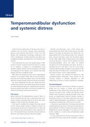
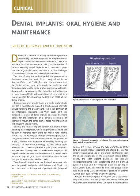
Figure 1: Comparison of crestal gingival fibre orientation<br />
Figure 2: Microscopic comparison of gingival fibre orientation (natural<br />
tooth on left, implant on right)<br />
Karring, 1994). Thus, personal <strong>oral</strong> <strong>hygiene</strong> must begin at the<br />
time of <strong>dental</strong> implant placement <strong>and</strong> should be modified<br />
using various adjuctive aids for <strong>oral</strong> <strong>hygiene</strong> to effectively clean<br />
the altered morphology of the peri-implant region before,<br />
during, <strong>and</strong> after implant placement. For instance,<br />
interproximal brushes can penetrate up to 3mm into a gingival<br />
sulcus or pocket <strong>and</strong> may effectively clean the peri-implant<br />
sulcus (Balshi, 1986). In addition to mechanical plaque control,<br />
daily rinses using 0.1% chlorhexidine gluconate or Listerine<br />
(Ciancio et al, 1995) provide a welcome adjunct.<br />
Hygiene with <strong>dental</strong> <strong>implants</strong> is so tedious <strong>and</strong> critical to their<br />
long-term success that the patient <strong>and</strong> <strong>dental</strong> professional<br />
must exercise considerable effort. During the <strong>maintenance</strong> visit,
the <strong>dental</strong> professional should concentrate on the peri-implant<br />
tissue margin, implant body, prosthetic abutment to implant<br />
collar connection, <strong>and</strong> the prosthesis (Garg, 1995).<br />
Clinical assessment<br />
Clinical inspection for signs of inflammation, i.e. bleeding on<br />
probing, exudate, mobility, probe-able pockets, <strong>and</strong> a<br />
radiographic evaluation of the peri-implant bony housing still<br />
remains the st<strong>and</strong>ard mode for evaluating the long-term status<br />
of endosseous <strong>dental</strong> <strong>implants</strong>. For instance, successful <strong>and</strong><br />
stable endosseous <strong>dental</strong> <strong>implants</strong> exhibit no mobility. But, if<br />
there is clinically perceptible mobility, then subsequent to<br />
radiographic evaluation of the implant <strong>and</strong> its surrounding<br />
bony housing, the abutment retaining screw (Lekholm et al,<br />
1986), <strong>and</strong>/or prosthetic abutment collar interface should be<br />
examined for looseness or breakage.<br />
All these modes of clinical assessment are used routinely,<br />
except for periodontal probing around peri-implant tissues that<br />
appear to be in a state of good health. The baseline data <strong>and</strong><br />
data from subsequent recare visits should be recorded in the<br />
daily progress notes to properly assess the peri-implant status<br />
logitudinally.<br />
Subsequent to a thorough intra<strong>oral</strong> examination, unless<br />
there is visual evidence of soft tissue changes, i.e. inflammation<br />
of peri-implant tissue with even slight attachment loss or<br />
mucositis, routine probing of the peri-implant tissue should not<br />
be performed.<br />
Usually during the first year subsequent to restoring <strong>dental</strong><br />
<strong>implants</strong>, a three-month recare schedule should be<br />
implemented, especially if the patient lost teeth because of<br />
periodontal disease. But if after 12 months, the patient’s<br />
<strong>implants</strong> are stable <strong>and</strong> peri-implant tissues are healthy, then a<br />
four to six-month recare regimen can be implemented<br />
(American Academy, 1996). However, be cognisant of each<br />
patient’s level of home care effectiveness, systemic health, <strong>and</strong><br />
periodontal status of the peri-implant tissue when determining<br />
these recare intervals.<br />
With <strong>dental</strong> implant patients, the <strong>dental</strong> professional must<br />
evaluate the prosthetic components for plaque, calculus, <strong>and</strong><br />
the stability of the implant abutment. Radiographs of <strong>dental</strong><br />
<strong>implants</strong> should be taken every 12 to 18 months during these<br />
<strong>maintenance</strong> visits (Baumgarten, 1995). For <strong>dental</strong> implant<br />
restorations that are screw retained, the <strong>dental</strong> professional<br />
needs to remove the prosthesis at least once a year to more<br />
easily assess the status of the peri-implant’s hard <strong>and</strong> soft<br />
tissues, the existence of acceptable mobility of the prosthetic<br />
components or the implant fixture itself, <strong>and</strong> the patient’s level<br />
of home care effectiveness (Meffert, 1995). Remember that the<br />
CLINICAL<br />
Figure 3: Gingival fibres between two natural teeth showing orientation<br />
perpendicular to the long axis of the teeth<br />
Figure 4: Gingival fibres between two <strong>implants</strong> showing orientation<br />
parallel with the long axis of the <strong>implants</strong><br />
presence of any symptoms of infection, radiographic evidence<br />
of peri-implant bone loss, <strong>and</strong>/or neuropathies may be<br />
indicative of an ailing or failing implant (Meffert, 1992).<br />
Implants vs natural teeth<br />
It is essential to underst<strong>and</strong> the periodontal relationship<br />
between the gingiva <strong>and</strong> the structure it attaches to be it a<br />
natural tooth or an implant. (Figures 1 <strong>and</strong> 2). The fibre<br />
orientation of the gingival cuff around a natural tooth attaches<br />
INTERNATIONAL DENTISTRY SA VOL. 10, NO. 1 57
CLINICAL<br />
Figure 5: Plastic curettes for scaling <strong>dental</strong> <strong>implants</strong> <strong>and</strong> demonstration of<br />
the implant surface after use. Note that there is no alteration to the<br />
surface<br />
perpendicular to the long axis of the tooth (Figure 3). This acts<br />
as a barrier when insertion of a periodontal probe within the<br />
sulcus. The probe tip advances apically till the tip contacts the<br />
perpendicular fibres <strong>and</strong> is halted. This orientation is not seen<br />
around <strong>implants</strong>. With an implant the gingival fibre orientation<br />
is parallel to the <strong>implants</strong> long axis (Figure 4). When a<br />
periodontal probe is inserted into the sulcus around an implant<br />
the probe tip advances passing between the fibres of the<br />
gingival cuff till the crestal bone prevents it from further<br />
advancement.<br />
The peri-implant mucosal seal may be less effective barrier to<br />
bacterial plaque than the periodontium around a natural tooth,<br />
tissue attachment (Wayant, 1994). There is less vasculature in<br />
the gingival tissue surrounding <strong>dental</strong> <strong>implants</strong> compared to<br />
natural teeth. This reduced vascularity concomitant with<br />
parallel-oriented collagen fibres adjecent to the body of any<br />
<strong>dental</strong> implant make <strong>dental</strong> <strong>implants</strong> more vulnerable to<br />
bacterial insult (Nevins <strong>and</strong> Langer 1995). During recare<br />
appointments, peri-implant periodontal probing should be<br />
performed only where signs of infection are present, i.e.<br />
exudate, swelling, bleeding on probing, inflamed peri-implant<br />
soft tissue, <strong>and</strong>/or radiographic evidence of peri-implant<br />
alveolar bone loss. Lastly, routine periodontal probing of <strong>dental</strong><br />
<strong>implants</strong> should not be performed, because this procedure<br />
could damage the weak epithelial attachment around <strong>dental</strong><br />
<strong>implants</strong>, possibly creating a pathway for the ingress of<br />
periodontal pathogens (Lang et al, 1994). Commercically<br />
available plastic probes should be used when investigating the<br />
crevicular depth around <strong>dental</strong> <strong>implants</strong>. The probing depth<br />
around <strong>dental</strong> <strong>implants</strong> may be related closely to the thickness<br />
<strong>and</strong> type of mucosa surrounding the implant. A healthy periimplant<br />
sulcus has been reported to range from 1.3 to 3.8mm,<br />
which is greater than those depths reported for natural teeth<br />
(van Steenberghe et al, 1993). In essence, the best indicator for<br />
evaluating an unhealthy site would be probing data gathered<br />
longitudinally (Quirynen et al, 1991).<br />
For all of these reasons, personal home care <strong>and</strong> consistent<br />
professional <strong>maintenance</strong> have proven to be critical to the<br />
success <strong>and</strong> longevity of endosseous <strong>dental</strong> <strong>implants</strong>. This is<br />
especially true in an environment with adjacent natural teeth,<br />
58 INTERNATIONAL DENTISTRY SA VOL. 10, NO. 1<br />
Figure 6: Plastic scaler used for recall <strong>maintenance</strong><br />
which if affected by periodontal disease, could act as a reservoir<br />
for pathogenic bacteria, ie. gram-negative anaerobic rods, <strong>and</strong><br />
seed the peri-implant sulcus (Mombelli et al, 1995).<br />
The physical characteristics of the peri-implant soft tissue are<br />
the focus of all <strong>oral</strong> <strong>hygiene</strong> instruction. The presence or<br />
absence of keratinised tissue in this critical area has not been<br />
unequivocally documented to state that peri-implant tissues are<br />
more vulnerable to the ingress of pathogenic bacteria with or<br />
without keratinised tissue being present around <strong>dental</strong><br />
<strong>implants</strong>. However, the ability of the patient to maintain good<br />
home care around <strong>dental</strong> <strong>implants</strong> is facilitated by the presence<br />
of keratinised tissue surrounding <strong>implants</strong>. Thus, if a patient<br />
has no keratinised tissue around an implant, <strong>and</strong> a pull from a<br />
frenum or a chronic peri-implant mucositis exists, then<br />
placement of a soft tissue autogenous or alloplastic connective<br />
tissue graft is recommended to facilitate proper mechanical<br />
<strong>oral</strong> <strong>hygiene</strong> <strong>maintenance</strong> (Artzi et al, 1993).<br />
Specific criteria for obtaining clinical data around <strong>dental</strong><br />
<strong>implants</strong> that would allow proper monitoring <strong>and</strong> detect early<br />
possible failure of osseointegrated <strong>dental</strong> <strong>implants</strong> has not<br />
been clearly defined. Presently, the presence of mobility is the<br />
best indicator for diagnosis of implant failure. As opposed to<br />
natural teeth, <strong>dental</strong> <strong>implants</strong> exhibit minimal clinically<br />
undetectable movement because of the absence of a<br />
periodontal ligament. Therefore, healthy <strong>implants</strong> should<br />
appear nonmobile, even in the presence of peri-implant bone<br />
loss, if an adequate amount of supporting alveolar bone still<br />
exists (Papaioannou et al, 1995).<br />
When monitoring the health of the peri-implant soft tissues,<br />
the practitioner should be cognisant of changes in soft tissue<br />
colour, contour, <strong>and</strong> consistency. The presence of a fistulous<br />
tract could indicate the presence of a pathologic process or<br />
implant fracture.<br />
Bleeding<br />
There is controversy in the literature as to the accuracy <strong>and</strong><br />
significance of bleeding upon probing around <strong>dental</strong> <strong>implants</strong>.
CLINICAL<br />
Figure 7: Alteration of implant surface after use of stainless steel scalers Figure 8: Demonstration of gouging of the implant surface that may occur<br />
following use of an ultrasonic scaler<br />
Presently, the literature advocates the use of bleeding on<br />
probing as an indicator of peri-implant disease, because it can<br />
occur prior to histologic signs of inflammation or concurrently<br />
with other signs of implant failure, i.e. bone loss. However, as<br />
previously mentioned, routine probing is not recommended.<br />
Radiographic evaluation<br />
Radiographic interpretation is one of the most useful clinical<br />
parameters for evaluating the status of an endosseous <strong>dental</strong><br />
implant. Invasion of biologic width, predictable remodeling, or<br />
so-called saucerisation, is an average marginal bone loss of 1.5<br />
during the first year following prosthetic rehabilitation followed<br />
by an average of 0.2mm of vertical bone loss every subsequent<br />
year. Thus, progressive bone loss around a <strong>dental</strong> implant that<br />
exceeds these averages may be indicative of an ailing or failing<br />
implant. Lastly, during radiographic evaluation, no evidence of<br />
a peri-implant radiolucency should be found, because such a<br />
rarefaction usually indicates infection or failure to<br />
osseointegration (Apse et al, 1989).<br />
Professional cleaning instrumentation<br />
Instruments made of metal, such as stainless steel, should be<br />
limited to natural teeth <strong>and</strong> not to be used to probe or scale<br />
<strong>dental</strong> <strong>implants</strong>. The rationale for this well-documented <strong>and</strong><br />
spoken conclusion is that this metal is so hard it can scratch,<br />
contaminate, or cause a galvanic reaction at the implantabutment<br />
interface (Speelman et al, 1992).<br />
Ideally, h<strong>and</strong> periodontal scalers for cleaning <strong>dental</strong> <strong>implants</strong><br />
can be plastic, Teflon, gold-plated, or made of wood (Figures 5<br />
<strong>and</strong> 6) (Gantes <strong>and</strong> Nilveus, 1991). When using gold-plated<br />
curettes, the manufacturer recommends not sharpening these<br />
<strong>hygiene</strong> instruments, as the gold surface could be chipped<br />
exposing the h<strong>and</strong> metal underneath this coating. Stainless<br />
steel scaling instruments may abraid the implant surface,<br />
stripping off any surface treatment such as hydroxyapatite (HA)<br />
as the instruments hardness is greater then the titanium alloy<br />
the implant is fabricated from (Figure 7).<br />
Other cleaning armamentarium contraindicated for use with<br />
<strong>dental</strong> <strong>implants</strong> are air powder abrasive units, flour or pumice<br />
for polishing, <strong>and</strong> sonic <strong>and</strong> ultrasonic scaling units (Rapley et<br />
al, 1990). Ultrasonic, piezo or sonic scaler tips may mar the<br />
60 INTERNATIONAL DENTISTRY SA VOL. 10, NO. 1<br />
<strong>implants</strong> surface leading to microroughness <strong>and</strong> plaque<br />
accumulation. The stainless steel tip may also lead to gouging<br />
of the <strong>implants</strong> polished collar (Figure 8). However, some<br />
clinicians advocate using a sonic instrument with a plastic<br />
sleeve over the tip for scaling <strong>dental</strong> <strong>implants</strong>. Air powder<br />
polishing units may also damage the implant surface <strong>and</strong><br />
should be avoided during <strong>hygiene</strong> appointments (Figure 9).<br />
Even the use of baking soda powder in these units may strip off<br />
any surface coating on the implant. Additionally, the air<br />
pressure may detach the soft tissue connection with the<br />
coronal of the implant leading to emphysema.<br />
Titanium or titanium alloy surfaces of <strong>dental</strong> <strong>implants</strong> can be<br />
polished using a rubber cup along with a nonabrasive polishing<br />
paste or a gauze strip with tin oxide. Not only is the <strong>hygiene</strong><br />
armamentarium important, but so are the home care<br />
techniques used to maintain endosseous <strong>dental</strong> <strong>implants</strong>.<br />
Patients should be taught the modified bass technique of<br />
brushing using a medium-sized head, soft-bristled toothbrush.<br />
The use of intra<strong>dental</strong> brushes should be used by implant<br />
patients after being shown their proper use. The plastic-coated<br />
wire brush is the only type to be used with <strong>dental</strong> <strong>implants</strong> to<br />
clean <strong>and</strong> not scratch the implant surface (Figure 10).<br />
Recently, automated mechanical toothbrushes have been<br />
advocated as a daily mode of tooth cleansing. These devices<br />
may be a rotary, circular, or sonic type. With these home care<br />
instruments, the key to their effectiveness is proper instruction<br />
on their use <strong>and</strong> then diligent daily use by the implant patient.<br />
As with natural dentition, adjuctive cleaning aids such as<br />
flossing are still valuable. As with dentated patients, an implant<br />
patient’s home care requirements should be individually<br />
tailored according to each patient’s needs. Individual needs are<br />
based on the location <strong>and</strong> angulation of the <strong>dental</strong> <strong>implants</strong>,<br />
the position <strong>and</strong> length of transmucosal abutments, the type of<br />
prosthesis, <strong>and</strong> the dexterity of each patient.<br />
The other popularised type of cleansing device is the use of<br />
<strong>oral</strong> irrigators with or without the addition of antimicrobial<br />
solutions. Also, <strong>oral</strong> rinses with antimicrobial properties such as<br />
Listerine or chlorhexidine have been widely advocated<br />
throughout the literature (Mombelli <strong>and</strong> Lang, 1992, Ciancio,<br />
1994, Garg et al, 1997).
CLINICAL<br />
Figure 9: Demonstration of alteration of the implant surface following<br />
application of an air polisher <strong>and</strong> baking soda. Note the change in surface<br />
texture<br />
Summary<br />
During the infancy years of <strong>dental</strong> implantology, the emphasis<br />
for long-term success of osseointegrated <strong>implants</strong> was the<br />
surgical phase of <strong>dental</strong> implantology. In the years that<br />
followed, the emphasis for success had switched from a purely<br />
surgical influence to focusing more on the proper fixture<br />
placement which would be dictated by the prosthetic <strong>and</strong><br />
aesthetic needs of each particular case.<br />
In more recent years, the <strong>dental</strong> professional has recognised<br />
professional implant <strong>maintenance</strong> <strong>and</strong> diligent patient home<br />
care as two critical factors for the long-term success of <strong>dental</strong><br />
<strong>implants</strong>. The microbiota <strong>and</strong> clinical presentation of periimplantitis<br />
is the same as periodontitis around a natural tooth.<br />
References<br />
Adell R, Lekholm U, Rockler B, et al (1981). A 15 year study of osseoinegrated<br />
<strong>implants</strong> in the treatment of the edentulous jaw. Int J Oral Surg 10: 387-416<br />
Cox JF, Zarb GA (1987). The logitudinal clinical efficacy of osseointegrated <strong>dental</strong><br />
<strong>implants</strong>: a 3 year report.Int J <strong>oral</strong> Maxillofac Implants 2: 91-100<br />
Albrektsson T, Branemark P,Hansson HA, et al. Osseointegrated titanium<br />
<strong>implants</strong>. Acta Orthop Sc<strong>and</strong>. 1981;52:155<br />
Orton GS, Steele DL, Wolinsky LE (1989). The <strong>dental</strong> professional’s role in<br />
monitoring <strong>and</strong> <strong>maintenance</strong> of tissue-integrated prostheses. Int J Oral axillofac<br />
Implants 4(4):305-310<br />
Bauman GR, Mills M, Rapley JW, et al (1992). Clinical parameters of evaluation<br />
during implant <strong>maintenance</strong>. Int J Oral Maxillofac Implants 7(2);220-227<br />
Rateischak KH, Wolf HF (1995). eds Color Atlas of Dental Medicine; Implantology.<br />
Stuttgart, NY: Thieme Medical Publishers 305-316<br />
Meffert RM, Langer4 B, Fritz ME (1992). Dental Implants: a review. J Periodontol<br />
63(11):859-870<br />
Meffert RM (1993). Contemporary Implant Dentistry. Carl E. Misch, ed. st Louis,<br />
Mo: Mosby Year Book; chap33<br />
Warrer K, Buser D, Lang NP, et al (1995). Plaque-induced peri-implantitis in the<br />
presence or absence of keratinized mucosa: an experimental study in monkeys.<br />
Clin Oral implant Res 6:131-138<br />
Lang NP, Karring T(1994). Proceedings of the 1st European Workshop on<br />
Periodontology. Chicago, IL: Quintessence<br />
Balshi TJ (1986). Hygiene <strong>maintenance</strong> procedures for patients treated with the<br />
tissue-integrated prothesis (osseointegration). Quintessence 17(2):95-102<br />
Ciancio SG, Lauciello C, Shibley O et al (1995). The effect of an antiseptic<br />
mouthrinse on implant <strong>maintenance</strong>: plaque <strong>and</strong> peri-implant gingival tissues. J.<br />
Periodontol 66(11):962-965<br />
Garg AK (1995). Practical Implant Dentistry. Dallas, TX: Taylor Publishing Co: 111-<br />
115<br />
Lekholm U, Ericsson I, Adell R, et al (1986). The condition of the soft tissues at<br />
tooth <strong>and</strong> fixture abutments supporting fixed bridges: a microbiological <strong>and</strong><br />
histological study. J Clin Periodontol 13:558-562<br />
American Academy of Periodontology. Annals of Periodontology. 1996 World<br />
62 INTERNATIONAL DENTISTRY SA VOL. 10, NO. 1<br />
Figure 10: Plastic-coated interproximal brush applied around implant<br />
abutments <strong>and</strong> under the superstructure for plaque removal<br />
Workshop in Periodontics 1(1):816-820<br />
Baumgarten HS, Chiche GJ (1995). Diagnosis <strong>and</strong> evaluation of complications<br />
<strong>and</strong> failures associated with osseointegrated <strong>implants</strong>. Compend Contin Educ<br />
Dent 16(8):814-822<br />
Meffert RM (1995). In the spotlight: Implantology <strong>and</strong> the <strong>dental</strong> hygienist’s role.<br />
J Prac Hygiene. September:12-14<br />
Meffert RM (1992). How to treat ailing <strong>and</strong> failing <strong>implants</strong>. Implant Dent; Spring<br />
1(1):25-33<br />
Weyant RJ (1994). Characteristics associated with the loss <strong>and</strong> peri-implant tissue<br />
health of endosseous <strong>dental</strong> <strong>implants</strong>. Int J Oral Maxillofac Implants. Jan-Feb;<br />
9(1): 95-102<br />
Nevins M, Langer B (1995). The successful use of osseointegrated <strong>implants</strong> for the<br />
treatment of the recalcitrant periodontal patient. J Periodontol. Feb; 66(2): 150-<br />
7<br />
Lang NP, Wetzel AC, Stich H, Caffesse RG (1994). Histologic probe penetration in<br />
healthy <strong>and</strong> inflamed peri-implant tissues. Clin Oral Implants Res. Dec; 5(4): 191-<br />
201<br />
van Steenberghe D, Klinge B, Linden U, Quirynen M, Herrmann I, Garpl<strong>and</strong> C<br />
(1993). Periodontal indices around natural <strong>and</strong> titanium abutments: a<br />
longitudinal multicenter study.<br />
J Periodontol. Jun; 64(6): 538-41<br />
Quirynen M, van Steenberghe D, Jacobs R, Schotte A, Darius P (1991). The<br />
reliability of pocket probing around screw-type <strong>implants</strong>. Clin Oral Implants Res.<br />
Oct-Dec; 2(4): 186-92<br />
Mombelli A, Marxer M, Gaberthuel T, Grunder U, Lang NP (1995). The microbiota<br />
of osseointegrated <strong>implants</strong> in patients with a history of periodontal disease. J<br />
Clin Periodontol. Feb; 22(2): 124-30<br />
Artzi Z, Tal H, Moses O, Kozlovsky A (1993). Mucosal considerations for<br />
osseointegrated <strong>implants</strong>. J Prosthet Dent. Nov; 70(5): 427-32<br />
Papaioannou W, Quirynen M, Nys M, van Steenberghe D (1995). The effect of<br />
periodontal parameters on the subgingival microbiota around <strong>implants</strong>. Clin Oral<br />
Implants Res. Dec; 6(4): 197-204<br />
Apse P, Ellen RP, Overall CM, Zarb GA (1989). Microbiota <strong>and</strong> crevicular fluid<br />
collagenase activity in the osseointegrated <strong>dental</strong> implant sulcus: a comparison of<br />
sites in edentulous <strong>and</strong> partially edentulous patients. J Periodontal Res. Mar;<br />
24(2): 96-105<br />
Speelman JA, Collaert B, Klinge B (1992). Evaluation of different methods to<br />
clean titanium abutments. A scanning electron microscopic study. Clin Oral<br />
Implants Res. Sep; 3(3): 120-7<br />
Gantes BG, Nilveus R (1991). The effects of different <strong>hygiene</strong> instruments on<br />
titanium surfaces: SEM observations. Int J Periodontics Restorative Dent<br />
11(3):225-39<br />
Rapley JW, Swan RH, Hallmon WW, et al (1990). The <strong>oral</strong> <strong>hygiene</strong> instruments<br />
<strong>and</strong> materials on titanium implant abutments. Int J <strong>oral</strong> Maxillofac Implants 5:47-<br />
52<br />
Mombelli A, Lang NP (1992). Antimicrobial treatment of peri-implant infections.<br />
Clin Oral Implants Res. Dec; 3(4): 162-8<br />
Ciancio S (1994). Exp<strong>and</strong>ed <strong>and</strong> future uses of mouthrinses. J Am Dent Assoc.<br />
Aug; 125 Suppl 2: 29S-32S<br />
Garg AK, Duarte F, Funari K (1997). Hygienic <strong>maintenance</strong> of <strong>dental</strong> <strong>implants</strong>: the<br />
key to success. J Pract Hygiene 6(2):13-20