Depletion of eosinophils in mice through the use - Journal of ...
Depletion of eosinophils in mice through the use - Journal of ...
Depletion of eosinophils in mice through the use - Journal of ...
You also want an ePaper? Increase the reach of your titles
YUMPU automatically turns print PDFs into web optimized ePapers that Google loves.
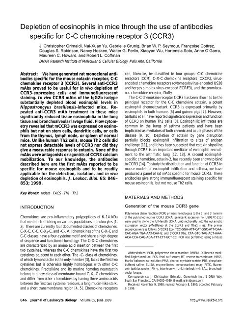
<strong>Depletion</strong> <strong>of</strong> <strong>eos<strong>in</strong>ophils</strong> <strong>in</strong> <strong>mice</strong> <strong>through</strong> <strong>the</strong> <strong>use</strong> <strong>of</strong> antibodies<br />
specific for C-C chemok<strong>in</strong>e receptor 3 (CCR3)<br />
J. Christopher Grimaldi, Nai-Xuan Yu, Gabrielle Grunig, Brian W. P. Seymour, Françoise Cottrez,<br />
Douglas S. Rob<strong>in</strong>son, Nancy Hosken, Walter G. Ferl<strong>in</strong>, Xiaoyan Wu, Hortensia Soto, Anne O’Garra,<br />
Maureen C. Howard, and Robert L. C<strong>of</strong>fman<br />
DNAX Research Institute <strong>of</strong> Molecular & Cellular Biology, Palo Alto, California<br />
Abstract: We have generated rat monoclonal antibodies<br />
specific for <strong>the</strong> mo<strong>use</strong> eotax<strong>in</strong> receptor, C-C<br />
chemok<strong>in</strong>e receptor 3 (CCR3). Several anti-CCR3<br />
mAbs proved to be <strong>use</strong>ful for <strong>in</strong> vivo depletion <strong>of</strong><br />
CCR3-express<strong>in</strong>g cells and immun<strong>of</strong>luorescent<br />
sta<strong>in</strong><strong>in</strong>g. In vivo CCR3 mAbs <strong>of</strong> <strong>the</strong> IgG2b isotype<br />
substantially depleted blood eos<strong>in</strong>ophil levels <strong>in</strong><br />
Nippostrongyus brasiliensis-<strong>in</strong>fected <strong>mice</strong>. Repeated<br />
anti-CCR3 mAb treatment <strong>in</strong> <strong>the</strong>se <strong>mice</strong><br />
significantly reduced tissue eos<strong>in</strong>ophilia <strong>in</strong> <strong>the</strong> lung<br />
tissue and bronchoalveolar lavage fluid. Flow cytometry<br />
revealed that mCCR3 was expressed on <strong>eos<strong>in</strong>ophils</strong><br />
but not on stem cells, dendritic cells, or cells<br />
from <strong>the</strong> thymus, lymph node, or spleen <strong>of</strong> normal<br />
<strong>mice</strong>. Unlike human Th2 cells, mo<strong>use</strong> Th2 cells did<br />
not express detectable levels <strong>of</strong> CCR3 nor did <strong>the</strong>y<br />
give a measurable response to eotax<strong>in</strong>. None <strong>of</strong> <strong>the</strong><br />
mAbs were antagonists or agonists <strong>of</strong> CCR3 calcium<br />
mobilization. To our knowledge, <strong>the</strong> antibodies<br />
described here are <strong>the</strong> first mAbs reported to be<br />
specific for mo<strong>use</strong> <strong>eos<strong>in</strong>ophils</strong> and to be readily<br />
applicable for <strong>the</strong> detection, isolation, and <strong>in</strong> vivo<br />
depletion <strong>of</strong> <strong>eos<strong>in</strong>ophils</strong>. J. Leukoc. Biol. 65: 846–<br />
853; 1999.<br />
Key Words: rodent · FACS · Th1 · Th2<br />
INTRODUCTION<br />
Chemok<strong>in</strong>es are pro-<strong>in</strong>flammatory polypeptides <strong>of</strong> 6–14 kDa<br />
that mediate traffick<strong>in</strong>g on various populations <strong>of</strong> leukocytes [1,<br />
2]. There are currently four documented classes <strong>of</strong> chemok<strong>in</strong>es:<br />
C-X-C, C-C, C-X 3-C, and -C-. All chemok<strong>in</strong>es <strong>of</strong> <strong>the</strong> C-X-C and<br />
C-C classes have a four-cyste<strong>in</strong>e motif and share a high degree<br />
<strong>of</strong> sequence and functional homology. The C-X-C chemok<strong>in</strong>es<br />
are characterized by an am<strong>in</strong>o acid <strong>in</strong>sertion between <strong>the</strong> first<br />
two cyste<strong>in</strong>es, whereas <strong>the</strong> C-C chemok<strong>in</strong>es have <strong>the</strong> first two<br />
cyste<strong>in</strong>es adjacent to each o<strong>the</strong>r. The -C- class <strong>of</strong> chemok<strong>in</strong>es,<br />
<strong>of</strong> which lymphotact<strong>in</strong> is <strong>the</strong> only member [3], lacks <strong>the</strong> first two<br />
cyste<strong>in</strong>es but is o<strong>the</strong>rwise highly homologous with o<strong>the</strong>r C-C<br />
chemok<strong>in</strong>es. Fractalk<strong>in</strong>e and its mur<strong>in</strong>e homolog neurotact<strong>in</strong><br />
belong to a new class <strong>of</strong> membrane-bound C-X 3-C chemok<strong>in</strong>es<br />
and differ from o<strong>the</strong>r chemok<strong>in</strong>es by hav<strong>in</strong>g three am<strong>in</strong>o acids<br />
between <strong>the</strong> first two cyste<strong>in</strong>e residues, a long muc<strong>in</strong>-like stalk,<br />
and a short transmembrane region [4, 5]. Chemok<strong>in</strong>e receptors<br />
can, likewise, be classified <strong>in</strong> four groups: C-C chemok<strong>in</strong>e<br />
receptors (CCR), C-X-C chemok<strong>in</strong>e receptors (CXCR), vir<strong>use</strong>ncoded<br />
chemok<strong>in</strong>e receptors (cytomegalovirus-encoded US28<br />
and herpes simplex virus-encoded ECRF3), and <strong>the</strong> promiscuous<br />
chemok<strong>in</strong>e receptor, Duffy.<br />
The C-C chemok<strong>in</strong>e receptor CCR3 has been shown to be <strong>the</strong><br />
pr<strong>in</strong>cipal receptor for <strong>the</strong> C-C chemok<strong>in</strong>e eotax<strong>in</strong>, a potent<br />
eos<strong>in</strong>ophil chemoattractant. CCR3 is expressed primarily by<br />
<strong>eos<strong>in</strong>ophils</strong> <strong>in</strong> both humans [6] and gu<strong>in</strong>ea pigs [7]. However,<br />
Sallusto et al. have reported significant expression and function<br />
<strong>of</strong> CCR3 on human Th2 cells [8]. Eos<strong>in</strong>ophilic <strong>in</strong>filtrates are<br />
common <strong>in</strong> <strong>the</strong> lungs <strong>of</strong> asthma patients and have been<br />
implicated as mediators <strong>of</strong> both chronic and acute phases <strong>of</strong> <strong>the</strong><br />
disease [9, 10]. <strong>Depletion</strong> <strong>of</strong> eotax<strong>in</strong> by gene disruption<br />
partially blocks eos<strong>in</strong>ophil <strong>in</strong>filtration to sites <strong>of</strong> antigen<br />
challenge [11], and it has been suggested that eotax<strong>in</strong> signal<strong>in</strong>g<br />
<strong>through</strong> CCR3 is an important mediator <strong>of</strong> eos<strong>in</strong>ophil recruitment<br />
to <strong>the</strong> asthmatic lung [12, 13]. A second <strong>eos<strong>in</strong>ophils</strong>pecific<br />
chemok<strong>in</strong>e, eotax<strong>in</strong>-2, has recently been shown to b<strong>in</strong>d<br />
to CCR3 [14]. To study <strong>the</strong> distribution and function <strong>of</strong> CCR3 <strong>in</strong><br />
mo<strong>use</strong> models <strong>of</strong> eos<strong>in</strong>ophil <strong>in</strong>filtration and asthma, we have<br />
produced a panel <strong>of</strong> rat mAbs specific for mo<strong>use</strong> CCR3. These<br />
antibodies give strong immun<strong>of</strong>luorescent sta<strong>in</strong><strong>in</strong>g specific for<br />
mo<strong>use</strong> <strong>eos<strong>in</strong>ophils</strong>, but not mo<strong>use</strong> Th2 cells.<br />
MATERIALS AND METHODS<br />
Generation <strong>of</strong> <strong>the</strong> mo<strong>use</strong> CCR3 gene<br />
Polymerase cha<strong>in</strong> reaction (PCR) primers homologous to <strong>the</strong> 5� and 3� term<strong>in</strong>i<br />
<strong>of</strong> <strong>the</strong> published mur<strong>in</strong>e CCR3 cDNA (genebank accession no. U29677) [15]<br />
were <strong>use</strong>d to clone <strong>the</strong> full-length cDNA unidirectionally <strong>in</strong>to <strong>the</strong> eukaryotic<br />
expression vector pMe18sneo at <strong>the</strong> EcoR1 and Xba1 sites. The primer<br />
sequences were as follows: 5�CCR3 Eco, TCC-GGA-ATT-CAT-GGC-ATT-CAA-<br />
CAC-AGA-TGA-AAT-CAA-G; and 3�CCR3 Xba, CTA-GTC-TAG-ACT-AAA-<br />
ACA-CCA-CAG-AGA-TTT-CTT-GCT-CC. PCR was performed us<strong>in</strong>g a mo<strong>use</strong><br />
Abbreviations: PCR, polymerase cha<strong>in</strong> reaction; DMEM, Dulbecco’s modified<br />
Eagle’s medium; FCS, fetal calf serum; RT, reverse transcriptase; HBSS,<br />
Hanks’ balanced salt solution; PMA, phorbol myristate acetate; PBS, phosphatebuffered<br />
sal<strong>in</strong>e; ELISA, enzyme-l<strong>in</strong>ked immunosorbent assay; FITC, fluoresce<strong>in</strong><br />
isothiocyanate; IFN-�, <strong>in</strong>terferon-�; IL-4, <strong>in</strong>terleuk<strong>in</strong>-4; BAL, bronchoalveolar<br />
lavage.<br />
Correspondence: J. Christopher Grimaldi, Genentech Inc., 1 DNA Way,<br />
South San Francisco, CA 94080-4990. E-mail: grim@gene.com<br />
Received November 9, 1998; revised February 8, 1999; accepted February<br />
9, 1999.<br />
846 <strong>Journal</strong> <strong>of</strong> Leukocyte Biology Volume 65, June 1999 http://www.jleukbio.org
Th2 cDNA library as template to amplify <strong>the</strong> 1079-bp CCR3 cod<strong>in</strong>g region. The<br />
PCR fragment was <strong>the</strong>n cloned <strong>in</strong>to <strong>the</strong> pMe18sNeo vector. The f<strong>in</strong>al construct<br />
pMe18sNeo/mCCR3 was sequenced on both strands to confirm that no<br />
PCR-generated mutations had occurred.<br />
Transfected cell l<strong>in</strong>es<br />
Stable mur<strong>in</strong>e CCR3 transfectants were generated <strong>in</strong> <strong>the</strong> mo<strong>use</strong> fibroblast cell<br />
l<strong>in</strong>e NIH3T3 and <strong>the</strong> rat cell myeloma l<strong>in</strong>e Y3 us<strong>in</strong>g <strong>the</strong> pMe18sNeo/mCCR3.<br />
NIH3T3 cells were transfected us<strong>in</strong>g lip<strong>of</strong>ectam<strong>in</strong>e (GIBCO-BRL, Gai<strong>the</strong>rsberg,<br />
MD) accord<strong>in</strong>g to <strong>the</strong> manufacturer’s protocol. Briefly, a 1:20 dilution <strong>of</strong><br />
lip<strong>of</strong>ectam<strong>in</strong>e <strong>in</strong> optiMEM � I medium (GIBCO) was mixed with 15 µg <strong>of</strong><br />
pMe18sNeo/mCCR3 for 30 m<strong>in</strong> at room temperature. The solution was <strong>the</strong>n<br />
added to 3 � 10 6 NIH3T3 cells at 37°C for 5 h. After 5 h <strong>the</strong> medium was<br />
exchanged with Dulbecco’s modified Eagle’s medium (DMEM) conta<strong>in</strong><strong>in</strong>g 10%<br />
fetal calf serum (FCS). G418 (GIBCO-BRL) at a f<strong>in</strong>al concentration <strong>of</strong> 1 mg/mL<br />
was added at 48 h to select for positive clones. The Y3 cell l<strong>in</strong>e was transfected<br />
by electroporation us<strong>in</strong>g <strong>the</strong> Gene Pulser (Bio-Rad, Hercules, CA). Briefly 1 �<br />
10 7 cells with 25 µg <strong>of</strong> pMe18sNeo/mCCR3 were electroporated at 300 mV and<br />
960 µF. The transfected cells were <strong>the</strong>n allowed to expand for 48 h <strong>in</strong> DMEM<br />
with 10% FCS before select<strong>in</strong>g <strong>in</strong> medium conta<strong>in</strong><strong>in</strong>g 1 mg/mL G418. Twenty<br />
s<strong>in</strong>gle G418-resistant clones were selected and analyzed for <strong>the</strong> presence <strong>of</strong><br />
CCR3 mRNA by reverse transcriptase-PCR.<br />
RT-PCR <strong>of</strong> stable G418-resistant transfectants<br />
RT-PCR analyses were performed on <strong>the</strong> clonal G418-resistant transfectants to<br />
determ<strong>in</strong>e <strong>the</strong> presence <strong>of</strong> CCR3 message. Total RNA was purified from 1 �<br />
10 7 cells us<strong>in</strong>g RNAzol B � (TEL-TEST, Inc., Friendswood, TX), per <strong>the</strong><br />
manufacturer’s protocol. The isolated total RNA was treated with DNase 1<br />
(Gene Hunter Inc., Nashville, TN) to elim<strong>in</strong>ate genomic DNA contam<strong>in</strong>ation.<br />
Oligo(dT)-primed reverse transcription was performed us<strong>in</strong>g 5 µg <strong>of</strong> DNase<br />
1-treated total RNA <strong>in</strong> 20-µL reactions. PCR was carried out us<strong>in</strong>g 5 µL <strong>of</strong> <strong>the</strong><br />
RT product and <strong>the</strong> above-mentioned CCR3 primers for 25 cycles <strong>in</strong> a standard<br />
50-µL reaction. The PCR conditions were 5 m<strong>in</strong> 94°C, <strong>the</strong>n 25 cycles at 94°C<br />
30 s, 55°C 45 s, and 72°C 90 s. The RT-PCR products were <strong>the</strong>n analyzed by<br />
agarose gel electrophoresis.<br />
Calcium flux and chemotaxis assays<br />
Calcium flux assays were performed on a Photon Technologies International<br />
fluorometer us<strong>in</strong>g <strong>the</strong> FeliX � fluorescence analysis s<strong>of</strong>tware version 1.11. Cells<br />
(1 � 10 7 ) were added to 1 mL <strong>of</strong> media conta<strong>in</strong><strong>in</strong>g 3 µg/mL Indo-1/AM<br />
(Molecular Probes, Eugene, OR) and allowed to <strong>in</strong>cubate with gentle agitation<br />
at room temperature for 45 m<strong>in</strong>. The labeled cells were <strong>the</strong>n centrifuged and<br />
resuspended <strong>in</strong> 1 mL <strong>of</strong> Hanks’ balanced salt solution (HBSS) with 1% FCS and<br />
kept at room temperature. For measurements <strong>of</strong> relative fluorescence, 1 � 10 6<br />
labeled cells were added to 1.9 mL <strong>of</strong> 37°C HBSS conta<strong>in</strong><strong>in</strong>g 1.6 mM CaCl2 and<br />
10 mM HEPES. Mo<strong>use</strong> and human eotax<strong>in</strong>, hMCP-2, hMCP-3, hMCP-4,<br />
mRANTES, and hEotax<strong>in</strong>-2 (Peprotech, Rocky Hill, NJ) were added at f<strong>in</strong>al<br />
concentrations rang<strong>in</strong>g from 1 nM to 1 µM. The most eotax<strong>in</strong>-responsive<br />
CCR3-transfected Y3 and NIH3T3 cell l<strong>in</strong>es were chosen for fur<strong>the</strong>r studies<br />
and named Y3/mCCR3 and NIH3T3/mCCR3, respectively. Chemotaxis <strong>of</strong><br />
phorbol myristate acetate (PMA)/ionomyc<strong>in</strong> activated 3� polarized Th2<br />
populations was performed us<strong>in</strong>g a range <strong>of</strong> eotax<strong>in</strong> concentrations as<br />
previously described [16].<br />
Anti-CCR3 mAb production<br />
Two-month-old Lewis rats (Harlan, Indianapolis, IN) were <strong>in</strong>jected <strong>in</strong>traperitoneally<br />
every 2 weeks for 10 weeks with 2 � 10 8 Y3/mCCR3 cells suspended <strong>in</strong><br />
phosphate-buffered sal<strong>in</strong>e (PBS) and irradiated with 10,000 rads. On <strong>the</strong> 12th<br />
week <strong>the</strong> same immunization was given <strong>in</strong>travenously and 4 days later <strong>the</strong><br />
spleen was removed for fusion. The splenocytes were f<strong>use</strong>d to SP2/0 cells and<br />
hybridomas selected us<strong>in</strong>g <strong>the</strong> ClonaCell-HY kit (Stem Cell Systems Inc.,<br />
Vancouver, Canada). Hybridomas were expanded <strong>in</strong> 96-well plates and <strong>the</strong><br />
supernatants tested by flow cytometry for <strong>the</strong>ir ability to sta<strong>in</strong> NIH3T3/mCCR3<br />
cells but not <strong>the</strong> NIH3T3 parent l<strong>in</strong>e. All positive hybridomas were recloned<br />
and rescreened to ensure that each hybridoma was clonal. Anti-CCR3 mAbs<br />
were isotyped us<strong>in</strong>g an Ouchterlony assay (ICN, Aurora, OH) as well as by<br />
enzyme-l<strong>in</strong>ked immunosorbent assay (ELISA) us<strong>in</strong>g <strong>the</strong> rat monoAB ID/SP kit<br />
(Zymed, South San Francisco, CA) as per <strong>the</strong> manufacturer’s protocol.<br />
Epitope mapp<strong>in</strong>g <strong>of</strong> anti-CCR3 mAbs<br />
Experiments to determ<strong>in</strong>e <strong>the</strong> anti-CCR3 mAb epitope consisted <strong>of</strong> two<br />
experimental methods: cross block<strong>in</strong>g by flow cytometry and peptide-specific<br />
ELISAs. Cross block<strong>in</strong>g experiments were carried out by saturat<strong>in</strong>g Y3/mCCR3<br />
cells with each <strong>of</strong> <strong>the</strong> anti-CCR3 mAbs at 100 µg/mL <strong>in</strong> PBS with 3% BSA on<br />
ice for 30 m<strong>in</strong> and <strong>the</strong>n sta<strong>in</strong><strong>in</strong>g with fluoresce<strong>in</strong> isothiocyanate (FITC)conjugated<br />
6SH2-59 anti-CCR3 mAb at 1 µg/mL for 30 m<strong>in</strong> followed by three<br />
washes <strong>in</strong> PBS with 3% BSA. The sta<strong>in</strong>ed cells were analyzed for <strong>the</strong> ability <strong>of</strong><br />
<strong>the</strong> unlabeled antibody to block 6SH2-59 CCR3-FITC sta<strong>in</strong><strong>in</strong>g.<br />
Peptide ELISAs were performed on each <strong>of</strong> <strong>the</strong> anti-CCR3 mAbs by coat<strong>in</strong>g<br />
96-well ELISA plates with syn<strong>the</strong>tic peptides correspond<strong>in</strong>g to <strong>the</strong> am<strong>in</strong>oterm<strong>in</strong>al<br />
peptide and <strong>the</strong> three extracellular loops <strong>of</strong> CCR3 (Research Genetics,<br />
Huntsville, AL). The sequences <strong>of</strong> <strong>the</strong> peptides were as follows: am<strong>in</strong>oterm<strong>in</strong>al,<br />
MAFNTDEIKTVVESFETTPHEYEWAPPCEKVRIKELG; loop 1,<br />
LWNEWGFGHYMC; loop 2, HESQDSFGEFSCSPRYPEGEEDSWKRF-<br />
HALR; and loop 3, AFHRTFLETSCEQSKHLDL. Each well was coated<br />
overnight at 4°C with 100 µL <strong>of</strong> peptide at 2 mg/mL <strong>in</strong> PBS. The plates were<br />
<strong>the</strong>n washed with PBS conta<strong>in</strong><strong>in</strong>g 0.05% Tween-20 followed by <strong>the</strong> addition <strong>of</strong><br />
100 µL <strong>of</strong> anti-CCR3 mAb supernatants at room temperature for 2 h. Plates<br />
were washed and horseradish peroxidase-conjugated rabbit anti-rat polyclonal<br />
antibody (Zymed) was added for 1 h. Plates were washed aga<strong>in</strong> and <strong>the</strong><br />
colorimetric substrate 2,2’-az<strong>in</strong>o-bis (3 ethylbenzthiazol<strong>in</strong>e-6-sulfonic acid)<br />
was added. Plates were read on a Molecular Devices 96-well plate reader<br />
(Mounta<strong>in</strong> View, CA).<br />
Characterization <strong>of</strong> <strong>the</strong> anti-CCR3 mAbs for<br />
agonistic and antagonistic activity<br />
Agonistic and antagonistic activity was assayed by calcium flux analysis.<br />
Y3/mCCR3 cells were labeled and calcium mobilization was assayed as<br />
described. For assay<strong>in</strong>g agonistic activity, 50 µg <strong>of</strong> purified anti-mCCR3 mAb<br />
was added to labeled cells at 37°C and assayed for calcium mobilization over 5<br />
m<strong>in</strong>. For antagonistic activity, 100 µg/mL <strong>of</strong> purified anti-CCR3 mAb was<br />
added to labeled cells at 37°C for 5 m<strong>in</strong> followed by <strong>the</strong> addition <strong>of</strong> eotax<strong>in</strong> at a<br />
f<strong>in</strong>al concentration <strong>of</strong> 100 µM. The calcium mobilization was assayed over 10<br />
m<strong>in</strong>, which <strong>in</strong>cluded <strong>the</strong> 5-m<strong>in</strong> mAb <strong>in</strong>cubation step.<br />
Preparation <strong>of</strong> cells<br />
The Th1 clone, D1.1, and <strong>the</strong> Th2 clone, CDC35, were stimulated and passaged<br />
with rabbit anti-mo<strong>use</strong> immunoglobul<strong>in</strong>, as described previously [17, 18].<br />
CD4 � Th1 and Th2 cells were generated <strong>in</strong> vitro from DO11.10 T cell receptor<br />
transgenic BALB/c <strong>mice</strong> as described previously [19]. Briefly, naive CD4 � T<br />
cells were cultured with OVA 323-339, <strong>the</strong> ovalbum<strong>in</strong> peptide for which <strong>the</strong><br />
transgenic T cells are specific, for ei<strong>the</strong>r 1 or 3 weekly stimulations (designated<br />
1� or 3� polarized). Successful polarization was confirmed by test<strong>in</strong>g for<br />
<strong>in</strong>terferon-� (IFN-�) and <strong>in</strong>terleuk<strong>in</strong>-4 (IL-4) production by activated Th1 and<br />
Th2 cells, respectively. Rest<strong>in</strong>g cells were harvested immediately after <strong>the</strong> last<br />
stimulation; activated cells were rest<strong>in</strong>g cells cultured at 37°C for 6h<strong>in</strong>PMA<br />
(50 ng/mL) plus ionomyc<strong>in</strong> (500 ng/mL). CD4 � and CD8 � splenic T cells were<br />
isolated with a FACSTAR � plus cell sorter and activated as described above.<br />
Stem cells were purified from bone marrow on <strong>the</strong> basis <strong>of</strong> positive sta<strong>in</strong><strong>in</strong>g for<br />
C-kit � and Sca-1 � and negative sta<strong>in</strong><strong>in</strong>g for a mixture <strong>of</strong> l<strong>in</strong>eage-specific cell<br />
surface markers (Ly6G, Mac1, B220, CD4, CD8, and Ter119). Splenic dendritic<br />
cells were purified by <strong>the</strong> method <strong>of</strong> Macatonia et al. [20].<br />
Expression <strong>of</strong> CCR3, both prote<strong>in</strong> and mRNA<br />
Immun<strong>of</strong>luorescent sta<strong>in</strong><strong>in</strong>g <strong>of</strong> cell suspensions was performed with FITCconjugated<br />
6SH2-59 anti-CCR3 mAb at 1 µg/mL. Total RNA was isolated from<br />
cell l<strong>in</strong>es and populations with RNAzol B (TEL-TEST). Nor<strong>the</strong>rn blots were<br />
performed with 10 µg total RNA and probed with <strong>the</strong> full-length cod<strong>in</strong>g region<br />
<strong>of</strong> mCCR3 cDNA us<strong>in</strong>g standard methods [21]. For quantitative RT-PCR, 4 µg<br />
RNA was treated with DNase 1 and reverse transcribed with SuperScript<br />
(GIBCO). Threefold dilutions <strong>of</strong> cDNA, represent<strong>in</strong>g 0.1–200 pg RNA, were<br />
PCR amplified for 35–40 cycles us<strong>in</strong>g conditions as described above for <strong>the</strong><br />
cDNA clon<strong>in</strong>g. The primers <strong>use</strong>d were as follows: 5� primer, ATG-GCA-TTC-<br />
Grimaldi et al. <strong>Depletion</strong> <strong>of</strong> mo<strong>use</strong> <strong>eos<strong>in</strong>ophils</strong> <strong>through</strong> <strong>the</strong> <strong>use</strong> <strong>of</strong> antibodies 847
AAC-ACA-GAT-GAA-ATC-AAG; 3� primer, GGA-TAG-CGA-GGA-CTG-CAG-<br />
GAA-AAC. PCR products were separated on agarose gels, sta<strong>in</strong>ed with SYBR �<br />
green I dye (Molecular Probes) and <strong>the</strong> CCR3 product quantitated by a Storm �<br />
860 phosphoimager (Molecular Dynamics, Sunnyvale, CA). The results were<br />
expressed as arbitrary units <strong>of</strong> CCR3 mRNA normalized to <strong>the</strong> content <strong>of</strong> HPRT<br />
and �-act<strong>in</strong> mRNA <strong>in</strong> each sample.<br />
In vivo depletion with anti-CCR3<br />
Eos<strong>in</strong>ophilia was <strong>in</strong>duced <strong>in</strong> 2-week-old female BALB/c <strong>mice</strong> by subcutaneous<br />
<strong>in</strong>jection <strong>of</strong> 500 Nippostrongylus brasiliensis stage 3 larvae (N. brasiliensis). A<br />
s<strong>in</strong>gle antibody treatment consisted <strong>of</strong> 0.5 mg purified anti-CCR3 mAb given<br />
<strong>in</strong>traperitoneally at day 12 after N. brasiliensis <strong>in</strong>fection. Circulat<strong>in</strong>g <strong>eos<strong>in</strong>ophils</strong><br />
were counted <strong>in</strong> hepar<strong>in</strong>ized blood taken from <strong>the</strong> tail ve<strong>in</strong> at 0, 24, 48,<br />
96, and 168 h after mAb adm<strong>in</strong>istration as previously described [22]. For<br />
repeated mAb treatment, <strong>the</strong> animals were given <strong>in</strong>traperitoneal <strong>in</strong>jections <strong>of</strong> 1<br />
mg anti-CCR3 mAb 48 h before N. brasiliensis <strong>in</strong>fection and <strong>the</strong>n 0.5 mg <strong>of</strong><br />
mAb on days 3, 8, and 11 after N. brasiliensis <strong>in</strong>fection. The animals were killed<br />
on day 12 for isolation <strong>of</strong> bronchioalveolar lavage fluid (BAL), lung tissue<br />
sampl<strong>in</strong>g, and eos<strong>in</strong>ophil count<strong>in</strong>g <strong>in</strong> peripheral blood. BAL cytosp<strong>in</strong>s and lung<br />
paraff<strong>in</strong> sections were sta<strong>in</strong>ed with May-Grünwald Giemsa. For <strong>in</strong> vitro<br />
restimulation, lung cell suspensions were prepared as described previously [23]<br />
and stimulated on plates previously coated with 10 µg/mL anti-CD3 (Pharm<strong>in</strong>gen,<br />
San Diego, CA). After 3 days <strong>the</strong> supernatants were removed and IL-4 and<br />
IL-5 concentrations measured by ELISA [23]. Paraff<strong>in</strong> sections from repeated<br />
antibody treatment experiments were analyzed for eos<strong>in</strong>ophil depletion by two<br />
<strong>in</strong>dependent pathologists. Sta<strong>in</strong>ed sections were scored <strong>in</strong> a bl<strong>in</strong>ded manner on<br />
a scale <strong>of</strong> 0–4, with 1 be<strong>in</strong>g <strong>the</strong> m<strong>in</strong>imum and 4 be<strong>in</strong>g <strong>the</strong> maximum eos<strong>in</strong>ophil<br />
<strong>in</strong>filtration.<br />
RESULTS AND DISCUSSION<br />
Much recent evidence suggests that eotax<strong>in</strong> and its receptor,<br />
CCR3, are important mediators <strong>of</strong> allergic <strong>in</strong>flammation by<br />
stimulat<strong>in</strong>g <strong>in</strong>filtration <strong>of</strong> <strong>eos<strong>in</strong>ophils</strong> and, possibly, Th2 cells<br />
<strong>in</strong>to <strong>the</strong> lung. In order to study CCR3 expression and function<br />
<strong>in</strong> well-characterized mo<strong>use</strong> models <strong>of</strong> eos<strong>in</strong>ophil <strong>in</strong>filtration<br />
and airway hyper-reactivity, we have produced monoclonal<br />
antibodies to mo<strong>use</strong> CCR3.<br />
Mo<strong>use</strong> CCR3 cDNA and stable transfectant<br />
generation<br />
The strategy chosen for produc<strong>in</strong>g mAbs to mo<strong>use</strong> CCR3 was<br />
immunization <strong>of</strong> rats with a transfected rat cell l<strong>in</strong>e express<strong>in</strong>g<br />
mo<strong>use</strong> CCR3. The gene for mo<strong>use</strong> CCR3 was isolated by PCR<br />
us<strong>in</strong>g oligonucleotides based on <strong>the</strong> published mo<strong>use</strong> CCR3<br />
sequence [15]. Thirty-four different mo<strong>use</strong> cDNA libraries were<br />
screened by PCR for <strong>the</strong> presence <strong>of</strong> CCR3 message. CCR3<br />
cDNA could be detected <strong>in</strong> three libraries: N. brasiliensis<strong>in</strong>fected<br />
lung, RAG-2 -/- spleen, and a polarized Th2 population<br />
<strong>of</strong> CD4 � T cells (data not shown). The presence <strong>of</strong> CCR3 cDNA<br />
<strong>in</strong> <strong>the</strong> latter two libraries was unexpected beca<strong>use</strong> several o<strong>the</strong>r<br />
T cell and spleen cDNA libraries were negative. The Th2<br />
library had <strong>the</strong> strongest signal, so it was <strong>use</strong>d as <strong>the</strong> template to<br />
generate <strong>the</strong> full-length mo<strong>use</strong> CCR3 cDNA. This cDNA was<br />
subcloned <strong>in</strong>to <strong>the</strong> eukaryotic expression vector pMe18sNeo.<br />
The resultant construct pMe18sNeo/mCCR3 was <strong>use</strong>d to transfect<br />
<strong>the</strong> rat cell l<strong>in</strong>e Y3 and <strong>the</strong> mo<strong>use</strong> fibroblast cell l<strong>in</strong>e<br />
NIH3T3.<br />
Stable CCR3 transfectants were isolated and screened us<strong>in</strong>g<br />
multiple procedures to ensure that <strong>the</strong> cell l<strong>in</strong>es were clonal<br />
and expressed high levels <strong>of</strong> native, functional mo<strong>use</strong> CCR3.<br />
Three separate selection steps were <strong>use</strong>d: (1) s<strong>in</strong>gle cell clon<strong>in</strong>g<br />
<strong>of</strong> G418 resistance colonies, (2) detection <strong>of</strong> CCR3 message by<br />
RT-PCR, and (3) test<strong>in</strong>g for positive signal transduction by<br />
eotax<strong>in</strong>-<strong>in</strong>duced calcium flux. Several clonal cell l<strong>in</strong>es express<strong>in</strong>g<br />
CCR3 from both NIH3T3 and Y3 transfections were<br />
isolated. A s<strong>in</strong>gle clonal transfectant was selected from both <strong>the</strong><br />
NIH3T3 and Y3 transfectants based on <strong>the</strong> highest calcium flux<br />
signal. The two stable mo<strong>use</strong> CCR3 cell l<strong>in</strong>es <strong>use</strong>d <strong>through</strong>out<br />
this work were named Y3/mCCR3 and NIH3T3/mCCR3. The<br />
Y3/mCCR3 cells were <strong>use</strong>d as <strong>the</strong> immunogen for mAb<br />
production.<br />
The Y3/mCCR3 cell l<strong>in</strong>e produced a robust calcium flux<br />
over a range <strong>of</strong> eotax<strong>in</strong> concentrations from 1 µM down to as<br />
little as 0.5 nM (Fig. 1A). The NIH3T3/mCCR3 cell l<strong>in</strong>e also<br />
produced a significant calcium flux that was approximately<br />
20% <strong>of</strong> that seen with <strong>the</strong> Y3/mCCR3 cell l<strong>in</strong>e; <strong>the</strong> parent cell<br />
l<strong>in</strong>es were both unresponsive (Fig. 1B). Both human and mur<strong>in</strong>e<br />
eotax<strong>in</strong> produced similar calcium flux responses on <strong>the</strong> tranfected<br />
l<strong>in</strong>es. The Y3/mCCR3 transfectant produced a detectable<br />
calcium flux <strong>in</strong> response to two o<strong>the</strong>r human CCR3<br />
ligands, human eotax<strong>in</strong>-2 and human MCP-4 (data not shown).<br />
However, nei<strong>the</strong>r <strong>the</strong> Y3/mCCR3 nor NIH/mCCR3 transfectants<br />
produced a calcium flux <strong>in</strong> response to mo<strong>use</strong> RANTES,<br />
human MCP-2, or MCP-3, even at concentrations as high as 1.5<br />
µM (data not shown). The above data confirmed that mo<strong>use</strong><br />
CCR3 was expressed by <strong>the</strong> Y3/mCCR3 cell l<strong>in</strong>e.<br />
Production and characterization<br />
<strong>of</strong> anti-mCCR3 mAbs<br />
Spleen cells from an immunized rat were f<strong>use</strong>d to SP2/0 cells.<br />
Supernatants <strong>of</strong> hybridomas were tested by flow cytometry for<br />
positive sta<strong>in</strong><strong>in</strong>g <strong>of</strong> mCCR3 on NIH3T3/mCCR3 transfectants<br />
and negative sta<strong>in</strong><strong>in</strong>g on NIH3T3 parent l<strong>in</strong>es. Hybridomas<br />
produc<strong>in</strong>g supernatants specific for <strong>the</strong> NIH3T3/mCCR3 transfectant<br />
and negative for <strong>the</strong> NIH3T3 parent l<strong>in</strong>e (Fig. 2A) were<br />
expanded for fur<strong>the</strong>r study. In total, 30 different anti-mCCR3<br />
mAbs were produced. All anti-mCCR3 mAbs were fur<strong>the</strong>r<br />
tested on <strong>the</strong> Y3/mCCR3 as well as <strong>the</strong> Y3 parent cell l<strong>in</strong>e to<br />
confirm specificity for mCCR3 (Fig. 2B).<br />
Us<strong>in</strong>g calcium flux analysis, <strong>the</strong> anti-mCCR3 mAbs were<br />
tested for agonistic as well as antagonistic activity on <strong>the</strong><br />
Y3/mCCR3 cell l<strong>in</strong>e. None <strong>of</strong> <strong>the</strong> antibodies showed any<br />
agonistic activity as demonstrated by <strong>the</strong> lack <strong>of</strong> calcium<br />
mobilization when saturat<strong>in</strong>g amounts <strong>of</strong> anti-mCCR3 mAb<br />
were added to Y3/mCCR3 cells. To test whe<strong>the</strong>r any <strong>of</strong> <strong>the</strong><br />
anti-CCR3 mAbs were antagonists, a saturat<strong>in</strong>g amount <strong>of</strong> mAb<br />
was added to Y3/CCR3 cells followed by a limit<strong>in</strong>g amount <strong>of</strong><br />
eotax<strong>in</strong> (100 nM). None <strong>of</strong> <strong>the</strong> anti-CCR3 mAbs ca<strong>use</strong>d a<br />
reduction <strong>in</strong> calcium mobilization stimulated by eotax<strong>in</strong>. Therefore,<br />
it was concluded that none <strong>of</strong> <strong>the</strong> anti-mCCR3 mAbs were<br />
agonistic or antagonistic.<br />
Anti-mCCR3 mAbs recognize<br />
<strong>the</strong> am<strong>in</strong>o-term<strong>in</strong>al peptide<br />
Anti-CCR3 mAbs were analyzed by two separate techniques to<br />
determ<strong>in</strong>e <strong>the</strong>ir epitope specificity on <strong>the</strong> mo<strong>use</strong> CCR3 prote<strong>in</strong>.<br />
First, competitive <strong>in</strong>hibition us<strong>in</strong>g unlabeled purified antimCCR3<br />
mAbs was <strong>use</strong>d to block <strong>the</strong> sta<strong>in</strong><strong>in</strong>g <strong>of</strong> a FITC-<br />
848 <strong>Journal</strong> <strong>of</strong> Leukocyte Biology Volume 65, June 1999 http://www.jleukbio.org
Fig. 1. Calcium mobilization <strong>of</strong> mCCR3 transfectants. (A) Calcium mobilization <strong>in</strong> Y3/mCCR3 cell l<strong>in</strong>e with various concentrations <strong>of</strong> eotax<strong>in</strong> from 1 µM to 0.5 nM<br />
as well as <strong>the</strong> Y3 parent l<strong>in</strong>e with 1 µM eotax<strong>in</strong>. (B) NIH3T3/mCCR3, Y3/mCCR3, and NIH3T3 parent l<strong>in</strong>e all stimulated with 1 µM eotax<strong>in</strong>.<br />
conjugated anti-mCCR3 mAb named 6SH2-59. Competitive<br />
<strong>in</strong>hibition showed that all 30 mAbs could block <strong>the</strong> sta<strong>in</strong><strong>in</strong>g <strong>of</strong><br />
6SH2-59. A representative example <strong>of</strong> <strong>the</strong>se experiments is<br />
shown <strong>in</strong> Figure 3. This observation suggested that all 30<br />
mAbs might share a common epitope.<br />
To more specifically def<strong>in</strong>e <strong>the</strong> epitope recognized by <strong>the</strong><br />
anti-CCR3 mAbs, peptides were generated correspond<strong>in</strong>g to<br />
<strong>the</strong> four extracellular portions <strong>of</strong> mCCR3, generally referred to<br />
as <strong>the</strong> am<strong>in</strong>o-term<strong>in</strong>al peptide, loop 1, loop 2, and loop 3 (Fig.<br />
4). An ELISA with <strong>the</strong>se four peptides showed that 25 <strong>of</strong> <strong>the</strong> 30<br />
mAbs bound only to <strong>the</strong> am<strong>in</strong>o-term<strong>in</strong>al peptide and that <strong>the</strong><br />
rema<strong>in</strong><strong>in</strong>g 5 did not b<strong>in</strong>d to any <strong>of</strong> <strong>the</strong> peptides. Given that all<br />
30 mAbs can cross-block <strong>the</strong> b<strong>in</strong>d<strong>in</strong>g to CCR3, and that 25 <strong>of</strong><br />
Fig. 2. Flow cytometry <strong>of</strong> mCCR3-transfected cell l<strong>in</strong>es<br />
us<strong>in</strong>g anti-mCCR3 mAb. (A) Flow cytometry <strong>of</strong> NIH3T3/<br />
mCCR3 (bold) and NIH3T3 parent l<strong>in</strong>e with FITC-6SH2-<br />
59. (B) Flow cytometry <strong>of</strong> Y3/mCCR3 (bold) and Y3<br />
parent l<strong>in</strong>e with FITC-6SH2-59.<br />
<strong>the</strong> 30 mAbs recognized <strong>the</strong> am<strong>in</strong>o-term<strong>in</strong>al peptide, it is clear<br />
that <strong>the</strong> epitope(s) recognized by those 25 mAbs is conta<strong>in</strong>ed<br />
with<strong>in</strong> <strong>the</strong> am<strong>in</strong>o-term<strong>in</strong>al peptide. The mAbs <strong>use</strong>d <strong>in</strong> subsequent<br />
studies, namely, 6SH2-59, 6SH2-88, and 6S2-19-4, all<br />
recognized <strong>the</strong> am<strong>in</strong>o-term<strong>in</strong>al peptide.<br />
Anti-mCCR3 mAbs specifically b<strong>in</strong>d<br />
to <strong>eos<strong>in</strong>ophils</strong><br />
The surface expression <strong>of</strong> CCR3 was analyzed on a variety <strong>of</strong><br />
mo<strong>use</strong> tissues and cell populations by immun<strong>of</strong>luorescent<br />
sta<strong>in</strong><strong>in</strong>g with FITC-conjugated 6SH2-59. Thymocytes, whole<br />
splenocytes, splenic T and B cells, dendritic cells, and bone<br />
Grimaldi et al. <strong>Depletion</strong> <strong>of</strong> mo<strong>use</strong> <strong>eos<strong>in</strong>ophils</strong> <strong>through</strong> <strong>the</strong> <strong>use</strong> <strong>of</strong> antibodies 849
marrow stem cells were negative for CCR3 sta<strong>in</strong><strong>in</strong>g. In <strong>the</strong> bone<br />
marrow, 0.5–2% <strong>of</strong> <strong>the</strong> cells sta<strong>in</strong>ed with anti-CCR3. CCR3positive<br />
bone marrow cells were purified by cell sort<strong>in</strong>g,<br />
centrifuged onto glass slides, and sta<strong>in</strong>ed with Wright-Giemsa<br />
sta<strong>in</strong>. This population was comprised <strong>of</strong> mature (62%), and<br />
juvenile (34%) <strong>eos<strong>in</strong>ophils</strong> as well as eos<strong>in</strong>ophilic myelocytes<br />
(4%). Less mature cells, such as promyelocytes and blasts, were<br />
not observed <strong>in</strong> <strong>the</strong> CCR3-positive fraction. Analysis <strong>of</strong> lungs<br />
revealed that few anti-CCR3 sta<strong>in</strong><strong>in</strong>g cells could be seen <strong>in</strong><br />
normal BAL (Fig. 5A), however, a 33% population <strong>of</strong> brightly<br />
sta<strong>in</strong>ed cells was observed <strong>in</strong> <strong>the</strong> BAL <strong>of</strong> <strong>mice</strong> <strong>in</strong>fected with N.<br />
brasiliensis (Fig. 5B). CCR3-positive and -negative cells <strong>in</strong> <strong>the</strong><br />
BAL <strong>of</strong> <strong>in</strong>fected <strong>mice</strong> were separated by cell sort<strong>in</strong>g and<br />
analyzed histologically with May Grünwald Giemsa sta<strong>in</strong>.<br />
Essentially all <strong>of</strong> <strong>the</strong> CCR3 positive cells were <strong>eos<strong>in</strong>ophils</strong> (Fig.<br />
5D), whereas CCR3-negative BAL cells conta<strong>in</strong>ed no <strong>eos<strong>in</strong>ophils</strong><br />
(data not shown).<br />
Anti-mCCR3 mAbs did not detect Th2 cells<br />
Beca<strong>use</strong> <strong>the</strong> <strong>in</strong>itial CCR3 PCR clone was generated from a Th2<br />
cDNA library, we undertook a detailed analysis <strong>of</strong> CCR3<br />
Fig. 3. Competitive <strong>in</strong>hibition us<strong>in</strong>g FITC 6SH2-59 vs.<br />
unlabeled anti-CCR3 mAb sta<strong>in</strong>ed on NIH3T3/mCCR3<br />
transfectants. Histograms <strong>in</strong> bold represent sta<strong>in</strong><strong>in</strong>g with<br />
FITC 6SH2-59 only. Th<strong>in</strong> l<strong>in</strong>e histograms represent<br />
blocked sta<strong>in</strong><strong>in</strong>g by add<strong>in</strong>g unlabeled CCR3 mAb followed<br />
by FITC 6SH2-59. (A) As a control, FITC 6SH2-59<br />
was blocked by 6SH2-59. (B) FITC 6SH2-59 blocked by<br />
6SH2-88; this experiment was conducted for all 30<br />
anti-mCCR3 mAbs and showed similar blockage <strong>of</strong> FITC<br />
6SH2-59 sta<strong>in</strong><strong>in</strong>g as seen here.<br />
expression <strong>in</strong> Th cell populations and clones. First, both Th1<br />
and Th2 cell l<strong>in</strong>es were assayed for <strong>the</strong>ir response to eotax<strong>in</strong> by<br />
calcium flux assay. Calcium mobilization was not detected on<br />
ei<strong>the</strong>r <strong>the</strong> CDC35 (Th2) or D1.1 (Th1) clones us<strong>in</strong>g eotax<strong>in</strong> at<br />
concentrations between 1 µM and 1 nM. Similarly, activated<br />
3� polarized primary Th2 populations generated from ovalbum<strong>in</strong>-specific,<br />
TCR-transgenic DO11.10 BALB/c <strong>mice</strong>[19, 24]<br />
produced no significant chemotaxis <strong>in</strong> response to eotax<strong>in</strong> (data<br />
not shown). Surface expression <strong>of</strong> CCR3 on <strong>the</strong>se same Th<br />
populations and cell l<strong>in</strong>es was assessed by flow cytometry us<strong>in</strong>g<br />
FITC-conjugated 6SH2-59 mAb. No CCR3-positive cells were<br />
detected <strong>in</strong> any <strong>of</strong> <strong>the</strong> Th cell l<strong>in</strong>es or populations tested<br />
whe<strong>the</strong>r <strong>the</strong>y were rest<strong>in</strong>g or activated. Thus, nei<strong>the</strong>r Th1 nor<br />
Th2 CD4 � T cells expressed CCR3, at least <strong>in</strong> amounts that<br />
could be detected by antibody sta<strong>in</strong><strong>in</strong>g, eotax<strong>in</strong>-<strong>in</strong>duced calcium<br />
mobilization, or chemotaxis. It is important to note,<br />
however, that a recent publication describes an anti-human<br />
CCR3 mAb that sta<strong>in</strong>ed human Th2-like T cells [8].<br />
This recent publication [8] led us to exam<strong>in</strong>e carefully <strong>the</strong><br />
expression <strong>of</strong> CCR3 mRNA <strong>in</strong> mo<strong>use</strong> T cell populations.<br />
Nor<strong>the</strong>rn analysis <strong>of</strong> total RNA from rest<strong>in</strong>g and activated 1�<br />
Fig. 4. Graphic presentation <strong>of</strong> <strong>the</strong> four syn<strong>the</strong>tic peptides<br />
<strong>of</strong> mo<strong>use</strong> CCR3 <strong>use</strong>d <strong>in</strong> ELISA experiments to<br />
determ<strong>in</strong>e which extracelluar portion <strong>the</strong> anti-mCCR3<br />
mAbs recognized.<br />
850 <strong>Journal</strong> <strong>of</strong> Leukocyte Biology Volume 65, June 1999 http://www.jleukbio.org
Fig. 5. Flow cytometry <strong>of</strong> N. brasiliensis (Nb) -<strong>in</strong>fected and -un<strong>in</strong>fected mo<strong>use</strong><br />
BAL sta<strong>in</strong>ed with FITC 6SH2-59. (A) Un<strong>in</strong>fected BALB/c, (B) Nb-<strong>in</strong>fected<br />
BALB/c, (C) May-Grünwald Giemsa-sta<strong>in</strong>ed, unsorted Nb-<strong>in</strong>fected BAL, (D)<br />
FACS-sorted CCR3-positive Nb-<strong>in</strong>fected BAL. The CCR3-positive sta<strong>in</strong><strong>in</strong>g<br />
cells represents a pure population <strong>of</strong> <strong>eos<strong>in</strong>ophils</strong>.<br />
polarized Th1 and Th2 populations, CD4 � and CD8 � T cells,<br />
and Y3 showed no appreciable CCR3 message, however, <strong>the</strong><br />
Y3/mCCR3-transfected cell l<strong>in</strong>e produced an easily detectable<br />
band (Fig. 6A). In a separate Nor<strong>the</strong>rn experiment, CCR3<br />
mRNA was not detected <strong>in</strong> rest<strong>in</strong>g or activated 3� polarized<br />
Th1 or Th2 cells. As a more sensitive assay <strong>of</strong> CCR3 mRNA, a<br />
quantitative RT-PCR analysis was performed with samples <strong>of</strong><br />
some <strong>of</strong> <strong>the</strong> same mRNA preparations <strong>use</strong>d <strong>in</strong> <strong>the</strong> Nor<strong>the</strong>rn blot<br />
analysis. RNA from activated Th2 cells did <strong>in</strong>deed conta<strong>in</strong><br />
CCR3 mRNA, but <strong>the</strong> amounts were approximately 80-fold less<br />
than <strong>in</strong> a comparable sample from <strong>the</strong> Y3/mCCR3-transfected<br />
l<strong>in</strong>e (Fig. 6B). If CCR3 were expressed on <strong>the</strong>se two cells <strong>in</strong> a<br />
ratio similar to <strong>the</strong>ir mRNA content, it would be quite<br />
consistent with <strong>the</strong> differences <strong>in</strong> fluorescent sta<strong>in</strong><strong>in</strong>g and<br />
functional responses between <strong>the</strong>se two cell populations. In a<br />
separate RT-PCR analysis, us<strong>in</strong>g a higher cycle number, both<br />
nonactivated Th2 and activated Th1 populations appeared to<br />
express approximately 50-fold lower levels <strong>of</strong> CCR3 mRNA<br />
than did activated Th2 cells (Fig. 6C). This result could<br />
represent lower levels <strong>of</strong> CCR3 mRNA expression by all cells or<br />
<strong>the</strong> presence <strong>of</strong> a small number <strong>of</strong> activated Th2 cells <strong>in</strong> <strong>the</strong>se<br />
populations. As a control, PCR analysis <strong>of</strong> parallel samples <strong>of</strong><br />
all mRNAs without reverse transcription did not show <strong>the</strong><br />
CCR3 band, demonstrat<strong>in</strong>g that contam<strong>in</strong>at<strong>in</strong>g genomic DNA<br />
was not be<strong>in</strong>g detected <strong>in</strong> <strong>the</strong>se PCR assays. Whe<strong>the</strong>r <strong>the</strong> low<br />
level <strong>of</strong> expression <strong>of</strong> CCR3 mRNA reflects a similar low level<br />
<strong>of</strong> surface CCR3 expression on <strong>the</strong>se cells is not yet known, but<br />
<strong>the</strong>se data make clear that, <strong>in</strong> <strong>the</strong> mo<strong>use</strong>, CCR3 expression is<br />
not a <strong>use</strong>ful basis for dist<strong>in</strong>guish<strong>in</strong>g Th2 cells from o<strong>the</strong>r T<br />
cells.<br />
Some anti-mCCR3 mAbs can specifically deplete<br />
<strong>eos<strong>in</strong>ophils</strong> <strong>in</strong> vivo<br />
Three anti-CCR3 mAbs were chosen for <strong>in</strong> vivo studies:<br />
6SH2-59 (IgG2a), 6SH2-88 (IgG2b), and 6S2-19-4(IgG2b).<br />
IgG2b is <strong>the</strong> most efficient complement-fix<strong>in</strong>g rat IgG subclass<br />
and it had been previously demonstrated that <strong>the</strong> rat IgG2b<br />
anti-mo<strong>use</strong> Ly-6G antibody, RB6-8C5, could be <strong>use</strong>d to<br />
specifically deplete <strong>eos<strong>in</strong>ophils</strong> and neutrophils <strong>in</strong> vivo [25]. To<br />
test whe<strong>the</strong>r anti-CCR3 mAbs <strong>of</strong> <strong>the</strong> IgG2b subclass could be<br />
<strong>use</strong>d to specifically deplete <strong>eos<strong>in</strong>ophils</strong> <strong>in</strong> a similar manner, N.<br />
brasiliensis-<strong>in</strong>fected <strong>mice</strong> were <strong>in</strong>jected with 0.5 mg <strong>of</strong> 6S2-<br />
19-4 or 6SH2-88. For comparison, o<strong>the</strong>r groups were <strong>in</strong>jected<br />
with <strong>the</strong> IgG2a anti-CCR3 mAb 6SH2-59 or <strong>the</strong> IgG2b<br />
anti-Ly-6G mAb, RB6-8C5. Blood <strong>eos<strong>in</strong>ophils</strong> were counted<br />
before and after adm<strong>in</strong>istration <strong>of</strong> <strong>the</strong> mAbs. Both <strong>of</strong> <strong>the</strong> IgG2b<br />
anti-CCR3 mAbs, 6S2-19-4 and 6SH2-88, depleted <strong>the</strong> circulat<strong>in</strong>g<br />
<strong>eos<strong>in</strong>ophils</strong> to below <strong>the</strong> levels <strong>of</strong> un<strong>in</strong>fected <strong>mice</strong> with<strong>in</strong><br />
24 h and <strong>the</strong>se levels rema<strong>in</strong>ed low for at least 7 days (Table 1).<br />
In subsequent experiments, as little as 100 µg <strong>of</strong> ei<strong>the</strong>r mAb<br />
gave maximal depletion <strong>of</strong> <strong>eos<strong>in</strong>ophils</strong> at <strong>the</strong> 24 h time po<strong>in</strong>t. In<br />
contrast, no significant depletion was observed with <strong>the</strong> IgG2a<br />
anti-CCR3 mAb 6SH2-59. Differential blood smears showed<br />
that <strong>the</strong> IgG2b anti-CCR3 mAbs depleted <strong>eos<strong>in</strong>ophils</strong>, but not<br />
neutrophils (data not shown), whereas <strong>the</strong> RB6-8C5 mAb<br />
depleted both cell types [25].<br />
Repeated anti-mCCR3 mAb treatment can<br />
partially deplete <strong>eos<strong>in</strong>ophils</strong> <strong>in</strong> N. brasiliensis<strong>in</strong>fected<br />
lung tissue and BAL fluid<br />
In <strong>the</strong> experiments discussed above, relatively little eos<strong>in</strong>ophil<br />
depletion was observed <strong>in</strong> <strong>the</strong> lung <strong>in</strong>filtrates or BAL <strong>of</strong> <strong>mice</strong><br />
given a s<strong>in</strong>gle <strong>in</strong>jection <strong>of</strong> anti-CCR3 mAb. To determ<strong>in</strong>e<br />
whe<strong>the</strong>r depletion <strong>in</strong> <strong>the</strong>se compartments would be more<br />
effective if begun at <strong>the</strong> <strong>in</strong>itiation <strong>of</strong> <strong>the</strong> <strong>in</strong>fection, <strong>mice</strong> were<br />
given 0.5 mg <strong>of</strong> 6S2-19-4 <strong>in</strong>traperitoneally 2 days before N.<br />
brasiliensis <strong>in</strong>fection and <strong>the</strong>n aga<strong>in</strong> on days 3, 8, and 11. Mice<br />
were killed on day 12 and <strong>the</strong> lungs, BAL fluid, and blood were<br />
collected for analysis. As expected, anti-CCR3 mAb-treated<br />
animals had low circulat<strong>in</strong>g eos<strong>in</strong>ophil levels comparable to<br />
un<strong>in</strong>fected control <strong>mice</strong> (data not shown). Total eos<strong>in</strong>ophil<br />
numbers <strong>in</strong> BAL fluid, as assessed by differential cell counts,<br />
were reduced from 3.55 � 10 6 � 1.42 � 10 6 <strong>in</strong> N. brasiliensis<strong>in</strong>fected<br />
<strong>mice</strong> to 1.07 � 10 6 � 0.46 � 10 6 <strong>in</strong> <strong>in</strong>fected,<br />
anti-CCR3-treated <strong>mice</strong> (n � 6 <strong>in</strong> each group). Anti-CCR3<br />
mAb treatment did not appear to deplete any cell type o<strong>the</strong>r<br />
than <strong>eos<strong>in</strong>ophils</strong> <strong>in</strong> ei<strong>the</strong>r blood or BAL cells. Paraff<strong>in</strong> sections<br />
<strong>of</strong> <strong>the</strong> lungs taken from animals treated with 6S2-19-4 showed a<br />
similar significant reduction <strong>in</strong> <strong>eos<strong>in</strong>ophils</strong>, <strong>in</strong> this case, to<br />
approximately 50% <strong>of</strong> that <strong>in</strong> <strong>in</strong>fected controls (data not shown).<br />
Thus <strong>the</strong> eos<strong>in</strong>ophil population <strong>in</strong> <strong>the</strong> lung tissue and BAL were<br />
partially protected from antibody-mediated kill<strong>in</strong>g. One possible<br />
explanation is that <strong>the</strong> expression <strong>of</strong> CCR3 on many<br />
Grimaldi et al. <strong>Depletion</strong> <strong>of</strong> mo<strong>use</strong> <strong>eos<strong>in</strong>ophils</strong> <strong>through</strong> <strong>the</strong> <strong>use</strong> <strong>of</strong> antibodies 851
Fig. 6. Expression <strong>of</strong> CCR3 mRNA <strong>in</strong> T cell populations. Total RNA from <strong>the</strong> cell populations <strong>in</strong>dicated was analyzed by Nor<strong>the</strong>rn blot (A) or by RT-PCR (B and C)<br />
for relative levels <strong>of</strong> CCR3 mRNA. (A) 10 µg <strong>of</strong> total RNA was run <strong>in</strong> each lane and <strong>the</strong>n blotted and probed for CCR3. (B) Relative levels <strong>of</strong> CCR3 mRNA <strong>in</strong> <strong>the</strong><br />
Y3/CCR3 cell l<strong>in</strong>e and <strong>in</strong> an activated Th2 population. (C) Relative levels <strong>of</strong> CCR3 mRNA <strong>in</strong> rest<strong>in</strong>g and activated Th1 and Th2 populations. The levels <strong>of</strong> PCR<br />
products <strong>in</strong> panels B and C are expressed as arbitrary phosphoimager units after normalization to HPRT and �-act<strong>in</strong> mRNA. The units <strong>in</strong> <strong>the</strong> two different RT-PCR<br />
experiments (B and C) should not be compared to each o<strong>the</strong>r.<br />
<strong>eos<strong>in</strong>ophils</strong> <strong>in</strong> <strong>the</strong> lung and BAL is low enough to evade<br />
antibody-mediated kill<strong>in</strong>g. A second possibility is that <strong>eos<strong>in</strong>ophils</strong><br />
arise from CCR3-negative precursors with<strong>in</strong> <strong>the</strong> lung and<br />
are not exposed to antibody or complement levels sufficient to<br />
mediate kill<strong>in</strong>g. Us<strong>in</strong>g <strong>the</strong>se anti-mCCR3 mAbs on various<br />
mur<strong>in</strong>e models <strong>of</strong> eos<strong>in</strong>ophil-mediated disease will help to<br />
elucidate <strong>the</strong> importance <strong>of</strong> CCR3 <strong>in</strong> disease as well as <strong>the</strong> role<br />
<strong>eos<strong>in</strong>ophils</strong> play <strong>in</strong> <strong>the</strong> progression <strong>of</strong> that disease.<br />
To determ<strong>in</strong>e whe<strong>the</strong>r <strong>the</strong> very low level <strong>of</strong> CCR3 expression<br />
by activated Th2 cells renders <strong>the</strong>m sensitive to anti-CCR3<br />
TABLE 1. <strong>Depletion</strong> <strong>of</strong> Eos<strong>in</strong>ophils <strong>in</strong> N. brasiliensis-Infected Mice<br />
Us<strong>in</strong>g Anti-CCR3 IgG2b Rat mAb<br />
mAb treatment<br />
(0 h)<br />
Day 10<br />
(24 h)<br />
Day 11<br />
(48 h)<br />
Day 12<br />
(96 h)<br />
Day 14<br />
(168 h)<br />
Day 17<br />
6S2-19-4 (Anti-mCCR3/IgG2b) 447 13 5 8 11<br />
6S2-88 (Anti-mCCR3/IgG2b) 471 8 20 20 13<br />
6S2-59 (Anti-mCCR3, IgG2a) 484 224 445 308 211<br />
RB6-8C5 (Anti-Ly-6G, IgG2b) 450 20 7 2 60<br />
N. brasiliensis (untreated) 297 302 353 253 177<br />
Eos<strong>in</strong>ophil counts <strong>in</strong> peripheral blood from N. brasiliensis-<strong>in</strong>fected BALB/c<br />
<strong>mice</strong> treated with 0.5 mg i.p. <strong>of</strong> mAb on day 10 after <strong>in</strong>fection (0 h). Counts were<br />
taken at 0, 24, 48, 96, and 168 h after <strong>the</strong> <strong>in</strong>traperitoneal <strong>in</strong>jection. N � 9, all<br />
values are <strong>eos<strong>in</strong>ophils</strong>/ml � 10 4 . Normal BALB/c <strong>mice</strong> have 30 � 10 4<br />
<strong>eos<strong>in</strong>ophils</strong>/mL.<br />
kill<strong>in</strong>g <strong>in</strong> vivo, IL-4 and IL-5 levels were determ<strong>in</strong>ed by<br />
restimulation assays <strong>in</strong> vitro on <strong>the</strong> lung cells taken on day 12<br />
from <strong>mice</strong> treated with anti-CCR3 mAb as described above.<br />
Serum was taken from <strong>the</strong>se same animals and assayed for IgE<br />
levels as a sensitive measure <strong>of</strong> Th2 help [23]. No significant<br />
difference was observed <strong>in</strong> ei<strong>the</strong>r IL-4 and IL-5 production <strong>in</strong><br />
vitro or <strong>in</strong> serum IgE levels between untreated and anti-CCR3treated<br />
<strong>mice</strong>. This demonstrates that IgG2b anti-CCR3 mAbs<br />
do not ca<strong>use</strong> any reduction <strong>in</strong> Th2 cytok<strong>in</strong>es or <strong>in</strong> Th2-mediated<br />
responses and would appear to only reduce <strong>eos<strong>in</strong>ophils</strong>.<br />
In summary, this study demonstrates that mo<strong>use</strong> CCR3<br />
functions similarly but not identically to <strong>the</strong> previously published<br />
human CCR3. Human CCR3 transfectants have been<br />
shown to respond to RANTES, MCP-2, MCP-3, MCP-4,<br />
eotax<strong>in</strong>, and eotax<strong>in</strong>-2 [14, 26]. This study has shown that<br />
eotax<strong>in</strong> can produce a robust calcium mobilization <strong>in</strong> cells<br />
transfected with mo<strong>use</strong> CCR3. In addition, eotax<strong>in</strong>-2 and<br />
MCP-4 can produce significant calcium mobilization <strong>in</strong> cells<br />
transfected with mo<strong>use</strong> CCR3 (data not shown), whereas<br />
RANTES, MCP-2, and MCP-3 showed no effect on mo<strong>use</strong><br />
CCR3 transfectants (data not shown). The selective expression<br />
<strong>of</strong> readily detectable mCCR3 on <strong>eos<strong>in</strong>ophils</strong> matches well with<br />
previous reports <strong>of</strong> huCCR3 expression and suggests utility <strong>of</strong><br />
<strong>the</strong>se antibodies for isolat<strong>in</strong>g and enumerat<strong>in</strong>g mo<strong>use</strong> eos<strong>in</strong>o-<br />
852 <strong>Journal</strong> <strong>of</strong> Leukocyte Biology Volume 65, June 1999 http://www.jleukbio.org
phils. However, <strong>the</strong> surface expression <strong>of</strong> CCR3 on mo<strong>use</strong> Th2<br />
cells, if present, was too low for detection by flow cytometry. In<br />
addition, our study shows that mo<strong>use</strong> Th2 cells are not<br />
responsive to eotax<strong>in</strong> ei<strong>the</strong>r by calcium mobilization assay or<br />
chemotaxis. This contrasts with a recent report <strong>of</strong> preferential<br />
sta<strong>in</strong><strong>in</strong>g with an anti-huCCR3 mAb as well as <strong>the</strong> eotax<strong>in</strong>mediated<br />
chemotaxis and calcium flux <strong>of</strong> human Th2 cells [8].<br />
Our observation <strong>of</strong> low levels <strong>of</strong> mCCR3 mRNA <strong>in</strong> activated<br />
Th2 cells, however, is consistent with a more recent report on<br />
CCR3 mRNA expression observed <strong>in</strong> human Th1 and Th2 cells<br />
[27]. It is quite possible that CCR3 expression on Th2<br />
population is tightly l<strong>in</strong>ked to an as yet unknown activation<br />
state and this could expla<strong>in</strong> <strong>the</strong> differences observed between<br />
different reports. We have also demonstrated that anti-mCCR3<br />
mAbs <strong>of</strong> <strong>the</strong> rat IgG2b isotype can also be <strong>use</strong>d to achieve<br />
partial to complete depletion <strong>of</strong> <strong>eos<strong>in</strong>ophils</strong> <strong>in</strong> vivo. Treated<br />
animals were <strong>use</strong>d <strong>in</strong> restimulation assays <strong>of</strong> lung cells, which<br />
showed no <strong>in</strong>crease <strong>in</strong> Th2-<strong>in</strong>duced cytok<strong>in</strong>e levels. In short,<br />
<strong>the</strong>se assays taken toge<strong>the</strong>r show that CCR3 is not a significant<br />
chemok<strong>in</strong>e receptor for mo<strong>use</strong> Th2 cells. Recent studies have<br />
shown that o<strong>the</strong>r chemok<strong>in</strong>e receptors such as CCR8 may have<br />
a far more significant role on Th2-specific cellular migration<br />
[28]. These antibodies should be <strong>use</strong>ful <strong>in</strong> future studies aimed<br />
at determ<strong>in</strong><strong>in</strong>g <strong>the</strong> roles <strong>of</strong> <strong>eos<strong>in</strong>ophils</strong> <strong>in</strong> <strong>in</strong>fectious and<br />
<strong>in</strong>flammatory diseases.<br />
ACKNOWLEDGMENTS<br />
DNAX Research Institute is supported by Scher<strong>in</strong>g-Plough<br />
Corporation. The authors would like to thank Debra Liggett for<br />
syn<strong>the</strong>sis <strong>of</strong> DNA, Dan Gorman, Allison Helms, and Connie<br />
Huff<strong>in</strong>e for DNA sequenc<strong>in</strong>g analysis, Dr. Terri McClanahan<br />
and Kar<strong>in</strong> Bacon for provid<strong>in</strong>g cDNA libraries, Dr. Donna<br />
Rennick for histology consultation, Dr. Joe Hedrick for chemotaxis<br />
analysis, Dr. Kenneth Soo for his helpful suggestions, and<br />
Gary Burget and Dr. Maribel Andonian for graphics work.<br />
REFERENCES<br />
1. Hedrick, J., Zlotnik, A. (1996) Chemok<strong>in</strong>es and lymphocytes. Curr. Op<strong>in</strong>.<br />
Immunol. 8, 343–347.<br />
2. Roll<strong>in</strong>s, B. (1997) Chemok<strong>in</strong>es. Blood 90, 909–928.<br />
3. Kelner, G. S., Kennedy, J., Bacon, K. B., Kleyensteuber, S., Largaespada,<br />
D. A., Jenk<strong>in</strong>s, N. A., Copeland, N. G., Bazan, J. F., Moore, K. W., Schall,<br />
T. J., Zlotnik, A. (1994) Lymphotact<strong>in</strong>: a cytok<strong>in</strong>e that represents a new<br />
class <strong>of</strong> chemok<strong>in</strong>e. Science 266, 1395–1399.<br />
4. Bazan, J. F., Bacon, K. B., Hardiman, G., Wang, W., Soo, K., Rossi, D.,<br />
Greaves, D. R., Zlotnik, A., Schall, T. J. (1997) A new class <strong>of</strong><br />
membrane-bound chemok<strong>in</strong>e with a CX3C motif. Nature 385, 640–644.<br />
5. Pan, Y., Lloyd, C., Zhou, H., Dolich, S., Deeds, J., Gonzalo, J. A., Vath, J.,<br />
Gossel<strong>in</strong>, M., Ma, J., Dussault, B., Woolf, E., Alper<strong>in</strong>, G., Culpepper, J.,<br />
Gutierrez-Ramos, J. C., Gear<strong>in</strong>g, D. (1997) Neurotact<strong>in</strong>, a membraneanchored<br />
chemok<strong>in</strong>e upregulated <strong>in</strong> bra<strong>in</strong> <strong>in</strong>flammation. Nature 387,<br />
611–617.<br />
6. Ponath, P. D., Q<strong>in</strong>, S., Post, T. W., Wang, J., Wu, L., Gerard, N. P., Newman,<br />
W., Gerard, C., Mackay, C. R. (1996) Molecular clon<strong>in</strong>g and characterization<br />
<strong>of</strong> a human eotax<strong>in</strong> receptor expressed selectively on <strong>eos<strong>in</strong>ophils</strong>. J.<br />
Exp. Med. 183, 2437–2448.<br />
7. Jose, P. J., Adcock, I. M., Griffiths-Johnson, D. A., Berkman, N., Wells,<br />
T. N., Williams, T. J., Power, C. A. (1994) Eotax<strong>in</strong>: clon<strong>in</strong>g <strong>of</strong> an eos<strong>in</strong>ophil<br />
chemoattractant cytok<strong>in</strong>e and <strong>in</strong>creased mRNA expression <strong>in</strong> allergenchallenged<br />
gu<strong>in</strong>ea-pig lungs. Biochem. Biophys. Res. Commun. 205,<br />
788–794.<br />
8. Sallusto, F., Mackay, C., Lanzavecchia, A. (1997) Selective expression <strong>of</strong><br />
<strong>the</strong> eotax<strong>in</strong> receptor CCR3 by human T helper 2 cells. Science 277,<br />
5005–5007.<br />
9. Lukacs, N. W., Strieter, R. M., Kunkel, S. L. (1995) Leukocyte <strong>in</strong>filtration<br />
<strong>in</strong> allergic airway <strong>in</strong>flammation. Am. J. Respir. Cell. Mol. Biol. 13, 1–6.<br />
10. Kay, A. B., Corrigan, C. J. (1992) Asthma. Eos<strong>in</strong>ophils and neutrophils. Br.<br />
Med. Bull. 48, 51–64.<br />
11. Ro<strong>the</strong>nberg, M. E., MacLean, J. A., Pearlman, E., Luster, A. D., Leder, P.<br />
(1997) Targeted disruption <strong>of</strong> <strong>the</strong> chemok<strong>in</strong>e eotax<strong>in</strong> partially reduces<br />
antigen-<strong>in</strong>duced tissue eos<strong>in</strong>ophilia. J. Exp. Med. 185, 785–790.<br />
12. Ganzalo, J. A., Jia, G. Q., Aguirre, V., Friend, D., Coyle, A. J., Jenk<strong>in</strong>s,<br />
N. A., L<strong>in</strong>, G. S., Katz, H., Lichtman, A., Copeland, N., Kopf, M.,<br />
Gutierrez-Ramos, J. C. (1996) Mo<strong>use</strong> eotax<strong>in</strong> expression parallels eos<strong>in</strong>ophil<br />
accumulation dur<strong>in</strong>g lung allergic <strong>in</strong>flammation but it is not restricted<br />
to a Th2-type response. Immunity 4, 1–14.<br />
13. Daugherty, B. L., Siciliano, S. J., DeMart<strong>in</strong>o, J. A., Malkowitz, L., Sirot<strong>in</strong>a,<br />
A., Spr<strong>in</strong>ger, M. S. (1996) Clon<strong>in</strong>g, expression, and characterization <strong>of</strong> <strong>the</strong><br />
human eos<strong>in</strong>ophil eotax<strong>in</strong> receptor. J. Exp. Med. 183, 2349–2354.<br />
14. Forssmann, U., Uguccioni, M., Loetscher, P., Dah<strong>in</strong>den, C. A., Langen, H.,<br />
Thelen, M., Baggiol<strong>in</strong>i, M. (1997) Eotax<strong>in</strong>-2, a novel CC chemok<strong>in</strong>e that is<br />
selective for <strong>the</strong> chemok<strong>in</strong>e receptor CCR3, and acts like eotax<strong>in</strong> on human<br />
eos<strong>in</strong>ophil and basophil leukocytes. J. Exp. Med. 185, 2171–2176.<br />
15. Post, T. W., Bozic, C. R., Ro<strong>the</strong>nberg, M. E., Luster, A. D., Gerard, N.,<br />
Gerard, C. (1995) Molecular characterization <strong>of</strong> two mur<strong>in</strong>e eos<strong>in</strong>ophil beta<br />
chemok<strong>in</strong>e receptors. J. Immunol. 155, 5299–5305.<br />
16. Hedrick, J. A., Saylor, V., Figueroa, D., Mizoue, L., Xu, Y., Menon, S.,<br />
Abrams, J., Handel, T., Zlotnik, A. (1997) Lymphotact<strong>in</strong> is produced by NK<br />
cells and attracts both NK cells and T cells <strong>in</strong> vivo. J. Immunol. 158,<br />
1533–1540.<br />
17. Tony, H. P., Parker, D. C. (1985) Major histocompatibility complexrestricted,<br />
polyclonal B cell responses result<strong>in</strong>g from helper T cell<br />
recognition <strong>of</strong> antiimmunoglobul<strong>in</strong> presented by small B lymphocytes. J.<br />
Exp. Med. 161, 223–241.<br />
18. Kurt-Jones, E. A., Hamberg, S., Ohara, J., Paul, W. E., Abbas, A. K. (1987)<br />
Heterogeneity <strong>of</strong> helper/<strong>in</strong>ducer T lymphocytes. I. Lymphok<strong>in</strong>e production<br />
and lymphok<strong>in</strong>e responsiveness. J. Exp. Med. 166, 1174–1187.<br />
19. Murphy, E., Shibuya, K., Hosken, N., Openshaw, P., Ma<strong>in</strong>o, V., Davis, K.,<br />
Murphy, K., O’Garra, A. (1996) Reversibility <strong>of</strong> T helper 1 and 2<br />
populations is lost after long-term stimulation. J. Exp. Med. 183, 901–913.<br />
20. Macatonia, S. E., Hosken, N. A., Litton, M., Vieira, P., Hsieh, C. S.,<br />
Culpepper, J. A., Wysocka, M., Tr<strong>in</strong>chieri, G., Murphy, K. M., O’Garra, A.<br />
(1995) Dendritic cells produce IL-12 and direct <strong>the</strong> development <strong>of</strong> Th1<br />
cells from naive CD4 � T cells. J. Immunol. 154, 5071–5079.<br />
21. Sambrook, J., Fritsch, E. F., Maniatis, T. (1989) Molecular Clon<strong>in</strong>g: a<br />
Laboratory Manual, Cold Spr<strong>in</strong>g Harbor, NY: Cold Spr<strong>in</strong>g Harbor Laboratory<br />
Press, 202–203.<br />
22. Discombe, G. (1946) Criteria <strong>of</strong> eos<strong>in</strong>ophilia. Lancet 1, 195.<br />
23. Seymour, B. W. P., P<strong>in</strong>kerton, K. E., Friebertsha<strong>use</strong>r, K. E., C<strong>of</strong>fman, R. L.,<br />
Gershw<strong>in</strong>, L. J. (1997) Second-hand smoke is an adjuvant for T helper-2<br />
responses <strong>in</strong> a mur<strong>in</strong>e model <strong>of</strong> allergy. J. Immunol. 159, 6169–6175.<br />
24. Murphy, K. M., Heimberger, A. B., Loh, D. Y. (1990) Induction by antigen<br />
<strong>of</strong> <strong>in</strong>trathymic apoptosis <strong>of</strong> CD4 � CD8 � TCRlo thymocytes <strong>in</strong> vivo. Science<br />
250, 1720–1723.<br />
25. Tepper, R. I., C<strong>of</strong>fman, R. L., Leder, P. (1992) An eos<strong>in</strong>ophil-dependent<br />
mechanism for <strong>the</strong> antitumor effect <strong>of</strong> <strong>in</strong>terleuk<strong>in</strong>-4. Science 257, 548–<br />
551.<br />
26. Heath, H., Q<strong>in</strong>, S., Rao, P., Wu, L., LaRosa, G., Kassam, N., Ponath, P. D.,<br />
Mackay, C. R. (1997) Chemok<strong>in</strong>e receptor usage by human <strong>eos<strong>in</strong>ophils</strong>.<br />
The importance <strong>of</strong> CCR3 demonstrated us<strong>in</strong>g an antagonistic monoclonal<br />
antibody. J. Cl<strong>in</strong>. Invest. 99, 178–184.<br />
27. Bonnechi, R., Bianchi, G., Bord<strong>in</strong>gnon, P., D’Ambrosio, D., Lang, R.,<br />
Borsatti, A., Sozzani, S., Allavena, P., Gray, P., Mantovani, A., S<strong>in</strong>igaglia, F.<br />
(1998) Differential expression <strong>of</strong> chemok<strong>in</strong>e receptors and chemotactic<br />
responsiveness <strong>of</strong> Type 1 T helper cells (Th1s) and Th2s. J. Exp. Med. 187,<br />
129–134.<br />
28. Z<strong>in</strong>goni, A., Soto, H., Hedrick, J. A., Stoppacciaro, A., Storlazzi, C. T.,<br />
S<strong>in</strong>igaglia, F., D’Ambrosio, D., O’Garra, A., Rob<strong>in</strong>son, D., Rocchi, M.,<br />
Santoni, A., Zlotnik, A., Napolitano, M. (1998) The chemok<strong>in</strong>e receptor<br />
CCR8 is preferentially expressed <strong>in</strong> Th2 but not Th1 cells. J. Immunol.<br />
161, 547–551.<br />
Grimaldi et al. <strong>Depletion</strong> <strong>of</strong> mo<strong>use</strong> <strong>eos<strong>in</strong>ophils</strong> <strong>through</strong> <strong>the</strong> <strong>use</strong> <strong>of</strong> antibodies 853


