e-Research: A Journal of Undergraduate Work - Chapman University
e-Research: A Journal of Undergraduate Work - Chapman University
e-Research: A Journal of Undergraduate Work - Chapman University

You also want an ePaper? Increase the reach of your titles
YUMPU automatically turns print PDFs into web optimized ePapers that Google loves.
A. Cyr, M. Weitzman<br />
Western Blotting<br />
10-well and 12-well 4-12% Bis-Tris Acetate gels with MOPS running buffer to run 18 ul samples <strong>of</strong> 30 ug samples<br />
and 60 ug samples respectively and full range rainbow recombinant protein marker. 1:5 loading dye prepared with<br />
LDS sample buffer (4X) and IM DTT (20X). Gels ran at 150 V for 1.5 hours. Transfers ran at 30 V for 2 hours in 20X<br />
NUPAGE transfer buffer, methanol, H2O, NUPAGE antioxidant. Membranes soaked in 5% milk with 100 ul NaN3<br />
after transfer in 4 degree C cold room. Membranes washed with 1X PBST. Membranes soaked with mouse ICP0,<br />
mouse GAPDH, or mouse Flag primary antibodies then horse-radish peroxidase-conjugated mouse secondary<br />
antibodies in 5% milk. Equal volumes <strong>of</strong> Western Lightening Plus-ECL oxidizing reagent and enhanced luminal<br />
reagent distributed over membranes for film development.<br />
RESULTS<br />
Cloning<br />
Cloning the two separate constructs <strong>of</strong> RNF168 was carried out as described in the methods section. Correct<br />
constructs <strong>of</strong> the clones were identified by restriction digest analysis and computer-based DNA sequencing. The<br />
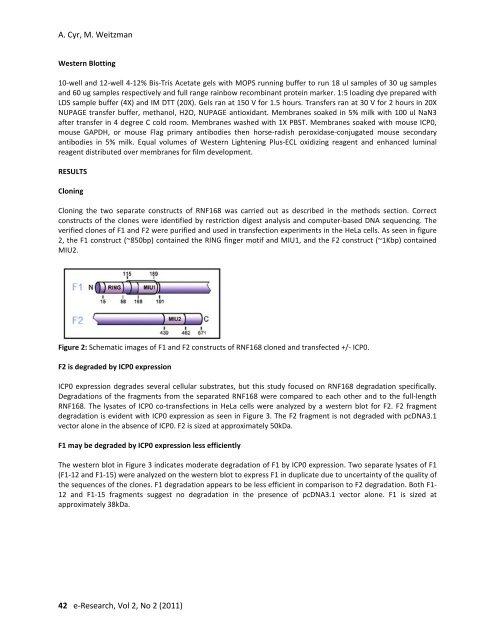
verified clones <strong>of</strong> F1 and F2 were purified and used in transfection experiments in the HeLa cells. As seen in figure<br />
2, the F1 construct (~850bp) contained the RING finger motif and MIU1, and the F2 construct (~1Kbp) contained<br />
MIU2.<br />
Figure 2: Schematic images <strong>of</strong> F1 and F2 constructs <strong>of</strong> RNF168 cloned and transfected +/- ICP0.<br />
F2 is degraded by ICP0 expression<br />
ICP0 expression degrades several cellular substrates, but this study focused on RNF168 degradation specifically.<br />
Degradations <strong>of</strong> the fragments from the separated RNF168 were compared to each other and to the full-length<br />
RNF168. The lysates <strong>of</strong> ICP0 co-transfections in HeLa cells were analyzed by a western blot for F2. F2 fragment<br />
degradation is evident with ICP0 expression as seen in Figure 3. The F2 fragment is not degraded with pcDNA3.1<br />
vector alone in the absence <strong>of</strong> ICP0. F2 is sized at approximately 50kDa.<br />
F1 may be degraded by ICP0 expression less efficiently<br />
The western blot in Figure 3 indicates moderate degradation <strong>of</strong> F1 by ICP0 expression. Two separate lysates <strong>of</strong> F1<br />
(F1-12 and F1-15) were analyzed on the western blot to express F1 in duplicate due to uncertainty <strong>of</strong> the quality <strong>of</strong><br />
the sequences <strong>of</strong> the clones. F1 degradation appears to be less efficient in comparison to F2 degradation. Both F1-<br />
12 and F1-15 fragments suggest no degradation in the presence <strong>of</strong> pcDNA3.1 vector alone. F1 is sized at<br />
approximately 38kDa.<br />
42 e-<strong>Research</strong>, Vol 2, No 2 (2011)