AutoDock Virtual Screening: Relaxed Complex scheme
AutoDock Virtual Screening: Relaxed Complex scheme
AutoDock Virtual Screening: Relaxed Complex scheme

Create successful ePaper yourself
Turn your PDF publications into a flip-book with our unique Google optimized e-Paper software.
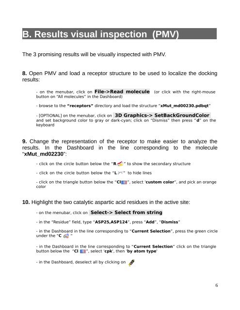
B. Results visual inspection (PMV)<br />
The 3 promising results will be visually inspected with PMV.<br />
8. Open PMV and load a receptor structure to be used to localize the docking<br />
results:<br />
- on the menubar, click on File->Read molecule (or click with the right-mouse<br />
button on “All molecules” in the Dashboard)<br />
- browse to the “receptors” directory and load the structure “xMut_md00230.pdbqt”<br />
- [OPTIONAL] on the menubar, click on 3D Graphics-> SetBackGroundColor<br />
and set background color to gray or dark-cyan; click on “Dismiss” then press “d” on the<br />
keyboard<br />
9. Change the representation of the receptor to make easier to analyze the<br />
results. In the Dashboard in the line corresponding to the molecule<br />
“xMut_md02230”:<br />
- click on the circle button below the “R ” to show the secondary structure<br />
- click on the circle button below the “L ” to hide lines<br />
- click on the triangle button below the “Cl ”, select 'custom color”, and pick an orange<br />
color<br />
10. Highlight the two catalytic aspartic acid residues in the active site:<br />
- on the menubar, click on Select-> Select from string<br />
- in the “Residue” field, type “ASP25,ASP124”, press “Add”, “Dismiss”<br />
- in the Dashboard in the line corresponding to “Current Selection”, press the green circle<br />
under the “C “<br />
- in the Dashboard in the line corresponding to “Current Selection” click on the triangle<br />
button below the “Cl ”, select 'cpk', then 'by atom type'<br />
- in the Dashboard, deselect all by clicking on<br />
6