

World Journal of Gastrointestinal Pathophysiology
World Journal of Gastrointestinal Pathophysiology
World Journal of Gastrointestinal Pathophysiology

You also want an ePaper? Increase the reach of your titles
YUMPU automatically turns print PDFs into web optimized ePapers that Google loves.
A<br />
B<br />
Nishida T et al . Treatment strategy for gastric intraepithelial neoplasia<br />
Table 3 Histological follow-up studies <strong>of</strong> gastric intraepithelial neoplasia through mild to severe dysplasia<br />
Reports (yr) LGIN (including mild to moderate dysplasia) HGIN (including severe dysplasia)<br />
bined ulceration (OR, 5.6) were significant predictive factors<br />
correlated with cancer after endoscopic resection <strong>of</strong><br />
gastric adenoma using multivariate analysis. For several<br />
decades, adenomatous polyps larger than 20 mm have<br />
been considered potentially malignant [30] and the rate<br />
<strong>of</strong> malignant transformations increased in accordance<br />
with an increase in the size. The depressed type is also<br />
reported to have more malignant potential. We, however,<br />
revealed that even patients with both smaller and elevated<br />
neoplasms had a greater than 40% under-diagnosis<br />
rate [27] . Similarly, cancer foci have been reported in hyperplastic<br />
polyps with a diameter <strong>of</strong> 5 mm [31] . Therefore, it is<br />
possible that a conventional endoscopic diagnosis based<br />
on size alone is not sufficient to make a precise preoperative<br />
diagnosis. Moreover, the surface appearance<br />
<strong>of</strong> an adenoma may also be an important factor for the<br />
diagnosis <strong>of</strong> malignancy [32] .<br />
WJGP|www.wjgnet.com<br />
Detection <strong>of</strong> carcinoma n (%) Interval (mean) n (%) Detection <strong>of</strong> carcinoma Interval (mean)<br />
Saraga et al [49] , 1987 1/64 (2) 4 yr 17/21 (81) 4 mo<br />
Lansdown et al [46] , 1990 0/7 (0) - 11/13 (85) 5 mo<br />
Rugge et al [50] , 1991 12/69 (17) 1 yr 6/8 (75) 4 mo<br />
Fertitta et al [51] , 1993 7/30 (23) 10 mo 25/31 (81) 5 mo<br />
Farinati et al [52] , 1993 - - 16/49 (33) 1<br />
-<br />
Di Gregorio et al [53] , 1993 6/89 (7) 2 yr 6/10 (60) 11 mo<br />
Bearzi et al [54] , 1994 8/81 (9.9) - 27/44 (61) -<br />
Rugge et al [55] , 1994 13/90 (14) 2 yr 14/18 (78) 9 mo<br />
Kolodziejczyk et al [56] , 1994 2/35 1 (5.7 2 ) - 7/7 (100) -<br />
Kokkola et al [57] , 1996 0/9 (0) - 2/3 (67) 1.5 yr<br />
Rugge et al [19] , 2003 8/90 (8.9) 4 yr 11/16 (69) 34 mo<br />
Yamada et al [58] , 2004 0/38 (0) - 1/10 (10) 54 mo<br />
Park et al [59] , 2008 3/26 (12) 58 mo 3<br />
1/1 (100) 58 mo 3<br />
Overall 60/628 (9.5) 145/231 (63)<br />
Proportion progressing to carcinoma and mean interval. 1 Moderate or severe; 2 Mild or moderate dysplasia; 3 Overall follow-up interval.<br />
LGIN: Low-grade intraepithelial neoplasia; IN: Intraepithelial neoplasia; HGIN: High-grade intraepithelial neoplasia.<br />
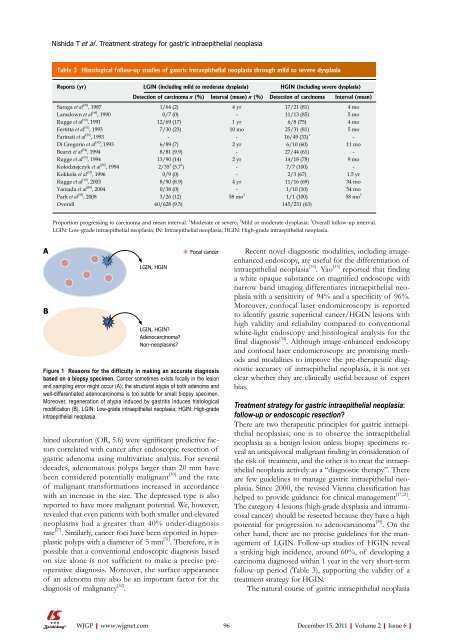
LGIN, HGIN<br />
LGIN, HGIN?<br />
Adenocarcinoma?<br />
Non-neoplasms?<br />
Focal cancer<br />
Figure 1 Reasons for the difficulty in making an accurate diagnosis<br />
based on a biopsy specimen. Cancer sometimes exists focally in the lesion<br />
and sampling error might occur (A); the structural atypia <strong>of</strong> both adenoma and<br />
well-differentiated adenocarcinoma is too subtle for small biopsy specimen.<br />
Moreover, regeneration <strong>of</strong> atypia induced by gastritis induces histological<br />
modification (B). LGIN: Low-grade intraepithelial neoplasia; HGIN: High-grade<br />
intraepithelial neoplasia.<br />
Recent novel diagnostic modalities, including imageenhanced<br />
endoscopy, are useful for the differentiation <strong>of</strong><br />
intraepithelial neoplasia [33] . Yao [33] reported that finding<br />
a white opaque substance on magnified endoscope with<br />
narrow band imaging differentiates intraepithelial neoplasia<br />
with a sensitivity <strong>of</strong> 94% and a specificity <strong>of</strong> 96%.<br />
Moreover, confocal laser endomicroscopy is reported<br />
to identify gastric superficial cancer/HGIN lesions with<br />
high validity and reliability compared to conventional<br />
white-light endoscopy and histological analysis for the<br />
final diagnosis [34] . Although image-enhanced endoscopy<br />
and confocal laser endomicroscopy are promising methods<br />
and modalities to improve the pre-therapeutic diagnostic<br />
accuracy <strong>of</strong> intraepithelial neoplasia, it is not yet<br />
clear whether they are clinically useful because <strong>of</strong> expert<br />
bias.<br />
Treatment strategy for gastric intraepithelial neoplasia:<br />
follow-up or endoscopic resection?<br />
There are two therapeutic principles for gastric intraepithelial<br />
neoplasias; one is to observe the intraepithelial<br />
neoplasia as a benign lesion unless biopsy specimens reveal<br />
an unequivocal malignant finding in consideration <strong>of</strong><br />
the risk <strong>of</strong> treatment, and the other is to treat the intraepithelial<br />
neoplasia actively as a “diagnostic therapy”. There<br />
are few guidelines to manage gastric intraepithelial neoplasia.<br />
Since 2000, the revised Vienna classification has<br />
helped to provide guidance for clinical management [17,21] .<br />
The category 4 lesions (high-grade dysplasia and intramucosal<br />
cancer) should be resected because they have a high<br />
potential for progression to adenocarcinoma [35] . On the<br />
other hand, there are no precise guidelines for the management<br />
<strong>of</strong> LGIN. Follow-up studies <strong>of</strong> HGIN reveal<br />
a striking high incidence, around 60%, <strong>of</strong> developing a<br />
carcinoma diagnosed within 1 year in the very short-term<br />
follow-up period (Table 3), supporting the validity <strong>of</strong> a<br />
treatment strategy for HGIN.<br />
The natural course <strong>of</strong> gastric intraepithelial neoplasia<br />
96 December 15, 2011|Volume 2|Issue 6|


