Weillcornellmedicine - Weill Medical College - Cornell University
Weillcornellmedicine - Weill Medical College - Cornell University
Weillcornellmedicine - Weill Medical College - Cornell University

Create successful ePaper yourself
Turn your PDF publications into a flip-book with our unique Google optimized e-Paper software.
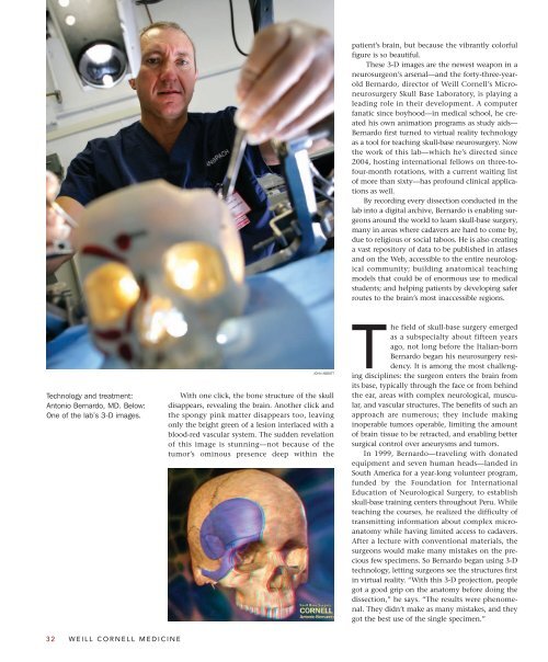
Technology and treatment:<br />
Antonio Bernardo, MD. Below:<br />
One of the lab’s 3-D images.<br />
32 WEILL CORNELL MEDICINE<br />
JOHN ABBOTT<br />
With one click, the bone structure of the skull<br />
disappears, revealing the brain. Another click and<br />
the spongy pink matter disappears too, leaving<br />
only the bright green of a lesion interlaced with a<br />
blood-red vascular system. The sudden revelation<br />
of this image is stunning—not because of the<br />
tumor’s ominous presence deep within the<br />
patient’s brain, but because the vibrantly colorful<br />
figure is so beautiful.<br />
These 3-D images are the newest weapon in a<br />
neurosurgeon’s arsenal—and the forty-three-yearold<br />
Bernardo, director of <strong>Weill</strong> <strong>Cornell</strong>’s Microneurosurgery<br />
Skull Base Laboratory, is playing a<br />
leading role in their development. A computer<br />
fanatic since boyhood—in medical school, he created<br />
his own animation programs as study aids—<br />
Bernardo first turned to virtual reality technology<br />
as a tool for teaching skull-base neurosurgery. Now<br />
the work of this lab—which he’s directed since<br />
2004, hosting international fellows on three-tofour-month<br />
rotations, with a current waiting list<br />
of more than sixty—has profound clinical applications<br />
as well.<br />
By recording every dissection conducted in the<br />
lab into a digital archive, Bernardo is enabling surgeons<br />
around the world to learn skull-base surgery,<br />
many in areas where cadavers are hard to come by,<br />
due to religious or social taboos. He is also creating<br />
a vast repository of data to be published in atlases<br />
and on the Web, accessible to the entire neurological<br />
community; building anatomical teaching<br />
models that could be of enormous use to medical<br />
students; and helping patients by developing safer<br />
routes to the brain’s most inaccessible regions.<br />
The field of skull-base surgery emerged<br />
as a subspecialty about fifteen years<br />
ago, not long before the Italian-born<br />
Bernardo began his neurosurgery residency.<br />
It is among the most challenging<br />
disciplines: the surgeon enters the brain from<br />
its base, typically through the face or from behind<br />
the ear, areas with complex neurological, muscular,<br />
and vascular structures. The benefits of such an<br />
approach are numerous; they include making<br />
inoperable tumors operable, limiting the amount<br />
of brain tissue to be retracted, and enabling better<br />
surgical control over aneurysms and tumors.<br />
In 1999, Bernardo—traveling with donated<br />
equipment and seven human heads—landed in<br />
South America for a year-long volunteer program,<br />
funded by the Foundation for International<br />
Education of Neurological Surgery, to establish<br />
skull-base training centers throughout Peru. While<br />
teaching the courses, he realized the difficulty of<br />
transmitting information about complex microanatomy<br />
while having limited access to cadavers.<br />
After a lecture with conventional materials, the<br />
surgeons would make many mistakes on the precious<br />
few specimens. So Bernardo began using 3-D<br />
technology, letting surgeons see the structures first<br />
in virtual reality. “With this 3-D projection, people<br />
got a good grip on the anatomy before doing the<br />
dissection,” he says. “The results were phenomenal.<br />
They didn’t make as many mistakes, and they<br />
got the best use of the single specimen.”