

Etude par Sonde Atomique Tomographique de la formation de nano ...
Etude par Sonde Atomique Tomographique de la formation de nano ...
Etude par Sonde Atomique Tomographique de la formation de nano ...

Create successful ePaper yourself
Turn your PDF publications into a flip-book with our unique Google optimized e-Paper software.
tel-00751814, version 1 - 14 Nov 2012<br />
Université <strong>de</strong> Rouen<br />
U.F.R. <strong>de</strong>s Sciences et Techniques<br />
Ecole doctorale « SPMII »<br />
THESE<br />
Discipline : Physique<br />
Spécialité : Sciences <strong>de</strong>s Matériaux<br />
Présentée <strong>par</strong><br />
Olena KALOKHTINA<br />
Pour l’obtention du gra<strong>de</strong> <strong>de</strong><br />
DOCTEUR DE L’UNIVERSITE DE ROUEN<br />
Le 5 juin 2012<br />
STUDY OF THE FORMATION OF NANO-PARTICLES IN<br />
ODS AND NDS STEELS BY ATOM PROBE TOMOGRAPHY<br />
Directeur <strong>de</strong> thèse : Philippe PAREIGE<br />
Encadrant : Bertrand RADIGUET<br />
Membres du jury :<br />
Mme. Marie-France BARTHE Directeur <strong>de</strong> Recherches – Orléans Prési<strong>de</strong>nte<br />
M. Michel PEREZ Professeur <strong>de</strong>s Universités - Lyon Rapporteur<br />
Mme. Brigitte DECAMPS Directeur <strong>de</strong> Recherches - Orsay Rapporteur<br />
Mme. Martine BLAT Ingénieur Chercheur EDF - Les Renardières<br />
M. Yann <strong>de</strong> CARLAN Ingénieur CEA - Sac<strong>la</strong>y<br />
M. Philippe PAREIGE Professeur <strong>de</strong>s Universités - Rouen<br />
M. Bertrand RADIGUET Maitre <strong>de</strong> Conférences - Rouen
tel-00751814, version 1 - 14 Nov 2012
tel-00751814, version 1 - 14 Nov 2012<br />
Study of the <strong>formation</strong> <strong>nano</strong>-<strong>par</strong>ticles in ODS and NDS steels<br />
by Atom Probe Tomography<br />
Abstract: Thanks to their good mechanical properties at high temperature and their<br />
stability un<strong>de</strong>r irradiation and thermal ageing, Oxi<strong>de</strong> Dispersion Strengthened (ODS) steels<br />
are good candidates for fuel c<strong>la</strong>ddings of several Generation ΙV nuclear reactors. Based on the<br />
same principle, ferritic/martensitic (FM) steels can also be reinforced by nitri<strong>de</strong>s (Nitri<strong>de</strong><br />
Dispersion Strengthened (NDS) steels).<br />
In both cases, e<strong>la</strong>boration processes have to be optimized to get a fine and <strong>de</strong>nse<br />
dispersion of <strong>nano</strong>-reinforcements. Such optimization requires characterizing the<br />
microstructure at the <strong>nano</strong>meter scale, in or<strong>de</strong>r to un<strong>de</strong>rstand the basic mechanisms of<br />
<strong>formation</strong> of the nitri<strong>de</strong>s and oxi<strong>de</strong>s during e<strong>la</strong>boration. In this work, Atom Probe<br />
Tomography has been used to investigate the <strong>nano</strong>structure of ODS and NDS steels at<br />
different steps of the e<strong>la</strong>boration.<br />
� An ODS mo<strong>de</strong>l alloy, with a high level of Y, Ti and O, as well as a ferritic ODS steel<br />
produced by mechanical alloying (MA) of Fe-18CrWTi and 0.5% Y2O3 (wt.%) pow<strong>de</strong>rs in<br />
industrial attritor were investigated in as milled state. In addition, the steel was characterized<br />
after annealing at 850°C during 1 hour and after extrusion at 1100°C. It is shown that milling<br />
results in <strong>par</strong>tial dissolution of Y and O in the matrix. However, Y, O and in a less extend Ti<br />
rich <strong>nano</strong>clusters are present in as-milled state. During subsequent annealing and<br />
consolidation, these <strong>nano</strong>clusters act as nuclei for <strong>formation</strong> of Y-Ti-O-rich <strong>nano</strong><strong>par</strong>ticles.<br />
� Ferritic NDS steels e<strong>la</strong>borated by two distinct ways, (i) mechanical alloying or (ii)<br />
nitriding of pre-alloyed pow<strong>de</strong>rs, followed by annealing at different temperatures or hot-<br />
extrusion, were investigated. In the first case a high <strong>de</strong>nsity of Ti and N rich <strong>nano</strong><strong>par</strong>ticles is<br />
observed since as-milled state. It is shown that consolidation should be performed at 800°C to<br />
get the highest <strong>de</strong>nsity of <strong>par</strong>ticles since coarsening occurs at higher temperatures. In the<br />
second case, Cr and Ti nitri<strong>de</strong>s are observed after annealing. This result is encouraging.<br />
However, the process needs to be improved, in or<strong>de</strong>r to get a more homogeneous<br />
microstructure.<br />
This research has been supported by the Agence Nationale <strong>de</strong> <strong>la</strong> Recherche within the<br />
aXtrem project.<br />
Key words: ferritic-martensitic ODS steels, mechanical alloying, nitriding, 3D atom-probe,<br />
phase trans<strong>formation</strong>s.
tel-00751814, version 1 - 14 Nov 2012
tel-00751814, version 1 - 14 Nov 2012<br />
Acknowledgements<br />
It would not have been possible to write this doctoral thesis without the help and support<br />
of the kind people around me who ma<strong>de</strong> my stay throughout my thesis so rich and valuable.<br />
Above all, I would like to express my <strong>de</strong>ep and sincere gratitu<strong>de</strong> to my supervisors, Pr.<br />
Philippe PAREIGE and Dr. Bertrand RADIGUET, who accepted me as their Ph.D. stu<strong>de</strong>nt,<br />
giving won<strong>de</strong>rful opportunity to perform this research. They offered me so much advice, good<br />
teaching and lots of good i<strong>de</strong>as, patiently supervising and allowing me to <strong>de</strong>velop the<br />
un<strong>de</strong>rstanding of the subject.<br />
I am very thankful to Mr. Michel PEREZ, Professeur <strong>de</strong>s Universités INSA Lyon and<br />
Mme Brigitte DECAMPS Directrice <strong>de</strong> Recherches CSNSM Orsay, who kindly accepted to<br />
review this manuscript in extremely short period of time. Their constructive comments and<br />
remarks improved the quality of my Ph.D. work.<br />
My sincere thanks also go to the presi<strong>de</strong>nt of the dissertation committee Mme Marie-<br />
France BARTHE, Directrice <strong>de</strong> Recherches CEMHTI/CNRS Orléans, for her time and efforts<br />
in the un<strong>de</strong>rstanding of this work.<br />
I would like to express my <strong>de</strong>ep gratitu<strong>de</strong> to Mr Yann <strong>de</strong> CARLAN Ingénieur CEA<br />
Sac<strong>la</strong>y and Mme Martine BLAT Ingénieur Chercheur EDF Les Renardières, for the<br />
organization of my work within my research project and <strong>par</strong>ticipation in the dissertation<br />
committee of the thesis.<br />
Appreciation also goes to Stéphanie Hollner, Ingénieur post-doc CEA Sac<strong>la</strong>y, for her<br />
timely help and advices concerning MatCalc simu<strong>la</strong>tions.<br />
There are all the other people who p<strong>la</strong>yed very special role over all those years.<br />
I am very grateful to Gerald DA COSTA and Francois VURPILLOT for their capability<br />
to quickly answer the questions and give their personal opinion on the experimental topics.<br />
I also would like to express my appreciation to Fabien CUVILLY, who patiently<br />
introduced me to the world of SEM/FIB tip pre<strong>par</strong>ation. It would be no exaggeration to say<br />
that this study would not have been completed today without his motivation and help.<br />
My thanks go to Constantinos HATZOGLOU, for his kind help with the mo<strong>de</strong>ling of<br />
evaporation behavior adapted to the case of materials studied in the thesis.<br />
I am very thankful to the technical staff of GPM <strong>la</strong>boratory for providing me a won<strong>de</strong>rful<br />
working environment. My thanks go to Sylvain CHAMBRELAND, Laurence CHEVALIER,
tel-00751814, version 1 - 14 Nov 2012<br />
Béatrice FOULON, Cécile GENEVOIS-MAZELIER, Jonathan HOUARD, Charly<br />
VAUDOLON, Romain VINCENT for their full-time assistance during my research work and<br />
timely help.<br />
I owe my <strong>de</strong>epest gratitu<strong>de</strong> to GPM office staff. In <strong>par</strong>ticu<strong>la</strong>r, Christine CLERGET,<br />
Agnès DALLE, Caroline JORRY, Germain MARTIGNY for their kind help in organization<br />
questions and in communication with state institutions, where their assistance was very<br />
valuable.<br />
I would like also to acknowledge my <strong>de</strong>ar friends with whom I shared my PhD years<br />
Irina, Victor, Alexan<strong>de</strong>r, S<strong>la</strong>va and Al<strong>la</strong>. Also, these three and a half years in Rouen would<br />
not been so good time without won<strong>de</strong>rful people Marilyne, A<strong>de</strong>line, Manuel, Nico<strong>la</strong>s, Maria,<br />
Thomas, Julien, Hefei, Wanghua and all others my colleagues and friends. Thank you for<br />
your support, assistance and friendship!<br />
The <strong>la</strong>st but not the least, I am greatly in<strong>de</strong>bted to my family, that <strong>de</strong>spite the<br />
geographical distance was always nearby. These thanks are for my <strong>par</strong>ents for their unlimited<br />
support and lots of wise words when it was nee<strong>de</strong>d. I am also very grateful to my husband for<br />
his incredible amount of patience, care and encouragements all the time.<br />
Though it will not be enough to express my gratitu<strong>de</strong> in words to all those people who<br />
support me all this time…Thank you!
tel-00751814, version 1 - 14 Nov 2012<br />
Table of content<br />
INTRODUCTION____________________________________________________________________1<br />
CHAPTER 1. DEVELOPMENT OF NANO-REINFORCED F/M STEELS_________4<br />
I. Generation IV nuclear reactors .................................................................................. 4<br />
II. Materials requirements and possible candidates for SFR .................................... 8<br />
II.1. Austenitic steels ............................................................................................................. 9<br />
II.2. Ferritic/Martensitic (F/M) steels ............................................................................... 10<br />
II.3. Nano-reinforced steels ............................................................................................... 10<br />
III. E<strong>la</strong>boration process of ODS ....................................................................................... 10<br />
III.1. Mechanical Alloying process ..................................................................................... 11<br />
III.2. Consolidation of pow<strong>de</strong>r and additional TMT ........................................................ 13<br />
III.3. Microstructure and influence of e<strong>la</strong>boration <strong>par</strong>ameters ....................................... 14<br />
a)Ferritic or ferritic/martenticitic (F/M) matrix ............................................................ 14<br />
b)Dispersion of oxi<strong>de</strong>s <strong>par</strong>ticles ................................................................................... 16<br />
c)Mechanism of <strong>formation</strong> of <strong>nano</strong><strong>par</strong>ticles ................................................................. 23<br />
d)Influence of the e<strong>la</strong>boration <strong>par</strong>ameters on the microstructure of ODS .................... 25<br />
III.4. Partial conclusion ........................................................................................................ 28<br />
IV. Nitri<strong>de</strong> Dispersion Strengthened (NDS) alloys...................................................... 30<br />
IV.1. NDS e<strong>la</strong>boration by nitriding .................................................................................... 30<br />
a)Principle ................................................................................................................. 30<br />
b)NDS production since 80`s ........................................................................................ 32<br />
c)Mechanism of <strong>formation</strong> of nitri<strong>de</strong>s .......................................................................... 35<br />
IV.2. NDS e<strong>la</strong>boration by Mechanical Alloying ................................................................ 37<br />
IV.3. Partial conclusions ...................................................................................................... 38<br />
V. Bibliography of Chapter 1 .......................................................................................... 39<br />
CHAPTER 2. MATERIALS, EXPERIMENTAL AND SIMULATION<br />
TECHNIQUES_______________________________________________________________ 46<br />
I. Materials ....................................................................................................................... 46<br />
I.1. ODS mo<strong>de</strong>l alloy ............................................................................................................ 47<br />
I.2. ODS steel ...................................................................................................................... 48<br />
I.3. Mechanically alloyed NDS steel (MA NDS) ............................................................... 49
tel-00751814, version 1 - 14 Nov 2012<br />
Table of contect<br />
I.4. Nitri<strong>de</strong>d NDS ................................................................................................................. 50<br />
II. Atom Probe Tomography (APT) .............................................................................. 52<br />
II.1. Principle ...................................................................................................................... 52<br />
II.2. Laser assisted field evaporation ................................................................................ 56<br />
II.3. Experimental <strong>de</strong>vices and their performances ......................................................... 57<br />
II.4. Particu<strong>la</strong>r case of ODS and NDS materials .............................................................. 60<br />
a)Determination of experimental <strong>par</strong>ameters ............................................................... 60<br />
b)Isotope over<strong>la</strong>ps ......................................................................................................... 64<br />
c)Methods for APT data treatment .............................................................................. .69<br />
d)Local magnification effect ......................................................................................... 71<br />
III. Mo<strong>de</strong>lling of precipitation kinetics with MatCalc and Thermocalc .................. 79<br />
III.1. Theory of precipitation kinetics ................................................................................ 79<br />
III.2. Mo<strong>de</strong>lling of precipitation kinetics with MatCalc ................................................... 80<br />
a)Precipitate nucleation .............................................................................................. ..80<br />
b)Evolution of radius and composition of precipitates ................................................. 81<br />
c)Evaluation of interfacial energies .............................................................................. 82<br />
III.3. Used <strong>par</strong>ameters for MatCalc calcu<strong>la</strong>tions .............................................................. 83<br />
III.4. ThermoCalc software ................................................................................................. 84<br />
IV. Conclusions of the Chapter 2. ................................................................................... 86<br />
V. Bibliography of the Chapter 2 ................................................................................... 87<br />
CHAPTER 3. OXIDE DISPERSION STRENGTHENED STEELS________________90<br />
I. ODS mo<strong>de</strong>l alloy ........................................................................................................... 91<br />
I.1. Investigation of as-milled state ..................................................................................... 91<br />
a)Microstructure ........................................................................................................... 91<br />
b)Evaporation artifacts .................................................................................................. 94<br />
c)Chemical composition of Y-Ti-O-rich and Fe-rich phases ....................................... 99<br />
I.2. Discussion .................................................................................................................... 103<br />
I.3.Partial conclusion ........................................................................................................ 107<br />
II. ODS industrial steel .................................................................................................. 108<br />
II.1. Global chemical composition ................................................................................... 108<br />
II.2. Nano<strong>par</strong>ticles study .................................................................................................. 110<br />
a)Distribution, number <strong>de</strong>nsity and size ..................................................................... 110<br />
b)Chemical composition ............................................................................................. 112<br />
c)Interfacial structure of <strong>nano</strong><strong>par</strong>ticles ....................................................................... 115<br />
II.3. Discussion .................................................................................................................. 117
tel-00751814, version 1 - 14 Nov 2012<br />
Table of contect<br />
a)Com<strong>par</strong>ison between APT, SANS and TEM results ............................................... 117<br />
b)Composition of <strong>nano</strong><strong>par</strong>ticles .................................................................................. 121<br />
c)Formation mechanism of <strong>nano</strong><strong>par</strong>ticles in ODS steel ............................................. 125<br />
II.4.Partial conclusion ...................................................................................................... 127<br />
III. Bibliography of Chapter 3 ........................................................................................ 128<br />
CHAPTER 4. NITRIDE DISPERSION STRENGTHENED STEELS____________131<br />
I. Study of MA NDS steel ............................................................................................... 131<br />
I.1. Matrix chemical composition ..................................................................................... 132<br />
I.2. Grain boundaries study .............................................................................................. 133<br />
a)Pow<strong>de</strong>r annealed at 600°C/1h .................................................................................. 133<br />
b)Pow<strong>de</strong>r annealed at 700°C/1h ................................................................................. 135<br />
c)Hot extru<strong>de</strong>d state .................................................................................................... 136<br />
d)Segregation intensity as a function of temperature ................................................. 137<br />
I.3. Nano<strong>par</strong>ticles characterization .................................................................................. 138<br />
a)As-milled condition ................................................................................................. 138<br />
b)Annealing at 600°C/1h ............................................................................................ 141<br />
c)Annealing at 700°C/1h ............................................................................................ 144<br />
d)Hot extru<strong>de</strong>d condition (800°C) ............................................................................. 146<br />
e) Annealing at 850°C/1h ........................................................................................... 148<br />
f)Annealing at 1000°C/1h ........................................................................................... 152<br />
I.4. Discussion .................................................................................................................... 153<br />
a)Chemical composition of <strong>nano</strong><strong>par</strong>ticles................................................................... 153<br />
b)Precipitation and kinetics ........................................................................................ 158<br />
c)Com<strong>par</strong>ison between ODS and NDS ....................................................................... 162<br />
I.5. Partial conclusion ........................................................................................................ 165<br />
II. Study of nitri<strong>de</strong>d NDS ............................................................................................... 166<br />
II.1. Characterization of pow<strong>de</strong>rs ................................................................................... 166<br />
a)Pow<strong>de</strong>r in as-nitri<strong>de</strong>d state ...................................................................................... 167<br />
b)Pow<strong>de</strong>r annealing at 600°C/1h ................................................................................ 168<br />
c)Pow<strong>de</strong>r annealing at 700°C/1h ................................................................................ 171<br />
II.2. Characterization of consolidated material ............................................................. 175<br />
a)Experiment 1. LATAP ............................................................................................. 175<br />
b)Experiment 2. LAWATAP ...................................................................................... 176<br />
c)Experiment 3. LAWATAP ...................................................................................... 178<br />
II.3. Discussion .................................................................................................................. 180<br />
II.4 Partial conclusion ........................................................................................................ 183<br />
III. Bibliography of the Chapter 4 ................................................................................. 184<br />
GENERAL CONCLUSIONS AND PERSPECTIVES______________________________186
tel-00751814, version 1 - 14 Nov 2012<br />
Table of contect<br />
APPENDIXES_____________________________________________________________________ 192<br />
Appendix 1.ANR AXTREM program ................................................................................... 193<br />
Appendix 2. 3DAP sample pre<strong>par</strong>ation methods ........................................................... 195<br />
a) “Double <strong>la</strong>yer” method ........................................................................................... 195<br />
b) “Micro-loop” method ............................................................................................. 196<br />
c) Pre<strong>par</strong>ation of APT tips from pow<strong>de</strong>rs ................................................................... 196<br />
d) Specimen pre<strong>par</strong>ation for neutron diffraction experiments .................................... 198<br />
Appendix 3. 3DAP data treatment methods .................................................................... 200<br />
a) Statistical tests ........................................................................................................ 200<br />
b) Method of “iso position” ....................................................................................... 201<br />
c) “Erosion“ method ................................................................................................... 203<br />
Appendix 3. Field evaporation mo<strong>de</strong>l ............................................................................... 205<br />
Appendix 4. MatCalc script .................................................................................................. 207<br />
Bibliography of Appendixes ................................................................................................ 211
tel-00751814, version 1 - 14 Nov 2012<br />
Introduction<br />
Introduction<br />
Today many research programs are <strong>de</strong>voted to the improvement of existing materials and<br />
to the <strong>de</strong>velopment of new ones that will be able to meet the different challenges for the new<br />
generations of nuclear reactors (Generation IV and fusion). The objective of Generation IV<br />
(GEN IV) reactors is to produce an abundant, reliable, proliferation resistant, safe and of<br />
course competitive energy. All these innovative systems are based on a higher running<br />
temperature associated to more severe irradiation conditions (higher irradiation dose, fast<br />
neutrons). As it is easily un<strong>de</strong>rstandable, the combination of high temperature, high neutron<br />
dose and severe environment is a major challenge for the viability of structural materials.<br />
Oxi<strong>de</strong>-Dispersion-Strengthened (ODS) ferritic / martensitic steels are <strong>de</strong>veloped in or<strong>de</strong>r<br />
to combine the swelling resistance of ferritic steels with a creep resistance in high temperature<br />
conditions at least equal to the austenitic steels. They found their application for fuel c<strong>la</strong>dding<br />
of several GEN ΙV reactors [1–4].<br />
With respect of the positive experience of ODS materials, new or alternate pathway to<br />
create iron-based alloys reinforced with a fine dispersion of nitri<strong>de</strong>s - Nitri<strong>de</strong>-Dispersion-<br />
Strengthened steels (NDS) – are envisaged and tested. In this case, the material could benefit<br />
of the stability of nitri<strong>de</strong>s <strong>par</strong>ticles [5]. In or<strong>de</strong>r to e<strong>la</strong>borate these new materials, several<br />
e<strong>la</strong>boration ways can be envisaged. Among them, nitriding could lead to a minimization of<br />
the production costs, thus avoiding expensive mechanical alloying step required in the case of<br />
ODS steels.<br />
An attempt to produce such NDS materials has been realised within the aXtrem project<br />
lea<strong>de</strong>d by CEA Sac<strong>la</strong>y and <strong>la</strong>unched by the Agence Nationale <strong>de</strong> <strong>la</strong> Recherche (ANR). The<br />
main objective of the project is the <strong>de</strong>velopment of advanced materials reinforced by stable<br />
dispersion of <strong>nano</strong><strong>par</strong>ticles (oxi<strong>de</strong>s and nitri<strong>de</strong>s) <strong>de</strong>dicated to c<strong>la</strong>dding elements of the so-<br />
called Sodium Fast Reactor. In or<strong>de</strong>r to improve or to give for the first time an un<strong>de</strong>rstanding<br />
of the basic mechanism at the origin of the <strong>formation</strong> of <strong>nano</strong>-reinforcement in Oxi<strong>de</strong>-<br />
Dispersion-Strengthened (ODS) and Nitri<strong>de</strong> Dispersion Strengthened (NDS) steels and to<br />
qualify their possible application (from research to <strong>de</strong>velopment), several research groups:<br />
CEA SRMA (Sac<strong>la</strong>y), EDF MMC (Les Renardières), Laboratoire Léon Brillouin (Sac<strong>la</strong>y),<br />
CNRS - Groupe <strong>de</strong> Physique <strong>de</strong>s matériaux (Rouen) and industrial <strong>par</strong>tners: AREVA NP<br />
(Lyon), Nitruvid Bodycote (Argenteuil), Sotep, (Issoudun), join their efforts in the project. In<br />
this program, complementary techniques such as Atom Probe Tomography (APT),<br />
1
tel-00751814, version 1 - 14 Nov 2012<br />
Transmission Electron Microscopy (TEM) and Small Angle Neutron Scattering (SANS) are<br />
used to fully characterize, at very fine scale ODS and NDS materials.<br />
The present Ph.D. work has been performed in this aXtrem framework and is mainly<br />
<strong>de</strong>voted to the characterization of ODS and NDS materials using the Atom Probe<br />
Tomography [6–9]. The characterization of the microstructure after each step of the<br />
e<strong>la</strong>boration process (such as mechanical milling, nitridation, extrusion etc… as well as after<br />
annealing at various temperatures), gives key in<strong>formation</strong> for the un<strong>de</strong>rstanding of the<br />
<strong>nano</strong>clusters <strong>formation</strong> mechanisms which is necessary for the optimization of the e<strong>la</strong>boration<br />
process.<br />
The present report is divi<strong>de</strong>d into 4 chapters. The first chapter <strong>de</strong>scribes the innovative<br />
nuclear systems issue from the Generation IV Forum, focusing on their severe service<br />
conditions. More <strong>de</strong>tails are given for the SFR concept and specificities for materials of<br />
c<strong>la</strong>dding elements. A first half of the chapter is <strong>de</strong>voted to ODS, their <strong>de</strong>velopment and<br />
physical metallurgy. More <strong>de</strong>tails are given on the oxi<strong>de</strong> <strong>nano</strong><strong>par</strong>ticles, structure, composition<br />
as well as their <strong>formation</strong> mechanism. The second half of the chapter is <strong>de</strong>voted to NDS steel.<br />
Possible ways of e<strong>la</strong>boration, experience in the past as well as today’s opportunities are<br />
discussed.<br />
The second chapter reports the <strong>de</strong>scription of the studied materials, their e<strong>la</strong>boration<br />
conditions, and the methods used in this work. The basic concepts of APT technique, the<br />
exp<strong>la</strong>nation of data treatment methods as well as the difficulties associated to the specific case<br />
of ODS steels, will be discussed. MatCalc and ThermoCalc softwares were also used in this<br />
work in or<strong>de</strong>r to estimate the expected microstructure as a function of the chemical<br />
composition and heat treatment patterns. Their basic principles and the <strong>par</strong>ameters that were<br />
used for mo<strong>de</strong>ling are <strong>de</strong>scribed.<br />
The third chapter provi<strong>de</strong>s an atomic scale <strong>de</strong>scription of two Oxi<strong>de</strong> Dispersion<br />
Strengthened ODS materials. The first one is a mo<strong>de</strong>l alloy that is characterized in the as-<br />
milled condition. As the second one is concerned, it is an industrial ODS steel <strong>de</strong>signed by<br />
CEA [10]. A more complete study of the three following states is presented: as-milled,<br />
annealed and consolidated. These results associated to results coming from different<br />
techniques (from other <strong>la</strong>boratories, TEM [10–12], SANS [12]) are com<strong>par</strong>ed. This general<br />
<strong>de</strong>scription of the material allows to give some scenario for the observed microstructural<br />
evolution.<br />
2
tel-00751814, version 1 - 14 Nov 2012<br />
Introduction<br />
The forth chapter <strong>de</strong>als with the microstructural data obtained on Nitri<strong>de</strong> Dispersion<br />
Strengthened steels (NDS). The characterisation of NDS material produced by mechanical<br />
alloying (MA NDS) is firstly reported. In a second <strong>par</strong>t, the results of the study of NDS<br />
material produced by nitriding process are given. Both materials were characterized after<br />
different treatments: initial state (as-milled or as-nitri<strong>de</strong>d), after subsequent annealing<br />
treatments (up to 1000°C) as well as in the hot extru<strong>de</strong>d state.<br />
Finally a conclusion summarises all this study, gives some ten<strong>de</strong>ncies for ODS or NDS<br />
structural <strong>formation</strong> and evolution and gives some perspectives on the e<strong>la</strong>boration ways used<br />
here and production of <strong>nano</strong>structured materials.<br />
References:<br />
[1] K.L. Murty, I. Charit, Journal of Nuclear Materials 383 (2008) 189–195.<br />
[2] S. Ukai, S. Mizuta, T. Yoshitake, T. Okuda, M. Fujiwara, S. Hagi, T. Kobayashi, Journal<br />
of Nuclear Materials 283-287 (2000) 702–706.<br />
[3] S. Ukai, M. Harada, H. Okada, M. Inoue, S. Nomura, S. Shikakura, K. Asabe, T.<br />
Nishida, M. Fujiwara, Journal of Nuclear Materials 204 (1993) 65–73.<br />
[4] J.S. Cheon, C.B. Lee, B.O. Lee, J.P. Raison, T. Mizuno, F. De<strong>la</strong>ge, J. Carmack, Journal<br />
of Nuclear Materials 392 (2009) 324–330.<br />
[5] L.E. Kindlimann, G.S. Ansell, Metall and Materi Trans B 1 (1970) 507–515.<br />
[6] D. B<strong>la</strong>vette, A. Bostel, J.M. Sarrau, B. Deconihout, A. Menand, Nature 363 (1993) 432–<br />
435.<br />
[7] D. B<strong>la</strong>vette, B. Deconihout, A. Bostel, J.M. Sarrau, M. Bouet, A. Menand, Rev. Sci.<br />
Instrum. 64 (1993) 2911.<br />
[8] M.K. Miller, R.G. Forbes, Materials Characterization 60 (2009) 461–469.<br />
[9] A. Menand, E. Ca<strong>de</strong>l, C. Pareige, D. B<strong>la</strong>vette, Ultramicroscopy 78 (1999) 63–72.<br />
[10] Y. <strong>de</strong> Car<strong>la</strong>n, J.-L. Becha<strong>de</strong>, P. Dubuisson, J.-L. Seran, P. Billot, A. Bougault, T.<br />
Cozzika, S. Doriot, D. Hamon, J. Henry, M. Ratti, N. Lochet, D. Nunes, P. Olier, T.<br />
Leblond, M.H. Mathon, Journal of Nuclear Materials 386-388 (2009) 430–432.<br />
[11] F. De<strong>la</strong>brouille, Rapport H-T27-2010-00646-FR. Caractérisation Au Microscope<br />
Electronique En Transmission D’alliages NDS Et ODS (ANR AXTREM), 2010.<br />
[12] M. Ratti, Développement De Nouvelles Nuances D’acier Ferritiques/martensitiques<br />
Pour Le Gainage D’élément Combustible Des Réacteurs à Neutrons Rapi<strong>de</strong>s Au<br />
Sodium. Thèse, Institut Polytechnique <strong>de</strong> Grenoble, 2009.<br />
3
tel-00751814, version 1 - 14 Nov 2012<br />
Chapter 1.<br />
Development of <strong>nano</strong>-reinforced F/M steels<br />
I. Generation IV nuclear reactors<br />
Grow of energy <strong>de</strong>mands and un<strong>de</strong>rstanding of the link between global warming and<br />
carbon emissions cause to search alternative <strong>la</strong>rge-scale power sources. From that point of<br />
view, nuclear energy is an attractive source of energy. It allows production of low-cost energy<br />
and simultaneously reduces greenhouse gas emissions in 20-75 times [1] (see Figure 1.1) in<br />
com<strong>par</strong>ison to natural gas power sources (the cleanest fossil fuel avai<strong>la</strong>ble).<br />
Figure 1.1. Greenhouse gas emissions from electricity generations by different sources [1].<br />
It meets more than 20% [2] of the world’s <strong>de</strong>mand for electricity today. Further advances<br />
in nuclear energy system <strong>de</strong>signs can broa<strong>de</strong>n the opportunities for the use of nuclear energy.<br />
To explore these opportunities, the U.S. De<strong>par</strong>tment of Energy's Office of Nuclear Energy,<br />
Science and Technology has engaged governments, industries, and the research community<br />
worldwi<strong>de</strong> in a wi<strong>de</strong>-ranging discussion on the <strong>de</strong>velopment of next-generation of nuclear<br />
energy systems, known as "Generation IV". This has resulted in the <strong>formation</strong> of the<br />
Generation-IV International Forum (GIF). Member countries of GIF are interested in jointly<br />
<strong>de</strong>fining the future of nuclear energy. Generation IV refers to the <strong>de</strong>velopment and<br />
<strong>de</strong>monstration of one or more nuclear energy systems that would satisfy to the following<br />
conditions [3]:<br />
� sustainability: the systems should offer efficiency in the use of the natural resources,<br />
minimize environmental impact and minimize wastes (in terms of mass, radio toxicity,<br />
residual power, etc.);<br />
4
tel-00751814, version 1 - 14 Nov 2012<br />
Chapter 1. Bibliography<br />
� economics: the generating cost should be competitive com<strong>par</strong>ed with other energy<br />
sources and the capital investment cost should be low enough for the nuclear system un<strong>de</strong>r<br />
<strong>de</strong>velopment to remain accessible to a <strong>la</strong>rge number of countries;<br />
� safety and reliability: it is obligatory that future reactors perform at least as well in<br />
terms of safety and reliability as current reactors. In <strong>par</strong>ticu<strong>la</strong>r, a key focus is elimination,<br />
as far as possible, of the need for public evacuations from areas outsi<strong>de</strong> nuclear sites in the<br />
event of an acci<strong>de</strong>nt;<br />
� resistivity to proliferation risks and likely to be easily protected from external attack.<br />
For the moment, six reactors <strong>de</strong>signs could reach the Gen IV goals [4]:<br />
Molten salt reactor (MSR) is one of the thermal reactors. In many <strong>de</strong>signs, the nuclear<br />
fuel is dissolved in the molten fluori<strong>de</strong> that serves as a coo<strong>la</strong>nt. The fluid becomes critical in a<br />
graphite core, which serves as mo<strong>de</strong>rator.<br />
Supercritical-Water-Cooled Reactor (SCWR) is also a thermal spectrum reactor. It uses<br />
supercritical water (water at a temperature and pressure above its critical point, containing<br />
both properties of the liquid and the gas) as mo<strong>de</strong>rator and coo<strong>la</strong>nt.<br />
Very-High-Temperature Reactor (VHTR) is a thermal reactor that uses a graphite<br />
mo<strong>de</strong>rator. The outlet temperature, ~ 1000°C envisaged by reactor <strong>de</strong>sign, allows to increase<br />
thermal efficiency.<br />
Gas-Cooled Fast Reactor (GFR) is a fast neutron spectrum helium-cooled reactor with<br />
outlet temperature of 850°C. Radiation to high doses does not make helium radioactive in<br />
com<strong>par</strong>ison to other possible coo<strong>la</strong>nts.<br />
Lead-Cooled Fast Reactor (LFR) is a fast neutron reactor with molten lead or lead-<br />
bismuth eutectic coo<strong>la</strong>nt.<br />
Sodium-cooled fast reactor (SFR) is fast neutron sodium-cooled reactor with closed fuel<br />
cycle for efficient management of actini<strong>de</strong>s and conversion of fertile uranium.<br />
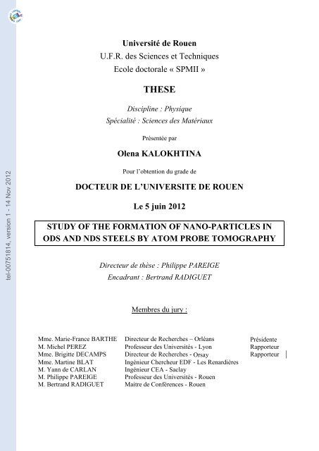
Schematic overview and relevant in<strong>formation</strong> about six selected reactors is represented in<br />
Figure 1.2. The <strong>de</strong>tailed <strong>de</strong>scription of the mentioned systems can be found in [4,5]. Most of<br />
these <strong>de</strong>signs are generally not expected to be avai<strong>la</strong>ble for commercial construction before<br />
2040 [6].<br />
5
tel-00751814, version 1 - 14 Nov 2012<br />
Chapter 1.Bibliography<br />
Thermal<br />
Fast<br />
Figure 1.2. Schematic overview of the different Generation IV reactors [4,6].<br />
Reference in<strong>formation</strong> is also presented [6].<br />
6
tel-00751814, version 1 - 14 Nov 2012<br />
Chapter 1. Bibliography<br />
Despite the differences among proposed concepts of Gen IV reactors (Figure 1.2.), the<br />
envisaged operation conditions for all these systems are rather challenging for structural<br />
materials. As it can be seen in Figure 1.3, the operating temperature of currently operating<br />
reactors (Gen II) does not exceed 400°C, whereas for Gen IV it is much higher. In addition,<br />
higher neutron doses than in Gen II, are expected for some of these systems (LFR, MSR,<br />
SFR).<br />
Figure 1.3. Overview of operating temperatures and disp<strong>la</strong>cement damage dose<br />
regimes for structural materials in current (generation II) and proposed future (Generation<br />
IV) fission energy systems. The six Gen IV fission systems are Very High Temperature<br />
Reactor (VHTR), Super Critical Water Reactor (SCWR), Lead Fast Reactor (LFR), Gas Fast<br />
Reactor (GFR), Sodium Fast Reactor (SFR) and Molten Salt Reactor (MSR) after [7].<br />
Among the other GEN IV <strong>de</strong>signs, SFR reactors may not require so much <strong>de</strong>velopments<br />
since significant experience already exist (prototypes have been operated: Joyo and Monju in<br />
Japan, BN600 in Russia and Phénix in France). SFR gained due to its closed fuel cycle and<br />
excellent potential for actini<strong>de</strong> management, including resource extension. Also, economics,<br />
safety and proliferation resistance are among its advantages. Recently, in the framework of<br />
Generation IV initiative, Fast Sodium Reactor is chosen as a priority reference <strong>de</strong>sign by<br />
many countries. France also has announced and confirmed the <strong>de</strong>cision to build a prototype of<br />
SFR scheduled for operation in 2020 [8].<br />
However, the operation conditions of SFR (as well as other Gen IV innovative systems)<br />
may be a major obstacle for the viability of existing materials employed for core elements.<br />
Core materials of concern are fuel components such as the c<strong>la</strong>dding (Figure 1.4). The<br />
c<strong>la</strong>dding keeps the fuel and products of fission reaction iso<strong>la</strong>ted from coo<strong>la</strong>nt (liquid Na). The<br />
7
tel-00751814, version 1 - 14 Nov 2012<br />
Chapter 1.Bibliography<br />
SFR c<strong>la</strong>dding will be subjected to temperatures from 400 to 600°C and neutron doses up to<br />
200 dpa. In addition c<strong>la</strong>dding is surroun<strong>de</strong>d by corrosive liquid Na that may have negative<br />
effect on material. The integrity of the fuel pins strongly <strong>de</strong>pends on whether the c<strong>la</strong>dding can<br />
withstand the irradiation environment.<br />
Figure 1.4. Fuel pin of Sodium Fast Reactor and its main components [9].<br />
In this context, the aXtrem program has been <strong>la</strong>unched by the Agence Nationale <strong>de</strong> <strong>la</strong><br />
Recherche (ANR). It is <strong>de</strong>voted to <strong>de</strong>velopment of advanced materials for application in<br />
extreme conditions such as c<strong>la</strong>dding elements of Sodium Fast Reactor. The present work is<br />
done in the framework of this program. More <strong>de</strong>tails about the <strong>par</strong>ticipants can be found in<br />
the Appendix 1.<br />
section.<br />
The possible candidates for c<strong>la</strong>dding elements of SFR are <strong>de</strong>scribed in the following<br />
II. Materials requirements and possible candidates for<br />
Sodium Fast Reactors<br />
Materials used in the core components, in the <strong>par</strong>ticu<strong>la</strong>r case of c<strong>la</strong>dding elements of<br />
SFR, should satisfy the following requirements [10–12]:<br />
� mechanical properties of material (tensile strength, ductility, creep resistance, fracture<br />
toughness) should remain acceptable after ageing;<br />
� dimensional stability un<strong>de</strong>r irradiation, whether un<strong>de</strong>r stress (irradiation creep or<br />
re<strong>la</strong>xation) or without stress (swelling, growth);<br />
� physical and chemical compatibility with the coo<strong>la</strong>nt;<br />
� other criteria for the materials are their costs to fabricate and to assemble.<br />
8
tel-00751814, version 1 - 14 Nov 2012<br />
Chapter 1. Bibliography<br />
All these requirements must be satisfied un<strong>de</strong>r normal operating as well as in acci<strong>de</strong>ntal<br />
conditions. These <strong>de</strong>mands are simi<strong>la</strong>r to materials used in the current commercial reactors,<br />
but are actually much more <strong>de</strong>manding for Gen IV [13].<br />
Different metallic materials are envisaged for c<strong>la</strong>dding application in SFR. They are<br />
discussed below.<br />
II.1. Austenitic steels<br />
This type of materials was selected for the c<strong>la</strong>dding elements of first generation of fast<br />
reactors [14,15]. For this purpose, 304 and 316 steels were used. They are chosen based on<br />
their good corrosion and thermal creep resistance. The high temperature mechanical strength,<br />
good fabrication technology and abundant experience are also among their advantages.<br />
However, they are subjected to significant void swelling induced by radiation [16]. It has been<br />
improved by adding stabilizing elements, varying chemical composition and applying cold<br />
work. For c<strong>la</strong>dding, as an example, 316Ti steel has been changed by austenitic 15/15Ti,<br />
exhibiting better swelling resistance. But use of <strong>la</strong>tter one is also limited by swelling at high<br />
doses, as it can be seen on Figure 1.5. In addition, influence of radiation provokes the<br />
<strong>de</strong>pletion of Cr from grain boundaries, making these materials sensitive to corrosion issues<br />
[17–19].<br />
Figure 1.5. Hoop <strong>de</strong><strong>formation</strong> of different gra<strong>de</strong>s of austenitic Phénix c<strong>la</strong>ddings and<br />
ferritic/martensitic (F/M) materials versus dose at temperatures between 675 and 825 K after<br />
[11].<br />
9
tel-00751814, version 1 - 14 Nov 2012<br />
Chapter 1.Bibliography<br />
II.2. Ferritic/Martensitic (F/M) steels<br />
These materials offer more advantages and are potential candidates for SFR c<strong>la</strong>ddings as<br />
well as for other Gen IV <strong>de</strong>signs [14,15]. Commercial ferritic/martensitic steels based on 9-<br />
12%Cr exhibit the highest swelling resistance in com<strong>par</strong>ison with austenitic steels (Figure<br />
1.5). This low swelling response appears to be a generic property of ferritic alloys [20,21]. In<br />
addition these materials have high thermal conductivity and low thermal expansion. A<br />
limitation to the use of ferritic–martensitic steels is their creep resistance at temperatures<br />
(400-600°C) <strong>de</strong>sired in the Gen IV systems. One approach to extend the range of operation<br />
temperatures of F/M steels is reinforcing the F/M <strong>la</strong>ttice by stable dispersion of <strong>nano</strong><strong>par</strong>ticles.<br />
II.3. Nano-reinforced steels<br />
One example of such <strong>nano</strong>-reinforced steels may be Oxi<strong>de</strong> Dispersion Strengthened steels<br />
(ODS). These materials are presently achieve increasing attention and currently consi<strong>de</strong>red as<br />
c<strong>la</strong>dding materials for SFR and several types of Gen IV systems [22,23].<br />
These materials show remarkable properties due to high <strong>de</strong>nsity (~10 23 to ~10 24 m -3 ) of<br />
<strong>nano</strong>metre scale oxi<strong>de</strong> <strong>par</strong>ticles (1-10 nm in diameter) dispersed in Fe-Cr matrix. These <strong>nano</strong>-<br />
oxi<strong>de</strong>s are found to be extremely resistant to coarsening (even during ageing at 900°C for<br />
times up to 3000 h [24,25]). They act as i) obstacles to dislocation motion [26], providing<br />
high creep strength in the range of interest [27–30] for SFR application and ii) sinks for<br />
radiation induced point <strong>de</strong>fects, providing good radiation resistance [31–34].<br />
Properties of such ODS steels strongly <strong>de</strong>pend on microstructure that requires close<br />
control over <strong>nano</strong><strong>par</strong>ticles <strong>de</strong>nsity, size, interfacial properties, etc.<br />
E<strong>la</strong>boration way, influence of chemical composition and e<strong>la</strong>boration <strong>par</strong>ameters on final<br />
microstructure, as well as study of <strong>nano</strong>-oxi<strong>de</strong>s will be discussed in next section.<br />
III. E<strong>la</strong>boration process of ODS<br />
ODS steels are usually produced by complex pow<strong>de</strong>r metallurgy technique: mechanical<br />
alloying (MA). This process is a dry, high energy ball-milling process, where elemental or<br />
pre-alloyed pow<strong>de</strong>rs (Fe, Cr etc…) are milled together with oxi<strong>de</strong> dispersoid and<br />
subsequently mechanically homogenized [31–35]. After MA the pow<strong>de</strong>r is consolidated by<br />
pow<strong>de</strong>r metallurgical processes, as Hot Isostatic Pressing (HIP) or Hot Extrusion (HE). Then<br />
10
tel-00751814, version 1 - 14 Nov 2012<br />
Chapter 1. Bibliography<br />
different recrystallization and cold- warm-working heat treatments are applied to produce<br />
final form such as tube [40,41]. Schematic e<strong>la</strong>boration route for iron-base ODS is shown on<br />
Figure 1.6.<br />
Elementary pow<strong>de</strong>rs<br />
(Fe, Cr, W, Ti…)<br />
Y2O3 pow<strong>de</strong>r<br />
Tubes<br />
Mechanical<br />
alloying<br />
Heat treatment<br />
Figure 1.6. Basic scheme of ODS production after [42].<br />
III.1. Mechanical Alloying process<br />
Rolling<br />
During MA, the pow<strong>de</strong>r <strong>par</strong>ticles are repeatedly <strong>de</strong>formed (f<strong>la</strong>ttened), fractured and cold<br />
wel<strong>de</strong>d. When two balls colli<strong>de</strong>, some amount of pow<strong>de</strong>r is trapped between them. This<br />
impact p<strong>la</strong>stically <strong>de</strong>forms the pow<strong>de</strong>r resulting in work har<strong>de</strong>ning and fracture. New surfaces<br />
are created, providing cold welding. In the early stages of MA, a <strong>la</strong>rge range of <strong>par</strong>ticles sizes<br />
<strong>de</strong>velops. They have a characteristic <strong>la</strong>yered structure consisting of different combinations of<br />
initial constituents. At certain stage, the fracture dominates over cold welding. As the<br />
<strong>de</strong><strong>formation</strong> caused by balls is still acting, the structure of <strong>par</strong>ticles is steadily refine, but the<br />
size do not change (stays constant). Finally, the interspacing between <strong>par</strong>ticles reduces and<br />
the number of <strong>la</strong>yers in the <strong>par</strong>ticles increases. A steady state <strong>par</strong>ticle size is reached when<br />
welding ba<strong>la</strong>nces fracture. At this stage, the internal structure of the <strong>par</strong>ticles is steadily<br />
refined. Within a short time, the crystallite (or grain) size is refined to the <strong>nano</strong>metre scale.<br />
In addition, high energy ball milling results in the <strong>formation</strong> of crystal <strong>de</strong>fects such as<br />
dislocations, vacancies, stacking faults. The presence of this <strong>de</strong>fect structure enhances the<br />
diffusivity of elements that increases alloying of the different elements.<br />
Can<br />
MA pow<strong>de</strong>r<br />
Hot extrusion<br />
11
tel-00751814, version 1 - 14 Nov 2012<br />
Chapter 1.Bibliography<br />
The structures that <strong>de</strong>velop during high energy ball-milling are highly <strong>de</strong>pen<strong>de</strong>nt on the<br />
process variables and the nature of pow<strong>de</strong>r components. These inclu<strong>de</strong> type and energy of<br />
mills, milling medium, temperature and atmosphere, the weight ratio of ball to the pow<strong>de</strong>r,<br />
etc. [37]. A brief <strong>de</strong>scription of the different <strong>par</strong>ameters is given below.<br />
→Atmosphere. To prevent the contamination of pow<strong>de</strong>r during alloying, it is usually<br />
performed in an inert atmosphere. Argon is the usual gas [39–42], however hydrogen is also<br />
used [47,48].<br />
→Temperature of milling. This is an important <strong>par</strong>ameter, since it enhances the diffusion<br />
process which is involved in <strong>formation</strong> of alloy phases. A slight increase of the temperature<br />
during milling may occur [37,39].<br />
→Time of mechanical alloying. It influences the final structure. Usually, the chosen time<br />
of milling is sufficient to achieve a steady state between fracture and cold welding of the<br />
pow<strong>de</strong>r <strong>par</strong>ticles. If the pow<strong>de</strong>r is milled for longer times, <strong>formation</strong> of un<strong>de</strong>sirable phases as<br />
well as increase of the contamination level of the pow<strong>de</strong>r may occur. The time of mechanical<br />
alloying is usually 48 hours [43,46,49] for conventional ODS.<br />
→Ball to pow<strong>de</strong>r mass ratio (BPR). This <strong>par</strong>ameter has significant influence on the rate<br />
of <strong>de</strong>crease of the crystallite size during MA process. Different values are reported in<br />
literature: 5:1 [50], 10:1 [51]. The 20:1 BPR [52] value is recommen<strong>de</strong>d to achieve the<br />
<strong>de</strong>sired alloying in a reasonable time.<br />
→Milling equipment. Different types of high energy milling equipment are used to<br />
produce mechanically alloyed pow<strong>de</strong>rs. More <strong>de</strong>tailed <strong>de</strong>scription of the different mills<br />
avai<strong>la</strong>ble for MA was done by Suryanarayana in [37]. An attritor (Figure 1.7 (a)) is very<br />
often used in conventional production of ODS [25,43,45,53]. It consists of a vertical drum<br />
with impellers insi<strong>de</strong>. Impellers are located at right angles with respect to each other. A motor<br />
rotates the impellers, which in turn cause to move milling balls in the drum. As a result,<br />
impacts between balls with pow<strong>de</strong>r, container wall, impellers, agitator shaft, lead to pow<strong>de</strong>r<br />
size reduction. In industrial attritor <strong>la</strong>rge quantities of pow<strong>de</strong>r (0.5-40 kg) can be milled at a<br />
time.<br />
Another popu<strong>la</strong>r mill is p<strong>la</strong>netary ball mill (Figure 1.7 (b)) [48,54]. It has such a name<br />
due to the p<strong>la</strong>net-like movement of its vials. It has another principle of work. In this system<br />
vials are located on rotating support disk. Special drive mechanism causes them to rotate<br />
around their own axes. The support disc rotates in opposite direction in re<strong>la</strong>tion to vials. The<br />
12
tel-00751814, version 1 - 14 Nov 2012<br />
Chapter 1. Bibliography<br />
centrifugal force caused by both: i) vials rotating around their own axes and ii) support disc<br />
rotating, acts on pow<strong>de</strong>r in the vials [37]. Centrifugal forces alternately act in like and<br />
opposite directions resulting in pow<strong>de</strong>r size reduction.<br />
(a) (b)<br />
Figure 1.7. Attritor (a) and p<strong>la</strong>netary (b) ball mill<br />
III.2. Consolidation of pow<strong>de</strong>r and additional TMT<br />
The consolidation procedure has a very important influence on the microstructure. As it<br />
was mentioned above, the consolidation of the pow<strong>de</strong>r usually involves either hot extrusion<br />
(HE) or hot isostatic pressing (HIP) [55].<br />
Before hot extrusion, the ground pow<strong>de</strong>rs are loa<strong>de</strong>d into a soft steel can, outgassed and<br />
sealed. The can, afterwards, is pre-heated to the extrusion temperature 850-1150°C [43,46,51]<br />
and then forced through a reducing die. Extrusion creates high shear and compressive forces<br />
which are responsible for the <strong>de</strong>nsification of the material.<br />
As in hot extrusion, HIP requires pow<strong>de</strong>rs to be contained in a vacuum sealed soft steel<br />
can. Then, isostatic pressure is applied to pow<strong>de</strong>rs at temperature 850-1150°C [56] resulting<br />
in nearly fully <strong>de</strong>nse pow<strong>de</strong>rs [57].<br />
From practical point of view hot extrusion is more attractive due to lower cost and<br />
possibility to produce <strong>la</strong>rger amounts of material. However, consolidation by hot extrusion<br />
produces anisotropic elongated (along hot extrusion direction) and textured grains [23,41].<br />
That leads to anisotropy in mechanical properties [41]. To <strong>de</strong>crease the anisotropy<br />
postextru<strong>de</strong>d Thermo Mechanical Treatment (TMT) are usually applied. They inclu<strong>de</strong><br />
sequences of cold and warm working following by softening-recrystallization heat treatments.<br />
13
tel-00751814, version 1 - 14 Nov 2012<br />
Chapter 1.Bibliography<br />
HIP is more interesting resulting in nearly isotropic characteristics of the material.<br />
III.3. Microstructure and influence of e<strong>la</strong>boration <strong>par</strong>ameters<br />
ODS steels is <strong>de</strong>signed with body-centered cubic (bcc) matrix (which is less sensitive to<br />
swelling issues in com<strong>par</strong>ison to face-centered cubic austenitic type matrix), reinforced by<br />
dispersion of <strong>nano</strong>-oxi<strong>de</strong>s. Presently, different ODS steels are being <strong>de</strong>veloped for fission and<br />
fusion applications in Japan –F4, K1, K4, K3 [22,43,58], in Europe - Fe-18Cr-1W-0.3Ti<br />
[54,59], Eurofer [60] and in United States 12YWT and 14YWT [27,51,61]. These ODS<br />
contain different amounts of Cr (9-20 wt.%). Major <strong>par</strong>t of them contains Ti and W as well as<br />
other alloying elements (Mo, Ni, Mn, V, C, Ta, Al).<br />
The current un<strong>de</strong>rstanding of the microstructures of current ODS steels, the effect of<br />
composition and key processing <strong>par</strong>ameters are discussed below.<br />
a) Ferritic or ferritic/martenticitic (F/M) matrix<br />
Due to the different chromium content, ODS steels may have different microstructures:<br />
→ODS steels that contain between 9 and 12 wt.% of Cr, (see Fe-Cr phase diagram in<br />
Figure 1.8) have martensitic structure. These steels are austenitic above 800°C. Martensitic<br />
trans<strong>formation</strong> occurs during the quench from the austenitic temperature down to room<br />
temperature. After such treatment, the microstructure consists of <strong>la</strong>ths with a very high<br />
<strong>de</strong>nsity of dislocations and the material is extremely hard (and brittle) [62]. A tempering heat<br />
treatment must be given to provi<strong>de</strong> a <strong>par</strong>tial recovery of the dislocation network. Martensitic<br />
9-12 %Cr ODS can be used for application in a temperature range below 800°C in or<strong>de</strong>r to<br />
avoid high temperature phase trans<strong>formation</strong> into austenite [13].<br />
→ODS steels containing more than 12 wt.% of Cr, present a fully ferritic matrix up to the<br />
melting temperature (see Fe-Cr phase diagram in Figure 1.8). This type of materials is<br />
envisaged for application in exten<strong>de</strong>d range of temperatures up to 1100°C in com<strong>par</strong>ison to<br />
martensitic ODS steels [59].<br />
In or<strong>de</strong>r to <strong>de</strong>crease the <strong>de</strong>gree of anisotropy in the direction of extrusion (if hot extrusion<br />
is used) for ferritic MA-ODS steel, recrystallization process may be used [64]. However, this<br />
is not so easily achievable. Up to now, all worldwi<strong>de</strong> efforts are directed to overcome<br />
“bamboo-like grain structure and a strong <strong>de</strong><strong>formation</strong> texture” [23] observed after hot<br />
extrusion in ferritic ODS steels.<br />
14
tel-00751814, version 1 - 14 Nov 2012<br />
Chapter 1. Bibliography<br />
From that point of view, martensitic ODS steels are more attractive since controlling the<br />
grain structure is possible by inducing α → γ phase trans<strong>formation</strong>. Such a way allows to<br />
significantly reduce the anisotropy of the microstructure after HE [23].<br />
Temperature (°C)<br />
9 Cr%<br />
12-18 Cr%<br />
Figure 1.8. Fe-Cr phase diagram [63]. Due to Cr content ODS steel are divi<strong>de</strong>d into<br />
martensitic (9-12wt.% Cr) and ferritic (>12wt.%Cr).<br />
In addition to that, Cr content in ODS materials is responsible for corrosion resistance.<br />
This is <strong>par</strong>ticu<strong>la</strong>rly important in the case of c<strong>la</strong>dding tube of SFR. A thin and stable <strong>la</strong>yer of<br />
chromium oxi<strong>de</strong> forms on the surface, thus increasing the resistance of the material to<br />
corrosion issues. The minimum concentration of chromium necessary to obtain an effective<br />
passive <strong>la</strong>yer is ~11 wt.% [65]. An increase of the chromium content beyond this level would<br />
result in a further increase of corrosion resistance. From that point of view, a ferritic material<br />
with high chromium content is preferred.<br />
Atomic Percent of Cr (at.%)<br />
Weight Percent of Cr (wt.%)<br />
However, high Cr ferritic ODS steel with Cr concentrations in the range of 14-22 wt.%<br />
can be subjected to har<strong>de</strong>ning and embrittlement after ageing in the region of ~400-550°C,<br />
temperatures potentially important for technological applications [66,67]. In<strong>de</strong>ed, an increase<br />
in yield and tensile strengths and reduction in ductility appear due to the <strong>formation</strong> of the α'<br />
phase (Cr-rich ferrite). This process is well known as the “475°C embrittlement”.<br />
Hence, the Cr content is one of the key <strong>par</strong>ameters to be further optimized in or<strong>de</strong>r to get<br />
the best corrosion resistance in corrosive environment with favourable mechanical properties.<br />
Concerning other alloying elements, the main objectives are to avoid <strong>formation</strong> of long<br />
life radioactive nucli<strong>de</strong>s. So normally, low activation elements are preferably used. Such<br />
15
tel-00751814, version 1 - 14 Nov 2012<br />
Chapter 1.Bibliography<br />
elements as Nb and Mo are not <strong>de</strong>sirable. They are rep<strong>la</strong>ced by W (and Ta in some cases).<br />
Alloying elements or impurities such as Ni, Cu, Co, Ag or Al also should be kept as low as<br />
possible. The content of C should be kept as low as possible in the case of ferritic ODS steels.<br />
A small amount of C (300 ppm 1 ) may result in austenite <strong>formation</strong> at high temperatures [65].<br />
The most commonly used alloying elements in ODS and their effects are summarised in Table<br />
1. 1.<br />
Table 1. 1. Effect of alloying additions (the most commonly used elements are shown)<br />
Cr Ferrite stabilizer, corrosion resistance<br />
Ti Grain refinement, <strong>par</strong>ticipation in <strong>nano</strong>clusters and solution strengthening<br />
W Solid solution strengthening [68], ferrite stabilizer [69].<br />
Mo<br />
Solid solution strengthening, ferrite stabilizer [69] less used due to high activation<br />
by neutrons<br />
Ni, Mn Austenite stabilizers [69]. Increase toughness [69,70].<br />
C Austenite stabilizer [65], enhance Ni effect [71].<br />
b) Dispersion of oxi<strong>de</strong>s <strong>par</strong>ticles<br />
In or<strong>de</strong>r to obtain a fine dispersion of <strong>nano</strong>meter scale oxi<strong>de</strong>s <strong>par</strong>ticles in a consolidated<br />
ODS material, 0.25-0.5 wt.% of Y2O3 [25,72–76,66,77] dispersoid is usually ad<strong>de</strong>d to Fe-Cr<br />
matrix during MA. Yttria is known as one of the most stable oxi<strong>de</strong>s. As an example, Y2O3 has<br />
the highest heat of <strong>formation</strong> which is 1906.7 kJ.mol -1 , in com<strong>par</strong>ison to other oxi<strong>de</strong>s such as<br />
Al2O3, SiO2, ThO2 TiO2 with heats of <strong>formation</strong> respectively of 1678.2, 910.9, 1227.6, 944.1<br />
kjmol -1 at 25°C [78]. In addition, iron and yttrium are immiscible, so it also contributes to the<br />
stability of Y2O3 oxi<strong>de</strong> in iron chromium alloys [71].<br />
Ukai et al. [23] have studied the effects of yttria content (up to 0.56 wt.%) on strength<br />
properties and ductility of hot extru<strong>de</strong>d Fe-13Cr-3W-0.5Ti ODS steel. The tensile strength<br />
along extrusion direction as well as creep rupture strength increases with increase in Y2O3<br />
content, whereas total elongation <strong>de</strong>creases. So the amount of Y2O3 (0-0.56 wt.%) should be a<br />
1 Ppm – one <strong>par</strong>t per million - is a unit to <strong>de</strong>scribe small values of miscel<strong>la</strong>neous<br />
dimensionless quantities. It <strong>de</strong>notes one <strong>par</strong>t in 10 6 or 1/1.000.000 × 100% = 0.0001%<br />
16
tel-00751814, version 1 - 14 Nov 2012<br />
Chapter 1. Bibliography<br />
compromise between strength and ductility characteristics of ODS steel. The creep rupture<br />
strength saturated at about 0.4 wt.% of Y2O3.<br />
The positive effect of Ti addition together with Y2O3 into Fe-Cr matrix during MA was<br />
shown by Ukai et al. [23]. Ti <strong>par</strong>ticipates with yttrium oxi<strong>de</strong> <strong>par</strong>ticles forming Y-Ti-O<br />
complex oxi<strong>de</strong>s resulting in finer size. As a consequence, an increase of the creep rupture<br />
strength is observed.<br />
This result was confirmed by different works. Ramar et al. [79] com<strong>par</strong>ed the distribution<br />
of <strong>par</strong>ticles by TEM in ODS Eurofer 97 (with and without Ti). He conclu<strong>de</strong>d that the addition<br />
of Ti results in smaller size of <strong>nano</strong><strong>par</strong>ticles (~20 nm and ~ 7 nm respectively without and<br />
with Ti addition). Miller et al. [61], using Atom Probe Tomography, com<strong>par</strong>ed the<br />
distribution of <strong>par</strong>ticles in 12YW and Ti-containing 12YWT ODS steel. He conclu<strong>de</strong>d that a<br />
higher number <strong>de</strong>nsity (1.4×10 24 m -3 ) of smaller sized (r=2.0 nm) <strong>par</strong>ticles is observed in Ti-<br />
containing 12YWT in com<strong>par</strong>ison to 12YW (3.9×10 23 m -3 , r=2.4 nm).<br />
Finally, Ratti et al. [80], using Small Angle Neutron Scattering, com<strong>par</strong>ed the behaviour<br />
of Fe-18Cr-1W ODS pow<strong>de</strong>rs, with addition of 0.8wt.% Ti and without, upon thermal<br />
treatments (850, 1000, 1300°C/1h). He showed that the <strong>nano</strong>-phases formed in pow<strong>de</strong>rs with<br />
some titanium are much more resistant to coarsening than the phases formed in the material<br />
without titanium.<br />
After MA and subsequent thermo-mechanical treatment (TMT), microstructure of ODS<br />
steel have been characterized by different techniques mainly by Transmission Electron<br />
Microscopy (TEM) and Atom Probe Tomography (APT). It is found that the nature<br />
(chemistry and structure) of dispersed oxi<strong>de</strong> <strong>par</strong>ticles in Fe-Cr matrix varies in a wi<strong>de</strong> range.<br />
Summary of observed oxi<strong>de</strong>s in different ODS steels are shown in Table 1. 2.<br />
In the case of Ti-containing ODS steels, TEM studies have shown variants of fine<br />
complex oxi<strong>de</strong>s, including pyrochlore Y2Ti2O7, Y2TiO5, and YTiO3. Y2O3 is found in ODS<br />
alloys that do not contain Ti. Nano<strong>par</strong>ticles also have been characterized by APT. As an<br />
example in MA 957 or Fe-14CrYWT they appear as clusters enriched in Y-Ti-O in<br />
com<strong>par</strong>ison to the matrix, but containing significant amount of Fe and Cr (Table 1. 2). The<br />
measured compositions by APT indicated that they differ from complex oxi<strong>de</strong>s Y2Ti2O7,<br />
Y2TiO5 and YTiO3. Their (Ti+Y)/O ratios range between 1.1 and 1.3.<br />
17
tel-00751814, version 1 - 14 Nov 2012<br />
Chapter 1.Bibliography<br />
Table 1. 2. Summary of data concerning the nature of oxi<strong>de</strong>s in different ODS steels.<br />
Equipment that is used for characterization, size of analysed <strong>nano</strong>-oxi<strong>de</strong>s and their structure<br />
is reported. In case of APT, chemical composition is reported (at.%, ba<strong>la</strong>nce is iron).<br />
Gra<strong>de</strong>s Equipment C, wt% Size, nm Structure<br />
Fe-18CrWTi [81]<br />
EFTEM<br />
HRTEM<br />
This disagreement between APT and TEM is mainly due to the difference of the size of<br />
the analyzed <strong>par</strong>ticles. APT observations generally focused on very small oxi<strong>de</strong>s <strong>par</strong>ticles less<br />
than few <strong>nano</strong>metres in diameter, whereas TEM (EDS) mainly focused on <strong>la</strong>rger oxi<strong>de</strong><br />
<strong>par</strong>ticles with about several tens of <strong>nano</strong>metres in diameter. Recent work of Sakasegawa by<br />
TEM on MA 957 tries to c<strong>la</strong>rify this point [86]. He reveals that different types of oxi<strong>de</strong>s are<br />
present in MA 957 and their nature <strong>de</strong>pends on their size (Figure 1.9 (a, b)):<br />
i) Big <strong>par</strong>ticles (>35 nm) are TiO2 <strong>par</strong>ticles, with a low Y content;<br />
ii) Particles with sizes from 15 nm to 35 nm are stoichiometric Y2Ti2O7 oxy<strong>de</strong>s<br />
(mainly observed by TEM).<br />
0.3 Ti 20-30 Y2O3, Y2Ti2O7, PyrOrtho<br />
Eurofer 97+Y2O3[79] HRTEM - 10-30 Y2O3<br />
Fe-9CrWVTaTi<br />
+Y2O3 [82]<br />
Eurofer 97+Y2O3<br />
[83]<br />
K1 [58]<br />
TEM<br />
(EELS)<br />
0.5 Ti ~10 ~ Y2Ti2O7<br />
HRTEM - ~12 Y2O3<br />
TEM<br />
(EDS),<br />
XRD<br />
MA 957 [84] APT 0.9 Ti 1.4<br />
14YWT [51] APT 0.4 Ti ~2<br />
0.28 Ti ~3.7 Y2Ti2O7<br />
Y-Ti-O rich <strong>nano</strong>clusters:<br />
Fe-1.7±1.7 Cr, 32.9±5.3 Ti, 15.4±7.3 Y,<br />
39.9±6.9 O, 0.02±0.2 Mo<br />
Y-Ti-O-rich <strong>nano</strong>clusters:<br />
Fe-1.2±1.1 Cr, 42.2±5.6 Ti, 7.5±4.3 Y,<br />
43.5±5.3 O<br />
14YWT [48] HRTEM 0.3 Ti ~10 YTiO3<br />
12YWT [49] APT 0.4 Ti 2-5<br />
Fe-16Cr0.1Ti [85]<br />
TEM<br />
(EDX)<br />
0.1 Ti ~9<br />
Eurofer [53] APT - ~2-10<br />
Y-Ti-O-rich <strong>nano</strong>clusters:<br />
Fe-13.66±0.52Cr, 3.03±0.26 Ti, 0.86±0.14<br />
Y, 2.89±0.25 O, 0.78±0.13 W<br />
Y2Ti2O7<br />
Y2TiO5<br />
Y-O rich <strong>par</strong>ticles:<br />
Fe-11.77±0.97Cr, 4.63±0.27Y,<br />
10.98±0.55O, 0.96±0.13Mn, 0.71±0.13<br />
Ta, 2.91±0.09V, 0.5±0.03W,<br />
0.92±0.08Si, 0.68±0.04N, 0.05±0.06 C,<br />
0.02P, 0.03Co<br />
18
tel-00751814, version 1 - 14 Nov 2012<br />
Chapter 1. Bibliography<br />
iii) Small <strong>par</strong>ticles (2 to 15 nm) are non-stoichiometric Y-,Ti-,O-enriched clusters<br />
(<strong>de</strong>signated as YxTiyOz and mainly characterized by APT).<br />
(a) (b)<br />
Figure 1.9. (a) Y over Ti concentration in oxi<strong>de</strong> <strong>par</strong>ticles <strong>de</strong>tected in MA 957 as a<br />
function of their size after TEM and XRD. Three regions are shown: 1) biggest <strong>par</strong>ticles are<br />
TiO2, >35 nm, 2) Y2Ti2O7 complex oxi<strong>de</strong>s with sizes between 15-30 nm and 3) the smallest<br />
TixYyOz <strong>nano</strong>clusters, with sizes from 2 to 15 nm (b) TEM image of extraction replica of MA<br />
957, showing different popu<strong>la</strong>tions of oxi<strong>de</strong>s [86].<br />
Further discussion will focus on these smallest non-stoichiometric YxTiyOz rich clusters<br />
(as <strong>de</strong>fined in [86]). It should be mentioned that different terminologies (<strong>par</strong>ticles, clusters,<br />
<strong>nano</strong>features, precipitates etc.) are used by different researchers or groups. This is sometime<br />
confusing. In this work and in future discussion, to se<strong>par</strong>ate them from <strong>la</strong>rger Y2Ti2O7,<br />
Y2TiO5, YTiO3 complex oxi<strong>de</strong>s, we <strong>de</strong>fine them as Ti-Y-O rich <strong>nano</strong><strong>par</strong>ticles or clusters (Y-<br />
O-rich <strong>par</strong>ticles or clusters in case of ODS steel without Ti).<br />
These <strong>nano</strong><strong>par</strong>ticles appear with significantly higher number <strong>de</strong>nsities (10 23 -10 24 m -3 in<br />
accordance to APT) and smaller sizes (less than 5 nm). It has been experimentally confirmed<br />
that they p<strong>la</strong>y a major role in oxi<strong>de</strong> dispersion strengthening of the steel [44]. Due to<br />
<strong>nano</strong>metric size they are mainly studied by APT. It is already mentioned that these <strong>nano</strong>-<br />
clusters contain a significant amount of Fe and Cr. The level of Fe estimated by APT and<br />
reported by different researches varies in wi<strong>de</strong> range. As an example, the same 12YWT ODS<br />
have been characterized in as-processed condition by different groups. The reported level of<br />
Fe in these <strong>nano</strong>clusters varies from ~78 at.% of Fe by Larson [49], ~56 at.% [61] and 4 at.%<br />
[25] by Miller.<br />
Another example is the APT study of the Eurofer ODS steel [53] (which does not contain<br />
Ti). Significant variations of chemical composition of <strong>nano</strong><strong>par</strong>ticles in the size range of 5 to<br />
10 nm are reported by the author. The Fe concentration insi<strong>de</strong> <strong>nano</strong><strong>par</strong>ticles <strong>de</strong>creases with<br />
19
tel-00751814, version 1 - 14 Nov 2012<br />
Chapter 1.Bibliography<br />
increasing size and no Fe is found at the core of <strong>la</strong>rge <strong>par</strong>ticles (10 nm). In addition, the core<br />
of 10 nm <strong>par</strong>ticles consists of Y, O, Mn and Si with M:O ratio ~0.72+/-0.15. The core is<br />
likely to have Y2O3 stoichiometry as it is also confirmed by TEM .<br />
Thus, at the moment it is unclear if these variations are coming from materials or APT<br />
technique (APT artefacts will be <strong>de</strong>scribed in Chapter 2) and the methods used to evaluate<br />
concentrations? Additional work is nee<strong>de</strong>d to c<strong>la</strong>rify the nature and composition of the<br />
<strong>nano</strong>clusters using advanced high resolution tools.<br />
Another interesting feature of these small <strong>nano</strong><strong>par</strong>ticles is observed. According to<br />
Marquis [87], <strong>nano</strong>-oxi<strong>de</strong>s observed by APT in different ODS steels (MA 957, Fe-12Cr<br />
mo<strong>de</strong>l alloy and Eurofer) may have a core shell structure (Figure 1.10).<br />
(a)<br />
(b)<br />
(c)<br />
Figure 1.10. 3D reconstructions of oxi<strong>de</strong> <strong>par</strong>ticles and concentration profiles through<br />
them. Particles with <strong>la</strong>rger sizes (~8 nm, left si<strong>de</strong>) and smaller (~4 nm, right si<strong>de</strong>) are shown<br />
for com<strong>par</strong>ison: (a) MA 957, after hot extrusion ~1150°C; (b) ODS Fe-12C alloy, HIPed at<br />
750°C/2h and annealed at 750°C/4h; and c) Eurofer, annealing at 1050°C/30min, tempered<br />
at 750/2h [87].<br />
The <strong>nano</strong><strong>par</strong>ticles in MA 957 consist of an Y, Ti, Al and O rich core and a Cr rich shell.<br />
In ODS Fe-12Cr, the core is composed of Y and O while shell is also enriched in Cr and<br />
finally in Eurofer, an yittria-rich core, surroun<strong>de</strong>d by Cr-rich and V-rich shell is found for all<br />
20
tel-00751814, version 1 - 14 Nov 2012<br />
Chapter 1. Bibliography<br />
oxi<strong>de</strong> <strong>par</strong>ticles. The presence of a shell is observed in<strong>de</strong>pen<strong>de</strong>ntly of the size (4-8 nm) of<br />
these <strong>nano</strong>features [87]. However, in the case of <strong>la</strong>rger <strong>par</strong>ticles (8 nm and more), the shell<br />
structure is visible all around the core. To rationalize the existence of these core–shell<br />
structures, Marquis suggested that since the structure of oxi<strong>de</strong> <strong>nano</strong><strong>par</strong>ticles is simi<strong>la</strong>r in three<br />
studied ODS (Y rich core with solute rich shells), they may be governed by common<br />
thermodynamic properties (interfacial energy between the oxi<strong>de</strong> <strong>nano</strong><strong>par</strong>ticle and surrounding<br />
matrix). The <strong>par</strong>ticles may have high nucleation barrier and the presence of the shell would<br />
<strong>de</strong>crease the interfacial energy, favoring their nucleation.<br />
Presence of a Cr rich shell in simi<strong>la</strong>r Fe-12Cr ODS mo<strong>de</strong>l alloy have been confirmed by<br />
TEM (STEM, EELS, EFTEM) investigations [88,89]. The reported thickness of the Cr-rich<br />
shell is ~1-3 nm. In addition, this shell is still present after prolonged annealing times (during<br />
24 and 96 h at 750°C) (Figure 1.11). EFTEM analyses performed on ODS annealed during 96<br />
h, do not revealed any significant difference in thickness of the Cr-rich shell in com<strong>par</strong>ison to<br />
<strong>par</strong>ticles after annealed during 4 hours.<br />
(a)<br />
(b)<br />
Figure 1.11. Core/shell structure of <strong>nano</strong>scale oxi<strong>de</strong> <strong>par</strong>ticles in an ODS Fe-12<br />
wt.%Cr alloy as revealed by EFTEM. Elemental maps (a) Y N2,3 and (b) Cr L2,3 for ODS<br />
mo<strong>de</strong>l alloy after annealing at 750°C during 96 hours. (c, d) EELS and (e) EDS O K, Cr L<br />
and Y L elemental profiles across <strong>nano</strong><strong>par</strong>ticles in the ODS alloy after annealing at 750°C<br />
during 24 hours [89].<br />
(c)<br />
(d)<br />
(e)<br />
21
tel-00751814, version 1 - 14 Nov 2012<br />
Chapter 1.Bibliography<br />
V-and Cr-enriched shells are also observed around all investigated <strong>par</strong>ticles by TEM<br />
(EDS/EELS) in Eurofer ODS [90].<br />
Taking into account difficulties of quantification of chemical composition by TEM, these<br />
results are in good agreement with results reported by E. Marquis [87].<br />
However, the same MA 957 ODS has been studied by Miller et al. [91] in the as-received<br />
state and after isothermal annealing treatments up to 24 h at 1300°C (by APT). This author<br />
does not report any in<strong>formation</strong> about the presence of a core shell structure!<br />
Hsiung et al [43,92,93] studied by HRTEM the interfacial structure of <strong>nano</strong><strong>par</strong>ticles in<br />
Fe-16Cr-4Al-2W-0.3Ti (<strong>de</strong>signated as K3) mechanically alloyed with 0.3 wt.% of Y2O3.<br />
After hot extrusion at 1100°C and additional annealing at 1050°C/1h, the sizes of the studied<br />
<strong>par</strong>ticles vary from 1.7 to 30 nm. A structure with an Y4Al2O9 core associated with a shell has<br />
been observed in both <strong>la</strong>rge and small <strong>nano</strong><strong>par</strong>ticles, Figure 1.12. However, in contrast to the<br />
previous reported core-shells structures (by Marquis [87], Castro [89], Klimenkov [90]), the<br />
author shows that the shell is amorphous. The author suggests that amorphous shell results in<br />
a <strong>par</strong>tial crystallization of amorphous <strong>nano</strong><strong>par</strong>ticles during consolidation, whereas<br />
amorphization of these oxi<strong>de</strong>s <strong>par</strong>ticles appear during MA process. The core/shell structure<br />
disappears after prolonged annealing treatment (900°C/168 h) for <strong>par</strong>ticle with diameter<br />
<strong>la</strong>rger than 20 nm, indicating that these core/shell structures are far from chemical<br />
equilibrium.<br />
The same structure of crystallize core and amorphous shell is found in hot extru<strong>de</strong>d<br />
(1107°C) Fe-20Cr-4.5Al-0.34Ti+0.5 wt.% of Y2O3 (<strong>de</strong>signated as MA 956) ODS steel. In this<br />
case significantly thicker shell is observed in com<strong>par</strong>ison to K3.<br />
By com<strong>par</strong>ing the HRTEM observations for K3 and MA 956 materials with results of<br />
Marquis [87], the author suggest that the content of these solute-enriched shells may differ<br />
and <strong>de</strong>pend on the initial composition of the ODS steel. He suggests that Y4Al2O9 and YAlO3<br />
core forms in the case of Al-containing K3 and MA materials, Y2TiO5 and Y2Ti2O7 may form<br />
in the case of Ti-containing ODS steel and Y2O3 may form in the case of Eurofer that do not<br />
contain either Al nor Ti. It is supposed that shell forms as a result of solutes <strong>de</strong>pletion from<br />
the core during crystallization of the <strong>par</strong>ticles upon annealing. The <strong>formation</strong> mechanism<br />
proposed by the author to rationalize the existence of these core-shell structures are discussed<br />
in <strong>de</strong>tails in next section.<br />
22
tel-00751814, version 1 - 14 Nov 2012<br />
Chapter 1. Bibliography<br />
As a conclusion, core shell/structure of <strong>nano</strong><strong>par</strong>ticles are observed by different research<br />
groups in different ODS materials and by different techniques (APT, HRTEM). However,<br />
there is no clear un<strong>de</strong>rstanding about the origin and the nature of these core/shell structures.<br />
At the moment the influence of <strong>par</strong>ticles sizes, times of ageing as well as different alloying<br />
elements in ODS steel on the stability of these dual core/shell structures is not clear. Further<br />
experimental work is nee<strong>de</strong>d.<br />
Figure 1.12. HRTEM images of a <strong>la</strong>rge <strong>nano</strong><strong>par</strong>ticle (> 20 nm) observed in K3:<br />
incoherent interface associated with a core/shell structure [43].<br />
c) Mechanism of <strong>formation</strong> of <strong>nano</strong><strong>par</strong>ticles<br />
Different analytical <strong>de</strong>vices such as X-ray diffraction (XRD), Small Angle Neutron<br />
Scattering (SANS), Transmission Electron Microscopy (TEM) and Atom Probe Tomography<br />
(APT) are used to study processes that occur during mechanical alloying and during<br />
subsequent TMT in ODS materials. Points of view of different research groups who studied<br />
the <strong>formation</strong> of <strong>nano</strong>clusters are reported in the following <strong>par</strong>agraphs.<br />
Some works show that the <strong>formation</strong> mechanism of <strong>nano</strong>clusters in ODS steel is a two<br />
stages process:<br />
i) Fragmentation, <strong>de</strong>composition/dissolution of initial Y2O3 during mechanical<br />
alloying step in the metallic matrix; The matrix is an homogeneous solid<br />
solution of Fe, Cr, Y and O;<br />
ii) Precipitation of the <strong>nano</strong>clusters occurs during annealing and following<br />
thermo-mechanical treatments.<br />
23
tel-00751814, version 1 - 14 Nov 2012<br />
Chapter 1.Bibliography<br />
An example is the work of Okuda et al. [94] who studied the change of behaviour of a<br />
Fe-13Cr alloy during mechanical alloying (with 3wt.% of Y2O3) and subsequent annealing<br />
treatments. Another example can be the study of Alinger et al. [56] on Fe-14Cr ODS steels.<br />
He confirmed that Y2O3 dissolution in Fe-14Cr alloy matrix is the dominating process during<br />
MA, resulting in a highly supersaturated solid solutions.<br />
Studies by Larson [49], Miller [51] and Yamashita [95] are also in this direction.<br />
However, it should be mentioned that in their works, APT investigations of ODS material are<br />
performed only in the final state, passing over investigation of material immediately after MA<br />
step. Their conclusions are mainly based on size differences of <strong>nano</strong> clusters in final state and<br />
the initial size of Y2O3 pow<strong>de</strong>r (~2 nm for <strong>nano</strong>clusters in as-received state and 17-31 nm for<br />
initial Y2O3 pow<strong>de</strong>r [51]) introduced during MA process. Another argument is that, the<br />
chemical composition of <strong>nano</strong>clusters estimated by APT, is more consistent with TiO than<br />
Y2O3 stoichiometry.<br />
However, some other groups of researchers show that the <strong>formation</strong> mechanism of these<br />
<strong>nano</strong>-oxi<strong>de</strong>s is not limited to dissolution/precipitation mechanisms as reported above. Among<br />
them, recent work by Brocq [96,97] on Fe-14Cr-2W-0.25Ti ODS showed that the role of<br />
mechanical alloying appears to be more complex. It could be summarised as follow:<br />
i) Dissolution of primary oxi<strong>de</strong> and nucleation of first <strong>nano</strong>clusters can occur<br />
simultaneously during ball-milling;<br />
ii) Nucleation and growth of <strong>nano</strong>clusters continue after subsequent annealing.<br />
The <strong>nano</strong>-oxi<strong>de</strong>s formed this way can be controlled through variation of ball-milling<br />
<strong>par</strong>ameters (intensity, temperature and atmosphere) and annealing <strong>par</strong>ameters (duration and<br />
temperature). However it should be mentioned that specific conditions of ball milling<br />
(reactive ball milling: YFe3 and Fe2O3 are used as reactants) are used for this study.<br />
Nevertheless, the possibility that <strong>nano</strong>clusters in conventional ODS can originate from the<br />
same mechanism are not exclu<strong>de</strong>d.<br />
An alternative <strong>formation</strong> mechanism of <strong>nano</strong>-oxi<strong>de</strong>s is suggested by Hsiung et al<br />
[43,92,93]. HRTEM investigations of Fe-16Cr-4Al-2W-0.3Ti ODS (<strong>de</strong>signated as K3) in its<br />
final state, revealed some Y2Al2O9 crystallite (in the core) surroun<strong>de</strong>d by an amorphous shell.<br />
Based on this observation, the author suggests that the <strong>formation</strong> mechanism of <strong>nano</strong><strong>par</strong>ticles<br />
may inclu<strong>de</strong> three stages (Figure 1.13.):<br />
24
tel-00751814, version 1 - 14 Nov 2012<br />
Chapter 1. Bibliography<br />
(a) During MA, fragmentation of initial Y2O3 <strong>par</strong>ticles occurs resulting in finely-<br />
dispersed (<strong>nano</strong> or sub-<strong>nano</strong>) fragments;<br />
(b) During continued MA process, agglomeration and amorphization of fragments<br />
mixed with matrix material occurs. This initiate the <strong>formation</strong> of some clusters and<br />
agglomerates (<strong>de</strong>signated as [MYO], M: alloying elements) in the Fe-Cr matrix;<br />
(c) Subsequent annealing leads to crystallization of the amorphous oxi<strong>de</strong> agglomerates<br />
<strong>la</strong>rger than 2 nm with the <strong>formation</strong> of an oxi<strong>de</strong> <strong>nano</strong><strong>par</strong>ticles (with a complex-oxi<strong>de</strong> core and<br />
solute-enriched (M) shell). Agglomerates or clusters that are smaller than 2 nm remain<br />
amorphous.<br />
(a)<br />
Figure 1.13. Schematic illustrations of (a) the three-stage mechanism for the<br />
<strong>formation</strong> of oxi<strong>de</strong> <strong>nano</strong><strong>par</strong>ticles containing a core/shell structure during MA and<br />
consolidation for Al as well as for Ti-containing ODS steel. (b) Size effect on the <strong>formation</strong> of<br />
core/shell structures in oxi<strong>de</strong> <strong>nano</strong><strong>par</strong>ticles. A solute-enriched shell can form in <strong>par</strong>ticles<br />
<strong>la</strong>rger than 2 nm when solute <strong>de</strong>pletion rate from the core is greater than solute diffusion rate<br />
from the oxi<strong>de</strong>/matrix interface during the crystallization stage; a <strong>par</strong>ticle smaller than 2 nm<br />
remains amorphous [92].<br />
d) Influence of the e<strong>la</strong>boration <strong>par</strong>ameters on the microstructure of ODS<br />
E<strong>la</strong>boration <strong>par</strong>ameters (time of milling, atmosphere, type of mill, conditions of<br />
consolidation etc.) may influence the final microstructure of ODS steel. It should be<br />
mentioned that e<strong>la</strong>boration <strong>par</strong>ameters for commercial alloys and for a <strong>la</strong>rge <strong>par</strong>t in scientific<br />
articles are only limited to the strict minimum, i.e. it is impossible to com<strong>par</strong>e! Thus a<br />
<strong>de</strong>tailed summary of the influence of <strong>par</strong>ameters at each stage of e<strong>la</strong>boration process is<br />
difficult.<br />
(b)<br />
25
tel-00751814, version 1 - 14 Nov 2012<br />
Chapter 1.Bibliography<br />
The effects of mechanical alloying (MA) conditions on oxygen contamination have been<br />
investigated by Ohtsuka et al [98]. It was shown that excess oxygen (<strong>de</strong>fined as the total<br />
content of oxygen in the steel minus the oxygen coming from Y2O3) is a very important<br />
<strong>par</strong>ameter along with Y2O3 and Ti additions that may result in coarser <strong>nano</strong><strong>par</strong>ticles and as a<br />
result reduce mechanical properties of the material. Oxygen content was drastically reduced<br />
by the application of a high purity Ar atmosphere during MA process.<br />
Also, he studied the influence of different amounts of excess O on the microstructure of a<br />
9Cr-ODS (Ti-containing) steel [45]. He showed that when the atomic ratio between excess<br />
oxygen and titanium in the steel is close to 1, finer and <strong>de</strong>nser distribution of oxi<strong>de</strong> <strong>par</strong>ticles<br />
(r=2 nm) can be achieved resulting in higher temperature strength.<br />
Miller et al. [51] studied by APT the influence of different milling times 40 and 80h on<br />
the microstructure of ball milled 14YWT ODS. The milling is performed in attritor (Zoz<br />
Simo<strong>la</strong>yer CM) un<strong>de</strong>r argon atmosphere. No residual <strong>par</strong>ts of Y2O3 pow<strong>de</strong>r are observed in<br />
the as-milled state indicating that, it has fully dissolved in Fe-Cr matrix during milling<br />
process. No significant difference is observed between 40 and 80 h of milling.<br />
The study of the influence of the consolidation temperature is reported in several works.<br />
A systematic study by SANS has been performed by Alinger et al. [56]. The effects of<br />
consolidation temperature on <strong>nano</strong>clusters size (r), number <strong>de</strong>nsity (N), volume fraction (f)<br />
and hardness (DPH) in MA 14YWT is shown on Figure 1.14 (a).<br />
(a) (b)<br />
Figure 1.14. SANS data for 14WYT (on the figures <strong>de</strong>signated as U14YWT and<br />
O14YWT indicating that Spex and attritor ball mills are respectively used) showing effect of:<br />
(a) HIP consolidation temperature (850, 1000 and 1150°C) on radius (r), volume fraction (f),<br />
number <strong>de</strong>nsity (N) and Vickers hardness (DPH) of <strong>nano</strong><strong>par</strong>ticles and (b) consolidation by<br />
HIP and HE at 850°C.<br />
26
tel-00751814, version 1 - 14 Nov 2012<br />
Chapter 1. Bibliography<br />
Consolidation is performed by HIP at different temperatures: 850, 1000 and 1150°C. As<br />
it can be seen from the figure, the <strong>nano</strong>clusters number <strong>de</strong>nsity as well as volume fraction<br />
<strong>de</strong>creases, whereas radius increases, with increasing consolidation temperature. The hardness<br />
<strong>de</strong>creases (from 635 to 344 kg mm -3 ) with increasing the HIP temperature from 850 to<br />
1150°C. In addition, the effects of consolidation by HIP and hot extrusion have been<br />
com<strong>par</strong>ed. The <strong>nano</strong>clusters number <strong>de</strong>nsity (3.5×10 24 m -3 ) is higher in hot extru<strong>de</strong>d alloy in<br />
com<strong>par</strong>ison to HIP one (1.2×10 24 m -3 ), whereas their size is approximately the same.<br />
Another study has been conducted by Hoelzer et al [44]. The Fe-14Cr-3W-0.4Ti wt.%<br />
ODS alloy was hot extru<strong>de</strong>d at different temperatures (850 and 1175°C). A high number<br />
<strong>de</strong>nsity of 2-nm size Ti-, Y-, and O-rich <strong>nano</strong>clusters was observed after HE at 850°C,<br />
whereas a lower number <strong>de</strong>nsity of predominantly Y2Ti2O7 oxi<strong>de</strong> <strong>par</strong>ticles are observed after<br />
extrusion at 1175°C. These results are, in general, consistent with Alinger´s conclusions,<br />
showing that higher consolidation temperature results in <strong>la</strong>rger radius as well as lower number<br />
<strong>de</strong>nsity of <strong>nano</strong><strong>par</strong>ticles.<br />
Dou et al [99] by TEM, also studied this point for hot extru<strong>de</strong>d Fe-15.5Cr-2W-0.1Ti-4Al<br />
ODS steel. His results are also consistent with Alinger’s [56]. He shows that reduction of hot<br />
extrusion temperature from 1150 to 1050°C lead to significant <strong>de</strong>crease of grain size, as well<br />
as <strong>nano</strong><strong>par</strong>ticles number <strong>de</strong>nsity increases (from 1.72×10 23 to 9.2×10 23 m -3 ) and their size<br />
<strong>de</strong>creases to about half (from 6.7 to 3.2 nm).<br />
All listed studies show a common trend. Lower temperature of consolidation results in<br />
higher number <strong>de</strong>nsity of <strong>nano</strong><strong>par</strong>ticles as well as smaller size. The other <strong>par</strong>ameters vary<br />
from study to study and general trends are difficult to give.<br />
Influence of Hot Isostatic Pressing (HIP) and Hot Extrusion (HE) on final microstructure<br />
and properties of ferritic Fe–14Cr, 2W, 0.3 Ti, 0.3Y2O3 ODS steel have been studied by<br />
Unifantowicz et al [48]. In both cases, mechanical alloying is performed in p<strong>la</strong>netary ball mill<br />
in hydrogen atmosphere. After the pow<strong>de</strong>r is <strong>de</strong>gassed at 400°C/2h, it is heated to<br />
consolidation temperature 1100°C/1h and finally extru<strong>de</strong>d in a 500 t press. In the case of HIP<br />
material, after canning and <strong>de</strong>gassing, consolidation is performed in Ar atmosphere by<br />
applying a pressure of 200MPa for 4 h at 1150°C. Afterwards both consolidated materials are<br />
subjected to the same heat treatment: 850°C/1h. The TEM investigations revealed, fairly<br />
equiaxed grains (having a mean diameter in the range of 40–80nm) and a high number <strong>de</strong>nsity<br />
of <strong>nano</strong>-sized oxi<strong>de</strong> <strong>par</strong>ticles in both materials. The <strong>nano</strong><strong>par</strong>ticles appear with higher number<br />
27
tel-00751814, version 1 - 14 Nov 2012<br />
Chapter 1.Bibliography<br />
<strong>de</strong>nsity (17.4×10 23 m -3 ) and higher radius (r=4 nm) in the case of the HE material in<br />
com<strong>par</strong>ison to the HIP one (9.3×10 23 m -3 , r=2 nm). This result is also consistent with<br />
Alinger’s [56] observation who also studied the influence of HIP and HE consolidation on the<br />
microstructure of 14YWT ODS. As for mechanical properties are concerned, the ODS steel<br />
produced by HE exhibits much higher tensile strength and hardness at low to mo<strong>de</strong>rate<br />
temperatures, as com<strong>par</strong>ed to the steel fabricated by Hot Isostatic Pressing. It is linked with an<br />
important work har<strong>de</strong>ning effects and also a higher number <strong>de</strong>nsity of <strong>nano</strong>-sized oxi<strong>de</strong>s<br />
<strong>par</strong>ticles in the HE state in com<strong>par</strong>ison to the HIP material (Figure 1.15).<br />
Figure 1.15. Ultimate tensile strength (UTS) and uniform elongation (εu) versus test<br />
temperature for ferritic Fe–14Cr-2W-0.3Ti-0.3Y2O3 ODS steels consolidated either by hot<br />
extrusion (HE) or hot isostatic pressing (HIP).<br />
However, at higher temperatures, both steels have simi<strong>la</strong>r tensile strength. It is important<br />
to note, that micropores are found in both consolidated materials. They are twice <strong>la</strong>rger in the<br />
case of the HIP steel (0.42 µm), as com<strong>par</strong>ed to the HE steel (0.26 µm).<br />
III.4. Partial conclusion<br />
Advanced nuclear reactors (Gen 4 and fusion) will operate at higher temperatures and<br />
higher irradiation doses in com<strong>par</strong>ison to nowadays reactors. Operation conditions of these<br />
advanced nuclear reactors are challenging for structural materials. Among the materials that<br />
are potentially capable to operate at high temperatures while being resistant to high neutron<br />
fluxes are Oxi<strong>de</strong> Dispersion Strengthened steels. These materials consist of<br />
ferritic/martensitic (F/M) matrix that is resistant to radiation swelling and fine dispersion of<br />
<strong>nano</strong>metre scale oxi<strong>de</strong>s. These <strong>nano</strong>metre scale <strong>par</strong>ticles are stable at elevated temperatures.<br />
They are responsible for high temperature creep strength properties of ODS steel (serve as an<br />
28
tel-00751814, version 1 - 14 Nov 2012<br />
Chapter 1. Bibliography<br />
obstacle for dislocation movement). In addition they are improving resistance to irradiation by<br />
providing a high number <strong>de</strong>nsity of trapping sites.<br />
Today many different ODS gra<strong>de</strong>s are e<strong>la</strong>borated. They are produced by mechanical<br />
alloying, using Y2O3 pow<strong>de</strong>r milled with Fe-Cr matrix, followed by consolidation and<br />
different thermo-mechanical treatments. It is wi<strong>de</strong>ly accepted that precipitation of Y-Ti-O rich<br />
<strong>nano</strong>phases occurs during Hot Extrusion (HE) or Hot Isostatic Pressing (HIP) processes while<br />
mechanical milling mainly causes the dissolution of the initial Y2O3 pow<strong>de</strong>r. However,<br />
limited amount of works were performed in or<strong>de</strong>r to characterise ODS pow<strong>de</strong>rs in the as-<br />
milled state. It can be expected that dissolution and nucleation mechanism may vary as a<br />
function of alloy chemistry and processing <strong>par</strong>ameters.<br />
ODS steels can contain various types of oxi<strong>de</strong>s: i) <strong>la</strong>rge TiO2 oxi<strong>de</strong>s, ii) stoichiometric<br />
Y2Ti2O7 oxi<strong>de</strong>s, iii) non-stoichiometric Ti-Y-O enriched <strong>nano</strong>-clusters,<br />
Reported results are very promising, however many questions still need to be answered.<br />
The nature of smallest <strong>nano</strong>-oxi<strong>de</strong>s is not fully un<strong>de</strong>rstood. Determination of the composition<br />
and structure of these <strong>par</strong>ticles would provi<strong>de</strong> insight into possible mechanisms of <strong>formation</strong><br />
and help to <strong>de</strong>velop methods to further optimise processing of <strong>de</strong>sired alloy structures. The<br />
<strong>nano</strong>scale character of the features requires a combination of complementary techniques such<br />
as Small Angle and Neutron Scattering (SANS), Transmission Electron Microscopy (TEM)<br />
and Atom Probe Tomography (APT).<br />
An attempt to answer some of these questions is performed in this current Ph.D work.<br />
From another point of view, alternative to ODS materials may give different advantages.<br />
One of such solution may be the Nitri<strong>de</strong> Dispersion Strengthened steel. These materials are<br />
<strong>de</strong>scribed in following section.<br />
29
tel-00751814, version 1 - 14 Nov 2012<br />
Chapter 1.Bibliography<br />
IV. Nitri<strong>de</strong> Dispersion Strengthened (NDS) alloys<br />
Taking into account the positive experience of ODS steels, <strong>de</strong>velopment of alternative<br />
solutions could be envisaged. In <strong>par</strong>ticu<strong>la</strong>r, the oxi<strong>de</strong>s could be rep<strong>la</strong>ced by another stable<br />
phase. Nitri<strong>de</strong> Dispersion Strengthened steels (NDS) can be obtained using a dispersion of<br />
<strong>nano</strong>metric nitri<strong>de</strong>s such as TiN. In this case, a high stability is expected at elevated<br />
temperatures [100,101].<br />
Dispersion strengthening of ferritic/martenstic (F/M) matrix by nitri<strong>de</strong>s can be obtained<br />
by different techniques, <strong>de</strong>pending the way nitrogen is introduced:<br />
(i) nitriding of a steel containing strong nitri<strong>de</strong> forming elements (Cr, Ti, V ect.);<br />
(ii) mechanical alloying with further thermo-mechanical treatments.<br />
These two approaches are <strong>de</strong>tailed in the next section.<br />
The procedure for the e<strong>la</strong>boration of materials can be significantly simplified (avoiding<br />
the expensive MA step), especially if the nitriding process is performed on the final tube. In<br />
that case, the amount of e<strong>la</strong>boration steps would be reduced resulting in a minimization of the<br />
fabrication cost. If a homogeneous NDS material could be produced, it may be significantly<br />
easier (in com<strong>par</strong>ison to ODS [102,103]) to control the recrystallization process during TMT.<br />
Therefore, NDS materials seem to be very promising. All advantages and weaknesses<br />
should be carefully evaluated in or<strong>de</strong>r to <strong>de</strong>fine if these materials can be worthy alternative to<br />
ODS.<br />
IV.1. NDS e<strong>la</strong>boration by nitriding<br />
a) Principle<br />
Nitriding involves the diffusion of nitrogen into the surface <strong>la</strong>yers of low carbon steel at<br />
elevated temperature. This surface treatment is usually applied to improve the fatigue<br />
resistance [104], wear resistance [105,106] and corrosion properties [106]. As it is shown on<br />
Figure 1.16, surface nitriding usually results in the <strong>formation</strong> of two <strong>la</strong>yers: a compound and a<br />
diffusion <strong>la</strong>yers. The compound <strong>la</strong>yer, located at the surface, is ma<strong>de</strong> of iron nitri<strong>de</strong>s [107].<br />
The improvement of the tribological and anticorrosion properties is mainly attributed to this<br />
<strong>la</strong>yer.<br />
30
tel-00751814, version 1 - 14 Nov 2012<br />
Chapter 1. Bibliography<br />
(a) (b)<br />
Figure 1.16. (a) Optical micrograph of the nitri<strong>de</strong>d zone observed in Fe–5 wt%Cr<br />
after [108]. (b) Hardness profile and nitrogen concentration at different positions from the<br />
surface. The positions of the interest are marked from (1) to (6) in the micrograph as in the<br />
profile.<br />
Enhancement of the fatigue properties is ascribed to the diffusion zone. In the diffusion<br />
zone, nitrogen is dissolved interstitially. If the iron matrix contains some alloying elements,<br />
their high affinity with nitrogen (Cr, Ti, Al, V) may promote nitri<strong>de</strong>s <strong>formation</strong>.<br />
Various nitriding processes exists, <strong>de</strong>pending on the medium used to bring nitrogen.<br />
Today the nitriding processes are:<br />
Gas nitriding. A nitrogen rich gas, usually ammonia (NH3) is used as a donor. When<br />
ammonia comes into contact with the heated work piece (>480°C), it dissociates into nitrogen<br />
and hydrogen. The nitriding potential, which <strong>de</strong>termines the rate of introduction of nitrogen<br />
into the surface, is <strong>de</strong>termined by the NH3 concentration at the work surface and its rate of<br />
dissociation.<br />
Salt bath nitriding. A nitrogen containing salt, such as cyani<strong>de</strong> (CNO), is used as a<br />
nitrogen donor medium. The component(s) is p<strong>la</strong>ced in a molten salt bath and is heated to get<br />
a hard nitri<strong>de</strong> <strong>la</strong>yer on the surface.<br />
P<strong>la</strong>sma nitriding. The work piece is p<strong>la</strong>ced in a vacuum vessel, where an atmosphere<br />
containing nitrogen is introduced. A voltage is applied between the work piece (catho<strong>de</strong>) and<br />
the vessel wall (ano<strong>de</strong>). It results in ionization of gas atoms which are accelerated toward the<br />
work piece. This ion bombardment cleans the surface and provi<strong>de</strong>s active nitrogen. Nitrogen<br />
can diffuse into the work piece un<strong>de</strong>r the effect of temperature which is controlled by the<br />
heating system. A key difference between gas and ion nitriding is the mechanism used to<br />
31
tel-00751814, version 1 - 14 Nov 2012<br />
Chapter 1.Bibliography<br />
generate nitrogen at the surface of the metal. A schematic representation of an ion nitriding<br />
furnace is shown in Figure 1.17.<br />
Vacuum process<br />
vessel<br />
Work piece<br />
Process gas<br />
Manifold<br />
(nitrogen, argon)<br />
Vacuum pump<br />
Figure 1.17. Simple schematic representation of a p<strong>la</strong>sma (ion) nitriding furnace<br />
system after [109].<br />
Among the several p<strong>la</strong>sma based surface modification processes, p<strong>la</strong>sma nitriding is<br />
consi<strong>de</strong>red as the most industrially adopted method.<br />
b) NDS production since 80`s<br />
Heating system<br />
Power<br />
source<br />
Nitri<strong>de</strong>-Dispersion-Strengthened Steels has been already envisaged for nuclear<br />
application in 1980s. Their <strong>de</strong>velopment was performed mainly by two companies. The first<br />
is United Kingdom Atomic Energy Authority (AEA, UK). Its activity was focused on<br />
<strong>de</strong>veloping reactor technologies for the nuclear power stations. The second one is the Garrett<br />
Corporation, (USA). Its activities are mainly concentrated on Aircraft Engineering and<br />
Aerospace Technology. Results of their investigations are mainly avai<strong>la</strong>ble in patents.<br />
Another scientific group – Swedish Institute of metals research - also performed several<br />
attempts in or<strong>de</strong>r to reinforced austenitic matrix with a fine dispersion of TiN <strong>par</strong>ticles.<br />
A summary of different ways used to produce Nitri<strong>de</strong> Dispersion Strengthened steels<br />
since 1980s is represented in Table 1. 3. Some of these studies were not primarily focused on<br />
nitri<strong>de</strong> investigation (morphology, microstructure, size etc.) but aimed improvement of<br />
mechanical properties (Patents). The nitriding was used for pow<strong>de</strong>r, strip, sheet as well as for<br />
32
tel-00751814, version 1 - 14 Nov 2012<br />
Chapter 1. Bibliography<br />
tubes. The austenitic, ferritic and F/M materials were nitri<strong>de</strong>d. Only gas nitriding was applied.<br />
It was always realised in two steps:<br />
i) Nitriding in the temperature range (815 to 1200°C);<br />
ii) Denitriding treatment at high temperature (T>1000°C) to remove excess of<br />
CrN and to convert Ti into TiN.<br />
Several important conclusions can be drawn:<br />
� Uniform distribution of TiN <strong>par</strong>ticles [100,110,111] have been obtained with<br />
re<strong>la</strong>tively small sizes (Table 1. 3.), producing a dispersion har<strong>de</strong>ned steel [110,111].<br />
� Resulting microstructure is strongly <strong>de</strong>pen<strong>de</strong>nt on nitriding <strong>par</strong>ameters. High<br />
nitriding temperatures (815 to 1200°C) result in an increased diffusion of alloying<br />
elements making difficult the control of the nitriding process. From one hand, if the<br />
nitriding time is exten<strong>de</strong>d greatly beyond the time nee<strong>de</strong>d to react all of the Ti excess<br />
CrN forms and further <strong>de</strong>nitriding treatment can lead to the pore <strong>formation</strong> [100,112].<br />
From another hand, if time was not sufficient, it resulted in non uniform microstructures<br />
through the cross-section (resulting in unnitri<strong>de</strong>d core).<br />
� The range of maximum service temperatures of nitri<strong>de</strong>d <strong>par</strong>ts have been<br />
limited by nitri<strong>de</strong>d temperature.<br />
Many ways of production, equipment and technology have been changed since then.<br />
Taking into account new possibilities, production way can be re-evaluated.<br />
As an example, p<strong>la</strong>sma nitriding can be an alternative to gas nitriding as proposed by<br />
AEA in the past. It offers many advantages such as close control, not only of temperature but<br />
also of gas composition, pressure heating rate and process time. Shorter process time is<br />
achievable, due to higher nitrogen concentration avai<strong>la</strong>ble on the surface. Due to such<br />
technique, nitrogen incorporation can be achieved at consi<strong>de</strong>rably lower temperature<br />
(~400°C). Further homogenization would occur during thermal treatment (before<br />
consolidation in case of pow<strong>de</strong>rs). So, precipitation of nitri<strong>de</strong>s (TiN) can be expected at lower<br />
temperature (600-700°C) in com<strong>par</strong>ison to what was already studied (800-1200°C).<br />
Such nitriding treatment has been realized in the framework of the ANR “aXtrem”<br />
program in col<strong>la</strong>boration with Nitruvid-Bodycote. P<strong>la</strong>sma nitriding of Fe-Cr-Ti pow<strong>de</strong>r have<br />
performed at T~400°C. The first results <strong>de</strong>voted to characterisation of this material are<br />
presented in the Ph.D.<br />
33
tel-00751814, version 1 - 14 Nov 2012<br />
Chapter 1.Bibliography<br />
Table 1. 3. Summary of nitriding ways for NDS production (research ~ 1980s). In<strong>formation</strong> is avai<strong>la</strong>ble from patents and conventional scientific literature.<br />
OM –Optical microscopy, X-Ray-X-ray diffraction, TEM- Transmission electron microscopy.<br />
Reference Composition/state Process<br />
Characterisation<br />
techniques<br />
Observation<br />
Kindlimann and<br />
Ansell [100]<br />
316 austenitic stainless<br />
steel (Fe-18Cr-12Ni-<br />
2.0Ti wt.%)<br />
(Strip)<br />
a) gas nitriding at 870-1210°C<br />
(NH3)<br />
b) <strong>de</strong>nitriding treatment<br />
TEM,<br />
X-ray<br />
Cubic TiN <strong>par</strong>ticles (non coherent with matrix),<br />
have Bain orientation re<strong>la</strong>tionship [113].<br />
Swedish institute<br />
for metals research<br />
[110]<br />
316L austenitic<br />
stainless steel (Fe-<br />
17.4Cr-12.8<br />
Ni+0.7Ti+1.2Mn wt.%)<br />
(Pow<strong>de</strong>r)<br />
a) gas nitriding at 900°C<br />
(N2+5%H);<br />
b) hot extrusion of cans filled<br />
with pow<strong>de</strong>r at 1150°C<br />
OM, TEM<br />
?<br />
Two homogeneous distributions of TiN nitri<strong>de</strong>s are<br />
observed [114]. Mean radii ~10 and 50 nm. The<br />
number <strong>de</strong>nsities are respectively 10 21 and 10 20 m -<br />
3<br />
Swedish institute<br />
for metals research<br />
[111].<br />
316L austenitic steel<br />
(Fe-<br />
18.13.9Ni+0.6wt.%Ti)<br />
(Pow<strong>de</strong>r)<br />
a) gas nitriding at 900-1100°C<br />
(pure N2); b) hot extrusion at<br />
1200°C.<br />
SEM, TEM<br />
(EDX)<br />
Nitriding at higher temperature (1000°C) gives two<br />
distributions of Ti-containing <strong>par</strong>ticles (R~150 and<br />
R~10 nm), nitriding at lower temperature (900°C)<br />
results in more homogeneous size distribution of<br />
<strong>par</strong>ticles (R~25 and R~1 nm).<br />
The Garett<br />
corporation.<br />
Patent4.464.207and<br />
0.008.228<br />
[112,115]<br />
Ferritic steel (type 409<br />
or 439) (Fe-11/19Cr-<br />
0.5Ti wt.%) (light<br />
gauge-strip or sheet)<br />
a) gas nitriding at 815-980°C<br />
(NH3 or N2);<br />
b) <strong>de</strong>nitriding treatment in H2 at<br />
1000°C<br />
a) gas nitriding at 1050-1200°C<br />
? P<strong>la</strong>te-like TiN shape<br />
F/M steel<br />
(in fully austenitic state);<br />
AEA [116]<br />
Patent 4.846.899<br />
(Fe-9/12Cr+2.5Ti wt.%)<br />
(strip, sheet, tube or<br />
precipitation of nitri<strong>de</strong> within<br />
matrix and b) converting to the<br />
?<br />
Resulting dispersoid is too coarse to make any<br />
strengthening effect<br />
pow<strong>de</strong>r)<br />
ferrtic, or ferritc/martensitic<br />
form during cooling<br />
34
tel-00751814, version 1 - 14 Nov 2012<br />
c) Mechanism of <strong>formation</strong> of nitri<strong>de</strong>s<br />
Chapter 1.Bibliography<br />
The <strong>formation</strong> of nitri<strong>de</strong>s in iron during nitriding is an issue with a long history.<br />
Precipitates shape, size, coherency with matrix, orientational re<strong>la</strong>tionship (OR) as well as<br />
p<strong>la</strong>stic and e<strong>la</strong>stic <strong>de</strong><strong>formation</strong>s of the matrix nee<strong>de</strong>d to nucleate and coarsen the precipitates<br />
have been studied by Jack [101,113,117] and Mittemeijer [118,119].<br />
Due to the <strong>la</strong>rge affinity of some alloying elements (Cr, Ti, V, Mo etc.) with nitrogen,<br />
these elements easily form thin and stable p<strong>la</strong>telets on the {100} faces of the bcc <strong>la</strong>ttice, thus<br />
influencing the mechanical properties. In most cases after nitriding of Fe-Cr and Fe-Ti<br />
systems, TiN and CrN p<strong>la</strong>te-like precipitates form. They <strong>de</strong>velop with the nitri<strong>de</strong> p<strong>la</strong>telet<br />
faces <strong>par</strong>allel to the {0 0 1}α-Fe ferritic p<strong>la</strong>nes [100,108,120,121]. The p<strong>la</strong>telets are oriented<br />
with respect to the ferrite matrix according to the Bain (also known as Baker-Nutting)<br />
orientation re<strong>la</strong>tionship (OR) given by:<br />
{0 0 1}α-Fe // {0 0 1}nitri<strong>de</strong>; [1 0 0] α-Fe // [1 1 0] nitri<strong>de</strong><br />
Schematic representation of the Bain OR is shown on Figure 1.18. Following the Bain<br />
OR, the expansion in the (011) α-Fe p<strong>la</strong>ne is typically less than 5% and is higher than 45% in<br />
the direction perpendicu<strong>la</strong>r to this p<strong>la</strong>ne. That <strong>de</strong>fines the p<strong>la</strong>te-like structure of precipitates.<br />
Figure 1.18. Schematic representation of the Bain (or Baker-Nutting) orientation<br />
re<strong>la</strong>tionship between a titanium nitri<strong>de</strong> <strong>par</strong>ticle and the ferrite matrix after [122].<br />
However, up to now, the initial stage of precipitate <strong>formation</strong> is un<strong>de</strong>r <strong>de</strong>bate. It was<br />
proposed by Jack [117], that there is a sequence of steps upon nitriding of Fe-Ti, Fe-Cr, Fe-<br />
Mo, Fe-W systems before equilibrium precipitates is formed. They are the following:<br />
35
tel-00751814, version 1 - 14 Nov 2012<br />
Chapter 1.Bibliography<br />
i. first, metastable intermediate Fe-M-N-enriched clusters (or mixed GP zones as <strong>de</strong>fined<br />
in [123]), where M=Cr, Ti, Mo or V are formed.<br />
ii. after nitriding these metastable clusters transforms into stoichiometric stable MN<br />
phase.<br />
Not all stages are observed in all alloys. As an example, in Fe-Cr alloys, equilibrium<br />
f.c.c. CrN precipitates have been i<strong>de</strong>ntified even in the initial stages of the nitriding [124].<br />
In or<strong>de</strong>r to c<strong>la</strong>rify that point, investigation of the initial stage of <strong>formation</strong> as well as the<br />
evolution of precipitates upon heat treatments in cold rolled Fe-4Ni-2Ti (wt.%) and Fe-4Ni-<br />
3Cr (wt.%) alloys have been performed by Chechenin et al [123]. The nitriding is performed<br />
on thin foils in NH3/H2 atmosphere, in the temperature range 300-600°C, during 1-24h. XRD<br />
and TEM (HRTEM) investigations revealed:<br />
� Small (dimensions 2.5 aTiN×2.5 aTiN ×0.5 aTiN) coherent Ti(Fe)N clusters (probably<br />
GP-zone type) are formed during nitriding at 300°C in Fe-4Ni-2Ti alloy.<br />
� Further annealing at higher temperatures result in growth of <strong>la</strong>teral dimensions of<br />
these clusters. In addition, these GP-like-clusters loose Fe and become stoichiometric<br />
TiN precipitates.<br />
However, no evi<strong>de</strong>nce of precipitate <strong>formation</strong> is observed in Cr-containing alloy at<br />
300°C, whereas at 400 °C thin (1 to 2 mono<strong>la</strong>yers) and wi<strong>de</strong> (more that 5 aCrN) p<strong>la</strong>telets are<br />
found. No evi<strong>de</strong>nce of <strong>formation</strong> of any GP-like clusters (pre-precipitation stage) is found at<br />
any stage for Fe-4Ni-3Cr alloy.<br />
These results are in agreement with the work of Jack in the case of the Fe-Ti system [117]<br />
and Mortimer [124] for Fe-Cr system.<br />
Another open question is the microstructure of nitri<strong>de</strong>d <strong>la</strong>yers. A number of<br />
investigations were conducted for Fe-Cr alloys [108,118–120,125–127], showing two<br />
precipitate morphologies:<br />
� In the initial stage very small (<strong>nano</strong>metric [108,123,125]), Cr-N precipitates appear.<br />
This stage is associated with high hardness, caused by field strains, initiated by misfit<br />
between nitri<strong>de</strong>s and α-Fe matrix.<br />
� On prolongated nitriding, coarsening of nitri<strong>de</strong>d zone appears. Either (a) the growth of<br />
nitri<strong>de</strong>d <strong>par</strong>ticles with loss of coherency at the matrix/cluster interface or (b) by<br />
discontinuous coarsening, where the fine Cr nitri<strong>de</strong> <strong>par</strong>ticles are rep<strong>la</strong>ced by a <strong>la</strong>mel<strong>la</strong>r<br />
36
tel-00751814, version 1 - 14 Nov 2012<br />
Chapter 1.Bibliography<br />
morphology consisting of nitri<strong>de</strong> and ferrite <strong>la</strong>mel<strong>la</strong>e [119]. It has been suggested that the<br />
driving force for the discontinuous coarsening of MeN nitri<strong>de</strong> <strong>de</strong>pends on the amount of<br />
Me [126]. Recently, <strong>la</strong>mel<strong>la</strong>r morphology is reported for Fe-V [128] alloy.<br />
Both coarsening reactions can occur simultaneously and may lead to a <strong>de</strong>crease of<br />
hardness, which is most pronounced for <strong>la</strong>mel<strong>la</strong>r microstructure.<br />
IV.2. NDS e<strong>la</strong>boration by Mechanical Alloying<br />
As it was already mentioned at the beginning of the section, nitriding is not the only way<br />
envisaged for NDS production. Mechanical alloying with subsequent consolidation of pow<strong>de</strong>r<br />
may also be consi<strong>de</strong>red as an alternative to nitriding way. Like in the case of ODS, the i<strong>de</strong>a of<br />
the method is the following:<br />
i) Introduction of “nitri<strong>de</strong> donor” (Y2O3 in case of ODS) into Fe-Cr matrix<br />
during mechanical alloying process.<br />
ii) Stimu<strong>la</strong>tion of precipitation of nitri<strong>de</strong>s (TiN) after further heat treatments<br />
(consolidation of a pow<strong>de</strong>r).<br />
An important question is the way of introduction of “nitri<strong>de</strong> donor” during MA process.<br />
Several possibilities can be suggested:<br />
a) MA of Fe-Cr that contains Ti in solid solution with CrN, Cr2N or FexN. Further heat<br />
treatment converts necessary amount of nitrogen to react with Ti and form TiN precipitates.<br />
This way has been proposed by Atomic Energy Authority (AEA, UK) [129].<br />
b) MA of Fe-Cr pow<strong>de</strong>r with TiN and further heat treatment could lead to <strong>formation</strong> of<br />
TiN <strong>par</strong>ticles.<br />
c) MA can be performed for Fe-Cr pow<strong>de</strong>r that contains nitrogen in solid solution with<br />
Ti pow<strong>de</strong>r (in the form of TiH2). Further heat treatments should result in precipitation of<br />
TiN. This way has never been explored before and represents a new solution. This way has<br />
been realised in the framework of the present ANR program and studied in the current Ph.D.<br />
work.<br />
The dissolution kinetics of FexN, Cr2N, TiN and TiH2 reinforcements in Fe-Cr matrix<br />
during MA have been studied by Ratti et al. [130] using neutron and X-ray diffraction. MA<br />
during 48 h is performed in all cases. It is conclu<strong>de</strong>d that the behaviour of all these<br />
reinforcements during MA is different. As an example, TiN do not dissolve in Fe-9Cr matrix<br />
37
tel-00751814, version 1 - 14 Nov 2012<br />
Chapter 1.Bibliography<br />
during milling. Concerning Cr2N, the author does not give any conclusions indicating that<br />
further study is nee<strong>de</strong>d. In the case of FeXN and TiH2, dissolution during 48 h occurs.<br />
In case of NDS e<strong>la</strong>boration by mechanical alloying several advantages can be envisaged:<br />
� At least several solutions for nitrogen incorporation during MA can be suggested<br />
(in contrast to ODS production, where commonly, only Y2O3 is used). These<br />
solutions can offer some additional advantages in term of stability, i.e. they can<br />
be less stable and dissolution may occur more easily (in com<strong>par</strong>ison to stable<br />
Y2O3);<br />
� Homogeneous dispersion of strengthening <strong>par</strong>ticles (well known attribute of<br />
MA).<br />
� Significant experience gained form ODS production.<br />
IV.3. Partial conclusions<br />
Nitri<strong>de</strong> Dispersion Strengthened Steels can have different advantages and could be an<br />
alternative to ODS, rep<strong>la</strong>cing oxi<strong>de</strong>s by a fine and <strong>de</strong>nse dispersion of nitri<strong>de</strong>s. NDS materials<br />
have been already envisaged for nuclear application in 1980s. Taking into account options<br />
suggested in the past and new possibilities that are avai<strong>la</strong>ble nowadays, dispersion of nitri<strong>de</strong>s<br />
insi<strong>de</strong> Fe-Cr matrix can be achieved by two ways: p<strong>la</strong>sma nitriding and mechanical alloying.<br />
Characterisation of such kind of materials in as-recieved state (as well as at elevated<br />
temperature and irradiation) is nee<strong>de</strong>d at all levels as almost no database is avai<strong>la</strong>ble at the<br />
moment in com<strong>par</strong>ison to ODS steel.<br />
In the framework of the ANR aXtrem program, ferritic Fe-18CrWT NDS steels are<br />
e<strong>la</strong>borated by both mechanical alloying and p<strong>la</strong>sma nitriding methods. The aim of the current<br />
work is to provi<strong>de</strong> a systematic study of the microstructural evolution at each step of the<br />
e<strong>la</strong>boration process (initial state and subsequent heat treatments up to 1000°C) by means of<br />
Atom Probe Tomography.<br />
The studied materials and experimental technique used in this program and Ph.D will be<br />
<strong>de</strong>scribed in the next chapter.<br />
38
tel-00751814, version 1 - 14 Nov 2012<br />
V. Bibliography of Chapter 1<br />
Chapter 1.Bibliography<br />
[1] Nuclear Energy Agency (NEA) ‘Nuclear Energy Today’: URL www.oecd-<br />
Nea.org/Html/Pub/Nuclearenergytoday/Welcome.html, (2005).<br />
[2] M.M. Abu-Kha<strong>de</strong>r, Progress in Nuclear Energy 51 (2009) 225–235.<br />
[3] European nuclear society, www.euronuclear.org/1-in<strong>formation</strong>/generation-IV.htm<br />
(2004).<br />
[4] T. Abram, S. Ion, Energy Policy 36 (2008) 4323–4330.<br />
[5] Gen IV International forum, http://www.gen-4.org/Technology/systems/in<strong>de</strong>x.htm<br />
(2010).<br />
[6] G. Van Goethem, M. Hugon, V. Bhatnagar, P. Mano<strong>la</strong>tos, M. Deffrennes, Nuclear<br />
Engineering and Design 237 (2007) 1486–1502.<br />
[7] J.G. Marques, Energy Conversion and Management 51 (2010) 1774–1780.<br />
[8] G.L. Fiorini, A. Vasile, Nuclear Engineering and Design 241 (2011) 3461–3469.<br />
[9] Clefs Cea n.55 ‘Nuclear systems of the future Generation IV’,<br />
http://www.cea.fr/var/cea/storage/static/gb/library/Clefs55/contents.htm (2007).<br />
[10] K.L. Murty, I. Charit, Journal of Nuclear Materials 383 (2008) 189–195.<br />
[11] P. Yvon, F. Carré, Journal of Nuclear Materials 385 (2009) 217–222.<br />
[12] B. Raj, K.B.S. Rao, Journal of Nuclear Materials 386-388 (2009) 935–943.<br />
[13] O.N.E. Agency, Structural Materials for Innovative Nuclear Systems (SMINS):<br />
Workshop Proceedings, Karlsruhe, Germany, 4-6 June 2007, OECD Publishing, 2008.<br />
[14] J.S. Cheon, C.B. Lee, B.O. Lee, J.P. Raison, T. Mizuno, F. De<strong>la</strong>ge, J. Carmack, Journal<br />
of Nuclear Materials 392 (2009) 324–330.<br />
[15] I. Shibahara, N. Akasaka, S. Onose, H. Okada, S. Ukai, Journal of Nuclear Materials<br />
212-215, Part 1 (1994) 487–491.<br />
[16] F.A. Garner, M.B. Toloczko, B.H. Sencer, Journal of Nuclear Materials 276 (2000)<br />
123–142.<br />
[17] P.R. Okamoto, L.E. Rehn, Journal of Nuclear Materials 83 (1979) 2–23.<br />
[18] J.M. Titchmarsh, S. Dumbill, Journal of Nuclear Materials 227 (1996) 203–219.<br />
[19] S. Bruemmer, E. Simonen, P. Scott, P. Andresen, G. Was, J. Nelson, Journal of Nuclear<br />
Materials 274 (1999) 299–314.<br />
[20] E. Little, Materials Science and Technology, 22 5 (2006).<br />
[21] E.A. Little, Journal of Nuclear Materials 87 (1979) 11–24.<br />
[22] S. Ukai, S. Mizuta, T. Yoshitake, T. Okuda, M. Fujiwara, S. Hagi, T. Kobayashi,<br />
Journal of Nuclear Materials 283-287 (2000) 702–706.<br />
[23] S. Ukai, M. Harada, H. Okada, M. Inoue, S. Nomura, S. Shikakura, K. Asabe, T.<br />
Nishida, M. Fujiwara, Journal of Nuclear Materials 204 (1993) 65–73.<br />
[24] P. Miao, G.R. O<strong>de</strong>tte, T. Yamamoto, M. Alinger, D. Klingensmith, Journal of Nuclear<br />
Materials 377 (2008) 59–64.<br />
[25] M.K. Miller, D.T. Hoelzer, E.A. Kenik, K.F. Russell, Intermetallics 13 (2005) 387–392.<br />
39
tel-00751814, version 1 - 14 Nov 2012<br />
Chapter 1.Bibliography<br />
[26] J. Ma<strong>la</strong>p<strong>la</strong>te, F. Mompiou, J.-L. Bécha<strong>de</strong>, T. Van Den Berghe, M. Ratti, Journal of<br />
Nuclear Materials In Press (2011).<br />
[27] R.L. Klueh, J.P. Shingle<strong>de</strong>cker, R.W. Swin<strong>de</strong>man, D.T. Hoelzer, Journal of Nuclear<br />
Materials 341 (2005) 103–114.<br />
[28] T. Hayashi, P.M. Sarosi, J.H. Schneibel, M.J. Mills, Acta Materialia 56 (2008) 1407–<br />
1416.<br />
[29] M.B. Toloczko, D.S. Gelles, F.A. Garner, R.J. Kurtz, K. Abe, Journal of Nuclear<br />
Materials 329-333 (2004) 352–355.<br />
[30] R.L. Klueh, P.J. Maziasz, I.S. Kim, L. Heatherly, D.T. Hoelzer, N. Hashimoto, E.A.<br />
Kenik, K. Miyahara, Journal of Nuclear Materials 307-311 (2002) 773–777.<br />
[31] I.-S. Kim, J.D. Hunn, N. Hashimoto, D.L. Larson, P.J. Maziasz, K. Miyahara, E.H. Lee,<br />
Journal of Nuclear Materials 280 (2000) 264–274.<br />
[32] P. Pareige, M. Miller, R. Stoller, D. Hoelzer, E. Ca<strong>de</strong>l, B. Radiguet, Journal of Nuclear<br />
Materials 360 (2007) 136–142.<br />
[33] H.S. Cho, R. Kasada, A. Kimura, Journal of Nuclear Materials 367-370 (2007) 239–<br />
243.<br />
[34] M.S. El-Genk, J.-M. Tournier, Journal of Nuclear Materials 340 (2005) 93–112.<br />
[35] C.C. Koch, J.D. Whittenberger, Intermetallics 4 (1996) 339–355.<br />
[36] C. Suryanarayana, Materials Park, OH: ASM International 7 (1998) 80.<br />
[37] C. Suryanarayana, Progress in Materials Science 46 (2001) 1–184.<br />
[38] C. Suryanarayana, E. Ivanov, V. Boldyrev, Materials Science and Engineering: A 304-<br />
306 (2001) 151–158.<br />
[39] P.R. Soni, Mechanical Alloying: Fundamentals and Applications, Cambridge<br />
International Science Publishing, 1999.<br />
[40] C. Cap<strong>de</strong>vi<strong>la</strong>, H.K.D.H. Bha<strong>de</strong>shia, Advanced Engineering Materials 3 (2001) 647–<br />
656.<br />
[41] S. Ukai, M. Harada, H. Okada, M. Inoue, S. Nomura, S. Shikakura, T. Nishida, M.<br />
Fujiwara, K. Asabe, Journal of Nuclear Materials 204 (1993) 74–80.<br />
[42] Japan Atomic Energy Agency,<br />
Http://www.jaea.go.jp/english/news/p06101302/in<strong>de</strong>x.shtml (2006).<br />
[43] L. Hsiung, M. Fluss, S. Tumey, J. Kuntz, B. El-Dasher, M. Wall, B. Choi, A. Kimura, F.<br />
Wil<strong>la</strong>ime, Y. Serruys, Journal of Nuclear Materials 409 (2011) 72–79.<br />
[44] D.T. Hoelzer, J. Bentley, M.A. Sokolov, M.K. Miller, G.R. O<strong>de</strong>tte, M.J. Alinger, Journal<br />
of Nuclear Materials 367-370 (2007) 166–172.<br />
[45] S. Ohtsuka, S. Ukai, M. Fujiwara, T. Kaito, T. Narita, Journal of Nuclear Materials<br />
329-333 (2004) 372–376.<br />
[46] S. Ukai, T. Nishida, H. Okada, T. Okuda, M. Fujiwara, K. Asabe, J. Nucl. Sci. Technol.<br />
34 (1997) 256–263.<br />
[47] Z. Oksiuta, N. Baluc, Journal of Nuclear Materials 386-388 (2009) 426–429.<br />
[48] P. Unifantowicz, Z. Oksiuta, P. Olier, Y. <strong>de</strong> Car<strong>la</strong>n, N. Baluc, Fusion Engineering and<br />
Design In Press (2011).<br />
[49] D. Larson, P. Maziasz, I. Kim, K. Miyahara, Scripta Materialia 44 (2001) 359–364.<br />
40
tel-00751814, version 1 - 14 Nov 2012<br />
Chapter 1.Bibliography<br />
[50] A. Ramar, Z. Oksiuta, N. Baluc, R. Schäublin, Fusion Engineering and Design 82<br />
(2007) 2543–2549.<br />
[51] M.K. Miller, K.F. Russell, D.T. Hoelzer, Journal of Nuclear Materials 351 (2006) 261–<br />
268.<br />
[52] C.-L. Chen, Y.-M. Dong, Materials Science and Engineering: A 528 (2011) 8374–8380.<br />
[53] C.A. Williams, E.A. Marquis, A. Cerezo, G.D.W. Smith, Journal of Nuclear Materials<br />
400 (2010) 37–45.<br />
[54] M. Couvrat, L. Chaffron, D. Nunes, P. Bonnaillie, M.H. Mathon, M. Perrut, Solid State<br />
Phenomena 172-174 (2011) 721–726.<br />
[55] H.V. Atkinson, S. Davies, Metallurgical and Materials Transactions A 31 (2000) 2981–<br />
3000.<br />
[56] M.J. Alinger, G.R. O<strong>de</strong>tte, D.T. Hoelzer, Acta Materialia 57 (2009) 392–406.<br />
[57] Z. Livne, A. Munitz, J.C. Rawers, R.J. Fields, Nanostructured Materials 10 (1998) 503–<br />
522.<br />
[58] R. Kasada, N. Toda, K. Yutani, H.S. Cho, H. Kishimoto, A. Kimura, Journal of Nuclear<br />
Materials 367-370 (2007) 222–228.<br />
[59] Y. <strong>de</strong> Car<strong>la</strong>n, J.-L. Becha<strong>de</strong>, P. Dubuisson, J.-L. Seran, P. Billot, A. Bougault, T.<br />
Cozzika, S. Doriot, D. Hamon, J. Henry, M. Ratti, N. Lochet, D. Nunes, P. Olier, T. Leblond,<br />
M.H. Mathon, Journal of Nuclear Materials 386-388 (2009) 430–432.<br />
[60] M. Klimiankou, R. Lindau, A. Mös<strong>la</strong>ng, Journal of Nuclear Materials 367-370 (2007)<br />
173–178.<br />
[61] M.K. Miller, E.A. Kenik, K.F. Russell, L. Heatherly, D.T. Hoelzer, P.J. Maziasz,<br />
Materials Science and Engineering A 353 (2003) 140–145.<br />
[62] L. Schäfer, M. Schirra, K. Ehrlich, Journal of Nuclear Materials 233-237 (1996) 264–<br />
269.<br />
[63] P.J. Cunat, Aciers inoxydables, critères <strong>de</strong> choix et structures, Techniques <strong>de</strong><br />
l’Ingénieur, M4540, (2000).<br />
[64] S. Ukai, M. Fujiwara, Journal of Nuclear Materials 307-311 (2002) 749–757.<br />
[65] M. Durand-Charre, Microstructure of Steels and Cast Irons, Springer, 2004, p.305,<br />
323.<br />
[66] C. Cap<strong>de</strong>vi<strong>la</strong>, M.K. Miller, I. Toda, J. Chao, Materials Science and Engineering: A 527<br />
(2010) 7931–7938.<br />
[67] J.S. Lee, C.H. Jang, I.S. Kim, A. Kimura, Journal of Nuclear Materials 367-370 (2007)<br />
229–233.<br />
[68] H. Hadraba, B. Fournier, L. Stratil, J. Ma<strong>la</strong>p<strong>la</strong>te, A.-L. Rouffié, P. Wi<strong>de</strong>nt, L. Ziolek,<br />
J.-L. Bécha<strong>de</strong>, Journal of Nuclear Materials 411 (2011) 112–118.<br />
[69] R.L. Klueh, Elevated-Temeperature Ferritic and Martensitic steels,<br />
Http://www.ornl.gov/~webworks/cppr/y2001/rpt/121054.pdf (2004).<br />
[70] V. Vodárek, A. Strang, Scripta Materialia 38 (1997) 101–106.<br />
[71] M. Alinger, On the Formation and Stability of Nanometer Scale Precipitates in Ferritic<br />
Alloys During Processing and High Temperature Service. Thesis, University of California,<br />
2004.<br />
[72] R. Lindau, A. Mös<strong>la</strong>ng, M. Schirra, P. Schlossmacher, M. Klimenkov, Journal of<br />
Nuclear Materials 307-311 (2002) 769–772.<br />
41
tel-00751814, version 1 - 14 Nov 2012<br />
Chapter 1.Bibliography<br />
[73] A. A<strong>la</strong>mo, V. Lambard, X. Averty, M.H. Mathon, Journal of Nuclear Materials 329-333<br />
(2004) 333–337.<br />
[74] M. Klimenkov, R. Lindau, A. Mös<strong>la</strong>ng, Journal of Nuclear Materials 386-388 (2009)<br />
557–560.<br />
[75] I. Monnet, P. Dubuisson, Y. Serruys, M.O. Ruault, O. KaI¨tasov, B. Jouffrey, Journal of<br />
Nuclear Materials 335 (2004) 311–321.<br />
[76] A. A<strong>la</strong>mo, J.L. Bertin, V.K. Shamardin, P. Wi<strong>de</strong>nt, Journal of Nuclear Materials 367-<br />
370 (2007) 54–59.<br />
[77] C. Cap<strong>de</strong>vi<strong>la</strong>, M.K. Miller, K.F. Russell, J. Chao, J.L. González-Carrasco, Materials<br />
Science and Engineering: A 490 (2008) 277–288.<br />
[78] T.C. Totemeier, C.J. Smithells, Smithells Metals Reference Book, Butterworth-<br />
Heinemann, 2004, p. 8-1.<br />
[79] A. Ramar, N. Baluc, R. Schäublin, Journal of Nuclear Materials 386-388 (2009) 515–<br />
519.<br />
[80] M. Ratti, D. Leuvrey, M.H. Mathon, Y. <strong>de</strong> Car<strong>la</strong>n, Journal of Nuclear Materials 386-<br />
388 (2009) 540–543.<br />
[81] C. Cayron, A. Montani, D. Venet, Y. Car<strong>la</strong>n, Journal of Nuclear Materials 399 (2010)<br />
219–224.<br />
[82] M. Klimiankou, R. Lindau, A. Mös<strong>la</strong>ng, Micron 36 (2005) 1–8.<br />
[83] M. Klimiankou, R. Lindau, A. Mös<strong>la</strong>ng, Journal of Crystal Growth 249 (2003) 381–<br />
387.<br />
[84] M.K. Miller, D.T. Hoelzer, E.A. Kenik, K.F. Russell, Journal of Nuclear Materials 329-<br />
333 (2004) 338–341.<br />
[85] H. Kishimoto, R. Kasada, O. Hashitomi, A. Kimura, Journal of Nuclear Materials 386-<br />
388 (2009) 533–536.<br />
[86] H. Sakasegawa, L. Chaffron, F. Legendre, L. Bou<strong>la</strong>nger, T. Cozzika, M. Brocq, Y. <strong>de</strong><br />
Car<strong>la</strong>n, Journal of Nuclear Materials 384 (2009) 115–118.<br />
[87] E.A. Marquis, Applied Physics Letters 93 (2008) 181904.<br />
[88] S. Lozano-Perez, V. <strong>de</strong> Castro Bernal, R.J. Nicholls, Ultramicroscopy 109 (2009)<br />
1217–1228.<br />
[89] V. <strong>de</strong> Castro, E.A. Marquis, S. Lozano-Perez, R. Pareja, M.L. Jenkins, Acta Materialia<br />
In Press (2011).<br />
[90] M. Klimenkov, R. Lindau, A. Mös<strong>la</strong>ng, Journal of Nuclear Materials 386-388 (2009)<br />
553–556.<br />
[91] M.K. Miller, D.T. Hoelzer, E.A. Kenik, K.F. Russell, Journal of Nuclear Materials 329-<br />
333 (2004) 338–341.<br />
[92] L.L. Hsiung, M.J. Fluss, S.J. Tumey, B.W. Choi, Y. Serruys, F. Wil<strong>la</strong>ime, A. Kimura,<br />
Phys. Rev. B 82 (2010) 184103.<br />
[93] L.L. Hsiung, M.J. Fluss, A. Kimura, Materials Letters 64 (2010) 1782–1785.<br />
[94] T. Okuda, M. Fujiwara, J Mater Sci Lett 14 (1995) 1600–1603.<br />
[95] S. Yamashita, S. Ohtsuka, N. Akasaka, S. Ukai, S. Ohnuki, Philosophical Magazine<br />
Letters 84 (2004) 525.<br />
42
tel-00751814, version 1 - 14 Nov 2012<br />
Chapter 1.Bibliography<br />
[96] M. Brocq, Synthèse Et Caractérisation D’un Acier ODS Pré<strong>par</strong>é Par Un Procédé<br />
Inspiré Du Broyage Réactif - <strong>Etu<strong>de</strong></strong> De L’influence Des Conditions De Broyage Et Recuit.<br />
Thèse, Université <strong>de</strong> Rennes, Ph.D, 2010.<br />
[97] M. Brocq, B. Radiguet, S. Poissonnet, F. Cuvilly, P. Pareige, F. Legendre, Journal of<br />
Nuclear Materials 409 (2011) 80–85.<br />
[98] S. Ohtsuka, S. Ukai, M. Fujiwara, T. Kaito, T. Narita, Journal of Physics and<br />
Chemistry of Solids 66 (2005) 571–575.<br />
[99] P. Dou, A. Kimura, T. Okuda, M. Inoue, S. Ukai, S. Ohnuki, T. Fujisawa, F. Abe,<br />
Journal of Nuclear Materials 417 (2011) 166–170.<br />
[100] L.E. Kindlimann, G.S. Ansell, Metall and Materi Trans B 1 (1970) 507–515.<br />
[101] D.H. Jack, Acta Metallurgica 24 (1976) 137–146.<br />
[102] Rapport CEA-R-5918. Lambard V. Développement D’une Nouvelle Nuance<br />
Martensitique ODS Pour Utilisation Sous Rayonnement à Haute Température, Thèse De<br />
Doctorat, Université Paris Sud Orsay, 2000.<br />
[103] Rapport CEA-R-5675. Réglé H. Alliages Ferritiques 14/20% De Chrome Renforcés Par<br />
Dispersion D’oxy<strong>de</strong>s, Thèse De Doctorat, Université Paris Sud Orsay, 1994.<br />
[104] Frank E., W. Rembges, H. Harig, Harterei-Tech. Mitt. 38 8 (1983) 110–115.<br />
[105] B. Podgornik, J. Vizintin, O. Wanstrand, M. Larsson, S. Hogmark, Surface and<br />
Coatings Technology 120-121 (1999) 502–508.<br />
[106] Y. Xi, D. Liu, D. Han, Surface and Coatings Technology 202 (2008) 2577–2583.<br />
[107] E.J. Mittemeijer, A.B.P. Vogels, P.J. Schaaf, Journal of Materials Science 15 (1980)<br />
3129–3140.<br />
[108] P. Jessner, R. Danoix, B. Hannoyer, F. Danoix, Ultramicroscopy 109 (2009) 530–534.<br />
[109] D. Pye, Practical Nitriding and Ferritic Nitrocarburizing, illustrated ed., ASM<br />
International, 2003.<br />
[110] L. Yue-Zhu, L. Arnberg, S. Savage, Materials Science and Engineering 98 (1988) 531–<br />
534.<br />
[111] A. Johansson, L. Arnberg, P. Gustafson, S. Savage, 46 (1991) 65.<br />
[112] L.E. Kindlimann, Dispersion Strengthened Ferritic Stainless Steel, U.S. Patent<br />
4464207, 1984.<br />
[113] D.H. Jack, Materials Science and Engineering 13 (1974) 19–27.<br />
[114] Y.Z. Li, L. Arnberg, Rep. IM-2143, Swedish Institute of Metals, Stockholm, (1986).<br />
[115] L.E. Kindlimann, Internally Nitri<strong>de</strong>d Ferritic Stainless Steels, and Methods of<br />
Producing Such Steels, U.S. Patent 0008228, 1979.<br />
[116] A.G. Wilson, Nitri<strong>de</strong> Dispersion Strengthened Steels and Method of Making, U.S.<br />
Patent 4846899, 1989.<br />
[117] K.H. Jack, Scan. J. Met., 1, 195 (1972).<br />
[118] E.J. Mittemeijer, J. Heat Treating 3 (1983) 114–119.<br />
[119] R.E. Schacherl, P.C.J. Graat, E.J. Mittemeijer, Metall and Mat Trans A 35 (2004)<br />
3387–3398.<br />
[120] G. Miyamoto, A. Yonemoto, Y. Tanaka, T. Furuhara, T. Maki, Acta Materialia 54<br />
(2006) 4771–4779.<br />
43
tel-00751814, version 1 - 14 Nov 2012<br />
Chapter 1.Bibliography<br />
[121] K.S. Jung, R.E. Schacherl, E. Bischoff, E.J. Mittemeijer, Surface and Coatings<br />
Technology 204 (2010) 1942–1946.<br />
[122] R.G. Nooning, Effect of stabilizing elements on the precipitation behavior and phase<br />
stability of type 409 ferritic stainless steel. Thesis, University of Pittsburg, (1999).<br />
[123] N.G. Chechenin, A.R. Chezan, C.B. Craus, D.O. Boerma, L. Niesen, P.M. Bronsveld,<br />
J.T.M. Hosson, Metall and Mat Trans A 33 (2002) 3075–3087.<br />
[124] Mortimer, B., Grieveson, P., Jack, K.H., 1, (1972) 203–209.<br />
[125] N.G. Chechenin, P.M. Bronsveld, A. Chezan, C.B. Craus, D.O. Boerma, J.T.M. <strong>de</strong><br />
Hosson, L. Niesen, Phys. Stat. Sol. (a) 177 (2000) 117–125.<br />
[126] N.E.V. Díaz, R.E. Schacherl, L.F. Zagonel, E.J. Mittemeijer, Acta Materialia 56 (2008)<br />
1196–1208.<br />
[127] G. Miamoto, A. Yonemoto, Y. Tanaka, T. Maki, T. Furuhara, ISIJ International 47<br />
(2007) 1491.<br />
[128] S.S. Hosmani, R.E. Schacherl, E.J. Mittemeijer, Acta Materialia 53 (2005) 2069–2079.<br />
[129] E.G. Wilson, Production of Nitri<strong>de</strong> Dispersion Strengthened Alloys, U.S. Patent<br />
4708742, 1987.<br />
[130] M. Ratti, Développement De Nouvelles Nuances D’acier Ferritiques/martensitiques<br />
Pour Le Gainage D’élément Combustible Des Réacteurs à Neutrons Rapi<strong>de</strong>s Au Sodium.<br />
Thèse, Institut Polytechnique <strong>de</strong> Grenoble, 2009.<br />
44
tel-00751814, version 1 - 14 Nov 2012<br />
Table of content<br />
Chapter 2. Materials, experimental and simu<strong>la</strong>tion techniques .......... 46<br />
I. Materials ......................................................................................................................... 46<br />
I.1. ODS mo<strong>de</strong>l alloy ................................................................................................... 47<br />
I.2. ODS steel ................................................................................................................. 48<br />
I.3. Mechanically alloyed NDS steel (MA NDS) .................................................. 49<br />
I.4. Nitri<strong>de</strong>d NDS .......................................................................................................... 50<br />
II. Atom Probe Tomography (APT) ............................................................................. 52<br />
II.1. Principle ................................................................................................................. 52<br />
II.2. Laser assisted field evaporation .................................................................... 56<br />
II.3. Experimental <strong>de</strong>vices and their performances ........................................ 57<br />
II.4. Particu<strong>la</strong>r case of ODS and NDS materials .................................................. 60<br />
a) Determination of experimental <strong>par</strong>ameters ............................................................. 60<br />
b) Isotope over<strong>la</strong>ps ................................................................................................................... 64<br />
c) Methods for APT data treatment ................................................................................... 69<br />
d) Local magnification effect ................................................................................................ 71<br />
III. Mo<strong>de</strong>lling of precipitation kinetics with MatCalc and Thermocalc<br />
softwares ...................................................................................................................................... 79<br />
III.1. Theory of precipitation kinetics .................................................................... 79<br />
III.2. Mo<strong>de</strong>lling of precipitation kinetics with MatCalc .................................... 80<br />
a) Precipitate nucleation ........................................................................................................ 80<br />
b) Evolution of radius and composition of precipitates ............................................ 81<br />
c) Evaluation of interfacial energies ................................................................................. 82<br />
III.3. Used <strong>par</strong>ameters for MatCalc calcu<strong>la</strong>tions ................................................. 83<br />
III.4. ThermoCalc software ......................................................................................... 84<br />
IV. Conclusions of the Chapter 2. ................................................................................... 86<br />
V. Bibliography of the Chapter 2 .................................................................................. 87<br />
45
tel-00751814, version 1 - 14 Nov 2012<br />
Chapter 2. Materials, experimental and simu<strong>la</strong>tion techniques<br />
Chapter 2. Materials, experimental and simu<strong>la</strong>tion<br />
techniques<br />
This work is a <strong>par</strong>t of the ANR aXtrem <strong>de</strong>voted to <strong>de</strong>velopment of ferritic steel reinforced<br />
by <strong>nano</strong>-<strong>par</strong>ticles, for nuclear application in extreme conditions. Basically, two kinds of<br />
materials are studied. The first one is an Fe-based Oxi<strong>de</strong> Dispersion Strengthened steel. ODS<br />
steels are currently un<strong>de</strong>r intense investigation all over the world as they are consi<strong>de</strong>red as<br />
reference solution for application such as c<strong>la</strong>dding elements in GEN IV reactors.<br />
Taking into account the positive experience of ODS materials, iron-based alloys<br />
reinforced by a fine dispersion of nitri<strong>de</strong>s (Nitri<strong>de</strong> Dispersion Strengthened steels) could also<br />
be promising. NDS steels, e<strong>la</strong>borated by two different ways: mechanical alloying and p<strong>la</strong>sma<br />
nitriding are investigated in this work. In the first section, studied materials, their production<br />
route and state in which they are characterized are <strong>de</strong>tailed.<br />
Characterization of microstructure at very fine scale, near atomic level, after different<br />
steps of e<strong>la</strong>boration process as well as after annealing at various annealing treatments is<br />
nee<strong>de</strong>d to get a better un<strong>de</strong>rstanding of basic <strong>la</strong>ws of <strong>nano</strong><strong>par</strong>ticle <strong>formation</strong> and evolution. To<br />
do so, Atom Probe Tomography is used [1–4]. Two types of Atom Probes are used to get<br />
accurate compositional measurements and rather good statistics for <strong>nano</strong><strong>par</strong>ticles<br />
characterization. Main principle, used <strong>de</strong>vises, chosen experimental conditions and appeared<br />
difficulties are <strong>de</strong>scribed in the second section.<br />
Thermodynamic and kinetic computer mo<strong>de</strong>lling has successfully established itself as an<br />
important tool for alloy <strong>de</strong>sign and predictions of precipitation behaviour. In this work,<br />
MatCalc and ThermoCalc softwares are used to estimate the expected microstructure as a<br />
function of chemical composition and heat treatment pattern. MatCalc and Thermocalc, their<br />
basic principles and used <strong>par</strong>ameters for mo<strong>de</strong>lling are presented is the third section.<br />
I. Materials<br />
Studied materials are mainly high chromium Fe-18Cr-1W ferritic steels reinforced either<br />
by small dispersion of oxi<strong>de</strong>s or nitri<strong>de</strong>s. In addition Fe-9Cr ODS mo<strong>de</strong>l alloy with higher<br />
level of Y2O3 is also studied.<br />
They are all obtained by introducing either Y or Ti and/or N or O in the pre-alloyed<br />
pow<strong>de</strong>r. All pre-alloyed pow<strong>de</strong>rs are e<strong>la</strong>borated by atomisation in Aubert & Duval Company.<br />
46
tel-00751814, version 1 - 14 Nov 2012<br />
Chapter 2. Materials, experimental and simu<strong>la</strong>tion techniques<br />
The e<strong>la</strong>boration ways used for production of ODS and NDS are <strong>de</strong>tailed below for each<br />
material. In all cases, a thermal treatment and consolidation step are performed in<br />
Commissariat à l'énergie atomique (CEA, SRMA/LTMEX), Sac<strong>la</strong>y.<br />
I.1. ODS mo<strong>de</strong>l alloy<br />
This mo<strong>de</strong>l alloy is e<strong>la</strong>borated in two steps. First, a Fe-9Cr-1W pre-alloyed pow<strong>de</strong>r is<br />
mechanically alloyed with 10 wt.% of titanium hydrate (TiH2) during 48 hours. Then, Fe-9Cr-<br />
1W-10Ti pow<strong>de</strong>r is again mechanically alloyed with 10 wt.% of Y2O3 during 48 hours.<br />
Mechanical alloying (MA) are performed in CEA (SRMA/LTMEX), Sac<strong>la</strong>y. In both cases<br />
MA is performed in p<strong>la</strong>netary ball mill (Fritsch P-6). Milling <strong>par</strong>ameters are:<br />
o Atmosphere: gas argon.<br />
o Milling speed: 400 rpm.<br />
o Ratio of the mass of the balls to the pow<strong>de</strong>r mballs/mpow<strong>de</strong>r: 1,20.<br />
o Direction of rotation is changed each cycle. One cycle consists in 15 minutes of<br />
milling and 20 minutes of vessel cooling. The final MA consists of 48 hours of<br />
milling and 64 hours of cooling (total sequence of 192 cycles).<br />
o Balls: diameter 20 mm ma<strong>de</strong> of stainless steel.<br />
The nominal composition of ODS mo<strong>de</strong>l alloy is given in the Table 2.1.<br />
Table 2.1. Nominal composition of ODS mo<strong>de</strong>l alloy. Ba<strong>la</strong>nce is iron.<br />
Cr W TiH2 Y O<br />
wt.% 9 1 10 10Y2O3<br />
at.% 7.45 0.29 11.0 4.8 7.1<br />
Such mo<strong>de</strong>l alloy with very high level of oxi<strong>de</strong>-like element can provi<strong>de</strong> useful<br />
in<strong>formation</strong> about dissolution mechanism of yittria and reaction occurring during milling.<br />
This alloy was firstly studied by X-ray and neutron diffraction in Laboratoire Léon Brillouin<br />
(LLB) in CEA Sac<strong>la</strong>y. X-ray diffraction analysis on single pow<strong>de</strong>r grain was performed in<br />
synchrotron Soleil [5–7], thanks to specific sample pre<strong>par</strong>ation method <strong>de</strong>veloped in GPM<br />
(see <strong>de</strong>tails in Appendix 2-d.). In addition, to these diffraction methods, Atom Probe<br />
Tomography was used to get in<strong>formation</strong> about solute distribution in the as-milled pow<strong>de</strong>r.<br />
47
tel-00751814, version 1 - 14 Nov 2012<br />
Chapter 2. Materials, experimental and simu<strong>la</strong>tion techniques<br />
I.2. ODS steel<br />
This ferritic ODS steel is e<strong>la</strong>borated by mechanical alloying of pre-alloyed Fe-18Cr-1W-<br />
0.3Ti pow<strong>de</strong>r with 0.6 wt.% of Y2O3 pow<strong>de</strong>r (SRMA co<strong>de</strong> F20). Mechanical alloying is<br />
performed by P<strong>la</strong>nsee Company in an industrial attritor. Mechanical alloying is performed<br />
un<strong>de</strong>r hydrogen atmosphere. Conditions of MA are confi<strong>de</strong>ntial so precision on experimental<br />
<strong>par</strong>ameters of the process is unknown.<br />
After, as-milled pow<strong>de</strong>r is pre-heated to the “consolidation temperature” of 1100°C and<br />
then hot extru<strong>de</strong>d into bars. The next step is hot rolling (20% reduction), performed at 700°C.<br />
Finally, hot extru<strong>de</strong>d bars are additionally annealed at 1050°C during 1h.<br />
In or<strong>de</strong>r to get in<strong>formation</strong> on clustering kinetics, the as-milled pow<strong>de</strong>r is also<br />
annealed at 850°C during 1 hour. Annealing is performed in hydrogen atmosphere. After heat<br />
treatment the pow<strong>de</strong>r is air cooled.<br />
Nominal compositions of ODS steel are <strong>de</strong>tailed in Table 2.2.<br />
Table 2.2. Nominal composition of ODS steel (F20). Ba<strong>la</strong>nce is iron.<br />
Cr Ti Si Mn W Ni Y O<br />
wt.% 18 0.3 0.15 0.3 1 0.3 0.6 Y2O3<br />
at.% 19.2 0.35 0.30 0.30 0.30 0.28 0.30 0.44<br />
In or<strong>de</strong>r to get in<strong>formation</strong> about the <strong>formation</strong> mechanism of oxi<strong>de</strong>s in this steel, the<br />
material was studied in the final state (so called “hot extru<strong>de</strong>d” sample) but also just after the<br />
mechanical alloying step (“as-milled” sample). It gives in<strong>formation</strong> about dissolution process<br />
during milling and the “initial” state for oxi<strong>de</strong> nucleation. The pow<strong>de</strong>r that was annealed at<br />
850°C/1h is called further “annealed”.<br />
The material has been characterized in the framework of the project by different<br />
techniques in three states: after MA, annealing and hot extrusion. Small-Angle Neutron<br />
Scattering experiments have been conducted by M.H. Mathon, M.Perrut and M.Ratti at<br />
Laboratoire Léon Brillouin in CEA Sac<strong>la</strong>y [7,8]. Investigations by Transmission Electron<br />
Microscopy [9–11] were also performed. Mechanical properties of the ODS steel have been<br />
extensively studied in the framework of the Ph.D. work of M. Ratti [7].<br />
48
tel-00751814, version 1 - 14 Nov 2012<br />
I.3. Mechanically alloyed NDS steel (MA NDS)<br />
Chapter 2. Materials, experimental and simu<strong>la</strong>tion techniques<br />
NDS steels were e<strong>la</strong>borated following two distinct ways. The first one is mechanical<br />
alloying. In this case, pre-alloyed pow<strong>de</strong>r Fe-18Cr-1W-0.14N is mechanically alloyed with<br />
0.5 wt.% of titanium hydrate (TiH2) in <strong>la</strong>boratory attritor ball mill. Mechanical alloying is<br />
performed in CEA (SRMA/LTMEX), Sac<strong>la</strong>y. Initial sizes of pre-alloyed pow<strong>de</strong>r and TiH2 are<br />
100 µm and 100 nm respectively. Parameters of milling are the following:<br />
o Atmosphere: gas argon.<br />
o Milling time: 10 hours.<br />
o Milling speed: 400 rpm.<br />
o The ratio of the mass of the balls to the pow<strong>de</strong>r mballs/mpow<strong>de</strong>r: 15.<br />
o Balls: stainless steel 440<br />
Nominal composition of MA NDS steel is <strong>de</strong>tailed in Table 2.3.<br />
Table 2.3. Nominal composition of Fe-18Cr-1W-0.14N NDS steel (J12). Ba<strong>la</strong>nce is iron.<br />
Cr W N Ti<br />
wt.% 18 1 0.14 0.5 (TiH2)<br />
at.% 19 0.30 0.54 0.57<br />
In or<strong>de</strong>r to study the precipitation process (nucleation, growth as well as coarsening<br />
stages) as-milled pow<strong>de</strong>r is subjected to different annealing treatments at 600, 700, 850,<br />
1000°C, during 1hour. Annealing is performed un<strong>de</strong>r hydrogen atmosphere. After annealing<br />
pow<strong>de</strong>rs are air cooled.<br />
In or<strong>de</strong>r to get bulk material, as-milled pow<strong>de</strong>r is <strong>de</strong>gassed at 300°C during 10 hours,<br />
then pre-heated at 800°C/1h and finally hot extru<strong>de</strong>d (SRMA co<strong>de</strong> J12).<br />
The APT characterisations were performed after on the following states:<br />
o as-milled state;<br />
o annealed at 600°C during 1h;<br />
o annealed at 700°C during 1h;<br />
o annealed at 850°C during 1h;<br />
o annealed at 1000°C during 1h;<br />
o consolidated state.<br />
49
tel-00751814, version 1 - 14 Nov 2012<br />
Chapter 2. Materials, experimental and simu<strong>la</strong>tion techniques<br />
Like ODS steel, the MA NDS was studied by both SANS [8] and TEM [11]. Since the<br />
e<strong>la</strong>boration way to produce NDS steel have never been performed before, a patent was created<br />
by M. Ratti and Y. <strong>de</strong> Car<strong>la</strong>n [12].<br />
I.4. Nitri<strong>de</strong>d NDS<br />
One of the advantages of NDS com<strong>par</strong>ed to ODS is the possibility to avoid complex step<br />
of mechanical alloying when nitrogen is introduced by simple nitriding process. In this work,<br />
a pre-alloyed Fe-18Cr-1W-0.75Ti pow<strong>de</strong>r is p<strong>la</strong>sma nitri<strong>de</strong>d by Bodycote-Nitruvid Company.<br />
The aim of the treatment is to introduce nitrogen in a quantity sufficient to reach the core<br />
of biggest <strong>par</strong>ticles. The level of nitrogen is a function of the temperature and duration of the<br />
treatment as well as the chemical activity of nitrogen and hydrogen mixture. The <strong>de</strong>sirable<br />
level of nitrogen ranges between 1000 and 2000 ppm in weight.<br />
Nitriding of 1.4 kg of pow<strong>de</strong>r has been performed in rotating drum, shown in Figure 2.1<br />
(a). In or<strong>de</strong>r to mix the pow<strong>de</strong>r during nitriding, impellers have been implemented in the<br />
drum, Figure 2.1 (b).<br />
(a)<br />
Figure 2.1. Rotary drum (inclined at 45°) where nitriding treatment is conducted [13,14] (a)<br />
full view, (b) impellers with pow<strong>de</strong>r.<br />
The nitriding conditions that were used by Bodycote-Nitruvid Company are:<br />
o Atmosphere of N2 (30%)/H2 (70%);<br />
o Nitriding time: 15 hours;<br />
o Temperature: 400°C;<br />
(b)<br />
50
tel-00751814, version 1 - 14 Nov 2012<br />
Chapter 2. Materials, experimental and simu<strong>la</strong>tion techniques<br />
The measurement of the temperature during nitriding of pow<strong>de</strong>r was performed by<br />
pyrometer 1 . Up to 1700 ppm in weight of nitrogen is incorporated during nitriding, value<br />
close to the expected one: 2000 ppm in weight. Nominal composition of nitri<strong>de</strong>d NDS steel is<br />
given in Table 2.4.<br />
iron.<br />
Table 2.4. Nominal composition of nitri<strong>de</strong>d NDS steel (SRMA co<strong>de</strong> J22). Ba<strong>la</strong>nce is<br />
Cr W Ti N<br />
wt.% 18 1 0.8 0.17<br />
at.% 19.1 0.30 0.86 0.67<br />
Before consolidation, as-nitri<strong>de</strong>d pow<strong>de</strong>r is sealed in a can and subjected to an<br />
homogenization treatment during 10 days at a temperature of 400°C. The purpose of the<br />
treatment is to get a more uniform distribution of nitrogen. The consolidated material is<br />
obtained after hot extrusion at 800°C (SRMA co<strong>de</strong> J22). After hot extrusion material is air<br />
cooled.<br />
The characterisation using the APT technique was performed on the following states:<br />
o as-nitri<strong>de</strong>d state;<br />
o annealed at 600°C during 1h;<br />
o annealed at 700°C during 1h;<br />
o consolidated state.<br />
1 Pyrometer – is a non-contacting <strong>de</strong>vice that measures thermal radiation. Can be used to<br />
<strong>de</strong>termine the temperature of an object's surface.<br />
51
tel-00751814, version 1 - 14 Nov 2012<br />
Chapter 2. Materials, experimental and simu<strong>la</strong>tion techniques<br />
II. Atom Probe Tomography (APT)<br />
II.1. Principle<br />
Atom Probe is <strong>de</strong>scribed as a field ion microscope (FIM) equipped with a time-of-flight<br />
mass spectrometer and position sensitive <strong>de</strong>tector (Figure 2.2.).<br />
Figure 2.2. Schematic representation of an APT (R – radius of the tip, T – temperature<br />
of analyse, V0- voltage applied to the tip, t1, t2, t3 –flight times of an atoms evaporated from<br />
the tip, MCP - micro channel p<strong>la</strong>tes, aDLD - advanced De<strong>la</strong>y Line Detector ).<br />
A sharp cryogenically-cooled needle-shaped specimen (end radius, R, smaller than 50<br />
nm) is introduced into an ultrahigh vacuum chamber (10 -10 mBar). A positive voltage V0,<br />
(several kilovolts) is then applied to the specimen. Thus, an electric field, E (given by E=<br />
V0/�R (equation 2.1)) is created at the apex of a specimen. � is a geometrical factor with a<br />
value between 2 and 8 [15].<br />
In the presence of an electric field, atoms at the surface of a tip are ionised, Figure 2.3.<br />
The field evaporation rate k due to the presence of the field is given by the Arrhenius-type<br />
equation [3]:<br />
k<br />
Q(<br />
E)<br />
exp<br />
k T<br />
�<br />
�� (2.2.)<br />
where Q(E) is the field evaporation activation energy, kb is the Boltzmann constant, T the<br />
applied temperature and υ is vibration frequency of the surface atoms on the tip.<br />
b<br />
52
tel-00751814, version 1 - 14 Nov 2012<br />
Chapter 2. Materials, experimental and simu<strong>la</strong>tion techniques<br />
Figure 2.3. Potential energy diagrams for atom (Ua) and ion (Ui) on the surface of a<br />
sample un<strong>de</strong>r an electric field [16].<br />
After the atom is ionized, it flies towards a <strong>de</strong>tector. The chemical nature of the<br />
evaporated ions is then <strong>de</strong>termined by time of flight mass spectrometry. The time of flight is<br />
the time between the electrical (or Laser) pulse that initiate the ionisation and the impact of<br />
the ion into the <strong>de</strong>tector (micro channel p<strong>la</strong>tes and aDLD).<br />
The mass-over-charge ratio, m/n, of ions is then <strong>de</strong>duced from the applied voltage,<br />
Vtotal=Vo+Vp (with Vo and Vp standing and pulse voltages) and the time of flight, t, by using<br />
the energy ba<strong>la</strong>nce re<strong>la</strong>tionship:<br />
m<br />
n<br />
�<br />
2<br />
� t �<br />
2e( V0<br />
�V<br />
) ��<br />
�<br />
� L �<br />
p (2.3.)<br />
where m is the ion mass, n is the ion charge state (1 + , 2 + ...), e is the electron elementary<br />
charge and t and L the flight time and length.<br />
The data collected during an experiment are represented in the form of a mass spectrum,<br />
showing the number of <strong>de</strong>tected ions as a function of their mass-over-charge ratio (given in<br />
atomic mass units, amu). Each peak could be i<strong>de</strong>ntified and associated to the corresponding<br />
element or its isotopes or combination (molecu<strong>la</strong>r ions). The concentration of an element i, in<br />
the analyzed volume is given in atomic percent by:<br />
N<br />
N<br />
i Ci � (2.4.)<br />
t<br />
53
tel-00751814, version 1 - 14 Nov 2012<br />
Chapter 2. Materials, experimental and simu<strong>la</strong>tion techniques<br />
where Ni is the number of atoms of the element i and Nt is the total number of atoms in the<br />
volume. The uncertainty of the concentration estimate due to counting statistics given by:<br />
C ( 1�<br />
C)<br />
2�<br />
� 2<br />
(2.5.)<br />
N<br />
The <strong>de</strong>tector on which evaporated ions arrive is a position sensitive Advanced De<strong>la</strong>y Line<br />
Detector (aDLD) [17]. It consists in two microchannel p<strong>la</strong>tes (located in front of aDLD<br />
<strong>de</strong>tector), which amplify the signals by creating electron bundle for each ion (see Figure 2.2.).<br />
This electron bundle creates electric signal in vertical and horizontal wire windings. The<br />
electrical signal propagates up to the end of each wire. The recording of signals in each wire<br />
end allows to measure difference of propagation <strong>de</strong><strong>la</strong>y between the top, the bottom, the left<br />
and the right of the <strong>de</strong>tector and to <strong>de</strong>duce accurately the position of impact. The aDLD also<br />
allows the <strong>de</strong>convolution of coordinates if group of ions arrive at the same time.<br />
Once the position of ion impacts (x, y) on the <strong>de</strong>tector is known, the coordinate of the<br />
atoms at the specimen surface may be computed by inverse projection. As shown in Figure<br />
2.4, the image magnification is given by:<br />
G<br />
L<br />
( m �1)<br />
R<br />
t<br />
� (2.6.)<br />
where m is the image compression factor re<strong>la</strong>ted to the position of the projection point P. R is<br />
the tip radius and L is the free flight length of an ion (the estimated specimen to <strong>de</strong>tector<br />
distance).<br />
Specimen<br />
P O<br />
(m+1)R<br />
R<br />
(x', y')<br />
Detector<br />
Figure 2.4. Shows that the <strong>de</strong>rivation of atom positions from impact coordinates<br />
simply involves a point projection P.<br />
L<br />
(x,y)<br />
d<br />
54
tel-00751814, version 1 - 14 Nov 2012<br />
Chapter 2. Materials, experimental and simu<strong>la</strong>tion techniques<br />
The x' and y' coordinates of the position of the ion in the specimen for an analysis along<br />
the specimen axis may be expressed as:<br />
x<br />
x �<br />
G<br />
� and<br />
y<br />
y �<br />
G<br />
� (2.7.)<br />
where x and y are the coordinates of an ion impact on the <strong>de</strong>tector. As the material is field<br />
evaporated <strong>la</strong>yer by <strong>la</strong>yer, a 3D reconstruction of the analyzed volume can be ma<strong>de</strong>. In<br />
practice the third coordinate, z, is <strong>de</strong>duced from the number of <strong>de</strong>tected ions, N. A small<br />
increment of <strong>de</strong>pth coordinate, Δz, corresponding to the ΔN evaporated atoms is given by<br />
formu<strong>la</strong>:<br />
�N�<br />
Qd<br />
2<br />
2 G i � z � �<br />
(2.8.)<br />
where i � is the atomic volume of the ith ion, Q is the <strong>de</strong>tection efficiency of the <strong>de</strong>tector and<br />
d is the diameter of the <strong>de</strong>tector.<br />
The typical <strong>de</strong>tection efficiency of an Atom Probe is about 50 to 60% (about 50 to 40%<br />
of arriving ions are lost). The loss is mainly due to some ions striking the interchannel regions<br />
of microchannel p<strong>la</strong>tes. These atoms are being lost in a random way, so this effect does not<br />
affect the quantitative measurements of composition.<br />
The <strong>de</strong>pth resolution is <strong>de</strong>termined by the screening distance of the electric field at the<br />
specimen surface. Usually, it is around 0.1 nm in conductive materials. This value is<br />
com<strong>par</strong>able to the typical <strong>la</strong>ttice interp<strong>la</strong>nar distances. The best spatial resolution is achieved<br />
along the <strong>de</strong>pth of analysis and <strong>la</strong>ttice p<strong>la</strong>nes of a crystalline materials are usually resolved if<br />
their normal is close to the direction of analysis.<br />
On the other hand, in a cristallographic p<strong>la</strong>ne, perpendicu<strong>la</strong>r to the direction of analysis,<br />
the image of the <strong>la</strong>ttice is “blurred”. The <strong>la</strong>teral resolution is of several angstroms. The<br />
physical limit is mainly controlled by the ion-optical aberrations. With the typical<br />
magnification G of an Atom Probe (about 10 6 ), an error in the position of ion impact equal to<br />
1 mm (caused for example by trajectory aberrations of the evaporating ions in the close<br />
vicinity of the specimen surface) lead to uncertainties in the range 0.2 − 0.5 nm. In addition to<br />
a <strong>de</strong>tection efficiency of 60%, these two effects are enough to blur the <strong>la</strong>ttice structure.<br />
Deeper discussion concerning spatial resolution of an Atom Probe can find in the recent work<br />
of Gault et al [18].<br />
55
tel-00751814, version 1 - 14 Nov 2012<br />
Chapter 2. Materials, experimental and simu<strong>la</strong>tion techniques<br />
Another important characteristic of an Atom Probe is its mass resolution. It characterizes<br />
the capability of an instrument to resolve two neighbouring peaks on the mass spectrum. It is<br />
convenient to <strong>de</strong>fine the mass resolution as the ratio between the peak position (in amu) and<br />
the peak width at 10% of its height. Mass resolution <strong>de</strong>pends on the type of AP and on<br />
experimental conditions. This will be discussed in a next section.<br />
II.2. Laser assisted field evaporation<br />
As it was shown in previous section, conventional Atom Probes use electrical pulses in<br />
or<strong>de</strong>r to evaporate surface atoms. In this case, analysis of materials that are brittle un<strong>de</strong>r the<br />
applied electric field (which is the case of ODS and NDS samples obtained from pow<strong>de</strong>r<br />
grains), could be very difficult. In<strong>de</strong>ed, these electrical pulses submit the material to cyclic<br />
stress that frequently leads to the rupture of the specimen [19].<br />
In the 1980`s Tsong and co-wokers <strong>de</strong>signed a pulsed Laser Atom Probe (PLAP) [20,21].<br />
This work was done on the basis of one-dimentional Atom Probe that differs from 3DAP by<br />
much smaller field of view and the use of the <strong>de</strong>tector non sensitive to the position. In the<br />
PLAP, because of duration of Laser pulses (<strong>nano</strong>second or sub<strong>nano</strong>second) the evaporation<br />
field of atoms was strongly thermally assisted. It was shown that the peak temperature of the<br />
specimen un<strong>de</strong>r <strong>nano</strong>second Laser pulse can reach 500 K [22]. In that case, the spatial<br />
resolution has been <strong>de</strong>gra<strong>de</strong>d due to surface diffusion of atoms prior their field evaporation.<br />
Since 2004, a new generation of Atom Probes, using femtosecond Laser pulses have been<br />
<strong>de</strong>veloped in the GPM <strong>la</strong>boratory [19,23]. In this case poor conductive materials (ceramics,<br />
oxi<strong>de</strong> <strong>la</strong>yers, etc…) as well as brittle samples can be analysed [24,25]. Another significant<br />
advantage is the higher mass resolution of the Laser assisted TAP in com<strong>par</strong>ison to electrical<br />
[26].<br />
As it was already mentioned, the evaporation of an atom is assisted either by i) increasing<br />
the electric field at the tip surface (case of applied electrical pulses) or ii) by increasing the<br />
temperature. Since few years, many efforts have been <strong>de</strong>voted to the un<strong>de</strong>rstanding of<br />
physical processes involved in field evaporation un<strong>de</strong>r action of femtosecond Laser. Up to<br />
now, two mo<strong>de</strong>ls are generally accepted:<br />
(i) Thermal pulse mo<strong>de</strong>l (TMP). According to this mo<strong>de</strong>l, a <strong>par</strong>t of the Laser pulse energy<br />
is absorbed in a fine area close to the tip apex, generating a localised thermal spike.<br />
56
tel-00751814, version 1 - 14 Nov 2012<br />
Chapter 2. Materials, experimental and simu<strong>la</strong>tion techniques<br />
This temperature rise can initiate field evaporation by thermal activation during 1 to<br />
10 ns [27,28].<br />
(ii) Optical rectification mo<strong>de</strong>l (OR). In this case, non linear response of the material is<br />
consi<strong>de</strong>red. Interaction of the Laser with the material generated non symmetric<br />
vibration of the electrons at the surface of the specimen. Thus, an electric field,<br />
sufficient to promote field evaporation of surface atoms is generated. The pulsed field<br />
induced by the OR may act exactly as a voltage pulse applied to the tip with sub-<br />
picoseconds duration [29,30].<br />
Recently, J. Houard and F. Vurpillot [31,32] have <strong>de</strong>monstrated that Laser assisted field<br />
evaporation is clearly a thermal process in case of metals. Mechanisms occurring in the<br />
insu<strong>la</strong>tors or semi-conductors are still un<strong>de</strong>r investigation.<br />
II.3. Experimental <strong>de</strong>vices and their performances<br />
Experiments are carried out with two different Atom Probes: Laser-Assisted Tomographic<br />
Atom Probe (LATAP) and Laser-Assisted Wi<strong>de</strong> Angle Tomographic Atom Probe (LAWATAP).<br />
Both of them are equipped with an advanced De<strong>la</strong>y Line position sensitive Detectors (aDLD).<br />
Laser-Assisted Tomographic Atom Probe (LATAP) is known for its high mass resolution<br />
that is achieved due to long distance between position sensitive <strong>de</strong>tector and the specimen<br />
(~40 cm). However, this technique allows a small volume of material to be analysed (up to<br />
20×20×200 nm 3 ), limiting statistics. This instrument is equipped with a Ti:Sa femtosecond<br />
Laser. Pulse length is 400 fs and the pulse repetition rate is equal to 2 kHz.<br />
As for the Laser-Assisted Wi<strong>de</strong> Angle Tomographic Atom Probe (LAWATAP), it is<br />
equipped with a dio<strong>de</strong>-pumped ytterbium femtosecond Laser with a pulse repetition rate of<br />
100 kHz [23]. Undoubted benefit of this modification of TAP is that <strong>la</strong>rger volume of<br />
materials can be analysed (up to 60×60×200 nm 3 ) in com<strong>par</strong>ison to LATAP. It is achieved by<br />
reduction of the distance between position sensitive <strong>de</strong>tector and the specimen (~20 cm).<br />
However, the shorter flight-path lengths of ions lead to the <strong>de</strong>gradation of the mass resolution.<br />
The com<strong>par</strong>ison of mass resolution of the LATAP and LAWATAP can be observed on<br />
Figure 2.5.<br />
57
tel-00751814, version 1 - 14 Nov 2012<br />
Chapter 2. Materials, experimental and simu<strong>la</strong>tion techniques<br />
Figure 2.5. Mass spectra obtained by Laser-Assisted Tomographic Atom Probe (LATAP)<br />
(dashed red line) and Wi<strong>de</strong> Angle Laser-Assisted Tomographic Atom Probe (LAWATAP)<br />
(blue line) for hot extru<strong>de</strong>d ODS steel (~330 000 at). For LATAP data<br />
and for LAWATAP data<br />
In Figure 2.5, mass resolution<br />
M 2+<br />
(56Fe )=100.<br />
M 10%<br />
M 2+<br />
(56Fe ) =215<br />
M 10%<br />
M 2+<br />
of the major iron isotope (56Fe ) is 215 for LATAP<br />
M 10%<br />
and 100 for LAWATAP experiments. In the case of LATAP, mass resolution is sufficient to<br />
se<strong>par</strong>ate most of the peaks. Only peaks of 56Fe 2+ and 57Fe 2+ are not se<strong>par</strong>ated. These<br />
conditions are sufficient to extract accurate measurements. In the case of LAWATAP, the<br />
mass resolution is not high enough and some difficulties may appear for 55Mn 2+ , 89Y 3+ ,<br />
58Fe 2+ , 58Ni 2+ peaks for example. Accurate global compositional measurements are not<br />
possible in majority of cases for these elements since their concentrations would be<br />
un<strong>de</strong>restimated.<br />
However, LAWATAP can be used to study chemical composition of iso<strong>la</strong>ted<br />
<strong>nano</strong><strong>par</strong>ticles. The mass spectrum corresponding to <strong>nano</strong><strong>par</strong>ticles is represented on Figure<br />
2.6.<br />
Number of atoms<br />
1000<br />
900<br />
800<br />
700<br />
600<br />
500<br />
400<br />
300<br />
200<br />
100<br />
0<br />
50Cr 2+<br />
50Ti 2+<br />
52Cr 2+<br />
53Cr 2+<br />
54Cr 2+<br />
54Fe 2+<br />
55Mn 2+<br />
56Fe 2+<br />
24 25 26 27 28 29 30<br />
Mass-over-charge ratio (amu)<br />
58Fe 2+<br />
58Ni 2+<br />
TAP<br />
57Fe 2+ LATAP<br />
LAWATAP<br />
LAWATAP<br />
89Y 3+<br />
58
tel-00751814, version 1 - 14 Nov 2012<br />
Chapter 2. Materials, experimental and simu<strong>la</strong>tion techniques<br />
Mass resolution in this case allows to in<strong>de</strong>ntify Y 3+ ions at 29.6 amu and TiO 2+ molecu<strong>la</strong>r<br />
ions at 31-33 amu located in the tail of the majoritary 56Fe 2+ peak. As a consequence,<br />
chemical composition of these <strong>nano</strong><strong>par</strong>ticles can be <strong>de</strong>fined.<br />
Number of atoms<br />
80<br />
70<br />
60<br />
50<br />
40<br />
30<br />
20<br />
10<br />
0<br />
Figure 2.6. Mass spectra represented for iso<strong>la</strong>ted <strong>nano</strong><strong>par</strong>ticles of hot extru<strong>de</strong>d ODS<br />
steel by Wi<strong>de</strong> Angle Laser-Assisted Tomographic Atom Probe (LAWATAP).<br />
Both techniques have also been com<strong>par</strong>ed. The chemical composition of <strong>nano</strong><strong>par</strong>ticles in<br />
hot extru<strong>de</strong>d ODS steel was calcu<strong>la</strong>ted with data coming from LATAP and LAWATAP mass<br />
spectra. The com<strong>par</strong>ison is shown in Figure 2.7. As it can be seen from the graph, there is a<br />
good agreement whatever is the Atom Probe.<br />
In this work, the combination of these two techniques was used. In or<strong>de</strong>r to get the most<br />
accurate composition measurements LATAP was preferably used. In or<strong>de</strong>r to get sufficient<br />
statistics for <strong>nano</strong><strong>par</strong>ticles size, number <strong>de</strong>nsity as well as chemical compositions LAWATAP<br />
was used.<br />
50Cr 2+<br />
50Ti 2+<br />
52Cr 2+<br />
54Cr 2+<br />
54Fe 2+<br />
53Cr 2+<br />
55Mn 2+<br />
56Fe 2+<br />
57Fe 2+<br />
89Y 3+<br />
24 25 26 27 28 29 30 31 32 33 34 35<br />
Mass-over-charge ratio (amu)<br />
64TiO 2+<br />
62TiO 2+<br />
63TiO 2+ 65TiO 2+<br />
68CrO 2+<br />
Observations and different hypothesis used for the calcu<strong>la</strong>tion of the chemical<br />
compositions in these ODS and NDS materials are now discussed in following sections.<br />
59
tel-00751814, version 1 - 14 Nov 2012<br />
Chapter 2. Materials, experimental and simu<strong>la</strong>tion techniques<br />
Measured concentration Ti, Y, O (at,%)<br />
20<br />
18<br />
16<br />
14<br />
12<br />
10<br />
8<br />
6<br />
4<br />
2<br />
0<br />
Figure 2.7. Com<strong>par</strong>ison of chemical composition estimated for <strong>nano</strong><strong>par</strong>ticles in ODS<br />
steel by Laser-Assisted Tomographic Atom Probe (LATAP) and Wi<strong>de</strong> Angle Laser-Assisted<br />
Tomographic Atom Probe (LAWATAP).<br />
II.4. Particu<strong>la</strong>r case of ODS and NDS materials<br />
a) Determination of experimental <strong>par</strong>ameters<br />
In or<strong>de</strong>r to get reproducible and reliable results during APT analysis, it is important to set<br />
correctly experimental conditions. Temperature, Laser wavelength and pulse fraction are<br />
important <strong>par</strong>ameters influencing results.<br />
LATAP<br />
LAWATAP<br />
core TAP<br />
core LAWATAP<br />
Ti Y O<br />
Laser wavelength. Two different Laser wavelengths were avai<strong>la</strong>ble on LATAP and<br />
LAWATAP at the beginning of this work: λ=1030 nm (infrared) and λ=515 nm (green). It is<br />
clearly established that for ferritic materials, green Laser pulses give <strong>la</strong>rgely better mass<br />
resolution than infrared. Thus, 515 nm wavelengths were used in all experiments.<br />
Temperature effect. The temperature of the sample during analysis may have different<br />
consequences. First, the risk of sample breaking increases when the temperature <strong>de</strong>creases.<br />
However, by increasing the temperature, mass resolution and spatial resolution are affected<br />
and additional effect such as preferential evaporation 2 may bias the chemical composition<br />
2 This effect happens when elements have different field of evaporation. If the standing voltage of the<br />
specimen is too high, elements with lower field of evaporation will preferentially evaporate during the<br />
time between the pulses. This effect can be avoi<strong>de</strong>d by increasing the pulse fraction (which results in a<br />
<strong>de</strong>crease of standing voltage) or <strong>de</strong>creasing the temperature (differences in evaporation fields are<br />
normally <strong>de</strong>creases with <strong>de</strong>creasing of the temperature).<br />
60
tel-00751814, version 1 - 14 Nov 2012<br />
Chapter 2. Materials, experimental and simu<strong>la</strong>tion techniques<br />
measurements. Best compromise between mass resolution and tip fracture are obtained for<br />
temperature in the range of 80K and 50K for LATAP and LAWATAP analysis, respectively.<br />
equal to<br />
Equivalent pulse fraction of the Laser. In the electrical mo<strong>de</strong>, the pulse fraction, α, is<br />
V p<br />
, where Vp and Vo are respectively pulse and standing voltages. In Laser mo<strong>de</strong>, an<br />
V<br />
o<br />
equivalent pulse fraction, giving the effect of the Laser pulse is given by:<br />
Vt �V<br />
V<br />
o<br />
where Vt and Vo are respectively electric voltages necessary to ionize surface atoms without<br />
or with pulse.<br />
o<br />
(2.9)<br />
Pulse fraction is <strong>de</strong>termined experimentally in the following way. At fixed value of<br />
evaporation rate, the standing voltage necessary to evaporate the sample at a given Laser<br />
energy is measured. This measurement is done for different values of the Laser energy, E. The<br />
<strong>de</strong>pen<strong>de</strong>nce V0 versus E is linear. The linear fit of experimental points gives an estimation of<br />
the voltage Vt (for zero Laser energy). The Laser energy applied to the tip <strong>de</strong>pends on the<br />
pulse repetition rate of a Laser (that differs for LATAP and LAWATAP and is respectively 2<br />
and 100 KHz) and size of Laser spot. An example of pulse fraction measurements (in terms of<br />
Laser power) is represented on Figure 2.8.<br />
Voltage (kV)<br />
3,9<br />
3,85<br />
3,8<br />
3,75<br />
3,7<br />
3,65<br />
3,6<br />
3,55<br />
3,5<br />
y = -0,0644x + 4,4962<br />
R 2 = 0,9969<br />
3,45<br />
4 6 8 10 12 14 16 18<br />
Laser power (mW)<br />
measured point<br />
fitted line<br />
Figure 2.8. Standing voltage necessary to evaporate ions as a function of the Laser<br />
power. Red points represent experimental values of the Laser power (LAWATAP). The cross<br />
of fitted line with y axis corresponds to the voltage, Vt, that is nee<strong>de</strong>d to evaporate an atom<br />
without Laser pulses.<br />
61
tel-00751814, version 1 - 14 Nov 2012<br />
Chapter 2. Materials, experimental and simu<strong>la</strong>tion techniques<br />
The obtained <strong>par</strong>ameters Vt (cross with y axis) and k (slope), gives the Laser power Pα. for<br />
a <strong>de</strong>sired pulse fraction α using formu<strong>la</strong>:<br />
P<br />
�<br />
�V0<br />
� �<br />
( � �1)<br />
k<br />
(2.10)<br />
The above procedure is common for LATAP and LAWATAP pulse fraction<br />
measurements.<br />
Different values of pulse fraction 5, 15, 25 % were tested in or<strong>de</strong>r to <strong>de</strong>termine the best<br />
experimental <strong>par</strong>ameters. A constant temperature of 80K was used. Mass spectra of three<br />
analyses obtained by LATAP are shown on Figure 2.9. As it can be seen, a pulse fraction of<br />
25% allows to give a high resolution mass spectrum.<br />
Number of atoms<br />
Nombre d'atomes<br />
200<br />
180<br />
160<br />
140<br />
120<br />
100<br />
80<br />
60<br />
40<br />
20<br />
5% - 80K<br />
15% - 80K<br />
25% - 80K<br />
0<br />
24 25 26 27 28 29 30<br />
Rapport Mass masse over charge sur charge ratio (uma)<br />
Figure 2.9. Mass spectra of three analyses performed on as-milled ODS steel by<br />
LATAP. Different conditions are used: pulse fraction of 5, 15 and 25% are tried. Temperature<br />
is fixed at 80K. Pulse fraction of 25% allows se<strong>par</strong>ate all the peaks.<br />
Further, in or<strong>de</strong>r to exclu<strong>de</strong> preferential evaporation effects, the influence of pulse<br />
fraction (15-25%) in the temperature range 80-50K on chemical compositions have been<br />
tested. The chemical compositions un<strong>de</strong>r these conditions are presented in Table 2.5. No<br />
preferential evaporation effects are observed. Reliable chemical compositions can be obtained<br />
in the temperature range T≤80K. Thus, following from different compromises pulse fraction<br />
of the Laser is set at 25% whereas the temperature in experiments ranges between 80-50 K.<br />
M �<br />
��<br />
ΔM<br />
10%<br />
104<br />
165<br />
215<br />
62
tel-00751814, version 1 - 14 Nov 2012<br />
Chapter 2. Materials, experimental and simu<strong>la</strong>tion techniques<br />
Table 2.5. Influence of analyse temperature and pulse fraction on chemical compositions<br />
measured in as-milled ODS steel by LATAP. Different analyse conditions are tested: 1) 25% at<br />
80K, 2) 15% at 80K and 25% at 50K. Ba<strong>la</strong>nce is Fe. Standard <strong>de</strong>viations are given by 2σ.<br />
Compositions are calcu<strong>la</strong>ted for 80 000 at.<br />
Cr W Ni Mn Si Y O Ti C<br />
wt.% 18 1 0.3 0.3 0.15 0.6 Y2O3 0.3 -<br />
at.% 18.7 0.295 0.28 0.30 0.29 0.28 0.43 0.3 -<br />
25%- 80K 18.4 0.09 0.16 0.35 0.88 0.25 0.70 0.38 0.03<br />
2σ 0.3 0.03 0.03 0.05 0.07 0.04 0.07 0.05 0.01<br />
15%-80K 18.8 0.25 0.18 0.31 1.0 0.35 0.54 0.28 0.05<br />
2σ 0.3 0.04 0.03 0.04 0.1 0.04 0.06 0.04 0.02<br />
25%- 50K 19.8 0.14 0.20 0.34 0.81 0.28 0.43 0.24 0.04<br />
2σ 0.3 0.03 0.03 0.05 0.07 0.04 0.05 0.04 0.02<br />
Parameters for 3D reconstruction. Parameters for the 3D reconstruction are also very<br />
important in or<strong>de</strong>r to obtain the best visualization of the analysed volume and estimate the<br />
best sizes and morphologies of the <strong>nano</strong><strong>par</strong>ticles.<br />
For the 3D reconstruction of the analysed volume, it is necessary to know the<br />
magnification, G, given by the expression (2.6). As the specimen is field evaporated, the<br />
radius R normally increases thus <strong>de</strong>creasing the magnification. The specimen radius is<br />
<strong>de</strong>duced from the evaporation voltage V owing to the re<strong>la</strong>tion:<br />
V<br />
R � (2.11)<br />
E�<br />
where E is the field of evaporation at the apex of the specimen and β is a constant re<strong>la</strong>ted<br />
to the shape of the specimen. So, in or<strong>de</strong>r to calcu<strong>la</strong>te the magnification, two <strong>par</strong>ameters have<br />
to be known: m and Eβ. These values can easily be <strong>de</strong>termined with the field ion microscopy<br />
or with results obtained near a well-known crystallographic pole ((h,k,l) known).<br />
In this <strong>la</strong>tter case, if one is able to in<strong>de</strong>x the crystallographic pole (and, hence, the Miller<br />
in<strong>de</strong>xes of p<strong>la</strong>nes), then the real interatomic spacing between two p<strong>la</strong>nes is known. As an<br />
example, the (110) pole is observed in MA NDS steel (bcc, white area on the picture) on<br />
Figure 2.10 (a).<br />
63
tel-00751814, version 1 - 14 Nov 2012<br />
Chapter 2. Materials, experimental and simu<strong>la</strong>tion techniques<br />
Figure 2.10. Reconstruction 3D of MA NDS steel, with reconstruction <strong>par</strong>ameters<br />
Eβ=14.5 and µ+1=1.45. (a) Top view showing the pole (110) and (b) crystallographic p<strong>la</strong>nes<br />
resolved near pole. Interp<strong>la</strong>ne distance in bcc Fe is ~2.02Å that is in good corresponds to the<br />
value, obtained by TAP (2.07Å).<br />
The reticu<strong>la</strong>r spacing in this case is 2.02 Å. Adjusting the distance between p<strong>la</strong>nes to the<br />
real one by p<strong>la</strong>ying with the reconstruction <strong>par</strong>ameters, allows to find the appropriate set of<br />
<strong>par</strong>ameters, Figure 2.10 (b). This method was used in the current work. The set of<br />
reconstruction <strong>par</strong>ameters for these materials using LATAP and LAWATAP is shown in<br />
Table 2.6.<br />
(a)<br />
10 nm<br />
(110) (110)<br />
Table 2.6. Reconstruction <strong>par</strong>ameters used in the work for ODS and NDS materials.<br />
Parameter<br />
LATAP<br />
ODS and NDS<br />
LAWATAP<br />
m+1 1.45 1.45<br />
Eβ 14.5-16.5 14.5-16.5<br />
Detection efficiency,% 0.58 0.60<br />
b) Isotope over<strong>la</strong>ps<br />
Isotope over<strong>la</strong>ps occur when two or more isotopes with the same mass-over-charge ratio<br />
are <strong>de</strong>tected. In this case it becomes impossible to distinguish them and special attention must<br />
be paid in or<strong>de</strong>r to avoid incorrect compositional measurements.<br />
The peaks that are i<strong>de</strong>ntified on mass spectra as well as isotope over<strong>la</strong>ps in ODS and<br />
NDS materials are reported in Table 2.7. The numbers are <strong>de</strong>duced from the natural<br />
(b)<br />
abundance when over<strong>la</strong>pping between peaks occurs.<br />
{110}<br />
2.07 Å<br />
Fe Cr<br />
<br />
64
tel-00751814, version 1 - 14 Nov 2012<br />
Chapter 2. Materials, experimental and simu<strong>la</strong>tion techniques<br />
Table 2.7. List of isotopes and molecu<strong>la</strong>r ions corresponding to peaks observed in ODS and NDS materials. Natural abundances are also<br />
shown. Over<strong>la</strong>pping peaks of different elements are <strong>de</strong>fined by the same colour.<br />
ODS NDS<br />
Elements Mass (amu) Natural abundance (%) Elements Mass (amu) Natural abundance(%)<br />
C + /C 2+ 6/12 100 C 2+ /C + 6/12 100<br />
Si 2+ 14/14.5/15.0 92.2/4.67/3.1 Al 3+ /Al 2+ 9/13.5 100<br />
O + or O2 2+ 16 100 Si 2+ 14/14.5/15.0 92.2/4.67/3.1<br />
Ti 2+ 23/23.5/24/24.5/25 8.0/7.3/73.8/5.5/5.4 O + or O2 2+ 16 100<br />
Cr 2+ 25/26/26.5/27 4.35/83.8/9.5/2.36 Ti 3+ 15.33/15.67/16.0/16.33/16.67 8.0/7.3/73.8/5.5/5.4<br />
Fe 2+ 27/28/28.5/29 5.8/91.72/2.2/0.28 AlO 2+ 21.5 100<br />
Mn 2+ 27.5 100 Ti 2+ 23/23.5/24/24.5/25 8.0/7.3/73.8/5.5/5.4<br />
Ni 2+ 29/30/30.5/31/32 68.1/26.2/1.14/3.63/0.93 Cr 2+ 25/26/26.5/27 4.35/83.8/9.5/2.36<br />
TiO 2+ 31/31.5/32/32.5/33 8.0/7.3/73.8/5.5/5.4 Fe 2+ 27/28/28.5/29 5.8/91.72/2.2/0.28<br />
O2 + 32 100 Ni 2+ 29/30/30.5/31/32 68.1/26.2/1.14/3.63/0.93<br />
CrO 2+ 33/34/34.5/35 4.35/83.8/9.5/2.36 TiN 30/30.5/31/31.5/32 8.0/7.3/73.8/5.5/5.4<br />
Y 3+ , Y 2+ 29.7/44.5 100 TiO 2+ 31/31.5/32/32.5/33 8.0/7.3/73.8/5.5/5.4<br />
YO 2+ /YO + 52.5/105 100 O2 + 32 100<br />
Cr + 50/52/53/54 4.35/83.8/9.5/2.36 CrN 2+ 32/33/33.5/34 4.35/83.8/9.5/2.36<br />
Fe + 54/56/57/58 5.8/91.72/2.2/0.28 Cr + 50/52/53/54 4.35/83.8/9.5/2.36<br />
Mn + /Mn 2+ 27.5/55 100 Fe + 54/56/57/58 5.8/91.72/2.2/0.28<br />
Ni + 58/60/61/62/64 68.1/26.2/1.14/3.63/0.93 Mn + /Mn 2+ 27.5/55 100<br />
TiO + 62/63/64/65/66 8.0/7.3/73.8/5.5/5.4 Ni + 58/60/61/62/64 68.1/26.2/1.14/3.63/0.93<br />
W 3+ 60/60.7/61/61.3/62 0.14/26.4/14.4/30.6/28.4 TiN + 60/61/62/63/64 8.0/7.3/73.8/5.5/5.4<br />
CrO + 66/68/69/70 4.35/83.8/9.5/2.36 TiO + 62/63/64/65/66 8.0/7.3/73.8/5.5/5.4<br />
FeO + 70/72/73/74 5.8/91.72/2.2/0.28 CrN + 64/66/67/68 4.35/83.8/9.5/2.36<br />
TiO 2+ 78/79/80/81/82 8.0/7.3/73.8/5.5/5.4 W 3+ 60/60.7/61/61.3/62 0.14/26.4/14.4/30.6/28.4<br />
W 2+ 90/91/91.5/92/93 0.14/26.4/14.4/30.6/28.4 W 2+ 90/91/91.5/92/93 0.14/26.4/14.4/30.6/28.4<br />
WO 2+ 98/99/99.5/100/101 0.14/26.4/14.4/30.6/28.4 WN 2+ 97/97.5/98.5/99/100 0.14/26.4/14.4/30.6/28.4<br />
65
tel-00751814, version 1 - 14 Nov 2012<br />
Chapter 2. Materials, experimental and simu<strong>la</strong>tion techniques<br />
Example: the simple case of isotopes over<strong>la</strong>p, common for both ODS and NDS materials<br />
are discussed. The same mass-over charge ratio 27 amu appears for 54Cr 2+ and 54 Fe 2+ (see<br />
Figure 2.11). In that case, the total number of ions at 27 amu (Xtotal), is equal to the number of<br />
Cr from the <strong>la</strong>st isotope (XCr 4 ) and atoms from the first isotope of Fe (XFe 1 ). XCr 4 can be<br />
estimated from other Cr 2+ isotopes at 25, 26, 26.5 amu, which represents respectively 4.35,<br />
83.8 and 9.5 % of chromium:<br />
XCr 4 =(XCr 1 + XCr 2 +XCr 3 )×2.4/(4.35+83.8+9.5),<br />
where XCr 1 , XCr 2 ,XCr 3 is the number of <strong>de</strong>tected ions located at 25.26 and 26.5 amu. Then the<br />
Cr isotopes at 27 amu can be calcu<strong>la</strong>ted as:<br />
XCr 4 =(Xtotal-XCr 4 ).<br />
Simi<strong>la</strong>r <strong>de</strong>convolution procedure is systematically used for 50Ti 2+ /50Cr 2+ and 58Fe 2+ /58Ni 2+<br />
isotope over<strong>la</strong>ps.<br />
Particu<strong>la</strong>r cases of isotopes over<strong>la</strong>ps in ODS and NDS materials are further <strong>de</strong>tailed. In<br />
ODS materials, the <strong>de</strong>convolution of peaks located just after the iron peak between 30 and 34<br />
amu is difficult. A mass spectrum obtained in an ODS steel is represented in Figure 2.11.<br />
As it can be seen, the difficulties are connected with the over<strong>la</strong>pping of peaks which are<br />
important for the compositions, such as: 62TiO 2+ /62Ni 2+ at 31, 64TiO 2+ /16O 2+ /64Ni 2+ at 32<br />
amu. An uncertainty may be introduced in such case concerning minor elements such as Ti, O<br />
important in this study.<br />
In this work, the chemical composition of <strong>nano</strong><strong>par</strong>ticles is calcu<strong>la</strong>ted following these<br />
assumptions and hypothesis:<br />
(a) the oxygen ions can be present at the 16 amu in the form of O + or O2 + . However,<br />
since there is no possibility to se<strong>par</strong>ate contributions of each (O + or O2 + ), the compositions<br />
have been calcu<strong>la</strong>ted following two assumptions: i) all ions at 16 correspond to O + or ii) to<br />
O2 + . However, the found difference between both assumptions does not exceed standard<br />
<strong>de</strong>viation. So, finally only one value (O + ) will be presented.<br />
<strong>nano</strong><strong>par</strong>ticles;<br />
b) no Ni 2+ atoms located at 30, 30.5, 31, 32 amu, since Ni is not associated with<br />
c) to se<strong>par</strong>ate over<strong>la</strong>ps at 32 amu, for as-milled pow<strong>de</strong>r, two hypotheses are used: i)<br />
only O2 + atoms are located at 32 amu without any TiO 2+ atoms, ii) only TiO 2+ is located at 32<br />
66
tel-00751814, version 1 - 14 Nov 2012<br />
Chapter 2. Materials, experimental and simu<strong>la</strong>tion techniques<br />
amu without O2 + . So, chemical compositions of <strong>nano</strong><strong>par</strong>ticles will be presented owing to both<br />
assumptions (showing minimum and maximum level of Ti). For annealed and hot extru<strong>de</strong>d<br />
pow<strong>de</strong>r chemical composition of <strong>par</strong>ticles will be calcu<strong>la</strong>ted taking into account TiO 2+<br />
molecu<strong>la</strong>r ions located at 32 amu (presence of TiO 2+ molecu<strong>la</strong>r ions at 31, 31.5, 32, 32.5, 33<br />
is clearly seen on mass spectrum, see on Figure 2.11.<br />
Number of atoms<br />
160<br />
140<br />
120<br />
100<br />
80<br />
60<br />
40<br />
20<br />
Figure 2.11. Mass spectrum of a hot extru<strong>de</strong>d ODS steel obtained with LATAP.<br />
As MA NDS steel are concerned, a mass spectrum of MA NDS steel annealed at<br />
850°C/1h is shown on Figure 2.12 (a, b).<br />
As it can be seen on Figure 2.12 (a, b), there is an over<strong>la</strong>p between<br />
62TiN 2+ /62TiO 2+ /62Ni 2 at 31, 63TiN 2+ /63TiO 2+ at 31.5, 64TiN 2+ /64TiO 2+ /64Ni 2+ at 32 amu<br />
(Figure 2.12 (b)). In this case, chemical composition of <strong>par</strong>ticles is calcu<strong>la</strong>ted following these<br />
assumptions:<br />
46Ti 2+<br />
47Ti 2+<br />
48Ti 2+<br />
49Ti 2+<br />
50Cr 2+ - 50Ti 2+<br />
52Cr 2+<br />
0<br />
22 23 24 25 26 27 28 29 30 31 32 33 34 35<br />
Mass-over-charge-ratio (amu)<br />
(a) Only N atoms are located at 14 amu (no Si atoms);<br />
53Cr 2+<br />
54Cr 2+ - 54Fe 2+<br />
55Mn 2+<br />
56Fe 2+<br />
57Fe 2+<br />
58Fe 2+ - 58Ni 2+<br />
89Y 3+<br />
60Ni 2+<br />
61Ni 2+<br />
62TiO 2+ - 62Ni 2+<br />
63TiO 2+<br />
64TiO 2+ - 16O2 + - 64Ni 2+<br />
65TiO 2+<br />
66TiO 2+<br />
68CrO 2+<br />
67
tel-00751814, version 1 - 14 Nov 2012<br />
Chapter 2. Materials, experimental and simu<strong>la</strong>tion techniques<br />
<strong>nano</strong><strong>par</strong>ticles.<br />
(b) No Ni atoms are located at 30, 30.5, 31, 32 amu. Ni is not associated to<br />
(c) TiN 2+ and TiO 2+ over<strong>la</strong>p between 30 and 33 amu is <strong>de</strong>convoluted in accordance to<br />
Ti natural abundances. The total number of ions at 31 amu (Xtotal), is equal to the number of<br />
TiN (XTiN 3 ) and the first isotope of TiO (XSi 1 ). XTiN 3 can be estimated from two others TiN 2+<br />
isotopes at 30 and 30.5 amu which represent respectively 8.0 and 7.3 %:<br />
XTiN 3 = (XTiN 1 +XTiN 2 ) × 79.3/(8.0+7.3),<br />
where XTiN 1 and XTiN 2 is a number of <strong>de</strong>tected ions located at 30 and 30.5 amu. Then the TiO<br />
isotopes located at 31 amu can be calcu<strong>la</strong>ted as:<br />
XTiO 1 =X total-XTiN 3 .<br />
Simi<strong>la</strong>rly, the TiN and TiO over<strong>la</strong>ps at 31.5 and 32 amu can be se<strong>par</strong>ated.<br />
(a) (b)<br />
Number of atoms<br />
80<br />
60<br />
40<br />
20<br />
27Al 2+<br />
14N + -14Si 2+<br />
29Si 2+<br />
30Si 2+<br />
46Ti 3+<br />
47Ti 3+<br />
48Ti 3+<br />
49Ti 3+<br />
50Ti 3+<br />
0<br />
13 14 15 16 17 18<br />
Mass-over-charge-ratio (amu)<br />
Figure 2.12. Mass spectrum of MA NDS steel after annealing at 850°C/1h. Intervals of<br />
mass spectrum (a) from 13 to 18 and (b) from 22 to 32 amu are represented.<br />
Concerning as-nitri<strong>de</strong>d NDS steel, a mass spectrum of a pow<strong>de</strong>r after annealing at<br />
700°C/1h is shown on Figure 2.13.<br />
Number of atoms<br />
The calcu<strong>la</strong>tion of chemical composition of <strong>nano</strong><strong>par</strong>ticles follows these assumptions:<br />
(a) Only N atoms are located at 14 amu (no Si atoms);<br />
(b) No Ni 2+ atoms are located at 30, 30.5, 31, 32 amu.<br />
160<br />
140<br />
120<br />
100<br />
80<br />
60<br />
40<br />
20<br />
46Ti 2+<br />
47Ti 2+<br />
48Ti 2+<br />
49Ti 2+<br />
50Cr 2+ -51Ti 2+<br />
52Cr 2+<br />
53Cr 2+<br />
54Cr 2+ - 54Fe 2+<br />
0<br />
22 23 24 25 26 27 28 29 30 31 32<br />
55Mn 2+<br />
56Fe 2+<br />
57Fe 2+<br />
Mass-over-charge-ratio (amu)<br />
58Fe 2+ - 58Ni 2+<br />
61Ni 2+<br />
60TiN 2 - 60Ni 2+<br />
61TiN 2+ - 61Ni 2+<br />
64TiN 2+ - 64TiO 2+ - 64Ni 2+<br />
62TiN 2+ - 62TiO 2+ -62Ni 2+<br />
63TiN 2+ - 63TiO 2+<br />
68
tel-00751814, version 1 - 14 Nov 2012<br />
Chapter 2. Materials, experimental and simu<strong>la</strong>tion techniques<br />
(c) Deconvolution of over<strong>la</strong>p between 64CrN 2+ and 64TiN 2+ is conducted due to<br />
natural abundances of Cr and Ti in agreement with the procedure discussed above<br />
(for 64TiO 2+ /64TiN 2+ or 54Cr 2+ /54 Fe 2+ ).<br />
Figure 2.13. Mass spectrum of nitri<strong>de</strong>d NDS steel after annealing at 700°C/1h by<br />
LAWATAP. Interval from 23 to 36 amu is shown.<br />
c) Methods for APT data treatment<br />
The data treatment procedure, in general, is simi<strong>la</strong>r for all materials and consists of<br />
several steps:<br />
Number of atoms<br />
3000<br />
2500<br />
2000<br />
1500<br />
1000<br />
500<br />
(a) Measurement of the global chemical composition of the analysed volume;<br />
(b) Investigation of solute distributions in analysed volumes. If clusters are not visible<br />
(simply by eyes), then statistical tests are used. The assumption of a random distribution of<br />
solute atoms (Cr, Ni, Mn, W, Si, etc.) is tested using the statistical test <strong>de</strong>scribed in Appendix<br />
3-a. Two results are possible:<br />
0<br />
(i) Statistical test confirms that the distribution of solute atoms in the analysed<br />
volume is random. In this case the analysed volume is consi<strong>de</strong>red as a <strong>par</strong>t of<br />
matrix.<br />
48Ti 2+<br />
50Cr 2+ -50Ti 2+<br />
52Cr 2+<br />
53Cr 2+<br />
54Cr 2+ - 54Fe 2+<br />
55Mn 2+<br />
56Fe 2+<br />
57Fe 2+<br />
58Fe 2+ - 58Ni 2+<br />
23 24 25 26 27 28 29 30 31 32 33 34 35 36<br />
Mass-over-charge ratio (amu)<br />
60TiN 2+ - 60Ni 2+<br />
61TiN 2+ - 61Ni 2+<br />
62TiN 2+ - 62Ni 2+<br />
63TiN 2+<br />
64CrN 2+<br />
66CrN 2+<br />
67CrN 2+<br />
68CrN 2+<br />
69
tel-00751814, version 1 - 14 Nov 2012<br />
Chapter 2. Materials, experimental and simu<strong>la</strong>tion techniques<br />
(ii) Statistical test indicates that the distribution of given solute atoms (Y, O in ODS<br />
as an example) is not random. In this case, a cluster <strong>de</strong>tection algorithm (see<br />
Appendix 3) is used.<br />
If <strong>nano</strong><strong>par</strong>ticles are <strong>de</strong>tected, they are subjected to further characterization in terms of<br />
size, chemical composition and number <strong>de</strong>nsity.<br />
Size of <strong>nano</strong><strong>par</strong>ticles is given by a Guinier radius [16]. Guinier radius represent the<br />
actual size of a feature and <strong>de</strong>fined by the formu<strong>la</strong> Rg= / 3lg<br />
5 , where lg is radius of gyration,<br />
i.e. radius of hypothetical body that have all his mass concentrated at a single distance from<br />
its centre of mass ( x, y , z ). In three dimensions, this hypothetical body is a spherical shell. In<br />
APT experiment it is usually <strong>de</strong>termined using the formu<strong>la</strong>:<br />
lg=<br />
n<br />
�i�1 ( x � x)<br />
i<br />
2<br />
� ( y � y)<br />
i<br />
n<br />
2<br />
� ( z<br />
where xi, yi, zi are the spatial coordinated of each atoms.<br />
i<br />
� z)<br />
2<br />
(2.12)<br />
The Guinier radius is <strong>de</strong>rived from 3DAP software for each individual <strong>nano</strong><strong>par</strong>ticle<br />
after “iso position” and “erosion” according to the procedure <strong>de</strong>tailed in Appendix 3-b,c.<br />
Chemical composition of <strong>nano</strong><strong>par</strong>ticles is calcu<strong>la</strong>ted from small sampling boxes p<strong>la</strong>ced<br />
well insi<strong>de</strong> the <strong>nano</strong><strong>par</strong>ticle (so called core measurements). The size of the box is adjusted<br />
taking into account the geometry of each individual <strong>par</strong>ticle. Such measurements allow to<br />
avoid the introduction of matrix atoms close to the <strong>nano</strong><strong>par</strong>ticle/matrix interface.<br />
Number <strong>de</strong>nsity of <strong>nano</strong><strong>par</strong>ticles in an analysed volume Nv, is estimated from the total<br />
number of atoms Nt, number of <strong>nano</strong><strong>par</strong>ticles Np, taking into account the <strong>de</strong>tection efficiency<br />
of the <strong>de</strong>tector, ζ, and the average atomic volume of the material, Ω, i.e. Nv=Npζ/ Ω Nt.<br />
Uncertainty Δ� of number <strong>de</strong>nsity Nv is estimated as:<br />
�� �<br />
N<br />
v<br />
N<br />
p<br />
(2.13)<br />
The chemical composition of the matrix is also estimated. It is <strong>de</strong>rived from the total<br />
number of atoms in the analysed volume minus the total number of atoms contained in<br />
<strong>nano</strong><strong>par</strong>ticles (if no other features are present in the volume (dislocation, grain boundary,<br />
etc…).<br />
70
tel-00751814, version 1 - 14 Nov 2012<br />
Chapter 2. Materials, experimental and simu<strong>la</strong>tion techniques<br />
One of the most common diagrams for data analysis is the construction of a composition<br />
profile. The first step in or<strong>de</strong>r to create a composition profile is to select a volume within the<br />
three-dimensional data set from which the composition profile is <strong>de</strong>sired. The shape of this<br />
selected volume may either be a cylin<strong>de</strong>r or a rectangu<strong>la</strong>r <strong>par</strong>allelepiped <strong>de</strong>pending on the<br />
type of the feature to analyze. The size and the position of this selected volume are then<br />
carefully adjusted by the user until the <strong>de</strong>sired volume is enclosed. Once the <strong>par</strong>allelepiped or<br />
cylin<strong>de</strong>r is correctly positioned, it is divi<strong>de</strong>d into small slices perpendicu<strong>la</strong>r to its long axe.<br />
The size of the slices may be <strong>de</strong>fined in terms of distance. The concentration of specie in each<br />
slice is then measured as following:<br />
N<br />
i<br />
c �<br />
i<br />
s<br />
(2.14)<br />
where N i s is the number of ions of solute i, and Nt is the total number of ions <strong>de</strong>tected in the<br />
sampling volume. The uncertainty (ΔC) on measured concentration <strong>de</strong>pends on the size of the<br />
sample (quantity of collected atoms (N)) and on the concentration of chemical specie. It is<br />
given by:<br />
�c � 2�<br />
where<br />
d) Local magnification effect<br />
N<br />
t<br />
c ( 1�<br />
c )<br />
N<br />
i i<br />
� �<br />
(2.15)<br />
Differences in fields of evaporation between the matrix and a second phase are usually<br />
observed in <strong>de</strong>composed materials. The difference of fields of evaporation between two<br />
phases provokes a local variation of the sample radius. This <strong>la</strong>tter variation may modify the<br />
ions trajectories during the very first moments of their flight from the specimen surface to the<br />
<strong>de</strong>tector [33,34]. It leads to trajectory aberrations, especially near interfaces and results in the<br />
so called local magnification effects. Trajectory aberrations and their influences on chemical<br />
compositions is the subject of numerous studies [34–36]. However, this artefact is still not<br />
fully un<strong>de</strong>rstood or more precisely not so well reproduced or simu<strong>la</strong>ted.<br />
The local magnification effect is illustrated in Figure 2.14 (a - d). It represents the result<br />
of the simu<strong>la</strong>tions of ion impacts on the <strong>de</strong>tector [33]. For a <strong>par</strong>ticle with a higher field of<br />
evaporation (com<strong>par</strong>ed to the field of evaporation of the matrix), ions from the <strong>par</strong>ticle fall<br />
outsi<strong>de</strong> the <strong>par</strong>ticle and a <strong>de</strong>pleted zone forms close to the interface (Figure 2.14 (c)). In<br />
t<br />
71
tel-00751814, version 1 - 14 Nov 2012<br />
Chapter 2. Materials, experimental and simu<strong>la</strong>tion techniques<br />
contrast, for a <strong>par</strong>ticle with a lower field of evaporation, the image of the <strong>nano</strong><strong>par</strong>ticle is<br />
compressed and ions coming from the surrounding matrix fall into the <strong>par</strong>ticle (Figure 2.14<br />
(d)). The local magnification can consequently modify the morphology, dimensions of<br />
precipitates and introduce bias in the measured chemical composition. These effects have<br />
been observed in the studied ODS and NDS materials and are discussed below.<br />
(a)<br />
(b)<br />
Figure 2.14. Schematic representation of local magnification effects in case of<br />
evaporation of (a) high-field precipitate; (b) low-field precipitate; Simu<strong>la</strong>tion of impact<br />
images for binary AB alloy containing a spherical β <strong>par</strong>ticle (eight interatomic distances). β<br />
phase is pure in B and its environment is pure in A. These two simu<strong>la</strong>ted images are cross<br />
sections of reconstructed precipitates with (c) Eprec > Ematrix and (d) Eprec < Ematrix: B atoms<br />
are in b<strong>la</strong>ck and A are in gray. The images are reproduced from [36,37].<br />
An indication of the presence of the local magnification effect during an analysis is the<br />
<strong>de</strong>tection of a local change in the atomic <strong>de</strong>nsity (i.e. number of atoms per unit of a volume)<br />
in the <strong>nano</strong><strong>par</strong>ticles com<strong>par</strong>ed to matrix. Nanofeatures in ODS steels generally exhibit a lower<br />
field of evaporation than the surrounding matrix (� p
tel-00751814, version 1 - 14 Nov 2012<br />
Chapter 2. Materials, experimental and simu<strong>la</strong>tion techniques<br />
evaporation than the surrounding matrix (� p>� m). In<strong>de</strong>ed, the atomic <strong>de</strong>nsity insi<strong>de</strong> the<br />
<strong>par</strong>ticles is lower than in the surrounding matrix (up to 5 times!).<br />
In or<strong>de</strong>r to evaluate the contributions of local magnification effects on the compositions<br />
in the case of “low and high field” <strong>par</strong>ticles, mo<strong>de</strong>lling was used. A mo<strong>de</strong>l <strong>de</strong>veloped by F.<br />
Vurpillot [38] and M. Gruber [39], allows to simu<strong>la</strong>te the field evaporation of atoms from a<br />
tip apex and to calcu<strong>la</strong>te ion trajectories. The general principle of the mo<strong>de</strong>l is <strong>de</strong>scribed in<br />
Appendix 4. The results are treated with the same tools as in APT. Calcu<strong>la</strong>tions adapted to the<br />
case of ODS and NDS were performed by Constantinos Hatzoglou in University of Rouen<br />
(Master and PhD work). The <strong>par</strong>ameters chosen for calcu<strong>la</strong>tions are:<br />
o Nano<strong>par</strong>ticles are spherical with different sizes, Rcore= 0.5 to 2.0 nm (Figure 2.15);<br />
o Two kinds of <strong>nano</strong><strong>par</strong>ticles are simu<strong>la</strong>ted with core-shell structure and without.<br />
Figure 2.15. Initial configuration of <strong>nano</strong><strong>par</strong>ticles before simu<strong>la</strong>ted field evaporation<br />
is shown. Rcore varies from 0.5 to 2.0 nm. Particles with and without core shell structure is<br />
shown.<br />
nm);<br />
Rcore=1 nm<br />
Rcore=1.5 nm<br />
Rcore=0.5 nm<br />
Core<br />
Shell<br />
Matrix<br />
Rcore=2 nm<br />
o The shell around the core consists of pure Cr atoms (100%);<br />
o The thickness of a shell is assumed to be simi<strong>la</strong>r for all sizes of <strong>nano</strong><strong>par</strong>ticles (0.5<br />
o The chemical species are <strong>de</strong>termined by their evaporation fields. However, the<br />
values of evaporation fields of oxygen, nitrogen as well as molecu<strong>la</strong>r ions (TiO, YO, TiN etc.)<br />
is not known. In addition, the values of evaporation fields of Fe, Ti, Y and Cr [16] are known<br />
only in the case of a pure Fe matrix. Since it is not possible to have accurate value of field of<br />
evaporation, the re<strong>la</strong>tive evaporation fields are estimated in com<strong>par</strong>ison to Fe (Table 2.8).<br />
Finally, it is consi<strong>de</strong>red that in first approximation, the simu<strong>la</strong>ted <strong>nano</strong><strong>par</strong>ticles consist of pure<br />
A element, dispersed in Fe-18Cr matrix, supposed pure Fe here.<br />
73
tel-00751814, version 1 - 14 Nov 2012<br />
Chapter 2. Materials, experimental and simu<strong>la</strong>tion techniques<br />
o Two types of <strong>nano</strong><strong>par</strong>ticles have been mo<strong>de</strong>lled: with lower evaporation field<br />
E A � A � � 0.<br />
7 and with higher evaporation field E A � A � � 1.<br />
4 , where EA and EFe are field of<br />
E<br />
E<br />
Fe<br />
Table 2.8. Field of evaporation of Fe, Cr, Y and Ti [16].<br />
Elements Fe Cr Ti Y<br />
Evaporation field: Ei (V.nm -1 ) 33 29 26 23.5<br />
Re<strong>la</strong>tive evaporation field: Ei 1 0.8 0.8 0.7<br />
evaporation of Fe and element A respectively. The values � (0.7 or 1.4) correspond to<br />
A<br />
<strong>nano</strong><strong>par</strong>ticles in ODS or <strong>nano</strong><strong>par</strong>ticles in NDS with atomic <strong>de</strong>nsity insi<strong>de</strong> <strong>nano</strong><strong>par</strong>ticles close<br />
to the experimentally observed ones (Figure 2.16).<br />
100 (a) (b)<br />
90<br />
Concentration (at,%)<br />
Density<br />
80<br />
70<br />
60<br />
50<br />
40<br />
30<br />
20<br />
10<br />
0<br />
0 0,5 1 1,5 2 2,5 3 3,5 4 4,5 5 5,5 6 6,5 7 7,5 8<br />
3,5<br />
3<br />
2,5<br />
2<br />
1,5<br />
1<br />
0,5<br />
Distance (nm)<br />
Fe(M)<br />
Shell<br />
Core<br />
0<br />
0 0,5 1 1,5 2 2,5 3 3,5 4 4,5 5 5,5 6 6,5 7 7,5 8<br />
Distance (nm)<br />
Figure 2.16. Profiles through simu<strong>la</strong>ted <strong>nano</strong><strong>par</strong>tilces with core-shell structure<br />
Rcore=2.0 nm, showing concentration (above) and re<strong>la</strong>tive atomic <strong>de</strong>nsity (below) for (a) low<br />
field with� � 0.<br />
7 and (b) high field with � �1.<br />
4 <strong>par</strong>ticles. Profiles build in evaporation<br />
A<br />
A<br />
direction with sampling box 1.0×1.0×1.0 nm 3 .<br />
Concentration (at,%)<br />
Density<br />
100<br />
90<br />
80<br />
70<br />
60<br />
50<br />
40<br />
30<br />
20<br />
10<br />
Fe<br />
0<br />
0 0,5 1 1,5 2 2,5 3 3,5 4 4,5 5 5,5 6 6,5 7 7,5 8 8,5 9 9,5 10<br />
1,4<br />
1,2<br />
1<br />
0,8<br />
0,6<br />
0,4<br />
0,2<br />
Distance (nm)<br />
Fe(M)<br />
Shell<br />
Core<br />
0<br />
0 0,5 1 1,5 2 2,5 3 3,5 4 4,5 5 5,5 6 6,5 7 7,5 8 8,5 9 9,5<br />
Distance (nm)<br />
74
tel-00751814, version 1 - 14 Nov 2012<br />
Chapter 2. Materials, experimental and simu<strong>la</strong>tion techniques<br />
The 3D representation of <strong>nano</strong><strong>par</strong>ticles before field evaporation and after are shown in<br />
Figure 2.17 (a-d) in the case of � � 0.<br />
7.<br />
(a)<br />
(b)<br />
(c)<br />
(d)<br />
A<br />
Nano<strong>par</strong>ticle with<br />
core-shell structure<br />
Nano<strong>par</strong>ticle without core shell structure<br />
Nano<strong>par</strong>ticle with core-shell structure<br />
Nano<strong>par</strong>ticle without<br />
core-shell structure<br />
Nano<strong>par</strong>ticle with core-shell structure<br />
Nano<strong>par</strong>ticle without core-shell structure<br />
Figure 2.17. Mo<strong>de</strong>lling of APT analyse for low-field ( � � 0.<br />
7)<br />
<strong>nano</strong><strong>par</strong>ticles with<br />
A<br />
Rcore (a) 0.5 nm, (b) 1.0 nm, (c) 1.5 nm and (d) 2.0 nm. Particles with and without core shell<br />
structure is shown. Cores of both are composed of pure element A. Initial configuration (left)<br />
and reconstruction after analyse (right).<br />
75
tel-00751814, version 1 - 14 Nov 2012<br />
Chapter 2. Materials, experimental and simu<strong>la</strong>tion techniques<br />
Before simu<strong>la</strong>ted field evaporation all <strong>par</strong>ticles have perfectly spherical shape. However,<br />
after field evaporation <strong>nano</strong><strong>par</strong>ticles (whatever the size is and with or without core shell<br />
structure) are <strong>de</strong>formed. Their <strong>la</strong>teral dimension is slightly compressed as it can be expected<br />
for low evaporation field <strong>par</strong>ticles. The chemical composition in the core of the <strong>par</strong>ticles is<br />
given in Table 2.9.<br />
Table 2.9. Core chemical composition of simu<strong>la</strong>ted low field ( � � 0.<br />
7)<br />
<strong>nano</strong><strong>par</strong>ticles<br />
A<br />
as a function of their size. Particles with core shell structure and without are treated.<br />
Composition is calcu<strong>la</strong>ted insi<strong>de</strong> of core in the box (0.7×0.7×0.7 nm 3 ). All compositions are<br />
in at.%.<br />
It is seen from the table, in the case of a low field <strong>nano</strong><strong>par</strong>ticles with Rcore=0.5 nm, that a<br />
significant amount of Fe and Cr is measured when the core shell structure is absent. When the<br />
radius increases up to 2 nm, their amount measured in the core <strong>de</strong>creases, however still some<br />
quantity is measured.<br />
In the case of a core-shell structure, significant amount of shell atoms is measured in the<br />
core of re<strong>la</strong>tively small <strong>nano</strong><strong>par</strong>ticle, Rcore=0.5 nm. Smaller amount of shell atoms is<br />
measured when Rcore=1.0 and no shell atoms is measured for <strong>nano</strong><strong>par</strong>ticle with Rcore=1.5 and<br />
2.0 nm.<br />
Rcore 0.5 nm 1 nm 1.5 nm 2.0 nm<br />
Composition without core shell<br />
A 35.0±12.3 86.0±5.3 91.8±3.7 94.0±3.4<br />
Fe 53.3±12.9 11.0±4.8 6.8±3.4 5.0±3.0<br />
Cr 11.7±8.3 2.9±2.6 1.3±1.6 1.0±1.4<br />
Composition with core shell<br />
A 22.2±7.5 87.8±5.2 100 100<br />
Fe - - - -<br />
Cr - - - -<br />
Shell 77.8±7.5 12.2±5.2 - -<br />
In all cases, for low field <strong>nano</strong><strong>par</strong>ticles (except <strong>nano</strong><strong>par</strong>ticles with core-shell structure<br />
with Rcore=1.5 and 2.0 nm) the core measurements may be biased.<br />
3D representations for as high-field <strong>nano</strong><strong>par</strong>ticles before and after field evaporation are<br />
shown in Figure 2.18 (a-d). As an example, <strong>nano</strong><strong>par</strong>ticles with Rcore=0.5 nm, with core shell<br />
and without, are <strong>de</strong>formed after evaporation (Figure 2.18 (a)). Their <strong>la</strong>teral dimensions are<br />
76
tel-00751814, version 1 - 14 Nov 2012<br />
Chapter 2. Materials, experimental and simu<strong>la</strong>tion techniques<br />
slightly elongated as it can be expected in high evaporation field <strong>par</strong>ticles. In <strong>par</strong>ticles with<br />
core shell structure, atoms belonging to shell and core are completely mixed. No more core<br />
shell structure can be <strong>de</strong>tected after simu<strong>la</strong>ted evaporation. Particle without core shell<br />
structure are also completely mixed with the matrix.<br />
(a) Initial configuration<br />
(b)<br />
(c)<br />
(d)<br />
Nano<strong>par</strong>ticle with core<br />
shell structure<br />
Nano<strong>par</strong>ticle without<br />
core-shell structure<br />
Nano<strong>par</strong>ticle with core<br />
shell structure<br />
Nano<strong>par</strong>ticle without<br />
core-shell structure<br />
Nano<strong>par</strong>ticle with core<br />
shell structure<br />
Figure 2.18. Mo<strong>de</strong>lling of APT analyse on high-filed ( � A � 1.<br />
4 ) <strong>nano</strong><strong>par</strong>ticles with<br />
Rcore (a) 0.5 nm, (b) 1.0 nm, (c) 1.5 nm and (d) 2.0 nm dispersed. Particles with and without<br />
core shell structure is shown. Cores of both are composed of pure element A. Initial<br />
configuration (left) and reconstruction after analyse (right).<br />
77
tel-00751814, version 1 - 14 Nov 2012<br />
Chapter 2. Materials, experimental and simu<strong>la</strong>tion techniques<br />
A field evaporation process of slightly bigger spherical <strong>nano</strong>clusters with Rcore=1.0, 1.5<br />
and 2 nm has been mo<strong>de</strong>lled (Figure 2.18 (b-d)). It is clearly seen that after field evaporation<br />
the shape is no more spherical. Both types of <strong>nano</strong><strong>par</strong>ticles are <strong>de</strong>formed like in previous<br />
case. In the case of core-shell structure, atoms belonging to shell and core are <strong>par</strong>tially mixed,<br />
but core shell structure is still seen. The chemical composition in the core as a function of the<br />
<strong>par</strong>ticle size is presented in Table 2.10.<br />
Table 2.10. Core chemical composition of simu<strong>la</strong>ted high field <strong>nano</strong><strong>par</strong>ticles ( � � 1.<br />
4 ) as a<br />
function of their size. Particles with core shell structure and without are treated. Composition<br />
is calcu<strong>la</strong>ted insi<strong>de</strong> of core in the box (0.7×0.7×0.7 nm 3 ). All compositions are in at.%.<br />
Rcore 0.5 nm 1 nm 1.5 nm 2.0 nm<br />
In the case of small <strong>nano</strong><strong>par</strong>ticles (Rcore=0.5 nm), with core shell structure or without, a<br />
significant amount of Fe and shell atoms is measured. Thus, composition measurements are<br />
not correct in these cases. The chemical composition measured in the <strong>la</strong>rger <strong>nano</strong><strong>par</strong>ticles<br />
(Rcore=1, 1.5 and 2 nm respectively), consists of 100 at.% of A in both cases (with or without<br />
core-shell structure).<br />
Composition without core shell<br />
A 50.0±29.0 100 100 100<br />
Fe 50.0±29.0 - - -<br />
Cr - - - -<br />
Composition with core shell<br />
A 42.8±37.4 100 100 100<br />
Fe - - - -<br />
Cr - - - -<br />
Shell 57.0±37.4 - - -<br />
As a conclusion, well-known artefacts appear during the field evaporation of low-field<br />
and high-field <strong>par</strong>ticles in APT analyses. These effects affect the reconstruction of the<br />
<strong>nano</strong><strong>par</strong>ticles leading to slight change of their dimensions. In addition, according to the<br />
mo<strong>de</strong>lling, the core compositions measured in low field <strong>nano</strong><strong>par</strong>ticles seems to be modified in<br />
almost all studied cases (except <strong>nano</strong><strong>par</strong>ticles with core-shell structure, having Rcore=1.5 and<br />
2.0 nm). The situation is completely different in the case of high field <strong>nano</strong><strong>par</strong>ticles with<br />
Rcore> 1nm, where concentration measurement seems unbiased (true composition).<br />
A<br />
78
tel-00751814, version 1 - 14 Nov 2012<br />
Chapter 2. Materials, experimental and simu<strong>la</strong>tion techniques<br />
III. Mo<strong>de</strong>lling of precipitation kinetics with MatCalc and<br />
Thermocalc softwares<br />
III.1. Theory of precipitation kinetics<br />
Precipitation occurs when the content of alloying elements, at a given temperature,<br />
exceeds the solubility limit. In that case, a second phase is formed. In the case of low<br />
supersaturation, the precipitation process can be divi<strong>de</strong>d into three main stages: nucleation,<br />
growth and coarsening. The chronological sequence of the evolution of precipitate<br />
characteristics (mean radius, volume fraction, and inter<strong>par</strong>ticle distance) during these three<br />
stages is schematically represented in Figure 2.19.<br />
Figure 2.19. The chronological sequence of the evolution of precipitate volume<br />
fraction f, diameter d and inter-<strong>par</strong>ticle spacing λ for isothermal annealing during the<br />
nucleation, growth and coarsening stage after [40] .<br />
Nucleation. During the nucleation stage, stable nuclei, bigger than a critical size r*, are<br />
formed. The time nee<strong>de</strong>d to form these critical nuclei is called incubation time, τ.<br />
Growth. Nuclei which have reached the critical size are thermodynamically stable and<br />
can continue to grow. The nuclei grow by attaching atoms diffusing towards its surface and<br />
therefore <strong>de</strong>pleting the matrix until the equilibrium volume fraction is reached. During the<br />
growth stage, both volume fraction and precipitate size increase with time. The growth of<br />
79
tel-00751814, version 1 - 14 Nov 2012<br />
Chapter 2. Materials, experimental and simu<strong>la</strong>tion techniques<br />
precipitates is mainly controlled either by the rate at which atoms are brought to the interface<br />
by diffusion, or the rate at which they cross the interface. This process usually follows a t 1/2<br />
<strong>de</strong>pen<strong>de</strong>ncy.<br />
Coarsening. Coarsening of precipitates, also called Ostwald ripening, is the <strong>la</strong>st stage of<br />
precipitation. It is very important for the long term creep strength. When the coarsening sets<br />
in, no new <strong>par</strong>ticles nucleate, but the <strong>la</strong>rgest ones grow at the expense of the smaller ones at a<br />
nearly constant volume fraction, f. This process results in fewer but <strong>la</strong>rger <strong>par</strong>ticles. Ostwald<br />
ripening is driven by the total <strong>de</strong>crease of the interfacial energy, as few coarse <strong>par</strong>ticles have a<br />
smaller surface area com<strong>par</strong>ed to a corresponding amount (in volume) of small <strong>par</strong>ticles. The<br />
asymptotic time <strong>de</strong>pen<strong>de</strong>ncy of precipitate size is usually proportional to t 1/3 .<br />
III.2. Mo<strong>de</strong>lling of precipitation kinetics with MatCalc<br />
A theoretical approach has been recently <strong>de</strong>veloped by Kozeschnik and his group for<br />
simu<strong>la</strong>tion of precipitation kinetics in multi-components alloys systems [41–43]. The mo<strong>de</strong>l<br />
of evolution of precipitates structure <strong>de</strong>rived by means of application of the thermodynamic<br />
extremum principle [44]. In combination of C<strong>la</strong>ssical Nucleation Theory (CNT), the evolution<br />
of the radii and chemical composition of individual precipitates in multi-component system<br />
can be obtained. This mo<strong>de</strong>l is implemented in the software MatCalc [41,43]. In addition, it<br />
provi<strong>de</strong>s access to thermodynamic and kinetic data, i.e. multi-component chemical potentials<br />
and mobilities based on CALPHAD type databases [137]. The numerical mo<strong>de</strong>l used in<br />
MatCalc is briefly reviewed here.<br />
The mo<strong>de</strong>l for simu<strong>la</strong>tion of precipitation kinetics that are implemented in software<br />
MatCAlc consists of two major <strong>par</strong>ts, i.e. module for precipitates nucleation and a module for<br />
the evolution of radius and chemical composition of precipitates.<br />
a) Precipitate nucleation<br />
Precipitate nucleation in solid-state systems can be <strong>de</strong>scribed by c<strong>la</strong>ssical nucleation<br />
theory (CNT). In accordance to this theory, the transient nucl eation rate J is given as:<br />
� �G<br />
* � � � � �<br />
Z�<br />
* N0<br />
exp��<br />
�exp�<br />
� � � J exp( � )<br />
� kT � � t � t<br />
J s<br />
� (2.16)<br />
where Js is steady state nucleation rate, N0 represents the total number of potential<br />
nucleation sites, Z is the Zeldovich factor, β* is the atomic attachment rate, k is the<br />
80
tel-00751814, version 1 - 14 Nov 2012<br />
Chapter 2. Materials, experimental and simu<strong>la</strong>tion techniques<br />
Boltzmann constant, T is the temperature, τ is the incubation time and t is time. G* is the<br />
critical nucleation energy.<br />
For application to multi-component systems, the mathematical formu<strong>la</strong>tion has been<br />
recently exten<strong>de</strong>d [41]. Table 2.11, summarizes the multi-component expressions for<br />
nucleation.<br />
Table 2.11. Expressions for multi-component precipitate nucleation after [45].<br />
Quantity <strong>de</strong>scription value comment<br />
Z<br />
(dimensionless)<br />
β* (s -1 )<br />
� G * (J)<br />
Zeldovich<br />
factor<br />
Atomic<br />
attachment<br />
rate<br />
Critical<br />
nucleation<br />
energy<br />
r* (m) Critical radius<br />
τ (s)<br />
Incubation<br />
time<br />
4�<br />
a<br />
�<br />
2<br />
�1<br />
� �G<br />
�<br />
2<br />
��<br />
2�kT<br />
�n<br />
� �<br />
т*<br />
�<br />
�<br />
��<br />
1/<br />
2<br />
2 2<br />
* ( )<br />
4 �<br />
� �1<br />
�n<br />
r cki<br />
coi<br />
i coiDoi<br />
�<br />
3<br />
16�<br />
�<br />
2<br />
3 F<br />
2�<br />
k<br />
F<br />
1<br />
*<br />
2�<br />
Z<br />
b) Evolution of radius and composition of precipitates<br />
2<br />
�<br />
�<br />
�<br />
�1<br />
n, number of atoms in the<br />
nucleus<br />
r*, radius of critical nucleus<br />
a, atomic distance<br />
Ω, mo<strong>la</strong>r volume,<br />
cki, c0i, element concentrations<br />
in precipitate/matrix interface,<br />
D0i, diffusivities in the matrix<br />
F– effective driving force,<br />
γ – interfacial energy<br />
Once a precipitate is nucleated, its further growth is evaluated from the evolution<br />
equations for multi-component precipitation <strong>de</strong>rived in ref [41]. These expressions have been<br />
summarized and discussed recently by Kozeschnik [42]. The total Gibbs free energy of a<br />
system with m spherical precipitates and n components is expressed as:<br />
3<br />
n<br />
m<br />
n<br />
m<br />
4�rk<br />
G N iµ<br />
�<br />
i � k ckiµ<br />
�<br />
2<br />
� � 0 0 ��<br />
� ��<br />
ki � � �4�r<br />
k � k<br />
i�1<br />
k�1<br />
3 � i�1<br />
� k�1<br />
(2.17)<br />
where, N0i and µoi are number of moles and chemical potential of component i in the matrix;<br />
µki and cki are the chemical potential and the concentration of component i in the precipitate k;<br />
rk is the radius of precipitate k, γk is the interfacial energy contributions. The first term from<br />
the left, is the sum of the chemical potentials of all atoms in the matrix. The middle term is<br />
81
tel-00751814, version 1 - 14 Nov 2012<br />
Chapter 2. Materials, experimental and simu<strong>la</strong>tion techniques<br />
the precipitate volume re<strong>la</strong>ted <strong>par</strong>t of the Gibbs energy. The left term takes the interface<br />
between precipitate and matrix into account.<br />
In thermodynamic equilibrium, the Gibbs free energy is a minimum. Since real systems<br />
during heat treatments are in a highly non-equilibrated state, driving forces exist for an<br />
evolution of the precipitate microstructure such that G is minimized. Three dissipative<br />
processes have been consi<strong>de</strong>red in MatCalc software:<br />
with<br />
Q1: Migration of interfaces with mobility, Mk,<br />
Q2: Diffusion of all atoms insi<strong>de</strong> the precipitates;<br />
Q3: Diffusion of all atoms in the matrix<br />
Q<br />
3<br />
� m<br />
n<br />
��<br />
k�1<br />
i�1<br />
Q<br />
2<br />
Q<br />
1<br />
� m<br />
�<br />
k�1<br />
� m<br />
n<br />
��<br />
k�1<br />
i�1<br />
2<br />
4� rk<br />
(2.18)<br />
M<br />
k<br />
5<br />
4�RTrk<br />
c<br />
45c<br />
D<br />
ki<br />
0i<br />
ki<br />
0i<br />
2<br />
ki<br />
3<br />
4�RTrk ( �k<br />
( cki<br />
� c0i<br />
) � rkc<br />
c D<br />
ki<br />
/ 3)<br />
The Gibbs free energy dissipation rate G, is given by sum of these three terms<br />
2<br />
(2.19)<br />
(2.20)<br />
–G=Q1+Q2+Q3. With the total Gibbs free energy and the corresponding dissipation terms, the<br />
thermodynamic extremum principle [44] is applied and a linear system of rate equations for<br />
the change of radius and chemical composition of each individual precipitate is obtained.<br />
Further <strong>de</strong>tails about the mo<strong>de</strong>l and the numerical treatment of the evolution equations are<br />
given in [41–43].<br />
c) Evaluation of interfacial energies<br />
The interfacial energy γ and effective driving force F are dominant quantities in the<br />
simu<strong>la</strong>tion of nucleation and growth of precipitates. These quantities appear during evaluation<br />
of critical nucleation energy ΔG* (see Table 2.11). In contrast to F, which can be estimated<br />
from thermodynamic databases, γ is not accessible by direct experimental measurement and is<br />
often consi<strong>de</strong>red as fitting <strong>par</strong>ameter. In MatCalc, γ is <strong>de</strong>termined from the “nearest-<br />
82
tel-00751814, version 1 - 14 Nov 2012<br />
Chapter 2. Materials, experimental and simu<strong>la</strong>tion techniques<br />
neighbour broken-bond mo<strong>de</strong>l” [46]. Based on assumption that pairwise bonding between<br />
neighbouring atoms, γ is re<strong>la</strong>ted to the solution enthalpy ΔEsol of precipitate with:<br />
n<br />
z<br />
s s � � ×ΔEsol (2.21)<br />
Nz<br />
where zL is the number of nearest neighbours per atom, zs the number of broken bonds across<br />
the interface per atom. ns <strong>de</strong>notes the number of atoms per m 2 interface and N is the Avogadro<br />
constant. Esol is estimated from thermodynamic databases with:<br />
L<br />
�H<br />
� Esol<br />
�<br />
(2.22)<br />
�f<br />
where H <strong>de</strong>notes the mo<strong>la</strong>r system enthalpy and fp is the precipitate phase fraction.<br />
III.3. Used <strong>par</strong>ameters for MatCalc calcu<strong>la</strong>tions<br />
Most quantities in eq. (2.16) are either thermodynamic quantities or kinetic quantities<br />
re<strong>la</strong>ted to the diffusivity of the atoms. Some of them can be obtained from in<strong>de</strong>pen<strong>de</strong>nt<br />
thermodynamic and kinetic databases. In contrast, the number of potential nucleation sites,<br />
N0, strongly <strong>de</strong>pends on the microstructure of the material and the type of heterogeneous<br />
nucleation sites. This feature requires consi<strong>de</strong>ration of the material microstructure, i.e. grain<br />
and subgrain size and dislocation <strong>de</strong>nsity. These <strong>par</strong>ameters are rarely known and present case<br />
is not an exception.<br />
Matcalc simu<strong>la</strong>tions were performed at CEA, Sac<strong>la</strong>y un<strong>de</strong>r guidance of Stéphanie<br />
HOLLNER (Ingénieur post-doc CEA Sac<strong>la</strong>y). To mo<strong>de</strong>l the precipitation kinetics with<br />
MatCalc the following <strong>par</strong>ameters have been used.<br />
Chemical composition that was used for mo<strong>de</strong>ling is Fe-18Cr-0.14N-0.5Ti (wt.%), that is<br />
a simple case of MA NDS steel. Real MA NDS steel was produced by mechanical alloying of<br />
Fe-18Cr-0.14N matrix with TiH2. In accordance to that, the simu<strong>la</strong>tions are based on the<br />
hypotheses that all nitrogen and titanium introduced during mechanical alloying are in solid<br />
solution. Nucleation sites are dislocations and grain boundaries. Since the dislocation <strong>de</strong>nsity<br />
in mechanically alloyed system is not known, the dislocation <strong>de</strong>nsity is chosen 10 14 (m -2 ) like<br />
in [40]. Since the real grain sizes are not known, the approximate values have been <strong>de</strong>rived<br />
from SEM pictures during APT sample pre<strong>par</strong>ation. Following grain sizes were tried:<br />
600°C/1h -80 nm, 700°C/1h- 100 nm, 850°C/1h – 900 nm, 1000°C/1h – 1000 nm. One step<br />
P<br />
83
tel-00751814, version 1 - 14 Nov 2012<br />
Chapter 2. Materials, experimental and simu<strong>la</strong>tion techniques<br />
thermal cycles that correspond to annealing during 1 hour at 600, 700, 850 or 1000°C were<br />
simu<strong>la</strong>ted. Script used for simu<strong>la</strong>tion of TiN precipitation is presented in Appendix 5. Results<br />
of calcu<strong>la</strong>tions will be discussed in Chapter IV.<br />
III.4. ThermoCalc software<br />
Thermocalc has been used for phase diagram calcu<strong>la</strong>tion of the studied systems [47].<br />
Thermocalc is based on the application of equilibrium thermodynamics. Basically, there are<br />
two approaches that can be followed: one based on the minimization of Gibbs energy, the<br />
other being a <strong>de</strong>rivative equivalent based on the solution of sets of non-linear equations. The<br />
main non-linear equations involved are the "equilibrium re<strong>la</strong>tionships": the re<strong>la</strong>tionships for<br />
ba<strong>la</strong>nced chemical reactions between the end-members of phases that are in equilibrium with<br />
each other:<br />
0 = ΔGo +RT ln K (2.24)<br />
In the equation (2.24), ΔGo is the Gibbs energy of the reaction between the pure end-<br />
members in the same structure as the phases in which they occur, K is the equilibrium<br />
constant, in terms of the activities of the end-members in their phases, T is temperature, and R<br />
is the gas constant.<br />
Thermocalc calcu<strong>la</strong>tions were performed by Dany Lemarchand and Nico<strong>la</strong>s Masquelier<br />
in GPM, Rouen. Compositions used for simu<strong>la</strong>tion are presented in Table 2.12. Calcu<strong>la</strong>tions<br />
were done for two temperatures: 600 and 1000°C.<br />
For MA NDS steel the composition used in simu<strong>la</strong>tion is Fe-19Cr-0.57Ti-0.54N-0.3W-<br />
0.3Mn-0.3Si (at.%). The results of simu<strong>la</strong>tions are reported in Table 2.12.<br />
Table 2.12. ThermoCalc calcu<strong>la</strong>tions for MA NDS pow<strong>de</strong>r. Two temperatures 600 and<br />
1000°C is used.<br />
Phases TiN (FCC)<br />
Laves-phase<br />
(Fe, Cr, W)<br />
σ phase (Fe, Cr) Ferrite (BCC)<br />
600°C 1% 0.3% 2.6% 96.1%<br />
1000°C 1% - - 99%<br />
Fcc phase that corresponds to TiN precipitates is expected after annealing at both<br />
temperatures (mass fraction is 1.0%). In addition significant amount of σ phase (mass fraction<br />
2.6%) as well as Laves-phase (mass fraction 0.3%) are also expected at low temperature<br />
84
tel-00751814, version 1 - 14 Nov 2012<br />
Chapter 2. Materials, experimental and simu<strong>la</strong>tion techniques<br />
(600°C/1h). TiN <strong>par</strong>ticles and ferrite, as predicted by ThermoCalc, can only be achieved at<br />
higher annealing temperature.<br />
For nitri<strong>de</strong>d NDS steel the composition used in ThermoCalc simu<strong>la</strong>tion is Fe-17.3Cr-<br />
0.26Ti-0.20W-0.6Si-N (at.%). In one case, the content of nitrogen used for simu<strong>la</strong>tion is 1<br />
at.%. In a second case, the content of nitrogen was ~11.0 at.%. This content corresponds to<br />
measured chemical composition of as-nitri<strong>de</strong>d pow<strong>de</strong>r after APT experiment. The results of<br />
simu<strong>la</strong>tions are reported in Table 2.13 and Table 2.14.<br />
Table 2.13. ThermoCalc calcu<strong>la</strong>tions for nitri<strong>de</strong>d NDS pow<strong>de</strong>r. Two temperatures 600<br />
and 1000°C is used. Nitrogen content is 1 at.%.<br />
Phases TiN (FCC) Cr2N (HCP) Ferrite (BCC) Austenite (FCC)<br />
600°C 0.5% 2% 97% -<br />
1000°C 0.6% - 25% 74%<br />
When small (~1 at.%) of nitrogen are introduced during nitriding treatment, the predicted<br />
microstructure of the material are composed of TiN (FCC) and Cr2N (HCP) phases dispersed<br />
in ferrite at low temperature. After annealing at higher temperature, slightly higher mass<br />
fraction of TiN is expected. However, the matrix in this case is a combination of ferrite and<br />
austenite.<br />
In the case of a <strong>la</strong>rge nominal amount of nitrogen in the Fe-18Cr matrix, a <strong>la</strong>rge content<br />
of CrN and Cr2N is predicted by ThermoCalc after annealing at lower temperature. The<br />
predicted microstructure of the material is fully austenitic with CrN precipitates after<br />
annealing at higher temperature.<br />
Table 2.14. ThermoCalc calcu<strong>la</strong>tions for nitri<strong>de</strong>d NDS pow<strong>de</strong>r. Two temperatures 600<br />
and 1000°C is used. Nitrogen content is ~11.0 at.%.<br />
Phases CrN (FCC) Cr2N (HCP) Ferrite (BCC) Austenite(FCC)<br />
600°C 14% 13% 73% -<br />
1000°C 22% - - 78%<br />
85
tel-00751814, version 1 - 14 Nov 2012<br />
Chapter 2. Materials, experimental and simu<strong>la</strong>tion techniques<br />
IV. Conclusions of the Chapter 2.<br />
Two types of materials are investigated in the work. The first one is a Fe-based Oxi<strong>de</strong><br />
Dispersion Strengthened steel that is currently un<strong>de</strong>r intense investigation worldwi<strong>de</strong><br />
(reference solution for application such as c<strong>la</strong>dding elements in GEN IV reactors). Based on<br />
ODS positive experiences, a second type of materials, Nitri<strong>de</strong> Dispersion Strengthened steels,<br />
have been e<strong>la</strong>borated in the framework of the ANR AXTREM program. Two different<br />
e<strong>la</strong>boration routes have been used: mechanical alloying and p<strong>la</strong>sma nitriding.<br />
To get a better un<strong>de</strong>rstanding of the basic processes at the origin of <strong>nano</strong><strong>par</strong>ticle<br />
<strong>formation</strong> and evolution in both ODS and NDS materials, characterization of microstructure<br />
at very fine scale after different steps of e<strong>la</strong>boration process is nee<strong>de</strong>d. The main experimental<br />
tool that will be used here is the Atom Probe Tomography. Two different types of Atom<br />
Probes will be used in this work and have been presented. As for any techniques, some<br />
artefacts may affect the results. They have been presented here and their mo<strong>de</strong>lling gives<br />
some in<strong>formation</strong> on the nature and the quantification of the bias.<br />
In addition, thermodynamic and kinetic computer mo<strong>de</strong>lling is also used in <strong>par</strong>allel to<br />
APT to follow the precipitation kinetics. A com<strong>par</strong>ison of experimental and mo<strong>de</strong>lling will<br />
thus be possible.<br />
In following chapter, experimental results <strong>de</strong>voted to characterisation of ODS materials<br />
are <strong>de</strong>scribed. NDS will be <strong>de</strong>scribed in the fourth chapter.<br />
86
tel-00751814, version 1 - 14 Nov 2012<br />
V. Bibliography of the Chapter 2<br />
Chapter 2. Materials, experimental and simu<strong>la</strong>tion techniques<br />
[1] D. B<strong>la</strong>vette, A. Bostel, J.M. Sarrau, B. Deconihout, A. Menand, Nature 363 (1993)<br />
432–435.<br />
[2] D. B<strong>la</strong>vette, B. Deconihout, A. Bostel, J.M. Sarrau, M. Bouet, A. Menand, Rev. Sci.<br />
Instrum. 64 (1993) 2911.<br />
[3] M.K. Miller, R.G. Forbes, Materials Characterization 60 (2009) 461–469.<br />
[4] A. Menand, E. Ca<strong>de</strong>l, C. Pareige, D. B<strong>la</strong>vette, Ultramicroscopy 78 (1999) 63–72.<br />
[5] L. Toualbi, M. Ratti, G. André, F. Onimus, Y. <strong>de</strong> Car<strong>la</strong>n, Journal of Nuclear Materials<br />
417 (2011) 225–228.<br />
[6] L. Toualbi-Bénévise, Mécanismes Mis En Jeu Lors De La Mécanosynthèse Des ODS :<br />
Apport De La SAT Et De La Diffraction De Rayons X Et De Neutrons. Présentation.<br />
Materiaux Conférence, Nant, 18-22 October, 2010.<br />
[7] M. Ratti, Développement De Nouvelles Nuances D’acier Ferritiques/martensitiques<br />
Pour Le Gainage D’élément Combustible Des Réacteurs à Neutrons Rapi<strong>de</strong>s Au Sodium.<br />
Thèse, Institut Polytechnique <strong>de</strong> Grenoble, 2009.<br />
[8] M.H. Mathon, Bi<strong>la</strong>n Des Caractérisation Des Alliages NDS Par Diffusion De Neutrons<br />
Aux Petits Angles. Présentation. ANR AXTREM Réunion, Sac<strong>la</strong>y, 27 Septembre, 2011.<br />
[9] C. Cayron, A. Montani, D. Venet, Y. Car<strong>la</strong>n, Journal of Nuclear Materials 399 (2010)<br />
219–224.<br />
[10] Y. <strong>de</strong> Car<strong>la</strong>n, J.-L. Becha<strong>de</strong>, P. Dubuisson, J.-L. Seran, P. Billot, A. Bougault, T.<br />
Cozzika, S. Doriot, D. Hamon, J. Henry, M. Ratti, N. Lochet, D. Nunes, P. Olier, T. Leblond,<br />
M.H. Mathon, Journal of Nuclear Materials 386-388 (2009) 430–432.<br />
[11] F. De<strong>la</strong>brouille, Rapport H-T27-2010-00646-FR. Caractérisation Au Microscope<br />
Electronique En Transmission D’alliages NDS Et ODS (ANR AXTREM), 2010.<br />
[12] M. Ratti, Y. De Car<strong>la</strong>n, Method for Manufacturing Reinforced Alloy Through<br />
Scattering of Nitri<strong>de</strong> Nano<strong>par</strong>ticles, Patent WO/2011/061435, 2011.<br />
[13] L. Poirier, B. Stau<strong>de</strong>r, Traitement Thermochimique Assisté P<strong>la</strong>sma En Vue Du<br />
Renforcement Par Dispersion De Nitrures: Cas De Poudre Et De Produit Massif.<br />
Présentation. ANR AXTREM Réunion, Sac<strong>la</strong>y, 27 Septembre, 2011.<br />
[14] Quatriéme réunion d’avancement du projet ANR aXtrem, Rouen. Rapport, 2009.<br />
[15] M.K. Miller, A. Cerezo, M.G. Hetherington, G.D.W. Smith, Atom Probe Field Ion<br />
Microscopy, C<strong>la</strong>rendon Press, 1996.<br />
[16] M. K. Miller, Atom-probe Tomography: Analysis at the Atomic Level, Kluwer<br />
Aca<strong>de</strong>mic/Plenum, New York, 2000.<br />
[17] G. Da Costa, F. Vurpillot, A. Bostel, M. Bouet, B. Deconihout, Rev. Sci. Instrum. 76<br />
(2005) 013304.<br />
[18] B. Gault, M.P. Moody, F. De Geuser, A. La Fontaine, L.T. Stephenson, D. Haley, S.P.<br />
Ringer, Microsc. Microanal. 16 (2010) 99–110.<br />
[19] B. Gault, F. Vurpillot, A. Vel<strong>la</strong>, M. Gilbert, A. Menand, D. B<strong>la</strong>vette, B. Deconihout,<br />
Rev. Sci. Instrum. 77 (2006) 043705.<br />
[20] G.Kellogg, T.Tsong, J. Appl. Phys. 51 (1980) 59.<br />
[21] T. Tsong, S. McLane, T.Kinkus, Rev. Sci.Instr 53 (1982).<br />
87
tel-00751814, version 1 - 14 Nov 2012<br />
Chapter 2. Materials, experimental and simu<strong>la</strong>tion techniques<br />
[22] H.F. Liu, T.T.Song, Rev.sci.Instr. 55 (1974).<br />
[23] B. Deconihout, F. Vurpillot, B. Gault, G. Da Costa, M. Bouet, A. Bostel, D. B<strong>la</strong>vette, A.<br />
Hi<strong>de</strong>ur, G. Martel, M. Brunel, Surf. Interface Anal. 39 (2007) 278–282.<br />
[24] B. Gault, A. Menand, F. <strong>de</strong> Geuser, B. Deconihout, R. Danoix, Appl. Phys. Lett. 88<br />
(2006) 114101.<br />
[25] M. Gilbert, F. Vurpillot, A. Vel<strong>la</strong>, H. Bernas, B. Deconihout, Ultramicroscopy 107<br />
(2007) 767–772.<br />
[26] B.Gault, Physique De L’évaporation Par Effet De Champ Assistée Par Impulsions<br />
Laser Ultracourtes - Application à La <strong>Son<strong>de</strong></strong> <strong>Atomique</strong>. Thèse. Université De Rouen, 2006.<br />
[27] H. Liu, T. Tsong, Rev. Sci. Instrum. 55 (1984).<br />
[28] A. Cerezo, G.D.W. Smith, P.H. Clifton, Appl. Phys. Lett. 88 (2006) 154103.<br />
[29] B. Gault, A. Vel<strong>la</strong>, F. Vurpillot, A. Menand, D. B<strong>la</strong>vette, B. Deconihout,<br />
Ultramicroscopy 107 (2007) 713–719.<br />
[30] A. Vel<strong>la</strong>, B. Deconihout, L. Marrucci, E. Santamato, Phys. Rev. Lett. 99 (2007) 046103.<br />
[31] J. Houard, A. Vel<strong>la</strong>, F. Vurpillot, B. Deconihout, Phys. Rev. B 81 (2010) 125411.<br />
[32] F. Vurpillot, J. Houard, A. Vel<strong>la</strong>, B. Deconihout, J. Phys. D: Appl. Phys. 42 (2009)<br />
125502.<br />
[33] F. Vurpillot, A. Bostel, D. B<strong>la</strong>vette, Appl. Phys. Lett. 76 (2000) 3127.<br />
[34] M.K. Miller, M.G. Hetherington, Surface Science 246 (1991) 442–449.<br />
[35] A. Morley, G. Sha, S. Hirosawa, A. Cerezo, G.D.W. Smith, Ultramicroscopy 109 (2009)<br />
535–540.<br />
[36] D. B<strong>la</strong>vette, F. Vurpillot, P. Pareige, A. Menand, Ultramicroscopy 89 (2001) 145–153.<br />
[37] E.A. Marquis, J.M. Hy<strong>de</strong>, Materials Science and Engineering: R: Reports 69 (2010)<br />
37–62.<br />
[38] F. Vurpillot. Thèse, Université <strong>de</strong> Rouen, 2001.<br />
[39] M. Gruber, Thèse. Université De Rouen. In Progress.<br />
[40] I. Holzer, Mo<strong>de</strong>lling and Simu<strong>la</strong>tion of Strengthening in Complex Martensitic 9-12%<br />
Cr Steel and a Binary Fe-Cu Alloy, Graz University of Technology, 2010.<br />
[41] J. Svoboda, F.D. Fischer, P. Fratzl, E. Kozeschnik, Materials Science and Engineering<br />
A 385 (2004) 166–174.<br />
[42] E. Kozeschnik, J. Svoboda, P. Fratzl, F.D. Fischer, Materials Science and Engineering<br />
A 385 (2004) 157–165.<br />
[43] E. Kozeschnik, J. Svoboda, F.D. Fischer, Calphad 28 (2004) 379–382.<br />
[44] L. Onsager, Phys. Rev. 37 (1931) 405.<br />
[45] E. Kozeschnik, I. Holzer, B. <strong>Son<strong>de</strong></strong>regger, Journal of Phase Equilibria and Diffusion 28<br />
(2007) 64–71.<br />
[46] B. <strong>Son<strong>de</strong></strong>regger, E. Kozeschnik, Metallurgical and Materials Transactions A 40 (2009)<br />
499–510.<br />
[47] ThermoCalc Software. User’s gui<strong>de</strong>.Stockholm, Swe<strong>de</strong>n, 1995-2008, (1995).<br />
88
tel-00751814, version 1 - 14 Nov 2012<br />
Table of content<br />
Chapter 3. Oxi<strong>de</strong> Dispersion Strengthened Steels ..................................... 90<br />
I. ODS mo<strong>de</strong>l alloy ............................................................................................................ 91<br />
I.1. Investigation of as-milled state ...................................................................... 91<br />
a) Microstructure ...................................................................................................................... 91<br />
b) Evaporation artifacts.......................................................................................................... 94<br />
c) Chemical composition of Y-Ti-O-rich and Fe-rich phases ................................... 99<br />
I.2. Discussion ............................................................................................................ 103<br />
I.3. Partial conclusions ............................................................................................ 107<br />
II. ODS industrial steel ................................................................................................... 108<br />
II.1. Global chemical composition ........................................................................ 108<br />
II.2. Nano<strong>par</strong>ticles study .......................................................................................... 110<br />
a) Distribution, number <strong>de</strong>nsity and size ...................................................................... 110<br />
b) Chemical composition ..................................................................................................... 112<br />
c) Interfacial structure of <strong>nano</strong><strong>par</strong>ticles ........................................................................ 115<br />
II.3. Discussion ............................................................................................................ 117<br />
a) Com<strong>par</strong>ison between APT, SANS and TEM results .............................................. 117<br />
b) Composition of <strong>nano</strong><strong>par</strong>ticles ....................................................................................... 121<br />
c) Formation mechanism of <strong>nano</strong><strong>par</strong>ticles in ODS steel ......................................... 125<br />
II.4. Partial conclusions ............................................................................................ 127<br />
III. Bibliography of Chapter 3 ....................................................................................... 128<br />
89
tel-00751814, version 1 - 14 Nov 2012<br />
Chapter 3. Oxi<strong>de</strong> Dispersion Strengthened Steels<br />
Chapter 3.<br />
Oxi<strong>de</strong> Dispersion Strengthened Steels<br />
As it was shown in Chapter 1, e<strong>la</strong>boration of ODS steels usually inclu<strong>de</strong>s a mechanical<br />
alloying step. During this process Y2O3 dispersoids are introduced into steel matrix. It is<br />
wi<strong>de</strong>ly accepted that during this process Y2O3 dissolved and nucleation of <strong>nano</strong>-oxi<strong>de</strong>s occurs<br />
during following annealing treatments [1, 2]. Despite on the fact that this schematic scenario<br />
is often used, it is not so clear whether and how far these single Y2O3 crystals get crushed<br />
during MA process. In this context, one of the objectives of the present work is to contribute<br />
to the study of the dissolution mechanism occurring during e<strong>la</strong>boration of ODS steels by<br />
mechanical alloying. For that purpose, ODS mo<strong>de</strong>l alloy with a high content of yttria (10<br />
wt.%) is studied. In addition to APT, this material also has been characterized by X-ray and<br />
neutron diffraction in Laboratoire Léon Brillouin (CEA/CNRS) [3–6].<br />
In addition to this mo<strong>de</strong>l alloy, an industrial ferritic ODS steel e<strong>la</strong>borated by CEA [7] is<br />
also studied. This material is studied in as-milled state, after annealing at 850°C during 1 hour<br />
and in hot extru<strong>de</strong>d state. As already mentioned, in the framework of ANR program, this ODS<br />
steel has been characterized by TEM and SANS [6]. Thus a com<strong>par</strong>ison between all<br />
techniques will be possible.<br />
90
tel-00751814, version 1 - 14 Nov 2012<br />
I. ODS mo<strong>de</strong>l alloy<br />
I.1. Investigation of as-milled state<br />
Chapter 3. Oxi<strong>de</strong> Dispersion Strengthened Steels<br />
ODS mo<strong>de</strong>l alloy has been pre<strong>par</strong>ed by ball milling in two steps: firstly FeCrW pre-alloy<br />
pow<strong>de</strong>r has been mechanically alloyed with 10 wt.% of TiH2 (48 h) and secondly 10 wt.% of<br />
Y2O3 pow<strong>de</strong>r has been ad<strong>de</strong>d by MA (48 h) to FeCrW-10Ti alloy. Three analyses were<br />
performed on three different pow<strong>de</strong>r grains. In all experiments the analyses are conducted at<br />
80K. The in<strong>formation</strong> about experiments is reported in Table 3.1. The observed<br />
microstructure is <strong>de</strong>tailed below.<br />
Table 3.1. Experiments performed for ODS mo<strong>de</strong>l alloy by APT.<br />
APT Number of analyses Number of analyzed atoms<br />
LATAP 1 240 000 at<br />
LATAP 1 230 000 at<br />
LAWATAP 1 5.10 6<br />
a) Microstructure<br />
3D reconstruction of ODS mo<strong>de</strong>l alloy is represented on Figure 3.1 (a-b). The figure<br />
reveals that two phases are present. Concentration profiles of Fe, Cr, Ti, Y, O elements are<br />
shown on Figure 3.1 (c). As it can be seen, one phase corresponds to a Fe+Cr rich phase. W,<br />
Si, Mn and C are also associated. The second phase corresponds to a Y+O rich phase with in a<br />
less extent Ti enrichment. Fe and Cr are clearly <strong>de</strong>pleted. This phase will be further <strong>de</strong>fined as<br />
the Y-Ti-O-rich phase.<br />
Owing to the 3D reconstruction, a geometrical characteristic of these phases (Fe-Cr-rich<br />
and Y-Ti-O-rich phases) could be their thicknesses (λ): λY-Ti-O for Y-Ti-O phase and λFe+Cr for<br />
the Fe+Cr-rich phase. The concentration profile (Figure 3.2) gives 2 < λY-Ti-O < 5.5 nm and 8<br />
< λFe < 10 nm. The thickness of the stacking sequence range between: 9 < λ < 12 nm.<br />
91
tel-00751814, version 1 - 14 Nov 2012<br />
Chapter 3. Oxi<strong>de</strong> Dispersion Strengthened Steels<br />
(a)<br />
(b)<br />
(c)<br />
40×40×130 nm 3<br />
Concentration of Y, Ti, O and Fe<br />
(at,%)<br />
Fe Cr W, WOx Y,YO, O/O2<br />
90<br />
80<br />
70<br />
60<br />
50<br />
40<br />
30<br />
20<br />
10<br />
0<br />
Ti, TiOx<br />
Direction of analyse<br />
Volume 18×18×60 nm 3<br />
Fe Y O Ti Cr<br />
0 10 20 30 40 50 60<br />
Distance (nm)<br />
FeO, CrOx<br />
Figure 3.1. 3D reconstruction of ODS mo<strong>de</strong>l alloy analysed by LAWATAP. (a) Only<br />
main elements (Fe, Cr, W, Ti, Y, O) and molecu<strong>la</strong>r ions (YO, TiOx, FeO, CrOx, WOx) are<br />
shown. Analyzed volume is 40×40×130 nm 3 . (b) Zoom on selected volume in (a). Y, YO, Ti,<br />
TiO, O/O2, FeO, CrOx elements are shown. (c) Concentration profiles generated from a<br />
volume in (b). Concentrations of main elements: Fe, Cr, Y, Ti and O are shown in at.%.<br />
Profile is obtained from a 12×12×60 nm 3 volume with steps of 0.2 nm.<br />
10<br />
9<br />
8<br />
7<br />
6<br />
5<br />
4<br />
3<br />
2<br />
1<br />
0<br />
Сoncentration of Cr (at.%)<br />
92
tel-00751814, version 1 - 14 Nov 2012<br />
Concentration Concentration (at,%) (at,%)<br />
100<br />
90<br />
80<br />
70<br />
60<br />
50<br />
40<br />
30<br />
20<br />
10<br />
0<br />
λ<br />
λ λFe+Cr Fe<br />
Chapter 3. Oxi<strong>de</strong> Dispersion Strengthened Steels<br />
λY-Ti-O λ YTiO<br />
Fe-Cr phase Y-Ti-O phase<br />
0 5 10 15 20 25 30 35 40 45 50 55 60<br />
Distance (at,%)<br />
Figure 3.2. Profile generated from a volume in Figure 3.1 (b). Characteristic lengths<br />
of Y-Ti-O – λY-Ti-O and Fe-Cr-rich phases and Fe-rich phases – λFe+Сr are shown. Fe, Cr, Mn,<br />
Si, C, W species are associated with Fe-Cr-rich phase while Y, YO, O/O2, Ti, TiOx, FeO,<br />
CrOx with Y-Ti-O phase. Profile is 12×12×60 nm 3 with step of 0.2 nm.<br />
A simi<strong>la</strong>r two-phase microstructure was also observed in a different analysis (LATAP<br />
analyse, see Figure 3.6) while a one phase structure, corresponding to Fe-Cr-rich phase has<br />
been observed in the <strong>la</strong>st experiment, Figure 3.3. This non homogeneous microstructure can<br />
be exp<strong>la</strong>ined simply looking at the microstructure observed during APT tip pre<strong>par</strong>ation in<br />
SEM / FIB (Figure 3.4). Different contrasts (bright and dark) clearly reveal that a two phase<br />
structure is present. The shape of both phases is not well <strong>de</strong>fined and irregu<strong>la</strong>r. From SEM<br />
picture it is clear that, the dimensions of these phases vary between tens of <strong>nano</strong>metres to<br />
several micrometers. So, the dimensions of these phases, can exp<strong>la</strong>in the different<br />
microstructures observed by 3DAP.<br />
Fe<br />
Cr<br />
W<br />
10×10×70 nm 3<br />
Figure 3.3. 3D reconstruction of ODS mo<strong>de</strong>l alloy analysed by LATAP. Fe, Cr, W are<br />
shown. No presence of Y-Ti-O-rich phase is observed. Analyzed volume is 10×10×70 nm 3 .<br />
93
tel-00751814, version 1 - 14 Nov 2012<br />
Chapter 3. Oxi<strong>de</strong> Dispersion Strengthened Steels<br />
Figure 3.4. SEM image of APT sample pre<strong>par</strong>ation by Focused Ion Beam (annu<strong>la</strong>r<br />
milling). Two contrasts are clearly observed indicating that two phases are observed.<br />
b) Evaporation artifacts<br />
In the 3DAP reconstructions of ODS mo<strong>de</strong>l alloy, a shift between Ti and Y atom<br />
positions is observed, as shown in Figure 3.1 (c). It should be mentioned that, the major <strong>par</strong>t<br />
of Y and almost all Ti are <strong>de</strong>tected as molecu<strong>la</strong>r ions. They are bon<strong>de</strong>d with O in the form of<br />
TiO, TiO2 and YO. In addition, CrO, CrO2 and FeO molecu<strong>la</strong>r ions are also observed. Thus,<br />
in or<strong>de</strong>r to study the Y/Ti shift in more <strong>de</strong>tails, concentration profiles showing only YO, TiOx,<br />
FeO and CrOx molecu<strong>la</strong>r ions are shown on Figure 3.5 (a-c).<br />
The YO and TiO+TiO2, FeO, CrO+CrO2 peaks correspond to the Y-Ti-O rich phase on<br />
the concentration profile. The shifts of TiO, TiO2 molecu<strong>la</strong>r ions re<strong>la</strong>tively to YO are clearly<br />
seen in the z direction (direction of analysis). The same observation is done for CrO, CrO2<br />
and FeO molecu<strong>la</strong>r ions.<br />
The shift between these ions is estimated as follow. First, the distance between YO and<br />
TiOx top peaks, d, are <strong>de</strong>termined (as indicated in Figure 3.5 (a)). Then, the width of each<br />
YO, TiO and TiO2 peaks (namely LYO and LTiOx) at half maximum height is estimated and the<br />
difference between these values (LTiOx - LYO) is calcu<strong>la</strong>ted. All these characteristics (d, LYO,<br />
LTiOx and LTiOx -LYO) are summarized in Table 3.2. d varies from 0.2 to 2.1 nm and (LTiOx -LYO)<br />
varies from -0.9 to 0.7 nm. As it can be seen from Figure 3.5 (a), in one case the TiOx peak is<br />
absent in com<strong>par</strong>ison to corresponding YO peak (peak 4 on the figure).<br />
94
tel-00751814, version 1 - 14 Nov 2012<br />
(b)<br />
(c)<br />
(a)<br />
Chapter 3. Oxi<strong>de</strong> Dispersion Strengthened Steels<br />
Figure 3.5. Profiles generated in a volume shown in Figure 3.1 (b). Only YO (in red)<br />
and (a) TiO+TiO2 (pink), (b) FeO (blue) and (c) CrO+CrO2 (<strong>de</strong>ep blue) molecu<strong>la</strong>r ions are<br />
shown. Decay of TiOx, FeO and CrOx molecu<strong>la</strong>r ions re<strong>la</strong>tively YO in z direction is clearly<br />
seen. d – <strong>de</strong>cay in z between tops of the YO and TiOx, FeO, CrOx peaks respectively. LYO,<br />
LTiOx, LFeO and LCrOx are widths of YO, TiOx, FeO and CrOx molecu<strong>la</strong>r ions measured at half of<br />
the maximum height of each peak on the profile. Profile is 12×12×60 nm 3 with step of 0.2 nm.<br />
95
tel-00751814, version 1 - 14 Nov 2012<br />
Chapter 3. Oxi<strong>de</strong> Dispersion Strengthened Steels<br />
As far as FeO, CrO and CrO2 are concerned on Figure 3.5 (b-c)), the characteristics are<br />
summarized in Table 3.3 and Table 3.4. In the case of FeO, d varies from 0.4 to 1.6 nm. These<br />
values show that the <strong>de</strong>cay between FeO and YO peaks is smaller than between TiOx and YO<br />
peaks. In addition, FeO peaks are slightly <strong>la</strong>rger than YO ones, which is not the case for TiOx.<br />
Finally, for CrO, CrO2 and YO shifts, d varies from 0.2 to 0.5 nm. One can note that the shifts<br />
between CrO, CrO2 and YO are smaller in com<strong>par</strong>ison to TiO, TiO2 and FeO.<br />
Table 3.2.Characteristics of TiO, TiO2 <strong>de</strong>cay re<strong>la</strong>tively YO molecu<strong>la</strong>r ions observed in ODS<br />
mo<strong>de</strong>l alloy. d is difference between YO and TiOx tops, LYO and LTiOx width of the peak at half<br />
of its maximum height.<br />
№ peak d (nm) L YO (nm) L TiOx (nm) LTiOx -LYO (nm)<br />
1 2.1 2.2 2.8 0.6<br />
2 0.9 1.1 1.25 0.15<br />
3 1 2.45 2.0 0.45<br />
4 - 2.3 - -<br />
5 2 1.2 1.9 0.7<br />
6 0.2 1.1 1.0 -0.1<br />
7 1 1.1 1.1 0<br />
8 1 3.0 2.1 -0.9<br />
Table 3.3. Characteristics of FeO molecu<strong>la</strong>r ions <strong>de</strong>cay re<strong>la</strong>tively YO molecu<strong>la</strong>r ions<br />
observed in ODS mo<strong>de</strong>l alloy. d is difference between peaks of YO and FeO peaks, LYO and<br />
LFeO width of the peak at half of its maximum height.<br />
№ peak d (nm) L YO (nm) L FeO (nm) L FeO - L YO (nm)<br />
1 1.6 2.2 4.1 1.9<br />
2 0.6 1.1 1.6 0.5<br />
3 0.4 2.45 1.9 -0.55<br />
4 0.9 2.3 2.1 0.2<br />
5 0.7 1.2 1.4 0.2<br />
6 0.9 1.1 2.3 1.2<br />
7 0.4 1.1 1.3 0.2<br />
8 0.5 3.0 2.6 -0.4<br />
Table 3.4.Characteristics of CrO and CrO2 molecu<strong>la</strong>r ions <strong>de</strong>cay re<strong>la</strong>tively YO molecu<strong>la</strong>r<br />
ions observed in ODS mo<strong>de</strong>l alloy. d is difference between peaks of YO and CrO, CrO2 peaks,<br />
LYO and LCrO width of the peak at half of its maximum height.<br />
№ peak d (nm) L YO (nm) L CrOx (nm) L CrOx - L YO (nm)<br />
1 0.2 2.2 4.2 2.0<br />
2 0.3 1.1 1.9 0.8<br />
3 0.5 2.45 2.2 -0.25<br />
4 - 2.3 - -<br />
5 0.5 1.2 1.6 0.4<br />
6 0.4 1.1 1.5 0.4<br />
7 0.3 1.1 1.3 0.2<br />
8 0.4 3.0 3.1 0.1<br />
96
tel-00751814, version 1 - 14 Nov 2012<br />
Chapter 3. Oxi<strong>de</strong> Dispersion Strengthened Steels<br />
Simi<strong>la</strong>r two phase structure is observed in LATAP experiment shown on Figure 3.6 (a).<br />
Composition profile built perpendicu<strong>la</strong>rly to Y-Ti-O-rich phase shown on Figure 3.6 (b) does<br />
not reveal any <strong>de</strong>cay of TiO, TiO2, CrO, FeO molecu<strong>la</strong>r ions re<strong>la</strong>tively YO. However, it<br />
should be keep in mind that in this case, the interfaces between Y-Ti-O and Fe-Cr rich phases<br />
are not perpendicu<strong>la</strong>r to the evaporation direction of the tip (like in case of LAWATAP<br />
analyse, Figure 3.1 (b)). As a result, the directions of composition profiles re<strong>la</strong>tively direction<br />
of analyse are not the same in both experiments. Thus, the <strong>de</strong>cay of TiO, TiO2, CrO, FeO may<br />
appear <strong>de</strong>pending on the orientation of Y-Ti-O-rich phase re<strong>la</strong>tively direction of analyse.<br />
(a) (b)<br />
Figure 3.6. ODS mo<strong>de</strong>l alloy analysed by LATAP. (a) 3D reconstruction of analysed<br />
volume. Two phase structure is observed: Fe-Cr phase and Y-Ti-O rich phases are shown.<br />
Only Fe, Cr and Y, YO, O, Ti, TiOx, FeO, CrOx is shown. (b) Concentration profile (above)<br />
generated in a volume as indicated by dashed rectangle in (a). The shift of TiO molecu<strong>la</strong>r<br />
ions in z direction re<strong>la</strong>tively YO is not observed in this case. Decrease of atomic <strong>de</strong>nsity<br />
(below) associated with Y-Ti-O-rich phase, indicating that the higher evaporation field is<br />
required in com<strong>par</strong>ison to matrix. Sampling size of profile is 5×5×0.1 nm 3 .<br />
On the overall, such TiOx and FeO, CrOx shifts in the z direction (seen only in one<br />
LAWATAP experiment) can be re<strong>la</strong>ted to the preferential retention effect. This effect arises<br />
when one element (or molecu<strong>la</strong>r ion) has a higher field of evaporation than others. In or<strong>de</strong>r to<br />
<strong>de</strong>scribe this effect, let us consi<strong>de</strong>r a binary alloy with A and B elements having different<br />
fields of evaporation such as EB
tel-00751814, version 1 - 14 Nov 2012<br />
Chapter 3. Oxi<strong>de</strong> Dispersion Strengthened Steels<br />
Figure 3.7. In the first case (case 1, on Figure 3.7), if the field at the tip apex is high enough,<br />
all ions have the same evaporation probability at (V0+Vp). This may be achieved by applying<br />
faster pulse repetition rates, lower temperature of analyse as well as lower standing field and<br />
higher pulse fraction. In second case, if the field at the tip apex (V0+Vp) is too low, then A<br />
atoms could stay at the surface while B elements will be evaporated (case 2, on the Figure<br />
3.7).<br />
A<br />
B<br />
Figure 3.7. Schematic diagram of the field evaporation rate as a function of specimen<br />
temperature for A-B binary alloy, after [8]. Constant temperature is consi<strong>de</strong>red. In case 1,<br />
conditions of analyse allowing to get equal field evaporation probability at (V0+Vp) for both<br />
elements are shown. In case 2, the increase of electric field (V0+Vp) is too low to induce<br />
evaporation of element A (that is more difficult to field evaporate in com<strong>par</strong>ison to B).<br />
The <strong>la</strong>tter effect could occur in the case of ODS mo<strong>de</strong>l alloy, when Y-Ti-O-rich phases<br />
reach the surface of the sample. The affinity between Ti, Y, Fe and Cr with O is high. As it is<br />
shown in Table 3.5, the enthalpy of <strong>formation</strong> of oxi<strong>de</strong> is higher for Y, Ti and slightly lower<br />
for Cr and Fe. As a consequence, their dissociation is difficult and they appear as molecu<strong>la</strong>r<br />
ions. The values of the field of evaporation are only known for Fe, Cr, Ti, Y ions embed<strong>de</strong>d in<br />
pure Fe matrix [8]. There is no data for molecu<strong>la</strong>r ions (TiO, YO, CrO, ...). From the<br />
observation of TiOx, CrOx and FeO behaviour in our experiments, it is suggested that the field<br />
required to evaporate TiOx as well as FeO and CrOx is higher in com<strong>par</strong>ison to YO molecu<strong>la</strong>r<br />
ions. Such a difference may be the reason of the shifts <strong>de</strong>scribed previously.<br />
The previously <strong>de</strong>scribed effects are taken into account for chemical composition<br />
measurements of Y-Ti-O –rich phases. In<strong>de</strong>ed, if care is not taken Ti as well as O atoms may<br />
be lost, resulting in their un<strong>de</strong>restimation. The methods used for calcu<strong>la</strong>tion of chemical<br />
composition are <strong>de</strong>tailed below.<br />
Vo<br />
Vp<br />
1 2<br />
10 6<br />
10 2<br />
10 -2<br />
10 -2<br />
10 2<br />
10 6<br />
Field evaporation<br />
rate (atom <strong>la</strong>yers/s)<br />
98
tel-00751814, version 1 - 14 Nov 2012<br />
Table 3.5. Heat of <strong>formation</strong> of pure binary oxi<strong>de</strong>s (at 25°C) [9].<br />
Chapter 3. Oxi<strong>de</strong> Dispersion Strengthened Steels<br />
Oxi<strong>de</strong> composition ΔHf (kJ/mol (Oxi<strong>de</strong>)) ΔHf (kJ/mol O)<br />
Y2O3 1906.7 636<br />
TiO 542 542<br />
TiO2 944.1 472<br />
CrO2 582.8 291<br />
Cr2O3 1130.40 376<br />
Fe3O4 1899.1 474<br />
Fe2O3 821.9 274<br />
c) Chemical composition of Y-Ti-O-rich and Fe-rich phases<br />
In or<strong>de</strong>r to ensure a reliable method for the composition measurements, chemical<br />
compositions have been measured in two different ways. In the first case, Y-Ti-O rich phases<br />
have been iso<strong>la</strong>ted from the matrix using the “iso position” method (see Appendix 3). The<br />
measurements were performed within regions where the concentration of Ti+Y+O (at.%)<br />
exceeds 30 to 35 at.% (Cthreshold). A schematic representation of the procedure is shown on<br />
Figure 3.8 (a).<br />
(a)<br />
YO<br />
TiO<br />
C threshold (30-35at.%)<br />
(b)<br />
Figure 3.8. Schematic representation of concentration measurements of Y-Ti-O rich<br />
<strong>nano</strong>phases in ODS mo<strong>de</strong>l alloy. Concentration measurements are performed (a) after “iso<br />
position” procedure, insi<strong>de</strong> regions where concentration of Ti+Y+O is more than Cthreshold<br />
and (b) in small boxes located in these Y-Ti-O phases. YO and TiO molecu<strong>la</strong>r ions are<br />
represented in red and in pink respectively.<br />
In the second case, small boxes were located in the <strong>par</strong>ticles as schematically indicated in<br />
Figure 3.8 (b). The size of the box has been adjusted to the dimensions of the <strong>par</strong>ticle, of the<br />
or<strong>de</strong>r 5×5×5 nm 3 on the average. Compositions obtained using these two methods are<br />
com<strong>par</strong>ed in Table 3.6 and Table 3.7. From both tables, a good agreement is observed<br />
YO<br />
TiO<br />
z z<br />
99
tel-00751814, version 1 - 14 Nov 2012<br />
Chapter 3. Oxi<strong>de</strong> Dispersion Strengthened Steels<br />
between both methods. So, two ways are further consi<strong>de</strong>red since both of them give reliable<br />
concentration measurements.<br />
Table 3.6. Chemical compositions of Y-Ti-O-rich phases in ODS mo<strong>de</strong>l alloy measured by<br />
LAWATAP. The composition is measured by “iso position” method. For com<strong>par</strong>ison the<br />
nominal composition is also reported. The composition is calcu<strong>la</strong>ted in accordance to the<br />
assumption that O2 ions located at 32 amu. The values are in at.%. Error bars are given by<br />
the standard <strong>de</strong>viation 2σ. Y/O and (Y+Ti)/O ratios are also shown.<br />
Elements Fe Cr Y Ti O Y/O (Y+Ti)/O<br />
Nominal (at.%) - 7.45 4.8 11.0 7.1 - -<br />
1 32.7 3.5 24.1 6.9 32.2 0.75 0.96<br />
2σ 0.4 0.2 0.3 0.2 0.4 0.03 0.05<br />
2 26.3 3.4 25.6 7.9 36.4 0.70 0.92<br />
2σ 0.2 0.1 0.2 0.1 0.2 0.01 0.02<br />
3 32.2 3.9 19.5 11.3 32.6 0.60 0.94<br />
2σ 0.3 0.2 0.3 0.2 0.3 0.02 0.03<br />
Mean 28.2 3.5 24.3 8.4 35.2 0.69 0.93<br />
2σ 0.1 0.1 0.2 0.1 0.2 0.02 0.03<br />
Table 3.7. Chemical compositions of Y-Ti-O-rich phases in ODS mo<strong>de</strong>l alloy measured by<br />
APT. Composition is measured in boxes located in Y-Ti-O-rich phases. 1-7 measurements<br />
correspond to LAWATAP (see Figure 3.1) and 8 to LATAP (Figure 3.6) analyses. For<br />
com<strong>par</strong>ison the nominal composition is also reported. The composition is calcu<strong>la</strong>ted in<br />
accordance to assumption that O2 ions located at 32 amu. The values are in at.%. Error bars<br />
are given by the standard <strong>de</strong>viation 2σ. Y/O and (Y+Ti)/O ratios are also shown.<br />
Elements Fe Cr Y Ti O Y/O (Y+Ti)/O<br />
Nominal (at.%) - 7.45 4.8 11.0 7.1 - -<br />
1 21.6 3.1 27.7 9.3 37.7 0.73 0.98<br />
2σ 1.6 0.8 1.7 1.2 1.6 0.10 0.25<br />
2 29.6 3.0 28.0 4.4 34.7 0.80 0.94<br />
2σ 1.6 0.6 1.6 0.7 1.6 0.10 0.23<br />
3 20.0 3.2 31.3 5.5 39.6 0.79 0.93<br />
2σ 1.3 0.6 1.3 0.7 1.4 0.10 0.20<br />
4 24.9 2.9 26.9 6.0 38.8 0.69 0.85<br />
2σ 1.3 0.5 1.3 0.7 1.4 0.10 0.20<br />
5 22.2 2.9 25.1 8.8 40.7 0.62 0.83<br />
2σ 1.3 0.6 1.4 0.9 1.5 0.10 0.20<br />
6 25.9 3.7 22.9 9.7 37.4 0.62 0.87<br />
2σ 1.2 0.6 1.2 0.8 1.3 0.10 0.20<br />
7 33.9 3.3 19.6 11.1 31.4 0.63 0.96<br />
2σ 1.7 0.8 1.5 1.1 1.6 0.11 0.22<br />
8 32.4 10.4 17.7 13.0 25.5 0.70 1.21<br />
2σ 4.4 2.9 3.6 3.2 4.1 0.37 0.61<br />
Mean 25.2 3.2 26.0 7.7 37.4 0.70 0.90<br />
2σ 0.5 0.2 0.5 0.3 0.6 0.05 0.10<br />
From these measurements, it can be seen that: some Cr and Fe is present in these Y-Ti-O-<br />
rich phases. Cr concentration is rather constant, about 3.0 at. %, and the level of Fe varies<br />
from 20 up to 33.9 at.%. As it was shown on concentration profiles (Figure 3.5 (b, c)), some<br />
CrO, CrO2 and FeO molecu<strong>la</strong>r ions are associated with Y-Ti-O-rich phases. It indicates that a<br />
<strong>par</strong>t of Fe and Cr atoms are originally present in the Y-Ti-O-rich phases. On the other hand,<br />
introduction of Fe and Cr due to 3DAP artefacts cannot be exclu<strong>de</strong>d (see Chapter 2, section<br />
100
tel-00751814, version 1 - 14 Nov 2012<br />
Chapter 3. Oxi<strong>de</strong> Dispersion Strengthened Steels<br />
ΙΙ.4 (d)) [10,11]. However, consi<strong>de</strong>ring the size of Y-Ti-O rich phases (�TiO > 2nm) and the<br />
results given by the field evaporation mo<strong>de</strong>l (chapter 2), the level of matrix atoms artificially<br />
incorporated in the core of Y-Ti-O rich phases could be rather low. In addition, other<br />
<strong>par</strong>ameters given in the tables, the ratios Y/O and (Y+Ti)/O, are not biased by field<br />
evaporation artefacts.<br />
Y and O concentrations vary from 17.7 up to 31.3 at.% and from 25.5 up to 40.7 at.%<br />
respectively. The <strong>de</strong>tected concentration of Ti varies from 4.4 up to 13.0 at.%. (Ti+Y)/O ratio<br />
varies from 0.83 up to 1.21 with a mean value of 0.93±0.02. The Y/O ratio varies from 0.60<br />
up to 0.80 with a mean value of 0.69 ± 0.01. This value is close to the Y/O ratio<br />
corresponding to the original Y2O3 pow<strong>de</strong>r (0.67). Thus, it seems that these Y-Ti-O rich<br />
phases correspond to remnants of the original Y2O3 pow<strong>de</strong>r with Fe, Cr and Ti that were<br />
incorporated during milling.<br />
In or<strong>de</strong>r to investigate the dissolution of Y and O, as well as Ti into Fe-Cr rich phase,<br />
concentrations of Y, O and also Ti have been measured in the matrix. The matrix<br />
measurements were done in boxes located at different distances from Y-Ti-O rich phases as<br />
indicated in Figure 3.9. The matrix concentrations, owing to their p<strong>la</strong>ce of measurement, are<br />
summarized in Table 3.8.<br />
130 nm<br />
(4) (6)<br />
(5)<br />
(7)<br />
Figure 3.9. Fe iso-concentration map of ODS mo<strong>de</strong>l alloy, showing the p<strong>la</strong>ces (red<br />
boxes) where measurements of matrix composition are done. The higher concentrations are in<br />
<strong>de</strong>ep blue and the lower in green and red (see scale on the right si<strong>de</strong> of the picture). Number<br />
of boxes (1-7) corresponds to the number of measurements in the Table 3.8.<br />
(2)<br />
(3)<br />
(1)<br />
101
tel-00751814, version 1 - 14 Nov 2012<br />
Chapter 3. Oxi<strong>de</strong> Dispersion Strengthened Steels<br />
Table 3.8. Chemical compositions of Fe-Cr rich phase measured by APT in ODS mo<strong>de</strong>l<br />
alloy. In total, the matrix composition is measured on three different pow<strong>de</strong>r grains.<br />
LAWATAP measurements in p<strong>la</strong>ces indicated on Figure 3.9, corresponds to 1-7 analyses.<br />
Matrix measurements are also performed for two LATAP analyses. In one (shown on Figure<br />
3.6), matrix composition is calcu<strong>la</strong>ted by simi<strong>la</strong>r way and corresponds to 8-11 analyses. In<br />
second one (shown on Figure 3.3), the composition is calcu<strong>la</strong>ted for Fe-Cr rich phase and<br />
corresponds to 12 analyse. For com<strong>par</strong>ison the nominal composition is also reported. The<br />
composition is calcu<strong>la</strong>ted in accordance to assumption that O2 ions located at 32 amu.<br />
Standard <strong>de</strong>viations are 2σ. The values are in at.%.<br />
Elements Fe Cr Si Mn W C Y Ti O<br />
Nominal (at.%) Bal. 7.45 - - - - 4.8 11.0 7.1<br />
1 88.7 9.9 0.04 0.33 0.22 0.08 0.11 0.32 0.70<br />
2σ 0.9 0.9 0.06 0.17 0.14 0.08 0.10 0.17 0.25<br />
2 90.3 7.7 0.16 0.31 0.15 0.08 0.39 0.08 0.80<br />
2σ 1.6 1.5 0.22 0.31 0.22 0.16 0.35 0.16 0.50<br />
3 89.9 7.2 0.24 0.33 0.24 0.06 0.24 0.34 1.5<br />
2σ 1.5 1.3 0.24 0.28 0.24 0.12 0.24 0.29 0.59<br />
4 90.2 8.6 0.19 0.18 0.13 0.03 0.11 0.10 0.36<br />
2σ 0.6 0.6 0.09 0.08 0.07 0.03 0.07 0.06 0.12<br />
5 90.1 8.6 0.19 0.26 0.25 0.05 0.07 0.08 0.35<br />
2σ 0.6 0.6 0.09 0.10 0.10 0.04 0.05 0.05 0.12<br />
6 90.9 8.2 0.22 0.27 0.14 0.04 0.01 0.01 0.28<br />
2σ 0.5 0.5 0.09 0.10 0.07 0.03 0.02 0.02 0.10<br />
7 90.7 8.1 0.10 0.16 0.18 0.02 0.09 0.08 0.54<br />
2σ 0.6 0.5 0.60 0.08 0.08 0.03 0.06 0.06 0.15<br />
8 86.4 10.1 0.22 0.22 0.22 0.04 0.39 0.33 1.50<br />
2σ 2.3 2.0 0.31 0.31 0.31 0.42 0.38 0.39 0.81<br />
9 88.6 9.4 0.37 0.16 0.19 0.11 0.06 0.22 0.80<br />
2σ 0.9 0.8 0.17 0.11 0.12 0.09 0.07 0.13 0.25<br />
10 88.8 8.70 0.38 0.25 0.47 0.09 0.09 0.12 1.18<br />
2σ 1.9 1.7 0.38 0.30 0.42 0.20 0.20 0.21 0.67<br />
11 85.1 11.7 0.22 0.43 0.11 0.22 0.01 0.40 1.70<br />
2σ 2.3 2.1 0.30 0.43 0.22 0.30 0.01 0.41 0.85<br />
12 89.5 9.5 0.27 0.15 0.16 0.13 0.01 0.05 0.05<br />
2σ 0.2 0.2 0.03 0.02 0.02 0.02 0.01 0.01 0.01<br />
Mean 89.7 8.85 0.20 0.23 0.19 0.07 0.08 0.12 0.48<br />
2σ 0.23 0.22 0.03 0.04 0.04 0.02 0.03 0.03 0.06<br />
As it can be seen in Table 3.8, the matrix concentration is different from the nominal one.<br />
A significant lower amount of Ti, O and Y are measured. Si, Mn, W and C are also <strong>de</strong>tected.<br />
In the Fe-Cr-rich phase, Fe varies from 85.1 to 90.9 at.% and Cr from 7.2 to 11.7 at.%.<br />
Fluctuations of Y, Ti and O elements are also observed from one p<strong>la</strong>ce to another. As an<br />
example, smaller amounts of Ti, Y and O are measured in box number 5 and 6 located far<br />
from Y-Ti-O rich phases in com<strong>par</strong>ison to boxes 1, 2 and 3 located closer (see Figure 3.9). In<br />
general, the Ti, Y and O concentrations vary from 0.01 to 0.40 at.%, 0.01 to 0.39 at.% and<br />
0.05 to 1.7 at.%, respectively. It must be mentioned that Y and O are almost immiscible in bcc<br />
iron (their equilibrium solubility limits are 0.029 at.% [12] and 0.06 [13] respectively at<br />
102
tel-00751814, version 1 - 14 Nov 2012<br />
Chapter 3. Oxi<strong>de</strong> Dispersion Strengthened Steels<br />
800°C). If to consi<strong>de</strong>r measured mean concentrations in the matrix (Table 3.8), they exceed<br />
the equilibrium values reported above. In contrast, Ti has a reasonable solubility in iron (>3.0<br />
at.% at 800°C [14]). However a significant smaller amount of Ti is <strong>de</strong>tected in the matrix of<br />
ODS mo<strong>de</strong>l alloy. It should be mentioned that in multicomponent system, the Ti solubility<br />
limit given above, may change. Supersaturation of the matrix in Y and O is not surprising<br />
since capacity of ball milling to extend the solubility limits have been <strong>de</strong>monstrated [15].<br />
Finally it should be mentioned that, the supersaturation of the matrix is more pronounced<br />
for O (0.48 ± 0.06 at.%) com<strong>par</strong>ing to Y (0.08 ± 0.03 at.%) and Ti (0.12 ± 0.03 at.%). First<br />
principles calcu<strong>la</strong>tions by Fu et al [1, 16], show that O form strong bonds with vacancies.<br />
Therefore, the higher solubility of O in bcc iron is possible due to its strong interaction with<br />
vacancies introduced during the MA process.<br />
As a comment, the results from the 12 th analyse in Table 3.8, show smaller Y and O<br />
concentrations. The values are more com<strong>par</strong>able to solubility limits, whereas again a<br />
significant smaller amount of Ti is measured. A good agreement with the nominal<br />
composition of the FeCrW pre-alloy pow<strong>de</strong>r without addition of TiH2 and Y2O3 is found. So,<br />
it can be assumed that, in that specific experiment, no TiH2 nor Y2O3 dissolution occur.<br />
I.2. Discussion<br />
The same ODS mo<strong>de</strong>l alloy was also analysed by neutron and X-ray diffraction (XRD) at<br />
different stages of mechanical alloying and subsequent annealing treatments [5, 6].<br />
Concerning the first stage, when FeCrW prealloyed pow<strong>de</strong>r is mechanically alloyed with<br />
TiH2, no characterisation by APT is avai<strong>la</strong>ble. The results of X-ray as well as neutron<br />
diffraction at this step show that peaks corresponding to TiH2 <strong>de</strong>crease with milling time and<br />
disappear after 48 hours. From these results the dissolution of a major <strong>par</strong>t of TiH2 in the Fe-<br />
9CrW matrix [6] is supposed.<br />
At second stage, FeCrW-Ti pow<strong>de</strong>r is mechanically alloyed with Y2O3. Both neutron<br />
diffraction [5,6] and APT results are avai<strong>la</strong>ble. Neutron diffraction shows that the intensity of<br />
the signal corresponding to the Y2O3 <strong>de</strong>creases with milling time. A single phase (alpha Fe) is<br />
observed after 24 h of milling. The authors suggest two possible mechanisms for Y2O3<br />
diffraction peaks diminution [5,6]: i) fragmentation with subsequent dissolution of Y2O3<br />
pow<strong>de</strong>r into Fe-rich matrix resulting in the <strong>formation</strong> of a supersaturated solid solution and ii)<br />
103
tel-00751814, version 1 - 14 Nov 2012<br />
Chapter 3. Oxi<strong>de</strong> Dispersion Strengthened Steels<br />
fragmentation of initial Y2O3 with amorphization of Y-O regions that do not contribute to<br />
diffraction.<br />
Our APT investigations clearly show that the microstructure is composed of two phases:<br />
i) Y and O enriched phases associated to Ti and ii) Fe-Cr rich phase supersaturated in Y and<br />
O. Therefore, the hypothesis of a complete dissolution of Y2O3 in the matrix [5, 6] is not<br />
supported here. The APT results cannot give in<strong>formation</strong> about the hypothesis that Y-Ti-O<br />
rich regions could be amorphous, as suggested by neutron diffraction results [5, 6]. However,<br />
it was shown by Kimura [17], that mechanical alloying up to 100 h of Fe-24Cr with 15 Y2O3<br />
wt.% results in a microstructure composed of <strong>nano</strong>-grains of iron surroun<strong>de</strong>d by amorphous<br />
<strong>la</strong>yer of Y and O atoms. The amorphization of Y2O3 pow<strong>de</strong>r (milled alone as well as<br />
dispersed in Ni-based alloy) has been also observed by Begin-Colin et al [18] and Ruhle and<br />
Steffens [19]. It must be keep in mind that in all listed works, different milling conditions<br />
(mills, intensities, ball to pow<strong>de</strong>r weight ratio, etc…) as well as different compositions of<br />
materials (mainly Y2O3, without Ti addition) are involved. To conclu<strong>de</strong> about amorphization<br />
of these Y-Ti-O-rich phases specific TEM investigation is nee<strong>de</strong>d.<br />
Another important observation can be given owing to APT results. As it can be seen from<br />
profiles on Figure 3.6 (b), a <strong>de</strong>crease of the atomic <strong>de</strong>nsity is <strong>de</strong>tected where Y-Ti-O-rich<br />
phase is present. It indicates, as previously exp<strong>la</strong>in, that Y-Ti-O rich phase is a high<br />
evaporation field phase com<strong>par</strong>ed to the matrix (Fe-Cr-rich phase). But, this is not the case for<br />
small <strong>nano</strong><strong>par</strong>ticles observed in the ODS steels MA with 0.2-0.5 wt.% of Y2O3. These<br />
<strong>nano</strong><strong>par</strong>ticles (as in MA957, Fe-12Cr ODS and Eurofer [20]) after heat treatment exhibit low<br />
evaporation field. This suggests, that Y-Ti-O-rich phases observed in as-milled ODS mo<strong>de</strong>l<br />
alloy and Ti-Y-O rich <strong>nano</strong><strong>par</strong>ticles in Ti-containing ODS alloys do not have the same nature.<br />
Concerning the second phase (Fe+Cr matrix), the thickness measured by APT varies<br />
from 8 to 10 nm and 15 nm is estimated from neutron diffraction. A significant reduction (up<br />
to 10 3 times) of the Fe pow<strong>de</strong>r is achieved (initial sizes (100 µm [6])) by mechanical alloying.<br />
As revealed by APT, the composition of this phase may vary <strong>de</strong>pending on the distance from<br />
Y-Ti-O-rich phase. On one hand, it may correspond to the nominal composition of the FeCrW<br />
pow<strong>de</strong>r without addition of TiH2 and Y2O3 reinforcements (0.01±0.01, 0.05±0.01 and<br />
0.05±0.01 at.% respectively for Y, Ti and O), i.e. indicating that MA does not occur. On the<br />
another hand, the composition of this Fe-Cr-rich phase may contain some Y (0.08±0.03 at.%),<br />
Ti (0.12±0.03 at.%) and O (0.48±0.06 at.%) in the surrounding of Y-Ti-O rich phases.<br />
104
tel-00751814, version 1 - 14 Nov 2012<br />
Chapter 3. Oxi<strong>de</strong> Dispersion Strengthened Steels<br />
It is reported from neutron diffraction that a small <strong>par</strong>t of Y and O can be dissolved in the<br />
bcc iron because of the slight change (0.00023 Å) of the <strong>la</strong>ttice <strong>par</strong>ameter (2,86800±0.00039<br />
and 2,86823±0.00005 Å respectively [5,6]) observed between 0 and 48h of MA. Since Y is<br />
significantly <strong>la</strong>rger than O, it may be assumed that the distortion of the bcc crystalline <strong>la</strong>ttice<br />
can be mainly caused by the Y dissolution. The application of the Vegard`s <strong>la</strong>w [21] indicates<br />
that the difference of 0.00023 Å, can be caused by a dissolution of approximately 0.03±0.02<br />
at.% of Y. Within experimental uncertainties, this is in good agreement with the APT value:<br />
0.08±0.03 at.%. It should be also mentioned that neutron diffraction suggests an average<br />
evolution of the <strong>la</strong>ttice <strong>par</strong>ameter <strong>de</strong>pending on the materials whereas APT shows that<br />
saturation of Y and O in Fe-Cr phase is not homogeneous and occurs next to Y-Ti-O rich<br />
phases. Therefore, these facts can further reduce the differences between neutron diffraction<br />
and APT results.<br />
The ODS mo<strong>de</strong>l alloy has been also studied by X-ray diffraction after annealing<br />
treatments (950-1300°C/1h) [6]. Clear signals have been <strong>de</strong>tected corresponding to YTiO3,<br />
Y2O3 and TiO phases. However, only Y-Ti-O-mixed regions have been observed by APT in<br />
as-milled condition (FeCrWTi+Y2O3). So, taking into account results of XRD it can be<br />
assumed that: i) during subsequent annealing Y-Ti-O rich phase transforms into YTiO3, Y2O3<br />
and TiO rich phases; ii) the microstructure observed in as-milled state is not homogeneous at<br />
micrometer scale. Along with Y-Ti-O rich phases, the existence of Ti-rich and Y-O-rich<br />
regions with low <strong>de</strong>nsity (>10 22 m -3 ) is possible. To c<strong>la</strong>rify this point, APT investigation of<br />
the material in as-milled state as well after annealing treatment should go further.<br />
The following discussion is <strong>de</strong>voted to the possible scenario to exp<strong>la</strong>in phase <strong>formation</strong><br />
in this ODS mo<strong>de</strong>l alloy un<strong>de</strong>r ball milling conditions. The evolution of the microstructure of<br />
materials un<strong>de</strong>r ball milling [22–24] can be <strong>de</strong>scribed from the theory of “driven alloys”<br />
<strong>de</strong>veloped for materials un<strong>de</strong>r irradiation [25]. The microstructure of MA ODS mo<strong>de</strong>l alloy<br />
may results of a dynamic competition between two phenomena that occurs at some stage of<br />
the MA process:<br />
i) on one hand, mechanical forces induced by p<strong>la</strong>stic <strong>de</strong><strong>formation</strong> and ballistic<br />
disp<strong>la</strong>cements tend to bring the system towards a random solid solution regardless the<br />
chemical nature of atoms;<br />
ii) on the other hand, thermodynamic driving forces tend to lead the system towards<br />
equilibrium.<br />
105
tel-00751814, version 1 - 14 Nov 2012<br />
Chapter 3. Oxi<strong>de</strong> Dispersion Strengthened Steels<br />
Here, the first process could lead to amorphization of Y2O3, <strong>par</strong>tial dissolution of Y and<br />
O in the ferritic matrix and incorporation of Fe and Cr into the Y and O rich phase. In addition<br />
to this chemical mixing, crystallites size <strong>de</strong>creases during MA. However, since the solubility<br />
limit of Y and O into Fe is very small, a <strong>la</strong>rge <strong>par</strong>t of Y and O remains in a se<strong>par</strong>ate phase. In<br />
addition, due to the strong affinity of Ti for O, this element can migrates to the Y and O rich<br />
phase. In<strong>de</strong>ed, it should be kept in mind that during mechanical alloying a high number of<br />
<strong>de</strong>fects is introduced [15,26,27] in the system and the temperature may increase [19,28]<br />
allowing diffusion. Finally, during subsequent annealing, <strong>formation</strong> of well-<strong>de</strong>fined oxi<strong>de</strong>s<br />
occurs. The presence of different types of oxi<strong>de</strong>s (as revealed by diffraction techniques) could<br />
be linked with the heterogeneous structure resulting from MA. These mechanisms are<br />
schematically represented in Figure 3.10.<br />
2) After mechanical alloying of<br />
FeCrW-10Ti matrix with<br />
Fe cristallites<br />
(~10 nm)<br />
+10wt.%Y2O3<br />
Yttrium oxi<strong>de</strong> is subjected to<br />
fragmentation, as a result<br />
microstructure consists of<br />
mixture Fe-Cr rich and Y-<br />
Ti-O-rich phases<br />
1) Before mechanical alloying<br />
Fe cristallites<br />
Y-Ti-O-rich (~90 nm)<br />
phases<br />
(thickness 2...5 nm)<br />
3) After thermal treatment<br />
YTiO3, Y2O3, TiO<br />
(~ 20 nm)<br />
Thermal treatment after<br />
MA leads to precipitation<br />
YTiO3, Y2O3 and TiO<br />
phases which become<br />
<strong>de</strong>tectable by diffraction.<br />
Figure 3.10.Shematic representation of Oxi<strong>de</strong>s <strong>formation</strong> mechanism in ODS mo<strong>de</strong>l<br />
alloy (after [6]).<br />
106
tel-00751814, version 1 - 14 Nov 2012<br />
I.3. Partial conclusions<br />
Chapter 3. Oxi<strong>de</strong> Dispersion Strengthened Steels<br />
ODS mo<strong>de</strong>l alloy was studied in the framework of the ANR aXtrem project by Atom<br />
Probe Tomography. Material was produced by mechanical alloying of FeCrW pre-alloyed<br />
pow<strong>de</strong>r with 10 wt.% of TiH2 and 10 wt.% of Y2O3. Characterization by APT was com<strong>par</strong>ed<br />
to results by X-ray and neutron diffraction [5, 6]. The main conclusions are the following:<br />
� APT revealed a two phase structure: i) Fe-Cr-rich phase and ii) Y-Ti-O-rich<br />
phase, with Y, O and in less extent Ti content close to the proportion introduced into<br />
material.<br />
� A <strong>formation</strong> mechanism for ODS mo<strong>de</strong>l alloy can be suggested: i) mechanical<br />
alloying results in fragmentation and amorphisation of initial Y2O3 pow<strong>de</strong>r associated to a<br />
surrounding supersaturation in Y and O into Fe-Cr rich matrix. ii) Strong affinity between<br />
Ti, Y and O results in the <strong>formation</strong> of Y-Ti-O-rich phases. iii) Further heat treatment<br />
stimu<strong>la</strong>tes the precipitation of TiYO3 and Y2O3 and TiO phases [5,6].<br />
107
tel-00751814, version 1 - 14 Nov 2012<br />
Chapter 3. Oxi<strong>de</strong> Dispersion Strengthened Steels<br />
II. ODS industrial steel<br />
ODS steel was produced by MA in an industrial attritor and was characterized by APT in<br />
as-milled, annealed at 850°C during 1h and hot-extru<strong>de</strong>d states. In<strong>formation</strong> about the<br />
different APT experiments performed on this steel are reported in Table 3.9.<br />
Table 3.9. APT experiments performed for ODS industrial steel.<br />
Material state APT<br />
As-milled pow<strong>de</strong>r<br />
Annealed at 850°C/1h pow<strong>de</strong>r<br />
Consolidated material<br />
Number of<br />
analyses<br />
Number of analyzed<br />
atoms<br />
LATAP 4 1.9 10 6<br />
LAWATAP 3 30.10 6<br />
LATAP 3 750 000<br />
LAWATAP 1 15.10 6<br />
LATAP 2 600 000<br />
LAWATAP 2 10.10 6<br />
In this first <strong>par</strong>t, the global chemical compositions calcu<strong>la</strong>ted in all studied conditions are<br />
presented and discussed. A more <strong>de</strong>tailed <strong>de</strong>scription of the microstructure is reported in a<br />
second section. In all studied conditions <strong>nano</strong>metre scale <strong>nano</strong><strong>par</strong>ticles are observed. The<br />
<strong>nano</strong><strong>par</strong>ticles are <strong>de</strong>scribed in terms of size, number <strong>de</strong>nsity and chemical composition. These<br />
APT results will be com<strong>par</strong>ed with SANS [6] and TEM [6, 29] results. At the end, <strong>formation</strong><br />
mechanism of <strong>nano</strong><strong>par</strong>ticles in ODS steel are discussed.<br />
II.1. Global chemical composition<br />
The global chemical composition corresponds to the chemical composition of the total<br />
analysed volume, i.e. including matrix and <strong>nano</strong><strong>par</strong>ticles. The mean global composition for<br />
each condition was measured from at least two different pow<strong>de</strong>r grains or bulk rods. Global<br />
chemical compositions of as-milled, annealed 850°C/1h pow<strong>de</strong>rs and hot extru<strong>de</strong>d material<br />
are shown in Table 3.10.<br />
As it can be seen from Table 3.10 fluctuations of Cr, Ni and W are <strong>de</strong>tected from one<br />
analyse to another in the as-milled pow<strong>de</strong>r. In addition, a small and constant amount of C is<br />
found. In the case of the annealed ODS material (850°C during 1h), fluctuations of Ti, Y, O<br />
and Si are observed from one experiment to another. In hot extru<strong>de</strong>d material fluctuations of<br />
Cr, Ti, Y, O and Si are also <strong>de</strong>tected.<br />
108
tel-00751814, version 1 - 14 Nov 2012<br />
Chapter 3. Oxi<strong>de</strong> Dispersion Strengthened Steels<br />
Table 3.10. Global chemical compositions of ODS steel in as-milled, annealed and hot<br />
extru<strong>de</strong>d state measured by LATAP. Nominal chemical composition is also shown for<br />
com<strong>par</strong>ison. Compositions are in at. %. Ba<strong>la</strong>nce is iron. Uncertainties are the standard<br />
<strong>de</strong>viation 2σ. All analyses are performed on different pow<strong>de</strong>r grains or bulk rods.<br />
Cr Ti Y O Si Ni Mn W C<br />
nominal 19.2 0.35 0.29 0.44 0.30 0.28 0.30 0.30 -<br />
As-milled pow<strong>de</strong>r<br />
1 19.0 0.33 0.25 0.34 0.82 0.13 0.32 0.13 0.02<br />
2σ 0.2 0.03 0.03 0.03 0.04 0.02 0.03 0.02 0.01<br />
2 19.5 0.28 0.21 0.32 1.00 0.17 0.29 0.19 0.03<br />
2σ 0.2 0.02 0.02 0.03 0.04 0.02 0.02 0.02 0.01<br />
3 19.8 0.28 0.25 0.38 0.99 0.20 0.30 0.24 0.03<br />
2σ 0.2 0.02 0.02 0.02 0.03 0.02 0.02 0.02 0.01<br />
4 18.7 0.35 0.25 0.37 1.0 0.17 0.29 0.17 0.04<br />
2σ 0.2 0.03 0.03 0.03 0.05 0.02 0.03 0.03 0.01<br />
Annealed at 850°C/1h<br />
1 19.2 0.16 0.18 0.51 0.41 0.18 0.29 0.20 0.04<br />
2σ 0.3 0.02 0.03 0.04 0.04 0.03 0.04 0.03 0.02<br />
2 18.8 0.25 0.32 0.54 0.98 0.18 0.29 0.22 0.03<br />
2σ 0.2 0.02 0.02 0.02 0.03 0.02 0.02 0.02 0.01<br />
3 19.0 0.16 0.21 0.42 0.87 0.17 0.27 0.24 0.03<br />
2σ 0.2 0.02 0.02 0.03 0.03 0.02 0.02 0.02 0.01<br />
Hot extru<strong>de</strong>d steel<br />
1 20.6 0.36 0.42 0.71 1.80 0.14 0.29 0.19 0.07<br />
2σ 0.2 0.03 0.03 0.03 0.05 0.02 0.02 0.03 0.01<br />
2 21.1 0.25 0.26 0.46 1.0 0.15 0.28 0.20 0.06<br />
2σ 0.2 0.02 0.02 0.02 0.40 0.02 0.02 0.02 0.01<br />
An average of all these compositions is reported in Table 3.11. In all cases, a significant<br />
excess of Si and <strong>de</strong>pletion of Ni and W are observed in com<strong>par</strong>ison to nominal values. In the<br />
as-milled pow<strong>de</strong>r, a slight <strong>de</strong>pletion in Ti, Y and O is measured. As far as all elements are<br />
concerned, the mean chemical composition of material is in re<strong>la</strong>tively good agreement with<br />
the nominal one.<br />
After annealing at 850°C during 1h, the Ti <strong>de</strong>pletion is more pronounced, in<br />
com<strong>par</strong>ison to as-milled condition. However, an excess in O is measured. This could be<br />
exp<strong>la</strong>ined by an oxidation of the pow<strong>de</strong>r during the annealing treatment. In the hot extru<strong>de</strong>d<br />
state, a slight excess in Cr and Si is found in com<strong>par</strong>ison to the previously reported cases. The<br />
excess in Ti and O is more pronounced than in the annealed state. As it will be shown <strong>la</strong>ter Y,<br />
Ti and O species form <strong>nano</strong><strong>par</strong>ticles. The average fluctuations of Ti, Y and O <strong>de</strong>pend on the<br />
size and the number <strong>de</strong>nsity of these <strong>nano</strong><strong>par</strong>ticles in the analysed volumes.<br />
109
tel-00751814, version 1 - 14 Nov 2012<br />
Chapter 3. Oxi<strong>de</strong> Dispersion Strengthened Steels<br />
Table 3.11. Mean chemical composition of as-milled, annealed and hot extru<strong>de</strong>d pow<strong>de</strong>r<br />
measured by LATAP. Compositions are in at. %. Ba<strong>la</strong>nce is iron. Uncertainties are the<br />
standard <strong>de</strong>viation 2σ.<br />
Cr Ti Y O Si Ni Mn W C<br />
nominal 19.2 0.35 0.29 0.44 0.30 0.28 0.30 0.30 -<br />
As-milled 19.6 0.30 0.25 0.36 0.98 0.18 0.30 0.21 0.03<br />
2σ 0.1 0.01 0.01 0.01 0.02 0.01 0.01 0.01 0.03<br />
Annealed at 850°C/1h 18.9 0.21 0.27 0.50 0.84 0.18 0.29 0.22 0.03<br />
2σ 0.1 0.02 0.02 0.02 0.02 0.02 0.02 0.02 0.01<br />
Hot extru<strong>de</strong>d state 20.9 0.31 0.34 0.59 1.45 0.14 0.28 0.22 0.07<br />
2σ 0.2 0.02 0.02 0.03 0.04 0.02 0.02 0.02 0.01<br />
II.2. Nano<strong>par</strong>ticles study<br />
As it was previously mentioned, <strong>nano</strong><strong>par</strong>ticles are observed in all studied conditions in<br />
the ODS industrial steel. These <strong>par</strong>ticles are characterized in terms of size, number <strong>de</strong>nsity<br />
and chemical composition below.<br />
a) Distribution, number <strong>de</strong>nsity and size<br />
The spatial distribution of the main elements in as-milled, annealed at 850°C/1h and<br />
extru<strong>de</strong>d conditions are shown on Figure 3.11 (a-c). As it can be seen from the figure, in all<br />
studied conditions the distributions of Y, YO, O, FeO and CrO species are not random. These<br />
species and molecu<strong>la</strong>r ions form <strong>nano</strong><strong>par</strong>ticles in the 3D images. Ti and TiO are also<br />
associated, excepted in the as-milled condition, where the contribution of this ions is less<br />
pronounced.<br />
An example of <strong>nano</strong><strong>par</strong>ticles <strong>de</strong>tected is shown in Figure 3.12 (1-3). It is worth noticing<br />
that the <strong>nano</strong>-<strong>par</strong>ticles in the as-milled state are diluted and not always well-<strong>de</strong>fined<br />
(roughness of the interface). In contrast, <strong>nano</strong>-<strong>par</strong>ticles observed after subsequent annealing<br />
and hot extrusion have spherical shape. As it was suggested by Klimiankou et al. [30], the<br />
solute diffusion occurring during heat treatments results in the minimisation of the surface<br />
energy of the <strong>nano</strong><strong>par</strong>ticles resulting in more spherical shape.<br />
110
tel-00751814, version 1 - 14 Nov 2012<br />
(a) 160×14×14 nm (b)<br />
3<br />
Iso<strong>la</strong>ted <strong>nano</strong><strong>par</strong>ticles<br />
(1)<br />
Chapter 3. Oxi<strong>de</strong> Dispersion Strengthened Steels<br />
Figure 3.11. 3D reconstructions of ODS steel studied by LATAP in (a) as-milled, (b)<br />
annealed at 850°C during 1h and (c) hot extru<strong>de</strong>d condition. Cr, W, Ti, TiO, Y, YO, O, FeO,<br />
CrO species are shown. In all cases distributions of Y, YO, O, FeO and CrO are associated<br />
with <strong>nano</strong><strong>par</strong>ticles. Iso<strong>la</strong>ted <strong>nano</strong><strong>par</strong>ticles using a cluster <strong>de</strong>tection algorithm are also shown.<br />
Zooms on <strong>par</strong>ticles indicated by arrows (1, 2, 3) are <strong>de</strong>picted respectively on Figure 3.12.<br />
10×10×10 nm 3<br />
(c)<br />
Iso<strong>la</strong>ted <strong>nano</strong><strong>par</strong>ticles<br />
Cr<br />
W<br />
Ti, TiO/O<br />
Y, YO, O<br />
CrO, FeO<br />
90×14×14 nm 3<br />
116×11×11 nm 3<br />
(1) (2)<br />
(3)<br />
Figure 3.12. Zoom on a typical <strong>par</strong>ticles <strong>de</strong>tected by APT in ODS steel (1) in as-milled<br />
state, (2) after annealing at 850°C during 1h and (3) in hot extru<strong>de</strong>d state. Cr and Fe atoms<br />
are represented in b<strong>la</strong>ck, W in orange, CrO and FeO in blue, Ti and TiO in pink and Y, YO<br />
and O in red. Solute atoms in the matrix are represented by spheres with re<strong>la</strong>tive size equal to<br />
1 and solute atoms in the <strong>nano</strong><strong>par</strong>ticles by spheres of the re<strong>la</strong>tive size equal to 4. (Cr and Fe<br />
are represented as dots). Volume of 10×10×10 nm 3 is shown for each <strong>par</strong>ticle.<br />
(3)<br />
Iso<strong>la</strong>ted <strong>nano</strong><strong>par</strong>ticles<br />
Cr<br />
W<br />
Ti, TiO<br />
Y, YO, O<br />
CrO, FeO<br />
(2)<br />
Cr<br />
W<br />
Ti, TiO<br />
Y, YO, O<br />
CrO, FeO<br />
111
tel-00751814, version 1 - 14 Nov 2012<br />
Chapter 3. Oxi<strong>de</strong> Dispersion Strengthened Steels<br />
The size distributions of the <strong>nano</strong><strong>par</strong>ticles as a function of their number <strong>de</strong>nsities are<br />
reported on Figure 3.13 for each condition.<br />
Number <strong>de</strong>nsity (m -3 )<br />
8E+23<br />
6E+23<br />
4E+23<br />
2E+23<br />
0<br />
as-milled<br />
annealed at 850°C/1h<br />
hot extru<strong>de</strong>d state<br />
0-0,5 0,5-1,0 1,0-1,5 1,5-2,0 2,0-2,5 2,5-3,0 3,0-3,5 3,5-4,0<br />
Rg (nm)<br />
Figure 3.13. Size distribution of <strong>nano</strong><strong>par</strong>ticles as the function of number <strong>de</strong>nsity is<br />
shown in ODS steel in as-milled, annealed at 850°C as well as in hot extru<strong>de</strong>d condition. (14,<br />
50 and 70 <strong>nano</strong><strong>par</strong>ticles are characterized respectively). Incertitu<strong>de</strong> on number <strong>de</strong>nsity, Nv, is<br />
given by<br />
�� �<br />
N<br />
v<br />
N<br />
p<br />
, where Np is number of <strong>de</strong>tected <strong>nano</strong><strong>par</strong>ticles.<br />
It is observed that the Guinier radii of <strong>nano</strong><strong>par</strong>ticles in all analysed conditions vary<br />
between 1 to 2.5 nm. The mean Guinier radius of <strong>nano</strong><strong>par</strong>ticles in as-milled state is 1.8 nm (-<br />
0.8/+1.2) and their mean number <strong>de</strong>nsity is (7 ± 2)×10 23 m -3 . The annealing treatment at<br />
850°C during 1h induces a slight <strong>de</strong>crease of the mean Guinier radius down to 1.35 nm (-<br />
0.35/+0.75) and a slight increase of their mean number <strong>de</strong>nsity (1.2±0.2)×10 24 m -3 . For the<br />
hot extru<strong>de</strong>d condition, the mean radius of <strong>nano</strong><strong>par</strong>ticles is 1.8 nm (-1.3/+0.5) and the number<br />
<strong>de</strong>nsity is (7 ± 0.6) × 10 23 m -3 , as in the as-milled condition. The characteristics of these<br />
<strong>par</strong>ticles (<strong>nano</strong>metre sizes and high number <strong>de</strong>nsities (10 23 -10 24 m -3 )) are com<strong>par</strong>able to those<br />
observed in different Ti-containing ODS alloys (for example MA 957 [31], 12 YWT [32] or<br />
14YWT [2]).<br />
b) Chemical composition<br />
The chemical compositions of <strong>nano</strong><strong>par</strong>ticles in the three states are reported in Table 3.12.<br />
The enrichment factor, is <strong>de</strong>fined as the ratio of concentration in the cluster over the<br />
concentration in the matrix is also given.<br />
112
tel-00751814, version 1 - 14 Nov 2012<br />
Chapter 3. Oxi<strong>de</strong> Dispersion Strengthened Steels<br />
Table 3.12. Nano<strong>par</strong>ticles and matrix compositions measured by APT in as-milled,<br />
annealed at 850°C during 1hour and hot extru<strong>de</strong>d ODS steel. Composition for <strong>nano</strong><strong>par</strong>ticles<br />
in as-milled state is calcu<strong>la</strong>ted in accordance to two assumptions (<strong>de</strong>pending on the peak O2<br />
/ TiO which is consi<strong>de</strong>red at 32 amu on mass spectrum). Enrichment factor, F (ratio of the<br />
concentration in the feature over the matrix concentration) is also reported. Compositions<br />
are in at. %. Uncertainties are the standard <strong>de</strong>viation 2σ.<br />
Elements<br />
As-milled Annealed at 850°C/1h Hot extru<strong>de</strong>d<br />
Matrix Nano<strong>par</strong>ticles<br />
TiO O2<br />
F Matrix Nano<strong>par</strong>ticles F Matrix Nano<strong>par</strong>ticles F<br />
Fe 77.7 69.9 0.90 79.7 54.2 0.70 76.8 43.3 0.60<br />
2σ 1.0 1.7 0.2 1.1 0.2 1.2<br />
Cr 19.8 19.0 0.94 18.3 26.6 1.5 20.8 26.3 1.3<br />
2σ 0.2 1.5 - 0.2 1.0 - 0.2 1.1 -<br />
Ti 0.25 1.6 0.55 6.4/2.3 0.05 4.1 59 0.03 8.9 297<br />
2σ 0.02 0.5 0.3 - 0.01 0.4 - 0.01 0.7 -<br />
Y 0.16 4.1 25 0.04 5.5 138 0.02 6.5 325<br />
2σ 0.02 0.7 - 0.01 0.5 - 0.01 0.6 -<br />
O 0.26 4.3 5.0 16/20 0.19 8.0 50 0.10 12.9 129<br />
2σ 0.02 0.8 0.8 - 0.02 0.6 - 0.01 0.9 -<br />
Si 0.98 0.91 1.0 0.97 1.0 1.0 1.20 1.20 0.9<br />
2σ 0.03 0.4 - 0.03 0.2 - 0.05 0.3 -<br />
C 0.03 n.s. 0.03 n.s. 0.05 n.s. -<br />
2σ 0.01 0.01 0.01 -<br />
Ti+Y+O<br />
at.%<br />
~10 at.% ~18 at.% ~28 at.%<br />
First of all, it appears that the levels of Fe and Cr measured insi<strong>de</strong> the <strong>nano</strong><strong>par</strong>ticles are<br />
always high (>70 at.%). This situation is simi<strong>la</strong>r for ordinary industrial ODS steels where<br />
different levels of Fe are reported by APT [33–35]. In<strong>de</strong>ed, the <strong>nano</strong><strong>par</strong>ticles contain about<br />
70, 55 and 44 at.% of Fe in as-milled, annealed and hot extru<strong>de</strong>d samples respectively.<br />
Secondly, the level of Cr is also high, but contrary to Fe, it is significantly higher in annealed<br />
and extru<strong>de</strong>d samples (~26 at.%) than in <strong>nano</strong><strong>par</strong>ticles <strong>de</strong>tected in as-milled pow<strong>de</strong>r (19<br />
at.%). As it has been exp<strong>la</strong>ined in Chapter 2 (section ΙΙ.4 (d)), at least <strong>par</strong>t of Fe and Cr<br />
<strong>de</strong>tected into these <strong>nano</strong><strong>par</strong>ticles can be due to trajectory over<strong>la</strong>ps. Thus, in addition to the<br />
measured level of Y, Ti and O in the <strong>nano</strong><strong>par</strong>ticles (Table 3.12.), the ratios Y/Ti and (Y+Ti)/O<br />
are given on Figure 3.14 (a, b).<br />
The first point is that <strong>nano</strong><strong>par</strong>ticles are enriched (F>1) in Y, O in all cases (whatever is<br />
the assumption about the peak at 32 amu in as-milled pow<strong>de</strong>r). The total level of Y, O and Ti<br />
is the highest in hot extru<strong>de</strong>d samples (~28 at.%). It is higher in annealed pow<strong>de</strong>r (~18 at.%)<br />
113
tel-00751814, version 1 - 14 Nov 2012<br />
Chapter 3. Oxi<strong>de</strong> Dispersion Strengthened Steels<br />
than in as-milled pow<strong>de</strong>r (~10 at.%). It is worth noticing that the behaviour of Ti differs<br />
slightly from the Y and O ones. In<strong>de</strong>ed, in the as-milled pow<strong>de</strong>r the enrichment factor of Ti is<br />
rather small (between 2 and 6 <strong>de</strong>pending on the assumption) com<strong>par</strong>ed to enrichment factor of<br />
Y (25) and O (16 to 20). On the contrary, the enrichment in Ti is com<strong>par</strong>able to the Y and O<br />
enrichment in annealed and hot extru<strong>de</strong>d samples. In other words, these <strong>nano</strong><strong>par</strong>ticles are<br />
mainly enriched in Y and O in as-milled pow<strong>de</strong>r, whereas they are enriched in Y, O and Ti in<br />
other samples. This clearly appears on the Figure 3.14, where the ratio O/Ti and Y/Ti strongly<br />
<strong>de</strong>crease with temperature of annealing treatment. The same behaviour is observed for the<br />
ratio Y/O. A consequence is that (Ti+Y)/O ratio remains constant.<br />
Ratios Y/Ti, Y/O, O/Ti and (Ti+Y)/O<br />
5<br />
4<br />
3<br />
2<br />
1<br />
0<br />
(a) (b)<br />
Y/Ti Y/O<br />
(Y+Ti)/O O/Ti<br />
as-milled 850C/1h hot extru<strong>de</strong>d<br />
Figure 3.14. Mean Y/Ti, Y/O and (Ti+Y)/O ratios calcu<strong>la</strong>ted for as-milled, annealed<br />
and hot extru<strong>de</strong>d ODS steel (a) chemical composition of as-milled pow<strong>de</strong>r calcu<strong>la</strong>ted in<br />
accordance to TiO assumption and (b) to O2 assumption. In both cases (a, b) the significant<br />
enrichment in Ti is evi<strong>de</strong>nt after subsequent heat treatments.<br />
As far as the matrix composition is concerned, most of the Y, O and Ti are still in solid<br />
solution in the matrix in the as-milled pow<strong>de</strong>r (64, 72 and 83% respectively). In annealed and<br />
hot extru<strong>de</strong>d samples, the higher the annealing temperature is, the lower remaining<br />
concentrations of Y, Ti and O in the matrix are. The concentrations of Y measured in the<br />
matrix of annealed (0.04±0.01 at.%) and hot extru<strong>de</strong>d (0.02±0.01 at.%) samples are near<br />
solubility limit (0.035 and 0.05 at.% at 850 and 1000°C respectively [12]). A significantly less<br />
amount of Ti is measured in the matrix in com<strong>par</strong>ison to its expected solubility limit (5.0 at.%<br />
850°C [14]). The <strong>de</strong>viation from equilibrium binary diagram is possible in multicomponent<br />
system. Significant <strong>de</strong>pletion of the matrix is corre<strong>la</strong>ted with <strong>nano</strong><strong>par</strong>ticles evolution during<br />
annealing and hot extrusion.<br />
Ratios Y/Ti, Y/O, O/Ti and (Ti+Y)/O<br />
However, as it can be seen in the Table 3.12, the O content in the matrix remains very<br />
high com<strong>par</strong>ed to other elements. In<strong>de</strong>ed, 40% of O <strong>de</strong>tected in annealed pow<strong>de</strong>r is still in<br />
10<br />
9<br />
8<br />
7<br />
6<br />
5<br />
4<br />
3<br />
2<br />
1<br />
0<br />
Y/Ti Y/O<br />
(Y+Ti)/O O/Ti<br />
as-milled 850C/1h hot extru<strong>de</strong>d<br />
114
tel-00751814, version 1 - 14 Nov 2012<br />
Chapter 3. Oxi<strong>de</strong> Dispersion Strengthened Steels<br />
solid solution (respectively 24 and 15 % for Ti and Y). In the hot extru<strong>de</strong>d state the ratios are<br />
respectively 10, 6 and 17 % for Ti, Y and O. The concentration of O measured in the matrix is<br />
<strong>la</strong>rgely higher than the solubility limit (which is 0.06 at.% at 800°C [13]). Simi<strong>la</strong>r observation<br />
is done by Miller et al. [36], reporting the oxygen concentration in the matrix of as-processed<br />
12YWT ODS to be
tel-00751814, version 1 - 14 Nov 2012<br />
Chapter 3. Oxi<strong>de</strong> Dispersion Strengthened Steels<br />
Concentration Concentration (at,%) (at,%)<br />
Concentration Concentration (at,%) (at,%)<br />
Concentration Concentration Concentration (at,%) (at,%) (at,%)<br />
40<br />
35<br />
30<br />
25<br />
20<br />
15<br />
10<br />
5<br />
0<br />
40<br />
35<br />
30<br />
25<br />
20<br />
15<br />
10<br />
5<br />
0<br />
60<br />
50<br />
40<br />
30<br />
20<br />
10<br />
0<br />
Along analyse direction<br />
O Y Ti Cr Fe<br />
0 2 4 6 8 10 12<br />
Distance (nm)<br />
O Y Ti Cr Fe<br />
core<br />
0 2 4 6 8 10 12<br />
Distance (nm)<br />
O Y Ti Cr Fe<br />
core<br />
core<br />
0 2 4 6 8 10<br />
Distance (nm)<br />
90<br />
80<br />
70<br />
60<br />
50<br />
40<br />
30<br />
20<br />
10<br />
0<br />
90<br />
80<br />
70<br />
60<br />
50<br />
40<br />
30<br />
20<br />
10<br />
0<br />
90<br />
80<br />
70<br />
60<br />
50<br />
40<br />
30<br />
20<br />
10<br />
0<br />
(a)<br />
Concentration Concentration of of Fe Fe (at,%) (at,%)<br />
Concentration Concentration of of Fe Fe (at,%) (at,%)<br />
Concentration Concentration Concentration of of of Fe Fe Fe (at,%) (at,%) (at,%)<br />
Concentration Concentration (at,%) (at,%)<br />
Concentration Concentration Concentration (at,%) (at,%) (at,%)<br />
45<br />
40<br />
35<br />
30<br />
25<br />
20<br />
15<br />
10<br />
5<br />
0<br />
Perpendicu<strong>la</strong>r Perpendicu<strong>la</strong>r to to analyse analyse direction<br />
direction<br />
Along analyse direction Perpendicu<strong>la</strong>r to analyse direction<br />
(b)<br />
Along analyse direction Perpendicu<strong>la</strong>r to analyse direction<br />
(c)<br />
Concentration Concentration (at,%) (at,%)<br />
40<br />
35<br />
30<br />
25<br />
20<br />
15<br />
10<br />
5<br />
0<br />
35<br />
30<br />
25<br />
20<br />
15<br />
10<br />
5<br />
O Y Ti Cr Fe<br />
0 2 4 6 8<br />
Distance (nm)<br />
O Y Ti Cr Fe<br />
0<br />
0 2 4 6 8 10<br />
Distance (nm)<br />
O Y Ti Cr Fe l<br />
0 2 4 6 8 10<br />
Distance (nm)<br />
Figure 3.15. Typical concentration profiles through Y-Ti-O enriched <strong>nano</strong><strong>par</strong>ticles<br />
observed in (a) as-milled and (b) annealed pow<strong>de</strong>rs as well as (c) in hot extru<strong>de</strong>d ODS steel.<br />
Concentration profiles are built along (left) and in the p<strong>la</strong>ne perpendicu<strong>la</strong>r to the evaporation<br />
direction (right) of the tip. Only Fe, Cr and Ti, Y, O species are shown. Sampling volume is<br />
1,5×1,5×0.1 nm 3 .<br />
Such Cr behaviour suggests the existence of a Cr rich shell around a Ti-Y-O-rich core.<br />
The difference between Cr profiles along and perpendicu<strong>la</strong>r to analysis direction can be<br />
qualitatively exp<strong>la</strong>ined by trajectory over<strong>la</strong>ps (has been discussed in Chapter 2, section II.4<br />
90<br />
80<br />
70<br />
60<br />
50<br />
40<br />
30<br />
20<br />
10<br />
0<br />
90<br />
80<br />
70<br />
60<br />
50<br />
40<br />
30<br />
20<br />
10<br />
0<br />
90<br />
80<br />
70<br />
60<br />
50<br />
40<br />
30<br />
20<br />
10<br />
0<br />
Concentration Concentration of of Fe Fe (at,%) (at,%)<br />
Concentration Concentration Concentration of of of Fe Fe Fe (at,%) (at,%) (at,%)<br />
Concentration Concentration of of Fe Fe (at,%) (at,%)<br />
116
tel-00751814, version 1 - 14 Nov 2012<br />
Chapter 3. Oxi<strong>de</strong> Dispersion Strengthened Steels<br />
(d)). As a consequence, the Cr atoms from the shell and Fe atoms from the matrix may be<br />
artificially disp<strong>la</strong>ced and mixed within <strong>nano</strong><strong>par</strong>ticles. Thus, no clear distinction between Y-<br />
Ti-O-rich core and Cr-rich shell is possible (Figure 3.15 (b-c)) on the profiles perpendicu<strong>la</strong>r<br />
to analyse axis.<br />
Moreover, the most recent first principles calcu<strong>la</strong>tions [37] performed for Ti-containing<br />
ODS steel, indicate that the presence of Cr in the core of <strong>nano</strong>cluster is not energetically<br />
favoured, i.e. the positive binding energies of Cr with Y and Ti suggest that they would<br />
interact repulsively in bcc iron resulting in Cr <strong>de</strong>pletion in the core.<br />
Several hypotheses can exp<strong>la</strong>in the existence of a core-shell structure: a) shell may act as<br />
interfacial phase, which reduces the surface energy of the <strong>par</strong>ticles favouring their nucleation<br />
[20,35]. b) Shell may result from a complex <strong>formation</strong> mechanism that inclu<strong>de</strong>s solid state<br />
amorphization during MA process and crystallization of <strong>nano</strong><strong>par</strong>ticle core during subsequent<br />
heat treatments. During the <strong>la</strong>tter process, the <strong>formation</strong> of solute-rich shells may occur by<br />
<strong>de</strong>pletion of the solutes that are not involved in crystallisation reaction [38–40].<br />
II.3. Discussion<br />
a) Com<strong>par</strong>ison between APT, SANS and TEM results<br />
ODS steels were also characterized by SANS [6] and TEM [6,29]. The Table 3.13,<br />
summarizes the different observations from each technique.<br />
In the as-milled pow<strong>de</strong>r, TEM shows that the intragranu<strong>la</strong>r microstructure is<br />
homogeneous and composed of small <strong>nano</strong>grains (several hundreds of <strong>nano</strong>metres in<br />
diameter) [29]. However, no <strong>nano</strong><strong>par</strong>ticle is visible. On the contrary, SANS [6] and APT<br />
reveal the presence of <strong>nano</strong><strong>par</strong>ticles. According to APT, these objects are not well-<strong>de</strong>fined<br />
<strong>nano</strong><strong>par</strong>ticles, but "dilute" features (Rg=1.8±0.2 nm) mainly enriched in Y and O. The SANS<br />
data does not give a clear <strong>de</strong>scription of their nature. They suggest: (a) remnants from original<br />
yttrium oxi<strong>de</strong>s, (b) “fragments” of material coming from matrix <strong>par</strong>ticles, (c) metastable<br />
aggregates of atoms or (d) <strong>nano</strong><strong>par</strong>ticles of point <strong>de</strong>fects caused by milling [6].<br />
The com<strong>par</strong>ison between SANS and APT techniques shows discrepancies in terms of size<br />
and number <strong>de</strong>nsity of these objects. There are several assumptions that can exp<strong>la</strong>in these<br />
discrepancies:<br />
i) SANS data are treated consi<strong>de</strong>ring that the shape of these features is spherical [6].<br />
117
tel-00751814, version 1 - 14 Nov 2012<br />
Chapter 3. Oxi<strong>de</strong> Dispersion Strengthened Steels<br />
ii) The radius estimated by APT is a Guinier radius. In the case of non-spherical <strong>par</strong>ticles<br />
the size can be slightly overestimated. In or<strong>de</strong>r to estimate the radius of these<br />
<strong>nano</strong><strong>par</strong>ticles in<strong>de</strong>pen<strong>de</strong>ntly of their shape, equivalent radius, Req, has been calcu<strong>la</strong>ted for<br />
each <strong>par</strong>ticle. It has been calcu<strong>la</strong>ted from the total number of atoms (N) inclu<strong>de</strong>d in the<br />
<strong>par</strong>ticle ( 3NV<br />
R � 3<br />
eq<br />
4�Q<br />
at<br />
, where Q is the <strong>de</strong>tection efficiency of the atom probe (0.58) and<br />
Vat=a 3 /2 is the atomic volume with the <strong>la</strong>ttice <strong>par</strong>ameter a=0.2866 nm). An average<br />
equivalent radius equal to 1.7±0.2 nm has been calcu<strong>la</strong>ted. So, the possibility that the size<br />
of these <strong>nano</strong><strong>par</strong>ticles estimated by Guinier radius (Rg=1.8±0.2 nm) can be significantly<br />
overestimated can be rejected.<br />
iii)SANS can “see” <strong>nano</strong><strong>par</strong>ticles (as observed by APT) but also another kind of<br />
<strong>nano</strong>features that remains invisible by APT (as an example point <strong>de</strong>fects clusters).<br />
Concerning pow<strong>de</strong>r subjected to annealing at 850°C during 1 hour, TEM reveals change<br />
in the internal microstructure in com<strong>par</strong>ison to the as-milled state [29]. A bimodal distribution<br />
of grains is observed in this case: small grains (few hundreds of <strong>nano</strong>metres in diameter)<br />
preferentially located in the core of the pow<strong>de</strong>r and <strong>la</strong>rge grains (several micrometers in<br />
diameter) located near the surface. Large oxi<strong>de</strong> <strong>par</strong>ticles with sizes from 50 to 100 nm in<br />
diameter and low number <strong>de</strong>nsity are observed in <strong>la</strong>rge grains. EDX investigation indicates<br />
that these <strong>par</strong>ticles are enriched in yttrium. No in<strong>formation</strong> has been obtained by APT on<br />
these oxi<strong>de</strong>s <strong>par</strong>ticles.<br />
The small grains contain a high <strong>de</strong>nsity of dislocations as well as <strong>nano</strong>metric <strong>par</strong>ticles<br />
[29]. However, these <strong>la</strong>tter are difficult to characterize because of their small size, near the<br />
resolution limit of TEM. Nevertheless, the presence of <strong>nano</strong>metric objects in this condition is<br />
reported by SANS and APT. From the point of view of APT, they are <strong>nano</strong>metric Y-Ti-O-rich<br />
<strong>nano</strong><strong>par</strong>ticles. Both techniques give com<strong>par</strong>able sizes (R=1.2nm for SANS and Rg=1.35±0.2<br />
nm for APT) and number <strong>de</strong>nsities (1.4×10 24 m -3 for SANS and 1.2±0.2×10 24 m -3 for APT).<br />
So, in contrast to the as-milled condition, the com<strong>par</strong>ison of sizes and number <strong>de</strong>nsities are<br />
fully consistent for both techniques.<br />
118
tel-00751814, version 1 - 14 Nov 2012<br />
Table 3.13. Summary of characterization of <strong>nano</strong>-reinforcements in ODS steel by TEM [6,7,29], SANS [6] and APT.<br />
As-milled<br />
Pow<strong>de</strong>r<br />
annealed at<br />
850°C/1h<br />
Hot extru<strong>de</strong>d<br />
(final state)<br />
Chapter 3. Oxi<strong>de</strong> Dispersion Strengthened Steels<br />
TEM SANS* APT<br />
� Microstructure insi<strong>de</strong> of pow<strong>de</strong>r grain is<br />
homogeneous, composed of small<br />
<strong>nano</strong>grains (several hundreds of<br />
<strong>nano</strong>metres).<br />
� No <strong>nano</strong><strong>par</strong>ticles are visible [29].<br />
� Bimodal grain distribution (several<br />
hundreds of <strong>nano</strong>metres located in the core<br />
and several micrometers located on the<br />
surface of the pow<strong>de</strong>r) [29].<br />
� Presence of precipitates (50-100 nm) in the<br />
<strong>la</strong>rge grains with a low number <strong>de</strong>nsity<br />
� Presence of <strong>nano</strong>metric <strong>nano</strong><strong>par</strong>ticles in<br />
small grains [29].<br />
� Elongated in extrusion direction grains<br />
(300 nm and 2 µm);<br />
� Presence of two popu<strong>la</strong>tions of <strong>par</strong>ticles:<br />
a) located on grain boundaries with sizes<br />
10-20 nm and b) homogeneously distributed<br />
insi<strong>de</strong> of grains with mean radius 1.8 nm<br />
and <strong>de</strong>nsity 0.8×10 23 m -3<br />
� Presence of small objects non<br />
i<strong>de</strong>ntified<br />
� Radius 0.8 nm<br />
� Volume fraction 0.4%<br />
� Number <strong>de</strong>nsity 1.9 × 10 24 m -3<br />
� Presence of <strong>nano</strong> phases<br />
� Radius 1.2 nm<br />
� Volume fraction 1%<br />
� Number <strong>de</strong>nsity 1.4 × 10 24 m -3<br />
� Presence of two popu<strong>la</strong>tions of <strong>nano</strong><br />
<strong>par</strong>ticles with a) mean radius 1.8 nm,<br />
number <strong>de</strong>nsity 3.5 ×10 23 m -3 and b)<br />
mean radius 5 nm and number <strong>de</strong>nsity ~<br />
5×10 21 m -3 ;<br />
� Volume fraction 1.1% (0.85 and<br />
0.25% respectively for R=1.8 and R=5<br />
nm).<br />
* From reported by SANS mean radius, R, and volume fractions, Fv, the number <strong>de</strong>nsity, Nv, was <strong>de</strong>duced in accordance to formu<strong>la</strong>:<br />
<strong>nano</strong><strong>par</strong>ticles and V is a volume of a spherical precipitate.<br />
� Presence of <strong>nano</strong><strong>par</strong>ticles enriched in Y and O<br />
� Shape is not well <strong>de</strong>fined<br />
� Mean radius 1.8 nm (-0.8/+1.2 nm)<br />
� Number <strong>de</strong>nsity 7±2×10 23 m -3<br />
� Presence of precipitates enriched in Y, Ti and O<br />
� Shape is spherical<br />
� Distribution in space homogeneous<br />
� Mean radius 1.35 nm (-0.35/+0.75 nm)<br />
� Number <strong>de</strong>nsity 1.2±0.2×10 24 m -3<br />
� Presence of precipitates enriched in Y, Ti and O<br />
� Shape is spherical<br />
� Distribution in space homogeneous<br />
� Mean radius 1.8 nm (-1.3/+0.5 nm)<br />
� Number <strong>de</strong>nsity 7±0.6×10 23 m -3<br />
fv<br />
Nv<br />
� where fv is volume fraction of<br />
V<br />
119
tel-00751814, version 1 - 14 Nov 2012<br />
Chapter 3. Oxi<strong>de</strong> Dispersion Strengthened Steels<br />
In the hot extru<strong>de</strong>d condition, TEM characterization [6] of the microstructure shows that<br />
it is ma<strong>de</strong> of very fine grains or ‘‘fibres”. Grains are elongated in the extrusion direction<br />
(several microns in longitudinal direction and about 300 nm in thickness), as expected after<br />
such a treatment. In addition, a high number <strong>de</strong>nsity of dislocations, 10 14 -10 15 m -2 , is reported<br />
[6, 41]. Such <strong>de</strong>nsities have the same or<strong>de</strong>r of magnitu<strong>de</strong> as in tempered martensitic ODS<br />
[42].<br />
Different c<strong>la</strong>sses of oxi<strong>de</strong>s are observed [6, 7]: (a) located mainly on the grain boundaries<br />
with sizes from several tens of <strong>nano</strong>metres, (b) spherical <strong>nano</strong><strong>par</strong>ticles (R=1.85 nm and<br />
<strong>de</strong>nsity 8×10 22 m -3 ) homogeneously distributed.<br />
As bigger <strong>par</strong>ticles are mainly located at grain boundaries and have a low number <strong>de</strong>nsity<br />
(
tel-00751814, version 1 - 14 Nov 2012<br />
Chapter 3. Oxi<strong>de</strong> Dispersion Strengthened Steels<br />
volume that cannot be accounted by APT and b) way of <strong>nano</strong><strong>par</strong>ticle treatment in APT as<br />
discussed above.<br />
From APT point of view, these smallest <strong>nano</strong>-oxi<strong>de</strong>s (Rg=1.8±0.2) are non-stoichiometric<br />
Ti-Y-O enriched <strong>par</strong>ticles.<br />
In general, for <strong>nano</strong><strong>par</strong>ticles observed in the pow<strong>de</strong>r annealed at 850°C during 1h and hot<br />
extru<strong>de</strong>d material, agreement between TEM, SANS and APT techniques is reasonable.<br />
b) Composition of <strong>nano</strong><strong>par</strong>ticles<br />
The Fe content reported by APT varies significantly in different ODS steels [32, 33,<br />
36]. Re<strong>la</strong>tive Fe concentration insi<strong>de</strong> <strong>par</strong>ticles in Eurofer ODS <strong>de</strong>creases with increasing<br />
<strong>par</strong>ticle size and no Fe is found by APT at the core of <strong>la</strong>rgest <strong>par</strong>ticles [35]. In our case, two<br />
possibilities could be consi<strong>de</strong>red: a) the <strong>de</strong>creasing content of Fe (70, 54 and 43 at.%) as a<br />
function of heat treatment could indicate thermodynamic reasons for the presence of the iron,<br />
b) some <strong>par</strong>t of Fe can be introduced due to APT artefacts [10,11].<br />
An attempt to quantify the contribution of matrix atoms artificially introduced in<br />
<strong>par</strong>ticles, due to trajectory over<strong>la</strong>ps in the vicinity of the interfaces is <strong>de</strong>scribed below. The<br />
calcu<strong>la</strong>tions are based on simple geometrical consi<strong>de</strong>rations and assumptions: a) the<br />
<strong>nano</strong><strong>par</strong>ticle contains only Y, Ti and O (CY+CTi+CO=100 at.%), b) <strong>nano</strong><strong>par</strong>ticles are<br />
surroun<strong>de</strong>d by Cr-rich shell (CCr=100 at.%) with R shell = 0.8 nm (estimated from<br />
concentration profile) and c) the matrix are composed only of Fe and Cr with experimentally<br />
measured concentrations.<br />
Two basic ways to introduce Fe and Cr are consi<strong>de</strong>red, as indicated in Figure 3.16 (a):<br />
i) Fe and Cr from matrix and ii) Cr from Cr-rich shell. The only exception is <strong>nano</strong><strong>par</strong>ticles in<br />
as-milled state, where existence of Cr shell is not evi<strong>de</strong>nt. Thus, the number of atoms coming<br />
from the matrix and Cr-rich shell into cluster<br />
artificial<br />
N can be <strong>de</strong>fined as:<br />
cluster<br />
121
tel-00751814, version 1 - 14 Nov 2012<br />
Chapter 3. Oxi<strong>de</strong> Dispersion Strengthened Steels<br />
(a)<br />
(c)<br />
Fe-Cr from matrix<br />
Cr from shell<br />
Fe-Cr matrix<br />
Y-Ti-O rich<br />
<strong>nano</strong>cluster<br />
Fe-Cr from matrix<br />
Cr from shell<br />
Cr rich shell<br />
(b)<br />
(d)<br />
Figure 3.16. (a). Schematic representation of bias introduced in the spherical<br />
<strong>nano</strong><strong>par</strong>ticle consisting of Y, Ti and O (CY+CTi+CO=100 at.%) during field evaporation. (b)<br />
Diagrams showing concentration of Fe, Cr that can be artificially introduced in Ti, Y, O rich<br />
<strong>nano</strong><strong>par</strong>ticles in (b) as-milled, (c) annealed at 850°C during 1h and (d) hot extru<strong>de</strong>d material<br />
as a function of re<strong>la</strong>tive atomic <strong>de</strong>nsity and zone of over<strong>la</strong>p (Rover<strong>la</strong>p indicated in brackets).<br />
where<br />
matrix<br />
Fe<br />
Table 3.12) and<br />
matrix<br />
matrix<br />
Shell<br />
artificial CFe<br />
CCr<br />
CCr<br />
Ncluster � � Vover<strong>la</strong>p<br />
��<br />
Vover<strong>la</strong>p<br />
��<br />
V<br />
(3.1)<br />
shell<br />
V<br />
V<br />
V<br />
matrix<br />
Cr<br />
at<br />
at<br />
C C , are measured concentrations of Fe and Cr in the matrix (taken from<br />
Shell<br />
C is the concentration of Cr in the shell (assumed here to be 100 at.%.).<br />
Cr<br />
V =a at<br />
3 /2 is the atomic volume with the <strong>la</strong>ttice <strong>par</strong>ameter a=0.2866 nm. η is the over<strong>la</strong>p rate,<br />
showing how many atoms can be artificially moved from the matrix to <strong>nano</strong><strong>par</strong>ticles, and is<br />
equal to 0.3, according to [10]. V is the over<strong>la</strong>pped zone, that corresponds to the region<br />
over<strong>la</strong>p<br />
in the matrix from where Fe and Cr can be introduced in the <strong>nano</strong><strong>par</strong>ticle. This <strong>par</strong>ameter is<br />
not known, so it will vary from R=0.4 to 1.2 nm. V shell is the volume of the Cr-rich shell. The<br />
at<br />
122
tel-00751814, version 1 - 14 Nov 2012<br />
Chapter 3. Oxi<strong>de</strong> Dispersion Strengthened Steels<br />
concentration of element, i, that is Fe, Cr, Ti, Y and O in the <strong>nano</strong><strong>par</strong>ticle then can be <strong>de</strong>duced<br />
from re<strong>la</strong>tion:<br />
where Ni is the number of chemical specie, i, and<br />
C<br />
N<br />
i<br />
� (3.2)<br />
i real<br />
artificial<br />
N cluster � N cluster<br />
real<br />
N cluster the number of atoms that form the<br />
<strong>nano</strong><strong>par</strong>ticle (Y, Ti and O). The results of calcu<strong>la</strong>tions for as-milled, annealed and hot<br />
extru<strong>de</strong>d <strong>nano</strong><strong>par</strong>ticles are reported in Figure 3.16 (b-d).<br />
As it was previously mentioned the artificially introduced matrix atoms, in <strong>par</strong>ticu<strong>la</strong>r Fe<br />
and Cr, result in an increase of the re<strong>la</strong>tive atomic <strong>de</strong>nsity insi<strong>de</strong> the <strong>nano</strong><strong>par</strong>ticles. In<br />
accordance with experimental results, the re<strong>la</strong>tive atomic <strong>de</strong>nsity increases by a factor 2 in<br />
<strong>nano</strong><strong>par</strong>ticles in as-milled state and factors 2 and 2.5 in annealed at 850°C during 1h and hot<br />
extru<strong>de</strong>d states. So, taking into account the increase of atomic <strong>de</strong>nsity, several conclusions<br />
can be done:<br />
a) the radius of over<strong>la</strong>pped zone, Rover<strong>la</strong>p, from 0.2 to 0.8 nm give the experimentally<br />
observed increase of re<strong>la</strong>tive atomic <strong>de</strong>nsity.<br />
b) around 36-42 at.% of Fe as well as 8.7-10.5 at.% of Cr can be introduced in<br />
<strong>nano</strong><strong>par</strong>ticles in as-milled state as a result of the local magnification effect. It should be<br />
mentioned that for as-milled <strong>nano</strong><strong>par</strong>ticles, calcu<strong>la</strong>tions are performed in accordance to<br />
assumption that there is no Cr-rich shell around Y-Ti-O-rich <strong>nano</strong><strong>par</strong>ticle (since no clear<br />
evi<strong>de</strong>nce was observed in experimental data). From these calcu<strong>la</strong>tions, the content of Fe and<br />
Cr that can be artificially introduced into <strong>nano</strong><strong>par</strong>ticles is slightly smaller in com<strong>par</strong>ison to the<br />
experimentally measured one (70 and 19 at.% respectively for Fe and Cr).<br />
c) about 10-33 at.% of Fe and 30-46 at.% of Cr can be introduced in <strong>nano</strong><strong>par</strong>ticles<br />
after annealing and hot extrusion (resulting in an increase of the re<strong>la</strong>tive atomic <strong>de</strong>nsity from<br />
1.8 to 3.2 times). It should be mentioned that the content of Cr estimated from these<br />
calcu<strong>la</strong>tions are higher than the experimentally observed ones (26.6 and 26.3 at.%<br />
respectively in annealed and hot extru<strong>de</strong>d samples). It should be keep in mind that in reality,<br />
the concentration of Cr rich shell may be lower than 100 at.%. As a consequence smaller<br />
content of Cr atoms would be introduced into Y-Ti-O-rich <strong>nano</strong><strong>par</strong>ticles, <strong>de</strong>creasing the<br />
difference with experimental results.<br />
Therefore, following these calcu<strong>la</strong>tions the measured level of Fe in the <strong>nano</strong><strong>par</strong>ticles<br />
can not be only exp<strong>la</strong>ined by trajectory over<strong>la</strong>ps. Some un<strong>de</strong>rp<strong>la</strong>ying level of Fe may be<br />
present originally in these <strong>nano</strong><strong>par</strong>ticles.<br />
123
tel-00751814, version 1 - 14 Nov 2012<br />
Chapter 3. Oxi<strong>de</strong> Dispersion Strengthened Steels<br />
Another kind of calcu<strong>la</strong>tions may also be done. The main i<strong>de</strong>a is to se<strong>par</strong>ate the mixed<br />
atoms in the <strong>nano</strong><strong>par</strong>ticles, those which initially contributed to <strong>nano</strong><strong>par</strong>ticle (Y, Ti, O) and<br />
those which were in the matrix (Fe, Cr). In the literature different methods for concentration<br />
correction were discussed and applied [11, 44]. In our calcu<strong>la</strong>tions, the increase of the re<strong>la</strong>tive<br />
atomic <strong>de</strong>nsity is a key <strong>par</strong>ameter, in accordance to which the number of excess atoms of Fe<br />
and Cr that should be removed from clusters is <strong>de</strong>termined. In or<strong>de</strong>r to perform a correction,<br />
we suppose that: a) the atomic <strong>de</strong>nsity into <strong>par</strong>ticles is the same than the BCC iron one, b) the<br />
<strong>nano</strong><strong>par</strong>ticle consists of Y, Ti, O and may contain some true level of Fe and Cr, c)<br />
<strong>nano</strong><strong>par</strong>ticles are surroun<strong>de</strong>d by Cr-rich shell (CCr=100 at.%) with R shell = 0.8 nm excepted<br />
in as-milled state and d) the matrix are composed only of Fe and Cr with experimentally<br />
measured concentrations.<br />
The compositions of <strong>nano</strong><strong>par</strong>ticles calcu<strong>la</strong>ted in accordance to the <strong>de</strong>scribed<br />
assumptions are represented in Table 3.14. After annealing and hot extrusion, the amount of<br />
Cr atoms that “artificially” entered into the <strong>nano</strong><strong>par</strong>ticles exceeds the number of atoms that<br />
has been measured by APT. This is the reason, why no Cr is present after correction in the<br />
table (Table 3.14). It may be conclu<strong>de</strong>d that removal of Cr that comes from Cr-rich shell<br />
allows keeping re<strong>la</strong>tive atomic <strong>de</strong>nsity equal to 1. In other words, the increase of the re<strong>la</strong>tive<br />
atomic <strong>de</strong>nsity observed in <strong>nano</strong><strong>par</strong>ticles may be caused by Cr that comes from the<br />
surrounding Cr-rich shell.<br />
Table 3.14. Corrected chemical composition of <strong>nano</strong><strong>par</strong>ticles in as-milled, annealed and hot<br />
extru<strong>de</strong>d ODS steel.<br />
As-milled Annealed state Hot extru<strong>de</strong>d state<br />
Fe 67 to 62 59.0 to 42.0 47.0 to 21.6<br />
Cr 18.0 to 17.0 - -<br />
Y 6.0 to 9.0 13.0 to 18.0 12.0 to 18<br />
Ti 2.4 to 3.5 9.5 to 13.5 17.0 to 25<br />
O 7.0 to 9.0 19.0 to 26.0 24.0 to 35.7<br />
Y+Ti+O 15-22 at% 41-58 at% 53-78 at%<br />
In this case, the amount of Fe that can be present in the <strong>nano</strong><strong>par</strong>ticles remains quite<br />
high: 67-62 at.%. It should be mentioned that there is a re<strong>la</strong>tively good agreement with the<br />
APT composition, indicating that just small <strong>par</strong>t of Fe is introduced due to local magnification<br />
effect. The major <strong>par</strong>t of the measured Fe is originally present in these <strong>nano</strong><strong>par</strong>ticles. The<br />
main enrichment of these <strong>nano</strong>clusters after subsequent heat treatments appears mainly in<br />
terms of Ti (in 4 and 7 times higher after annealing and hot extrusion respectively in<br />
com<strong>par</strong>ison to as-milled state) and O (~3 and ~4 times higher respectively for annealed and<br />
124
tel-00751814, version 1 - 14 Nov 2012<br />
Chapter 3. Oxi<strong>de</strong> Dispersion Strengthened Steels<br />
hot extru<strong>de</strong>d state) incorporation, whereas just slight enrichment in Y is observed (~2 times<br />
higher for both annealed and hot extru<strong>de</strong>d state). As it will be shown below, this can be linked<br />
with the fact that Y is the slowest diffuser. In addition, some <strong>par</strong>t of Y (36% owing to APT) is<br />
located in Y-O rich <strong>nano</strong>clusters just after mechanical alloying step.<br />
c) Formation mechanism of <strong>nano</strong><strong>par</strong>ticles in ODS steel<br />
Different <strong>formation</strong> mechanisms of <strong>nano</strong><strong>par</strong>ticles have been <strong>de</strong>tailed in Chapter 1<br />
(Section ΙΙΙ.3 (c)). In present case, <strong>nano</strong>metric <strong>nano</strong><strong>par</strong>ticles are observed in the ODS steel<br />
just after mechanical alloying step.<br />
Consi<strong>de</strong>ring the materials that have been studied in this work, a <strong>formation</strong> mechanism for<br />
oxi<strong>de</strong> <strong>nano</strong>clusters could be suggested. Mechanical alloying results in: a) dissolution of Y2O3<br />
initial oxi<strong>de</strong> inducing supersaturation of Y and O in the matrix (64 and 72 % of Y, O<br />
respectively) and b) <strong>formation</strong> of Y-O-enriched aggregates. These Y-O enriched phases could<br />
contain some amount of Ti (up to 1.6±0.5 at.%). However, the origin of YO-rich aggregates is<br />
still not clear. Two possibility can be consi<strong>de</strong>red: (i) they are produced by the direct<br />
trans<strong>formation</strong> (un<strong>de</strong>r ball milling conditions) of Y2O3 oxi<strong>de</strong> towards Y-O-rich phases or (ii)<br />
complete dissolution of Y2O3 occurs first and then Y-O-rich phase nucleates. Since Y2O3 is<br />
very stable oxi<strong>de</strong> (heat of <strong>formation</strong> of - 1907 kJ.mol -1 [9]), milling time and intensity of<br />
industrial attritor may not be sufficient fully dissolve it in Fe-Cr matrix. From this point, the<br />
hypothesis that MA results in only a <strong>par</strong>tial dissolution of original Y2O3 pow<strong>de</strong>r seems to be<br />
probable. Ten<strong>de</strong>ncy of Y2O3 to amorphization during ball milling process has been wi<strong>de</strong>ly<br />
<strong>de</strong>monstrated [17–19].<br />
Subsequent annealing or hot extrusion, results in significant <strong>de</strong>pletion of matrix in Y, O<br />
and Ti, whereas <strong>nano</strong><strong>par</strong>ticles become more enriched. Y-O <strong>nano</strong><strong>par</strong>ticles in as-milled state,<br />
act as nuclei for the <strong>formation</strong> of the <strong>nano</strong>-oxi<strong>de</strong> <strong>par</strong>ticles. Y, O and Ti are incorporated to<br />
these nuclei, resulting in their enrichment. After annealing or hot extrusion, most of Ti and Y<br />
are located into <strong>nano</strong><strong>par</strong>ticles. The schematic representation of <strong>nano</strong><strong>par</strong>ticles <strong>formation</strong> is<br />
summarized in Figure 3.17.<br />
125
tel-00751814, version 1 - 14 Nov 2012<br />
Chapter 3. Oxi<strong>de</strong> Dispersion Strengthened Steels<br />
Fe-9/18Cr-Ti (Ø = 100 µm)<br />
0,5 wt.% Y 2O 3 (Ø = 20 nm)<br />
1. Before mechanical alloying<br />
Fe<br />
Y-O<br />
Size ~ 2-3 nm<br />
100 µm<br />
Fe cristallite (15 nm)<br />
Fe cristallite (100 nm)<br />
Fe<br />
(YTiO 2)<br />
Size 3 nm<br />
2. After mechanical alloying 3. After mechanical alloying<br />
+<br />
heat treatment<br />
MA provi<strong>de</strong>s two simultaneous<br />
mechanisms: i) dissolution of Y2O3<br />
initial oxi<strong>de</strong> with supersaturation of Y<br />
and O in the matrix (64, and 72 at.%<br />
respectively for Y, O) and ii) <strong>formation</strong><br />
of Y-O-enriched aggregates by a)<br />
direct trans<strong>formation</strong> Y2O3 to Y-O rich<br />
<strong>par</strong>ticles or b) dissolution and<br />
subsequent nucleation.<br />
After subsequent annealing<br />
(850°C/1h) or hot extrusion<br />
Y-O <strong>nano</strong><strong>par</strong>ticles act as a<br />
nuclei for <strong>formation</strong> of Ti-<br />
Y-O <strong>nano</strong><strong>par</strong>ticles. Y, O<br />
and mostly Ti are<br />
incorporated into these<br />
nuclei resulting in their<br />
enrichment.<br />
Figure 3.17. Mechanism of <strong>nano</strong><strong>par</strong>ticles <strong>formation</strong> in ODS steel after [6].<br />
126
tel-00751814, version 1 - 14 Nov 2012<br />
II.4. Partial conclusions<br />
Chapter 3. Oxi<strong>de</strong> Dispersion Strengthened Steels<br />
ODS steel was produced by mechanical alloying of FeCrWTi pre-alloyed pow<strong>de</strong>r with<br />
0.5wt.% of Y2O3 in industrial attritor mill. The microstructure and especially <strong>nano</strong><strong>par</strong>ticles<br />
were studied in as-milled, annealed and final hot extru<strong>de</strong>d states by APT. Results are<br />
com<strong>par</strong>ed to SANS [6] and TEM [6, 7, 41] data. The main conclusions of the work are the<br />
following:<br />
� Nanometric <strong>par</strong>ticles enriched in Y and O are found in as-milled state. They are<br />
<strong>nano</strong>metric in sizes and have a high <strong>de</strong>nsity;<br />
� Subsequent annealing or hot extrusion results in the precipitation of ~3 nm in<br />
diameter Ti-, Y-, O-enriched <strong>nano</strong><strong>par</strong>ticles.<br />
� Reasonable agreement was found between SANS and APT results in case of<br />
annealed at 850°C/1h and extru<strong>de</strong>d pow<strong>de</strong>r.<br />
A <strong>formation</strong> mechanism for ODS steel can be suggested: a) mechanical alloying provi<strong>de</strong>s<br />
two simultaneous mechanisms: i) dissolution of Y2O3 initial oxi<strong>de</strong> (inducing supersaturation<br />
of Y and O in BCC matrix) and ii) <strong>formation</strong> of Y-O-enriched aggregates (more probable<br />
remnants of original Y2O3 pow<strong>de</strong>r); b) during subsequent annealing or hot extrusion, Y-O act<br />
as nuclei for the <strong>formation</strong> of the <strong>nano</strong>-oxi<strong>de</strong> <strong>par</strong>ticles. Y, O and Ti are incorporated to these<br />
nuclei, resulting in their enrichment. After annealing or hot extrusion, most of Ti and Y are<br />
located in the <strong>nano</strong><strong>par</strong>ticles.<br />
127
tel-00751814, version 1 - 14 Nov 2012<br />
Chapter 3. Oxi<strong>de</strong> Dispersion Strengthened Steels<br />
III. Bibliography of Chapter 3<br />
[1] M.K. Miller, C.L. Fu, M. Krcmar, D.T. Hoelzer, C.T. Liu, Front. Mater. Sci. China 3<br />
(2008) 9–14.<br />
[2] M.J. Alinger, G.R. O<strong>de</strong>tte, D.T. Hoelzer, Acta Materialia 57 (2009) 392–406.<br />
[3] L. Toualbi-Bénévise, Mécanismes Mis En Jeu Lors De La Mécanosynthèse Des ODS :<br />
Apport De La SAT Et De La Diffraction De Rayons X Et De Neutrons. Présentation.<br />
Materiaux Conférence, Nant, 18-22 October, 2010.<br />
[4] L. Toualbi, M. Ratti, G. André, F. Onimus, J. Ma<strong>la</strong>p<strong>la</strong>te, Y. <strong>de</strong> Car<strong>la</strong>n, Proceedings of<br />
the 14th International Conference on Fusion Reactor Materials, (2009).<br />
[5] L. Toualbi, M. Ratti, G. André, F. Onimus, Y. <strong>de</strong> Car<strong>la</strong>n, Journal of Nuclear<br />
Materials 417 (2011) 225–228.<br />
[6] M. Ratti, Développement De Nouvelles Nuances D’acier Ferritiques/martensitiques<br />
Pour Le Gainage D’élément Combustible Des Réacteurs à Neutrons Rapi<strong>de</strong>s Au Sodium.<br />
Thèse, Institut Polytechnique <strong>de</strong> Grenoble, 2009.<br />
[7] Y. <strong>de</strong> Car<strong>la</strong>n, J.-L. Becha<strong>de</strong>, P. Dubuisson, J.-L. Seran, P. Billot, A. Bougault, T.<br />
Cozzika, S. Doriot, D. Hamon, J. Henry, M. Ratti, N. Lochet, D. Nunes, P. Olier, T. Leblond,<br />
M.H. Mathon, Journal of Nuclear Materials 386-388 (2009) 430–432.<br />
[8] M. K. Miller, Atom-probe Tomography: Analysis at the Atomic Level, Kluwer<br />
Aca<strong>de</strong>mic/Plenum, New York, 2000.<br />
[9] T.C. Totemeier, C.J. Smithells, Smithells Metals Reference Book, Butterworth-<br />
Heinemann, 2004.<br />
[10] D. B<strong>la</strong>vette, F. Vurpillot, P. Pareige, A. Menand, Ultramicroscopy 89 (2001) 145–153.<br />
[11] F. Vurpillot, A. Bostel, D. B<strong>la</strong>vette, Appl. Phys. Lett. 76 (2000) 3127.<br />
[12] H. Okamoto, JPE 16 (1995) 473–473.<br />
[13] M. Hansen, K. An<strong>de</strong>rko, 2nd ed, McGraw Hill New York, 1958.<br />
[14] H. Okamoto, Journal of Phase Equilibria 17 (1996) 369–369.<br />
[15] C. Suryanarayana, Progress in Materials Science 46 (2001) 1–184.<br />
[16] C.L. Fu, M. Krcmar, G.S. Painter, X.-Q. Chen, Phys. Rev. Lett. 99 (2007) 225502.<br />
[17] Y. Kimura, S. Takaki, R. Suejima, R. Uemori, H. Tamehiro, ISIJ Int (Iron Steel Inst<br />
Jpn) 39 (1999) 176–182.<br />
[18] S. Begin-Colin, G. Le Caër, M. Zandona, E. Bouzy, B. Ma<strong>la</strong>man, Journal of Alloys<br />
and Compounds 227 (1995) 157–166.<br />
[19] M. Ruhle, T. Steffens, Zeitschrift Für Metallkun<strong>de</strong> 83 (1992) 436–440.<br />
[20] E.A. Marquis, Applied Physics Letters 93 (2008) 181904.<br />
[21] A.R. Denton, N.W. Ashcroft, Phys. Rev. A 43 (1991) 3161–3164.<br />
[22] P. Bellon, R.S. Averback, Phys. Rev. Lett. 74 (1995) 1819.<br />
[23] Y. Chen, M. Bibole, R. Le Hazif, G. Martin, Phys. Rev. B 48 (1993) 14.<br />
[24] P. Pochet, P. Bellon, L. Chaffron, G. Martin, MSF 225-227 (1996) 207–216.<br />
[25] P. Bellon, F. Soisson, G. Martin, 3 (1993) 1977–1979.<br />
128
tel-00751814, version 1 - 14 Nov 2012<br />
Chapter 3. Oxi<strong>de</strong> Dispersion Strengthened Steels<br />
[26] P.R. Soni, Mechanical Alloying: Fundamentals and Applications, Cambridge<br />
International Science Publishing, 1999.<br />
[27] B.Q. Zhang, L. Lu, M.O. Lai, Physica B: Con<strong>de</strong>nsed Matter 325 (2003) 120–129.<br />
[28] R.B. Schwarz, MSF 269-272 (1998) 665–674.<br />
[29] F. De<strong>la</strong>brouille, Rapport H-T27-2010-00646-FR. Caractérisation Au Microscope<br />
Electronique En Transmission D’alliages NDS Et ODS (ANR AXTREM), 2010.<br />
[30] M. Klimiankou, R. Lindau, A. Mös<strong>la</strong>ng, Journal of Crystal Growth 249 (2003) 381–<br />
387.<br />
[31] M.K. Miller, D.T. Hoelzer, E.A. Kenik, K.F. Russell, Journal of Nuclear Materials<br />
329-333 (2004) 338–341.<br />
[32] M.K. Miller, E.A. Kenik, K.F. Russell, L. Heatherly, D.T. Hoelzer, P.J. Maziasz,<br />
Materials Science and Engineering A 353 (2003) 140–145.<br />
[33] D. Larson, P. Maziasz, I. Kim, K. Miyahara, Scripta Materialia 44 (2001) 359–364.<br />
[34] M.K. Miller, K.F. Russell, D.T. Hoelzer, Journal of Nuclear Materials 351 (2006)<br />
261–268.<br />
[35] C.A. Williams, E.A. Marquis, A. Cerezo, G.D.W. Smith, Journal of Nuclear Materials<br />
400 (2010) 37–45.<br />
[36] M.K. Miller, D.T. Hoelzer, E.A. Kenik, K.F. Russell, Intermetallics 13 (2005) 387–<br />
392.<br />
[37] D. Murali, B.K. Panigrahi, M.C. Valsakumar, S. Chandra, C.S. Sundar, B. Raj,<br />
Journal of Nuclear Materials 403 (2010) 113–116.<br />
[38] L.L. Hsiung, M.J. Fluss, S.J. Tumey, B.W. Choi, Y. Serruys, F. Wil<strong>la</strong>ime, A. Kimura,<br />
Phys. Rev. B 82 (2010) 184103.<br />
[39] L.L. Hsiung, M.J. Fluss, A. Kimura, Materials Letters 64 (2010) 1782–1785.<br />
[40] L. Hsiung, M. Fluss, S. Tumey, J. Kuntz, B. El-Dasher, M. Wall, B. Choi, A. Kimura,<br />
F. Wil<strong>la</strong>ime, Y. Serruys, Journal of Nuclear Materials 409 (2011) 72–79.<br />
[41] J. Ma<strong>la</strong>p<strong>la</strong>te, F. Mompiou, J.-L. Bécha<strong>de</strong>, T. Van Den Berghe, M. Ratti, Journal of<br />
Nuclear Materials In Press (2011).<br />
[42] R.A. Renzetti, H.R.Z. Sandim, R.E. Bolmaro, P.A. Suzuki, A. Mös<strong>la</strong>ng, Materials<br />
Science and Engineering: A (2011).<br />
[43] H. Sakasegawa, L. Chaffron, F. Legendre, L. Bou<strong>la</strong>nger, T. Cozzika, M. Brocq, Y. <strong>de</strong><br />
Car<strong>la</strong>n, Journal of Nuclear Materials 384 (2009) 115–118.<br />
[44] A. Morley, G. Sha, S. Hirosawa, A. Cerezo, G.D.W. Smith, Ultramicroscopy 109<br />
(2009) 535–540.<br />
129
tel-00751814, version 1 - 14 Nov 2012<br />
Table of content<br />
Chapter 4. Nitri<strong>de</strong> Dispersion Strengthened Steels ................................ 131<br />
I. Study of MA NDS steel ............................................................................................... 131<br />
I.1. Matrix chemical composition ....................................................................... 132<br />
I.2. Grain boundaries study .................................................................................. 133<br />
a) Pow<strong>de</strong>r annealed at 600°C/1h ....................................................................... 133<br />
b) Pow<strong>de</strong>r annealed at 700°C/1h ....................................................................... 135<br />
c) Hot extru<strong>de</strong>d state ............................................................................................... 136<br />
d) Segregation intensity as a function of temperature ............................... 137<br />
I.3. Nano<strong>par</strong>ticles characterization ................................................................... 138<br />
a) As-milled condition ............................................................................................. 138<br />
b) Annealing at 600°C/1h ...................................................................................... 141<br />
c) Annealing at 700°C/1h ...................................................................................... 144<br />
d) Hot extru<strong>de</strong>d condition (800°C) .................................................................... 146<br />
e) Annealing at 850°C/1h ...................................................................................... 148<br />
f) Annealing at 1000°C/1h .................................................................................... 152<br />
I.4. Discussion ............................................................................................................ 153<br />
a) Chemical composition of <strong>nano</strong><strong>par</strong>ticles ...................................................... 153<br />
b) Precipitation and kinetics ................................................................................. 158<br />
c) Com<strong>par</strong>ison between ODS and NDS ............................................................. 162<br />
I.5. Partial conclusion ............................................................................................. 164<br />
II. Study of nitri<strong>de</strong>d NDS ............................................................................................... 165<br />
II.1. Characterization of pow<strong>de</strong>rs ......................................................................... 166<br />
a) Pow<strong>de</strong>r in as-nitri<strong>de</strong>d state.............................................................................. 167<br />
b) Pow<strong>de</strong>r annealing at 600°C/1h ...................................................................... 168<br />
c) Pow<strong>de</strong>r annealing at 700°C/1h ...................................................................... 171<br />
II.2. Characterization of consolidated material .............................................. 175<br />
a) Experiment 1. LATAP ......................................................................................... 175<br />
b) Experiment 2. LAWATAP .................................................................................. 176<br />
c) Experiment 3. LAWATAP .................................................................................. 178<br />
II.3. Discussion ............................................................................................................ 180<br />
II.4. Partial conclusions ........................................................................................... 183<br />
III. Bibliography of Chapter 4 ....................................................................................... 184<br />
130
tel-00751814, version 1 - 14 Nov 2012<br />
Chapter 4. Nitri<strong>de</strong> Dispersion Strengthened Steels<br />
Chapter 4.<br />
Nitri<strong>de</strong> Dispersion Strengthened Steels<br />
This fourth chapter <strong>de</strong>scribes Atom Probe results obtained on the two types of Nitri<strong>de</strong><br />
Dispersion Strengthened steels: the first one is e<strong>la</strong>borated by mechanical alloying of Fe-<br />
18Cr-1W-0.14N pow<strong>de</strong>r with TiH2 (named MA NDS steel) and the second one by p<strong>la</strong>sma<br />
nitriding of Fe-18Cr-1W-0.75Ti pow<strong>de</strong>r (named nitri<strong>de</strong>d NDS steel). In both cases, APT<br />
results will be com<strong>par</strong>ed to SANS [1,2] and TEM [3] results obtained in the framework of<br />
the ANR project and the <strong>formation</strong> mechanisms of nitri<strong>de</strong>s will be discussed.<br />
I. Study of MA NDS steel<br />
Pow<strong>de</strong>r grains of MA NDS steel are studied in the as-milled state (i.e. directly after<br />
mechanical alloying) and after different heat treatments up to 1000°C during 1h, in or<strong>de</strong>r to<br />
study the <strong>formation</strong> of nitri<strong>de</strong>s and their stability. In addition, MA NDS steel is studied in the<br />
consolidated state, after hot extrusion at 800°C.<br />
As for ODS steel, two complementary Atom Probes were used: the LATAP and the<br />
LAWATAP. The summary of different analyses realised are given in Table 4.1.<br />
Table 4.1. APT experiments performed on the MA NDS steel in each state.<br />
Materials APT<br />
Number of<br />
analyses<br />
Number of analyzed atoms<br />
As-milled pow<strong>de</strong>r<br />
TAP<br />
LAWATAP<br />
1<br />
2<br />
500 000<br />
12×10 6<br />
Pow<strong>de</strong>r annealed at 600°C/1h<br />
TAP<br />
LAWATAP<br />
1<br />
1<br />
200 000<br />
16×10 6<br />
Pow<strong>de</strong>r annealed at 700°C/1h<br />
TAP<br />
LAWATAP<br />
1<br />
1<br />
900 000<br />
15×10 6<br />
Pow<strong>de</strong>r annealed at 850°C/1h<br />
TAP<br />
LAWATAP<br />
2<br />
2<br />
900 000<br />
7×10 6<br />
Pow<strong>de</strong>r after hot extrusion at TAP 1 500 000<br />
(800°C)<br />
LAWATAP 1 15×10 6<br />
Pow<strong>de</strong>r annealed at TAP 1 400 000<br />
1000°C/1h<br />
LAWATAP 1 15×10 6<br />
In all analysed conditions, the precipitation of a second phase was <strong>de</strong>tected. In addition,<br />
several grain boundaries were observed as well. Results are presented in following way.<br />
Firstly, matrix compositions are provi<strong>de</strong>d. Afterwards, the results of grain boundary<br />
investigations are shown. Then, the in<strong>formation</strong> about <strong>par</strong>ticles (sizes, number <strong>de</strong>nsities and<br />
131
tel-00751814, version 1 - 14 Nov 2012<br />
Chapter 4. Nitri<strong>de</strong> Dispersion Strengthened Steels<br />
compositions) are given. All along the chapter, the results will always be presented in the<br />
following or<strong>de</strong>r: as-milled pow<strong>de</strong>r, annealed pow<strong>de</strong>rs at 600 and 700 during 1h, hot extru<strong>de</strong>d<br />
steel (at 800°C) and annealed pow<strong>de</strong>rs at 850 and 1000°C during 1h.<br />
I.1. Matrix chemical composition<br />
Chemical compositions of the matrix for each state are reported in the Table 4.2.<br />
Table 4.2. Matrix composition of MA NDS pow<strong>de</strong>rs (as-milled, annealed at 600, 700, 850<br />
and 1000°C during 1h) and consolidated steel (by hot extrusion at 800°C) measured by<br />
LATAP. Concentrations are in at.%. Ba<strong>la</strong>nce is iron. O and Al traces are <strong>de</strong>tected but values<br />
(not reported) are of the or<strong>de</strong>r of the background noise.<br />
Ti N Cr Ni Mn W Si C<br />
Bulk 0.57 0.54 19.0 - - 0.30 - -<br />
as-milled pow<strong>de</strong>r<br />
at .% 0.46 0.41 18.2 0.20 0.23 0.21 0.40 0.05<br />
2σ 0.02 0.02 0.2 0.03 0.02 0.02 0.02 0.01<br />
Annealed at 600°C/1h pow<strong>de</strong>r<br />
at .% 0.10 0.11 14.9 0.18 0.12 0.13 0.40 0.02<br />
2σ 0.02 0.02 0.20 0.03 0.02 0.02 0.03 0.01<br />
Annealed at 700°C/1h pow<strong>de</strong>r<br />
at .% 0.14 0.13 16.9 0.17 0.20 0.22 0.41 0.03<br />
2σ 0.01 0.01 0.01 0.02 0.02 0.02 0.02 0.01<br />
Hot extru<strong>de</strong>d state<br />
at.% 0.04 0.06 17.2 0.17 0.18 0.19 0.31 0.01<br />
2σ 0.01 0.01 0.20 0.02 0.02 0.02 0.02 0.003<br />
Annealed at 850°C/1h pow<strong>de</strong>r<br />
at.% 0.04 0.05 19.1 0.13 0.16 0.25 0.39 0.09<br />
2σ 0.01 0.01 0.12 0.01 0.02 0.02 0.02 0.01<br />
Annealed at 1000°C/1h pow<strong>de</strong>r<br />
at .% 0.03 0.05 18.8 0.18 0.21 0.23 0.28 0.05<br />
2σ 0.01 0.01 0.20 0.20 0.02 0.01 0.02 0.01<br />
The matrix composition of as-milled pow<strong>de</strong>r is slightly <strong>de</strong>pleted in Ti, N and W in<br />
com<strong>par</strong>ison to nominal values. A small amount of C (not expected) is <strong>de</strong>tected.<br />
After annealing at 600 and 700 °C during 1h and after hot extrusion, matrix becomes<br />
strongly <strong>de</strong>pleted in Ti and N in com<strong>par</strong>ison to nominal values. This <strong>de</strong>pletion is more<br />
132
tel-00751814, version 1 - 14 Nov 2012<br />
Chapter 4. Nitri<strong>de</strong> Dispersion Strengthened Steels<br />
pronounced in the matrix of pow<strong>de</strong>rs annealed at 850 and 1000°C during 1h. As it will be<br />
shown <strong>la</strong>ter, Ti as well as N species are involved in the precipitation of the second phase.<br />
The matrixes of pow<strong>de</strong>rs annealed at 600, 700°C and of the consolidated material are<br />
<strong>de</strong>pleted in Cr whereas the concentration of Cr in the matrix of as-milled, annealed at 850<br />
and 1000°C pow<strong>de</strong>rs is in good agreement with nominal values. These <strong>de</strong>pletions are linked<br />
to segregation on grain boundaries <strong>de</strong>scribed <strong>la</strong>ter.<br />
As far as W is concerned, lower concentrations are measured in the matrix for all<br />
conditions in com<strong>par</strong>ison to nominal values. Concerning the rest of elements, variations of<br />
Ni, Mn and C concentrations are measured from one state to another.<br />
I.2. Grain boundaries study<br />
Grain boundaries (GB) are non-equilibrium features, characterised by an increased<br />
energy com<strong>par</strong>ed to the perfect <strong>la</strong>ttice of the grain [4]. GB can interact with point <strong>de</strong>fects and<br />
solute atoms, resulting in inter-granu<strong>la</strong>r segregation or <strong>de</strong>pletion. Such changes in local<br />
chemistry may alter properties of GB. So, investigations of such segregations are of<br />
technological interest.<br />
Several grain boundaries were observed during APT characterisation of MA NDS steel.<br />
They were i<strong>de</strong>ntified thanks to Cr segregation. Before presenting the results, the method used<br />
for the quantification of the GB segregation is <strong>de</strong>fined:<br />
a) Concerning the chemical analysis, composition profiles through GB are<br />
plotted perpendicu<strong>la</strong>rly to the GB p<strong>la</strong>ne. These profiles represent the re<strong>la</strong>tive excess of<br />
solutes (Cboundary/Cmatrix). When <strong>par</strong>ticles are present at GB, attention is paid to avoid their<br />
presence along the profile.<br />
spectrum.<br />
b) Si and N species are presented together as N(Si) over<strong>la</strong>p on the mass<br />
c) No quantitative data on Ni are given on GB because of the low mass<br />
resolution and the too small number of atoms corresponding to each point on the profile (see<br />
section ΙΙ.4 of the Chapter 2).<br />
The segregations observed in MA NDS pow<strong>de</strong>rs annealed at 600, 700°C during 1h as<br />
well as in hot extru<strong>de</strong>d state are <strong>de</strong>scribed below<br />
a) Pow<strong>de</strong>r annealed at 600°C/1h<br />
133
tel-00751814, version 1 - 14 Nov 2012<br />
Chapter 4. Nitri<strong>de</strong> Dispersion Strengthened Steels<br />
Individual atom maps, showing GB as well as <strong>nano</strong><strong>par</strong>ticles (dashed grey circles) in<br />
pow<strong>de</strong>r annealed at 600° during 1h are <strong>de</strong>picted on Figure 4.1 (a). From visual inspection, Cr<br />
enrichment is clearly seen and traces of W, Ti and N(Si) can be distinguished.<br />
(a)<br />
(b)<br />
Figure 4.1. Grain boundary observed in MA NDS pow<strong>de</strong>r annealed at 600°C during<br />
1h. (a) 3D atom maps are shown for Cr, Ti, W and N(Si) species; Ti-enrichment<br />
corresponding to <strong>nano</strong><strong>par</strong>ticles can be seen in Ti map (dashed circles). (b) Variation of<br />
Cboundary/Cmatrix measured across the grain boundary p<strong>la</strong>ne as indicated by arrow in (a). (Cr<br />
and Fe are on the left profile and N(Si), Ti, and W species on the right one). Cboundary<br />
corresponds to the concentration measured in the sampling volume 6×8×1 nm 3 . Cmatrix is<br />
equal to the concentration of the element measured in the matrix.<br />
Composition profile drawn across this grain boundary (as indicated in Figure 4.1 (a))<br />
confirms Cr enrichment (Figure 4.1 (b)) as well as enrichment in Ti, N(Si) and W. However,<br />
it should be mentioned that concentration profile exhibit asymmetric behaviour with respect<br />
to Cr, which segregates up to 10 nm in the grain 1. The maximum peak of W is slightly<br />
shifted in com<strong>par</strong>ison to Ti, N(Si) and Cr peaks (Figure 4.1 (b)). Normally, the highest solute<br />
concentrations correspond to GB`s core, however, e<strong>la</strong>stically strained region near GB also<br />
can result in significant segregation outsi<strong>de</strong> the core [5, 6].<br />
134
tel-00751814, version 1 - 14 Nov 2012<br />
Chapter 4. Nitri<strong>de</strong> Dispersion Strengthened Steels<br />
Cr segregation is <strong>de</strong>tected for all studied GBs within this temperature condition. The<br />
re<strong>la</strong>tive excess of Cr (Cboundary/Cmatrix) varies from 1.44 to 1.6. As for Ti, N(Si) and W,<br />
enrichments are clearly visible at several GB but not in all cases.<br />
b) Pow<strong>de</strong>r annealed at 700°C/1h<br />
3D atom maps showing GBs and <strong>nano</strong><strong>par</strong>ticles in pow<strong>de</strong>r annealed at 700°C during 1h<br />
are shown Figure 4.2 (a). The enrichment of Cr, N(Si), Ti, in less extend W and Mn is<br />
visible. Composition profiles shown on Figure 4.2 (b), clearly revealed Cr, Ti, N(Si), W and<br />
Mn enrichment on GB as well. As for Cr, like in the previous case (600°C), the re<strong>la</strong>tive<br />
excess varies from one GB to another. Variations from 1.4 to 2.1 are observed.<br />
(a)<br />
(b)<br />
Figure 4.2. Grain boundaries observed in MA NDS pow<strong>de</strong>r annealed at 700°C during<br />
1h. (a) 3D atom maps are shown for Cr, Ti, W, Mn and N(Si) species; Ti-enrichment<br />
corresponding to <strong>nano</strong><strong>par</strong>ticles can be seen in Ti map (dashed circles) (b) Variation of<br />
Cboundary/Cmatrix measured across the grain boundary as indicated by the arrow in (a). (Cr and<br />
Fe are on the left profile and N(Si), Ti, Mn and W species on the right one). Cboundary<br />
corresponds to the concentration measured in a sampling volume of 6×8×1 nm 3 . Cmatrix is<br />
equal to the concentration of the element measured in the matrix.<br />
135
tel-00751814, version 1 - 14 Nov 2012<br />
Chapter 4. Nitri<strong>de</strong> Dispersion Strengthened Steels<br />
Concerning Ti, W, Mn and N(Si) species (Figure 4.2 (b)), concentration profile<br />
confirmed their enrichment. However, the maximum peak of Ti is shifted up to 1 nm in the<br />
direction of Grain 1 re<strong>la</strong>tively GB position. Simi<strong>la</strong>r shift of maximum peak can be noticed<br />
for N(Si) up to 1.5 nm in the direction of Grain 2. As it was mentioned previously, the solute<br />
segregation may occur not only for GB core but also as a result of solute interaction with<br />
strained regions next to GB [5, 6]. Taking into account standard <strong>de</strong>viations, Ti shows the<br />
higher excess, in com<strong>par</strong>ison to W, Mn and N(Si) species.<br />
c) Hot extru<strong>de</strong>d state<br />
Only one GB was intercepted in this state. 3D atom maps showing GB observed in<br />
hot extru<strong>de</strong>d pow<strong>de</strong>r is shown on Figure 4.3 (a).<br />
(a)<br />
(b)<br />
Cr N(Si) Ti W Mn<br />
Figure 4.3. Grain boundary observed in MA NDS steel extru<strong>de</strong>d at 800°C. (a) 3D atom maps<br />
are shown for Cr, Ti, W and N(Si) species; (b) Variation of Cboundary/Cmatrix measured across<br />
the grain boundary as indicated by the arrow in (a). (Cr and Fe are on the left profile and<br />
N(Si), Ti, and W species on the right one). Cboundary corresponds to the concentration<br />
measured in a sampling volume 6×8×1 nm 3 . Cmatrix is the concentration of the element<br />
measured in the matrix.<br />
136
tel-00751814, version 1 - 14 Nov 2012<br />
Chapter 4. Nitri<strong>de</strong> Dispersion Strengthened Steels<br />
The figure as well as composition profiles (shown on Figure 4.3 (b)), revealed<br />
enrichment of Cr, W, Mn, N(Si), Ti on GB. Taking into account standard <strong>de</strong>viations, the<br />
enrichment of N(Si) on the GB is significantly higher in com<strong>par</strong>ison to Mn, W and Ti.<br />
d) Segregation intensity as a function of temperature<br />
Intergranu<strong>la</strong>r Cr segregation is observed in pow<strong>de</strong>rs annealed at 600 and 700°C during<br />
1h as well as in hot extru<strong>de</strong>d state. This result is consistent with simi<strong>la</strong>r observations done in<br />
14YWT and Fe-12Cr wt.% ODS steels [7–9]. In addition to Cr, segregations of W, N, C and<br />
O elements on GBs are also reported in 14YWT ODS steels [7,8].<br />
The mean re<strong>la</strong>tive excess of Cr (Cboundary/Cmatrix) on GBs in MA NDS samples, in the<br />
different states is shown on Figure 4.4. The mean re<strong>la</strong>tive excess is averaged over 4 and 5<br />
GBs for pow<strong>de</strong>r annealed at 600 and 700°C respectively. Error bars on the graph represent<br />
maximum as well as minimum value of re<strong>la</strong>tive excess. Deviations are not shown for hot<br />
extru<strong>de</strong>d material since only one GB was observed.<br />
Excess of Cr (Cboundary/Cmatrix)<br />
2,2<br />
2,1<br />
2<br />
1,9<br />
1,8<br />
1,7<br />
1,6<br />
1,5<br />
1,4<br />
1,3<br />
1,2<br />
600°C/1h 700°C/1h h.e.(800°C)<br />
Heat treatment (°C)<br />
Figure 4.4. Variation of Cboundary/Cmatrix for different GBs observed in MA NDS<br />
pow<strong>de</strong>r. Different heat treatments are shown. Reported data are averaged from 4 and 5 GBs<br />
in pow<strong>de</strong>rs annealed at 600 and 700°C and one GB in the case of the hot extru<strong>de</strong>d state.<br />
As it can be seen on Figure 4.4, there is an increase of the Cr re<strong>la</strong>tive excess after<br />
annealing at 700°C, in com<strong>par</strong>ison to 600°C. However, significantly higher variations of Cr<br />
segregation intensities are measured between different GBs for the same annealing<br />
temperature (700°C). It is well known that segregation intensity not only <strong>de</strong>pends of the<br />
temperature, but also on the GB type (tilt, twist) and misorientation [4]. Variations of GB<br />
characteristics are responsible therefore for non homogeneous GB segregations in the same<br />
137
tel-00751814, version 1 - 14 Nov 2012<br />
Chapter 4. Nitri<strong>de</strong> Dispersion Strengthened Steels<br />
metallurgical state. In the present case, since there was no systematic study of the nature and<br />
misorientation of GB, the effect of annealing temperature on Cr segregation intensity can not<br />
be <strong>de</strong>duced.<br />
In addition, MA NDS pow<strong>de</strong>rs are subjected to mechanical alloying, which is known as<br />
a highly non-equilibrium process. During this process, non-equilibrium point <strong>de</strong>fects are<br />
introduced into material. This super-saturation of point <strong>de</strong>fects (vacancies) can lead to non<br />
equilibrium intergranu<strong>la</strong>r segregation. In<strong>de</strong>ed, two basic segregation mechanisms can be<br />
distinguished: (a) equilibrium and (b) non-equilibrium [4, 10]. The basis of equilibrium<br />
segregation relies on the existence of crystalline disor<strong>de</strong>red region, boundary or interface,<br />
where misfitting impurity atoms can find low-energy sites [11]. Non equilibrium segregation<br />
is due to the excess of vacancies which can migrate toward GB where they annihi<strong>la</strong>te.<br />
Vacancy fluxes can induce solute fluxes by solute dragging. If the time of annealing is<br />
enough, so that diffusion process is allowed to reach full equilibrium, equilibrium<br />
redistribution of solutes appears. In the present case, such phenomena can occur during<br />
annealing of as-milled pow<strong>de</strong>r. Since as-milled pow<strong>de</strong>r is subjected to high temperature<br />
annealing within short time (1 hour), this time might not be sufficient to reach equilibrium in<br />
the system. The observed segregation behaviour in MA NDS steel may be combination of<br />
both equilibrium as well as non-equilibrium aspects.<br />
To c<strong>la</strong>rify these points a specific experimental study is nee<strong>de</strong>d (but not in the program of<br />
this PhD work) to: (i) evaluate segregation intensity <strong>de</strong>pending on GBs nature; ii) study<br />
segregation kinetics as a function of longer ageing times as well as varying temperatures.<br />
I.3. Nano<strong>par</strong>ticles characterization<br />
a) As-milled condition<br />
Small <strong>nano</strong><strong>par</strong>ticles are observed in the as-milled pow<strong>de</strong>r (Figure 4.5 (a, b)). The<br />
shape of these <strong>nano</strong><strong>par</strong>ticles is roughly spherical. However, slight elongation in the p<strong>la</strong>ne<br />
perpendicu<strong>la</strong>r to the evaporation direction can be noted. Such a shape can result from local<br />
magnification effects [12, 13] occurring during field evaporation of a spherical <strong>par</strong>ticle with<br />
an evaporation field higher than the matrix one (high field <strong>par</strong>ticle). To c<strong>la</strong>rify this point,<br />
concentration profiles were plotted through <strong>par</strong>ticles. Such profile, oriented <strong>par</strong>allel to the<br />
specimen axis, is shown on Figure 4.6 (b). An enrichment in Ti, N and O can be clearly seen<br />
(Figure 4.6 (a)). In the Ti, N and O enriched area, the atomic <strong>de</strong>nsity is 5 times lower than in<br />
138
tel-00751814, version 1 - 14 Nov 2012<br />
Chapter 4. Nitri<strong>de</strong> Dispersion Strengthened Steels<br />
the surrounding matrix (Figure 4.6 (b)). This clearly <strong>de</strong>monstrates that the <strong>par</strong>ticles have<br />
high evaporation field.<br />
Figure 4.5. 3D representation of MA NDS pow<strong>de</strong>r in as-milled state (a) Particles<br />
iso<strong>la</strong>ted using <strong>de</strong>tection algorithm are shown (Ti, TiN, N, TiO and FeN, CrN species) into the<br />
matrix (Cr atoms). The analysed volume is 100×50×50 nm 3 . (b) Zoom on indicated<br />
<strong>nano</strong><strong>par</strong>ticle in (a) is presented.<br />
As it is exp<strong>la</strong>ined in Chapter II, local magnification effects may bias chemical<br />
compositions, by mixing matrix and cluster atoms near the interface. However, consi<strong>de</strong>ring<br />
that the only difference between <strong>par</strong>ticles and matrix is their evaporation fields, the mo<strong>de</strong>l<br />
<strong>de</strong>scribed in Chapter II (section II.4. (d)) suggests that the measured chemical composition of<br />
the core of high field <strong>par</strong>ticles bigger than 1 nm in radius is not strongly biased. As it is<br />
shown on the profile, the core of <strong>par</strong>ticles, even the <strong>la</strong>rgest one (Rg=2.2 nm in radius),<br />
contains Fe (about 40 at.%). Thus, it can be conclu<strong>de</strong>d that <strong>par</strong>ticles actually contains Fe in<br />
addition to N, Ti and O.<br />
Concentration (at,%)<br />
a)<br />
a)<br />
100<br />
90<br />
80<br />
70<br />
60<br />
50<br />
40<br />
30<br />
20<br />
10<br />
0<br />
100×50×50 nm 3<br />
Fe Cr Ti+N+O<br />
0 2 4 6 8 10 12<br />
Distance (nm)<br />
(1)<br />
Figure 4.6. Concentration profile drawn through Ti-N rich <strong>par</strong>ticle in MA NDS<br />
pow<strong>de</strong>r (as-milled state). (a) Profile has been drawn along evaporation direction. Ti, N, O<br />
species are shown together, Cr and Fe elements are also presented. The Guinier radius of<br />
the <strong>par</strong>ticle is 2.2 nm. (b) Profile showing a reduction of the re<strong>la</strong>tive atomic <strong>de</strong>nsity insi<strong>de</strong> of<br />
<strong>par</strong>ticle. Sampling size is 1.5×1.5×1 nm 3 .<br />
b)<br />
Atomic <strong>de</strong>nsity<br />
1,4<br />
1,2<br />
1,0<br />
0,8<br />
0,6<br />
0,4<br />
0,2<br />
0,0<br />
b)<br />
2 nm<br />
0 2 4 6 8 10 12<br />
Distance (nm)<br />
139
tel-00751814, version 1 - 14 Nov 2012<br />
Chapter 4. Nitri<strong>de</strong> Dispersion Strengthened Steels<br />
The measured chemical composition in the core of each cluster (in terms of Ti, N, Cr, O<br />
and Fe) as a function of cluster radius is <strong>de</strong>picted on Figure 4.7.<br />
Figure 4.7. Measured composition of <strong>nano</strong><strong>par</strong>ticles observed in MA NDS steel in asmilled<br />
state as a function of their size. Compositions are measured in the core of <strong>par</strong>ticles.<br />
Only Ti, N, O and Fe, Cr (at.%) are shown. For com<strong>par</strong>ison mean chemical composition is<br />
also presented.<br />
The measured concentration varies from 7 to 37 at.% for Ti, from 1 to 26 at.% for N and<br />
reaches up to 5 at.% for O. This element is probably incorporated into pow<strong>de</strong>r grains during<br />
e<strong>la</strong>boration process. The mean measured chemical composition of <strong>nano</strong>clusters is (in at.%):<br />
19.3±3.7 Ti, 10.6±2.9 N, 16.8±3.5 Cr, 46±5.6 Fe, 4.2±2.0 O, 1.8±1.2 W. It is worth<br />
noticing, that measured chemical composition of these <strong>nano</strong>clusters, does not correspond to<br />
stoichiometry of TiN nitri<strong>de</strong>. Titanium over nitrogen ratio (Ti/N) is 1.8±0.5.<br />
The Guinier radius of these <strong>nano</strong>clusters varies from 1.0 to 2.2 nm with a mean value of<br />
1.8 nm. The number <strong>de</strong>nsity of <strong>de</strong>tected clusters is (1.2±0.3)×10 23 m -3 . These values are in<br />
agreement with SANS data 1 on smaller sized popu<strong>la</strong>tion of <strong>nano</strong><strong>par</strong>ticles (which are<br />
respectively 1.6 nm and 7×10 22 m -3 ) [2]. As for second popu<strong>la</strong>tion of <strong>nano</strong><strong>par</strong>ticles, they<br />
1<br />
SANS investigations of the MA NDS steel are consistent with two size popu<strong>la</strong>tion of <strong>nano</strong><strong>par</strong>ticles in<br />
almost all analysed conditions : as-milled state, annealing up to 850 C and consolidated state.<br />
Number <strong>de</strong>nsity of <strong>nano</strong><strong>par</strong>ticles, Nv, was <strong>de</strong>duced in accordance to the formu<strong>la</strong> : Nv � fv<br />
/ V , where<br />
V is a volume of spherical <strong>par</strong>ticle and fv is the precipitated volume fraction.<br />
140
tel-00751814, version 1 - 14 Nov 2012<br />
Chapter 4. Nitri<strong>de</strong> Dispersion Strengthened Steels<br />
appear with <strong>la</strong>rger sizes (R=4 nm) and lower <strong>de</strong>nsities (2.2 × 10 21 m -3 ). Less than half of<br />
<strong>nano</strong><strong>par</strong>ticle may be intersected by APT volume of analyse with such <strong>de</strong>nsity. Thus, no<br />
in<strong>formation</strong> has been obtained on this popu<strong>la</strong>tion of <strong>nano</strong><strong>par</strong>ticles with APT.<br />
b) Annealing at 600°C/1h<br />
Like in the previous case, <strong>nano</strong>metric <strong>par</strong>ticles are observed in the pow<strong>de</strong>r after<br />
annealing at 600°C during 1h. 3D reconstruction of iso<strong>la</strong>ted <strong>nano</strong><strong>par</strong>ticles is shown on<br />
Figure 4.8 (a). The shape of these <strong>par</strong>ticles can be <strong>de</strong>fined as spherical with a slight<br />
elongation in directions perpendicu<strong>la</strong>r to the analyse axis, as in the as-milled pow<strong>de</strong>r. In this<br />
sample, GBs are visible thanks to solute segregation. Thus, it appears that about 87 % of the<br />
<strong>de</strong>tected <strong>nano</strong><strong>par</strong>ticles are located on GBs. It is known that GBs are favourable nucleation<br />
sites, as they reduce activation barrier for nucleation and are diffusion pipes thus enhancing<br />
solute diffusion [14]. Representative <strong>nano</strong><strong>par</strong>ticles located on GB and in the matrix are<br />
presented on Figure 4.8 (b).<br />
a)<br />
(1)<br />
100×54×54 nm 3<br />
(2)<br />
Figure 4.8. 3D reconstructions of MA NDS pow<strong>de</strong>r after annealing at 600°C/1h. (a)<br />
Nano<strong>par</strong>ticles that are iso<strong>la</strong>ted using <strong>de</strong>tection algorithm are presented. Cr, Ti, TiN, N, TiO,<br />
Al, AlO and FeN, CrN species are shown. The analysed volume is 100×54×54nm 3 . (b) Zoom<br />
on indicated <strong>par</strong>ticles in (a) are presented: (1) located at grain boundary (GB), (2) located<br />
in the matrix (M).<br />
No significant difference in size is observed between <strong>par</strong>ticles located along GB and into<br />
the matrix. Their Guinier radius varies from 0.9 to 2.7 nm with a mean value equal to 1.6<br />
nm. A slightly higher number <strong>de</strong>nsity ((1.4±0.2)×10 23 m -3 ) is observed in com<strong>par</strong>ison to the<br />
previous case (pow<strong>de</strong>r annealed at 600°C/1h).<br />
GB<br />
b)<br />
(1)<br />
4 nm<br />
(2)<br />
GB<br />
2 nm<br />
141
tel-00751814, version 1 - 14 Nov 2012<br />
Chapter 4. Nitri<strong>de</strong> Dispersion Strengthened Steels<br />
In the case of SANS results, two popu<strong>la</strong>tions of <strong>nano</strong>-objects are observed. Radius and<br />
number <strong>de</strong>nsity of the first popu<strong>la</strong>tion are 1.4 nm and 1.3×10 24 m -3 respectively [2].<br />
Concerning the size of this popu<strong>la</strong>tion there is a good agreement between both techniques.<br />
However, their number <strong>de</strong>nsities are very different. Several possibilities can be suggested to<br />
exp<strong>la</strong>in this discrepancy: a) it may be linked with local inhomogeneities in the material<br />
(in<strong>formation</strong> from APT is very local whereas SANS data are averaged over <strong>la</strong>rger volumes);<br />
b) as it was shown by mo<strong>de</strong>lling (see Chapter ΙΙ. section ΙΙ.4 (d)), the field evaporation of<br />
high evaporation field <strong>nano</strong><strong>par</strong>ticles results in a mixing of the <strong>par</strong>ticle atoms with<br />
surrounding matrix atoms. It is possible that this effect results in the “dissolution” of the<br />
smallest <strong>par</strong>ticles (
tel-00751814, version 1 - 14 Nov 2012<br />
Chapter 4. Nitri<strong>de</strong> Dispersion Strengthened Steels<br />
(containing true level of iron) and the mixed region (containing iron from cluster and from<br />
matrix) cannot be clearly i<strong>de</strong>ntified. Thus, Fe coming from the matrix is also counted.<br />
Figure 4.9. Chemical composition of <strong>par</strong>ticles in MA NDS steel after annealing at<br />
600°C during 1h as a function of their size. Compositions are measured in the core of<br />
<strong>par</strong>ticles. Only Ti, N, Al, O and Fe, Cr (at.%) are shown. (M) indicates that the <strong>par</strong>ticle<br />
located in the matrix (otherwise it associated with grain boundary). For com<strong>par</strong>ison mean<br />
chemical composition is shown.<br />
To avoid this problem, concentration profile can be plotted through the biggest <strong>par</strong>ticles.<br />
A profile drawn across a Ti-N rich <strong>par</strong>ticle with Rg=2.7 nm is shown on Figure 4.10. As it<br />
can be seen from the profile, the Fe content insi<strong>de</strong> core reaches 10-15 at.%. This result is<br />
consistent with SANS data. Another important observation also can be done from the profile.<br />
A Cr-rich region is observed next to Ti-N <strong>par</strong>ticles and matrix interface. Such behaviour of<br />
Cr, suggest existence of Cr-rich shell around Ti-N-rich <strong>par</strong>ticles. Such core-shell structures<br />
have been already observed in ODS [15].<br />
143
tel-00751814, version 1 - 14 Nov 2012<br />
Chapter 4. Nitri<strong>de</strong> Dispersion Strengthened Steels<br />
Figure 4.10. Concentration profile drawn through Ti-N rich <strong>par</strong>ticle in MA NDS<br />
pow<strong>de</strong>r (annealed at 600°C/1h). (a) Profile has been drawn along evaporation direction. Ti,<br />
N, O species are shown together, Cr and Fe elements are also presented. The Guinier radius<br />
of the <strong>par</strong>ticle is 2.7 nm. (b) Profile showing a reduction of the re<strong>la</strong>tive atomic <strong>de</strong>nsity insi<strong>de</strong><br />
of <strong>par</strong>ticle. Sampling size is 1.5×1.5×1 nm 3 .<br />
c) Annealing at 700°C/1h<br />
3D reconstruction of the microstructure observed in a MA NDS pow<strong>de</strong>r after annealing<br />
at 700°C during 1h is shown on Figure 4.11 (a). As in previous cases, <strong>nano</strong><strong>par</strong>ticles are<br />
observed. Like in pow<strong>de</strong>r annealed at 600°C during 1h, about 90% of them are located on<br />
GBs. Here again, these <strong>par</strong>ticles have a roughly spherical shape with a slight elongation in<br />
directions perpendicu<strong>la</strong>r to analysis axis. Examples of Ti-N rich <strong>par</strong>ticles located in the<br />
matrix and along the GB are <strong>de</strong>picted on Figure 4.11 (b-1,2). The <strong>la</strong>rgest Ti-N rich <strong>par</strong>ticle<br />
located along GB is shown in Figure 4.11 (b-3).<br />
Concerning the sizes, it is worth noticing that no significant differences between Guinier<br />
radii of <strong>par</strong>ticles located along the grain boundaries and in the matrix are observed. They<br />
vary from 1.3 to more than 5 nm (the <strong>la</strong>rgest <strong>par</strong>ticle in analysed volume is cut by the edge<br />
of analysed volume, thus the precise size cannot be <strong>de</strong>fined) with a mean value of 2.0 nm.<br />
The number <strong>de</strong>nsity, (2.7±0.2)×10 23 m -3 , is significantly higher than in as-milled pow<strong>de</strong>r and<br />
pow<strong>de</strong>r annealed at 600°C. So, nucleation rate seems to be higher at 700°C than at 600°C.<br />
As in the previous case (annealing at 600°C), the mean radius of <strong>par</strong>ticles estimated by<br />
APT (Rg=2.0 nm) is in reasonable agreement with the SANS measurement (R=1.7 nm) for<br />
the small size popu<strong>la</strong>tion of <strong>par</strong>ticles [2]. Also, there is slight discrepancy for number<br />
<strong>de</strong>nsities. A higher number <strong>de</strong>nsity (8.8×10 23 m -3 ) is reported by SANS in com<strong>par</strong>ison to<br />
APT ((2.7±0.2)×10 23 m -3 ). Such discrepancies were already discussed previously for<br />
annealed at 600°C pow<strong>de</strong>r. As for second popu<strong>la</strong>tion of <strong>nano</strong><strong>par</strong>ticles reported by SANS,<br />
144
tel-00751814, version 1 - 14 Nov 2012<br />
Chapter 4. Nitri<strong>de</strong> Dispersion Strengthened Steels<br />
their radius and number <strong>de</strong>nsity are respectively: R=4.4 nm and 8.4 ×10 21 m -3 . Consi<strong>de</strong>ring<br />
the low number <strong>de</strong>nsity of these <strong>nano</strong><strong>par</strong>ticles and the volume analysed by APT, less than<br />
one such big <strong>par</strong>ticle is expected in APT volumes. Luckily, only one <strong>par</strong>ticle (the <strong>la</strong>rgest, Rg<br />
>5 nm) has been intercepted during APT experiments.<br />
a)<br />
b)<br />
(1)<br />
(2)<br />
(1) (2)<br />
94×60×60 nm 3<br />
4 nm 8 nm<br />
Figure 4.11. 3D reconstructions of MA NDS pow<strong>de</strong>r after annealing at 700°C during<br />
1h. (a) Filtered image showing atoms and molecu<strong>la</strong>r ions (Ti, TiN, N, TiO, Al, AlO and FeN,<br />
CrN) belonging to <strong>par</strong>ticles i<strong>de</strong>ntified by <strong>de</strong>tection algorithm and Cr atoms representing<br />
matrix. The analysed volume is 94×60×60 nm 3 . (b) Zoom on indicated <strong>par</strong>ticles in (a) are<br />
shown: (1) located in the matrix, (2) associated with grain boundary (GB) and (3) the <strong>la</strong>rgest<br />
<strong>de</strong>tected Ti-N rich <strong>par</strong>ticle located along grain boundary.<br />
The chemical composition of each analysed <strong>nano</strong><strong>par</strong>ticle measured in the core as a<br />
function of their sizes is shown on Figure 4.12. There is no significant difference between<br />
<strong>par</strong>ticles located in the matrix and on the grain boundaries. The measured concentrations vary<br />
from 7 to 48 at.% for Ti, from 8 to 29 at.% for N and goes up to 9 at.% for O. The mean<br />
composition is: 24.2±1.5 Ti, 15.9±1.3 N, 24.4±1.5 Cr, 30.5±1.5 Fe, 4.0±0.7 O, 0.5±0.3 Al,<br />
0.41±0.22 W at.%. The chemical composition is almost simi<strong>la</strong>r to the one measured in the<br />
pow<strong>de</strong>r annealed at 600°C. The titanium over nitrogen ratio is equal to 1.5+/-0.2.<br />
GB<br />
(3)<br />
GB<br />
(3)<br />
GB<br />
145
tel-00751814, version 1 - 14 Nov 2012<br />
Chapter 4. Nitri<strong>de</strong> Dispersion Strengthened Steels<br />
As in previous case, concentration profile drawn across a Ti-N rich <strong>par</strong>ticle with<br />
Rg=3.0 nm (Figure 4.13) revealed, that the Fe content insi<strong>de</strong> core varies from 12 to 20 at.%.<br />
This result is consistent with SANS data. Also, like in previous case (pow<strong>de</strong>r annealed at<br />
600ºC/1h), Cr enrichment is seen next to <strong>par</strong>ticle interface.<br />
Figure 4.12. Composition of <strong>par</strong>ticles in MA NDS steel after annealing at 700°C<br />
during 1h as a function of their sizes. Compositions are measured in the core of <strong>par</strong>ticles.<br />
Only Ti, N, Al, O and Fe, Cr (at.%) are shown. Letter M indicates that the <strong>par</strong>ticle is located<br />
in the matrix (otherwise it is associated to grain boundary). For com<strong>par</strong>ison the mean<br />
chemical composition is also shown.<br />
a) b)<br />
Figure 4.13. Concentration profile drawn through Ti-N rich <strong>par</strong>ticle in MA NDS<br />
pow<strong>de</strong>r (annealed at 600°C/1h). (a) Profile has been drawn along evaporation direction. Ti,<br />
N, O species are shown together, Cr and Fe elements are also presented. The Guinier radius<br />
of the <strong>par</strong>ticle is 3.0 nm. (b) Profile showing a reduction of the re<strong>la</strong>tive atomic <strong>de</strong>nsity insi<strong>de</strong><br />
of <strong>par</strong>ticle. Sampling size is 1.5×1.5×1 nm 3 .<br />
d) Hot extru<strong>de</strong>d condition (800°C)<br />
146
tel-00751814, version 1 - 14 Nov 2012<br />
Chapter 4. Nitri<strong>de</strong> Dispersion Strengthened Steels<br />
As shown on Figure 4.14 (a), here also Ti and N enriched <strong>nano</strong><strong>par</strong>ticles are observed. As<br />
in pow<strong>de</strong>rs, they are roughly spherical. All <strong>de</strong>tected <strong>par</strong>ticles are located in the matrix. Their<br />
Guinier radius varies from 1 to 3.7 nm with a mean value of 2.1 nm. The number <strong>de</strong>nsity is<br />
higher than the one measured in samples annealed at 700°C: (3.1±0.4)×10 23 m -3 . These<br />
values are in good agreement with SANS results: a mean radius equal to 1.9 nm and a<br />
number <strong>de</strong>nsity equal to 5.6×10 23 m -3 [2] are reported. As for second popu<strong>la</strong>tion of these<br />
<strong>nano</strong><strong>par</strong>ticles, they appear with sizes: R=4.1 nm and number <strong>de</strong>nsities 4.2 ×10 22 m -3 .<br />
Approximately 9 <strong>nano</strong><strong>par</strong>ticles with such sizes should be intersected by APT volume,<br />
however no <strong>nano</strong><strong>par</strong>ticles with sizes more than 3.7 nm has been <strong>de</strong>tected by APT.<br />
130×50×50 nm 3<br />
a) b)<br />
(1)<br />
Figure 4.14. 3D reconstructions of MA NDS steel after hot extrusion ( 800°C). (a) Cr<br />
atoms and Ti, TiN, N, TiO, Al, AlO, FeN, CrN species belonging to the <strong>nano</strong><strong>par</strong>ticles<br />
i<strong>de</strong>ntified using <strong>de</strong>tection algorithm are shown. The analysed volume is 130×50×50 nm 3 . (b)<br />
Zoom on representative <strong>nano</strong><strong>par</strong>ticle indicated in (a).<br />
The chemical composition of each <strong>de</strong>tected <strong>nano</strong><strong>par</strong>ticle is shown on Figure 4.15 as a<br />
function of size. These <strong>par</strong>ticles are enriched in Ti, N, Cr, O and Al. The mean measured<br />
chemical composition is: 22.1±1.1 Ti, 16.7±1.0 N, 23.8±1.1 Cr, 34.1±1.2 Fe, 2.4±0.4 O,<br />
0.8±0.3 Al at.%. It is com<strong>par</strong>able to the ones measured in <strong>nano</strong><strong>par</strong>ticles in pow<strong>de</strong>rs annealed<br />
at 600 and 700°C. The Ti/N ratio in that case of consolidated sample is equal to 1.3+/-0.1.<br />
A <strong>de</strong>pen<strong>de</strong>nce of Ti and N concentrations as a function of <strong>nano</strong><strong>par</strong>ticles size is visible on<br />
Figure 4.15: the smallest <strong>par</strong>ticles seem to contain more iron than the biggest ones. The<br />
reasons for that have been exp<strong>la</strong>ined for MA NDS annealed at 600°C. Concentration profiles<br />
drawn through <strong>nano</strong><strong>par</strong>ticles (Rg >2.5 nm) revealed that the Fe content varies between 10 and<br />
20 at.%. These values are in agreement with SANS [2]. Also, Cr-rich region can be i<strong>de</strong>ntified<br />
next to <strong>nano</strong><strong>par</strong>ticle interface as in previous conditions (annealing at 600 and 700ºC/1).<br />
4 nm<br />
147
tel-00751814, version 1 - 14 Nov 2012<br />
Chapter 4. Nitri<strong>de</strong> Dispersion Strengthened Steels<br />
Figure 4.15. Composition of <strong>nano</strong><strong>par</strong>ticles in MA NDS steel after hot extrusion<br />
(800°C) as a function of their sizes. Only Ti, N, Al, O and Fe, Cr (at.%) are shown. For<br />
com<strong>par</strong>ison the mean chemical composition is reported.<br />
a) b)<br />
Figure 4.16. Concentration profile drawn through Ti-N rich <strong>par</strong>ticle in MA NDS<br />
pow<strong>de</strong>r (consolidated by hot extrusion). (a) Profile has been drawn along evaporation<br />
direction. Ti, N, O species are shown together, Cr and Fe elements are also presented. The<br />
Guinier radius of the <strong>par</strong>ticle is 3.7 nm. (b) Profile showing a reduction of the re<strong>la</strong>tive<br />
atomic <strong>de</strong>nsity insi<strong>de</strong> of <strong>par</strong>ticle. Sampling size is 1.5×1.5×1 nm 3 .<br />
e) Annealing at 850°C/1h<br />
3D reconstruction of a small volume containing <strong>nano</strong><strong>par</strong>ticles in pow<strong>de</strong>rs annealed at<br />
850°C during 1h is shown on Figure 4.17 (a, b). Two distinct families of <strong>nano</strong><strong>par</strong>ticles are<br />
observed in this analyse: Ti-N-rich and Ti-Al-O rich <strong>par</strong>ticles. It must be mentioned that<br />
148
tel-00751814, version 1 - 14 Nov 2012<br />
Chapter 4. Nitri<strong>de</strong> Dispersion Strengthened Steels<br />
these two families of <strong>par</strong>ticles have been observed in only one experiment. No grain<br />
boundaries have been <strong>de</strong>tected in all experiments (within this temperature condition)<br />
suggesting that no Cr segregation occurs at this temperature or that the grains became so<br />
<strong>la</strong>rge that the probability to be analysed during APT experiment tends to zero.<br />
a) b)<br />
Figure 4.17. 3D reconstructions of MA NDS steel after annealing at 850°C during 1h.<br />
(a) Particles iso<strong>la</strong>ted using <strong>de</strong>tection algorithm are presented. Cr, Ti, TiN, N, TiO, Al, AlO<br />
and FeN, CrN species are shown. Two families of <strong>nano</strong><strong>par</strong>ticles (Ti-N- and Ti-Al-O-rich) can<br />
be distinguished. The analysed volume is 60×40×40 nm 3 . (b) Zoom on indicated<br />
<strong>nano</strong><strong>par</strong>ticles in (a): Ti-N-rich and Ti-Al-O-rich <strong>par</strong>ticles.<br />
The shape of <strong>par</strong>ticles is roughly spherical but a) a slight elongation of Ti-N rich<br />
<strong>par</strong>ticles as well as b) slight compression of Ti-Al-O rich <strong>par</strong>ticles in the p<strong>la</strong>ne perpendicu<strong>la</strong>r<br />
to analysis direction is noticed. These effects can be exp<strong>la</strong>ined by the local magnification<br />
effect during the field evaporation of respectively high-field (Ti-N) and low-field (Ti-Al-O)<br />
<strong>par</strong>ticles.<br />
As for Ti-N-rich <strong>nano</strong><strong>par</strong>ticles, their Guinier radius varies from 2 to 4.4 nm with a mean<br />
value of 3.3 nm. Their number <strong>de</strong>nsity is equal to (1.0±0.3)×10 23 m -3 . Concerning Ti-Al-rich<br />
oxi<strong>de</strong>s, their Guinier radii vary from 1.3 to 2.5 nm with a mean value of 1.9 nm. Their<br />
number <strong>de</strong>nsity is equal to (1.2±0.5)×10 23 m -3 and it is almost equal to the value obtained for<br />
Ti-N rich <strong>nano</strong><strong>par</strong>ticles.<br />
60×40×40 nm 3 Ti-Al-rich oxy<strong>de</strong>s<br />
Ti-N-rich <strong>par</strong>ticles<br />
Ti-N-rich <strong>par</strong>ticles<br />
There is a good agreement between SANS and APT results for number <strong>de</strong>nsities of Ti-<br />
N-rich <strong>nano</strong><strong>par</strong>ticles ~10 23 m -3 . This is not the case for <strong>nano</strong><strong>par</strong>ticles sizes. The mean radius<br />
estimated by SANS (R=2.0 nm) is slightly lower than the one measured by APT (Rg=3.3<br />
nm). This could be attributed to the poor statistics, since a re<strong>la</strong>tively small number of Ti-N<br />
rich <strong>par</strong>ticles were intersected during APT analyses. At the same time, 3DAP revealed the<br />
4 nm<br />
Ti-Al-O-rich <strong>par</strong>ticles<br />
2 nm<br />
149
tel-00751814, version 1 - 14 Nov 2012<br />
Chapter 4. Nitri<strong>de</strong> Dispersion Strengthened Steels<br />
presence a second popu<strong>la</strong>tion: Ti-Al-O-rich <strong>par</strong>ticles. SANS investigation do not report<br />
anything about this popu<strong>la</strong>tion of <strong>nano</strong><strong>par</strong>ticles. Thus, probably the SANS signal have been<br />
mixed for both popu<strong>la</strong>tions, influencing the value of mean radius.<br />
The composition of each <strong>nano</strong><strong>par</strong>ticle as a function of the size in the case of Ti-N-rich<br />
<strong>par</strong>ticles is <strong>de</strong>picted on Figure 4.18 (a).<br />
(a)<br />
(b)<br />
Figure 4.18. Composition of <strong>nano</strong><strong>par</strong>ticles in MA NDS steel annealed at 850°C<br />
during 1h as a function of their size. (a) Only Ti, N, Al, O and Fe, Cr (at.%) are shown in<br />
case of Ti-N- rich <strong>par</strong>ticles and (b) only Ti, Al, O and Fe, Cr in shown in case of Ti-Al-Orich<br />
<strong>par</strong>ticles. For com<strong>par</strong>ison mean chemical compositions are also shown for both types of<br />
<strong>par</strong>ticles.<br />
A slight <strong>de</strong>pen<strong>de</strong>nce of the Fe level on <strong>par</strong>ticles size is noticed. The size of these<br />
<strong>nano</strong><strong>par</strong>ticles is <strong>la</strong>rge (Rg > 2 nm) enough to measure core composition not affected by<br />
trajectory aberrations. Thus, at this temperature, real size <strong>de</strong>pen<strong>de</strong>nce governed by<br />
thermodynamic is expected. The measured Ti, N and O concentrations vary from 38 to 48<br />
at.%, from 17 to 37 at.% and from 1.5 to 5 at.% respectively. Traces of Al are observed in<br />
few <strong>nano</strong><strong>par</strong>ticles. The mean core chemical composition is 48.6±2.4 Ti, 34.5±2.3 N, 7.0±1.3<br />
Cr, 7.5±1.3 Fe, 2.3±0.8 O. It is worth nothing that the concentration of Ti and N are<br />
significantly higher here, in com<strong>par</strong>ison to all previous conditions. Annealing at higher<br />
temperature leads to enrichment of these phases in terms of Ti, N as well as significant<br />
<strong>de</strong>pletion in Fe and Cr. This is confirmed by composition profiles showing about 5 at.% of<br />
Fe and Cr in the cluster core (Figure 4.19 (a)). The level of oxygen is not significantly<br />
changed. The Ti/N ratio is 1.4+/- 0.15. It should be pointed out, that this ratio does not<br />
150
tel-00751814, version 1 - 14 Nov 2012<br />
Chapter 4. Nitri<strong>de</strong> Dispersion Strengthened Steels<br />
change in com<strong>par</strong>ison to previous conditions. Here again, Cr-rich shell is clearly seen all<br />
around the <strong>par</strong>ticles (Figure 4.19 (a, b)).<br />
The composition of each Ti-Al-O rich <strong>par</strong>ticle as a function of their size is shown on<br />
Figure 4.18 (b). Measured concentrations varies from 6 to 15 at.% for Ti, from 4 to 14 at.%<br />
for O and up to 6 at.% for Al. The mean measured composition is 10.2±2.0 Ti, 1.4±0.8 N,<br />
30.3±3.1 Cr, 10.5±2.1 O, 4.7±1.4 Al and 42.7±3.3 Fe at.%. The calcu<strong>la</strong>ted Ti/O ratio is<br />
1.0+/-0.4.<br />
(a)<br />
Figure 4.19. Concentration profiles drawn through Ti-N rich <strong>par</strong>ticle in MA NDS<br />
steel (annealing at 850°C/1h). (a) Profile has been drawn along evaporation direction. Ti, N,<br />
O, Al species are shown together, Cr and Fe elements are presented. Guinier radius is 4.4<br />
nm. Cr rich area at the <strong>par</strong>ticle/matrix interface can be clearly seen. Sampling volume is 2.0-<br />
2.0-1.5 nm 3 . (b) Isoconcentration profile for Cr. Cr rich region (green) between Cr <strong>de</strong>pleted<br />
matrix and Ti-N rich core (red). (c) Profile showing a reduction of the re<strong>la</strong>tive atomic<br />
<strong>de</strong>nsity insi<strong>de</strong> of <strong>par</strong>ticle. Sampling size is 2.0×2.0×1.5 nm 3 .<br />
The driving force for precipitate <strong>formation</strong> is governed by the change in the Gibbs free<br />
energies, G° of the system. The Table 4.3, gives some examples (T=850°C) in the case of<br />
Al2O3, TiO2 and TiN <strong>formation</strong> [16]. Hence, when sufficient amount of O is avai<strong>la</strong>ble, the<br />
nucleation of Ti-N phase is less favourable (the value of G° is less negative) in com<strong>par</strong>ison to<br />
Al2O3 or TiO2 phases. This suggests that to promote nucleation of TiN, the level of O should<br />
be strongly controlled.<br />
b)<br />
151
tel-00751814, version 1 - 14 Nov 2012<br />
Chapter 4. Nitri<strong>de</strong> Dispersion Strengthened Steels<br />
Table 4.3 Change in the Gibbs free energies, G°, associated with Al2O3, TiO and TiN<br />
<strong>formation</strong> [16].<br />
Nucleated phase G° (J mol -1 ) G°at T=850°C<br />
2Al + 3O2 2Al2O3 G° = -16.9 × 10 5 – 51.1 T - 1 747 397 (J mol -1 )<br />
Ti + O2 TiO2 G° = -9.4 × 10 5 – 50.2 T - 996 374 (J mol -1 )<br />
Ti +N TiN G° = -3.4 × 10 5 – 30.1 T - 373 802 (J mol -1 )<br />
f) Annealing at 1000°C/1h<br />
A backscattered electrons SEM picture of an APT needle, pre<strong>par</strong>ed in a NDS pow<strong>de</strong>r<br />
annealed at 1000°C during 1h, is shown on Figure 4.20 (a). Grains are clearly visible with<br />
sizes about 700 nm. Small dots that are visible correspond to <strong>nano</strong><strong>par</strong>ticles. It is clearly seen,<br />
that the number <strong>de</strong>nsity of these <strong>nano</strong><strong>par</strong>ticles is very low. So the probability to intercept<br />
such <strong>par</strong>ticle during APT analyse is small. However, an iso<strong>la</strong>ted <strong>nano</strong><strong>par</strong>ticle was luckily<br />
observed and is presented on Figure 4.20 (b). Since only a <strong>par</strong>t of the <strong>par</strong>ticle was<br />
intercepted, its size cannot be given. It is bigger than 10 nm. The estimated number <strong>de</strong>nsity is<br />
about 4×10 21 m -3 .<br />
(a)<br />
200 nm<br />
APT volume<br />
Particles<br />
72×70×70 nm 3<br />
Figure 4.20. MA NDS steel annealed at 1000°C/1h. (a) SEM picture obtained during<br />
APT tip pre<strong>par</strong>ation; (b) iso<strong>la</strong>ted <strong>nano</strong><strong>par</strong>ticle using <strong>de</strong>tection algorithm. Cr, Ti, TiN, N,<br />
TiO, Al, AlO and FeN, CrN species are shown. The analysed volume is 72×70×70 nm 3 .<br />
The measured chemical composition of the <strong>par</strong>ticle is 52.5±2.0 Ti, 36.3±2.0 N, 4.4±0.8<br />
Cr, 5.6±1.0 Fe, 1.0±0.4 O at.%. The composition is not significantly changed in com<strong>par</strong>ison<br />
to the one measured in the samples annealed at 850°C. In com<strong>par</strong>ison with previous cases, no<br />
(b)<br />
152
tel-00751814, version 1 - 14 Nov 2012<br />
Chapter 4. Nitri<strong>de</strong> Dispersion Strengthened Steels<br />
significant evolution is observed in terms Ti/N ratio: 1.45+/-0.10. A concentration profile<br />
(Figure 4.21 (a)) drawn across the Ti-N rich <strong>par</strong>ticle reveals 5 at.% of Fe as well as Cr insi<strong>de</strong><br />
the core of the <strong>par</strong>ticle. Cr rich region is not observed in this condition at <strong>nano</strong><strong>par</strong>ticle/matrix<br />
interface. It is possible that the temperature is too high to promote <strong>formation</strong> of Cr-rich shell.<br />
(a) (b)<br />
Figure 4.21. Concentration profile drawn through Ti-N rich <strong>par</strong>ticle in MA NDS steel<br />
(annealing at 1000°C/1h). (a) Profile has been drawn along evaporation direction. Ti, N, O,<br />
Al species are shown together, Cr and Fe elements are presented. (b) Profile showing a<br />
reduction of the re<strong>la</strong>tive atomic <strong>de</strong>nsity insi<strong>de</strong> of <strong>par</strong>ticle. Sampling size is 2.0×2.0×1 nm 3 .<br />
I.4. Discussion<br />
An exhaustive investigation of NDS steel produced by mechanical alloying of pre-<br />
alloyed (Fe, Cr, W, N) pow<strong>de</strong>r with titanium hydri<strong>de</strong> was performed. The different heat<br />
treatments favoured the <strong>formation</strong> of Ti and N-rich <strong>nano</strong><strong>par</strong>ticles. In this <strong>par</strong>t, a summary of<br />
the data is exposed, followed by a discussion on their <strong>formation</strong> mechanism and com<strong>par</strong>ed<br />
with MatCalc calcu<strong>la</strong>tions.<br />
a) Chemical composition of <strong>nano</strong><strong>par</strong>ticles<br />
Before, discussing about the <strong>formation</strong> mechanisms of Ti-N rich <strong>par</strong>ticles, it is necessary<br />
to have a clear i<strong>de</strong>a of their nature. In <strong>par</strong>ticu<strong>la</strong>r, the evolution of the Ti over N ratio and the<br />
actual level of iron have to be discussed. The measured chemical composition of Ti-N-rich<br />
<strong>par</strong>ticles observed in MA NDS steel as a function of annealing temperature is <strong>de</strong>picted on<br />
Figure 4.22 (a, b). As it can be seen, the O concentration in <strong>par</strong>ticles remains almost constant<br />
in all experimental conditions. It does not exceed 5 at.%. Concerning Ti and N (Figure 4.22<br />
(a)), the lowest concentrations are measured in the as-milled sample. Ti and N levels are<br />
quite constant in <strong>par</strong>ticles observed in pow<strong>de</strong>rs annealed at 600 and 700°C as well as in hot<br />
153
tel-00751814, version 1 - 14 Nov 2012<br />
Chapter 4. Nitri<strong>de</strong> Dispersion Strengthened Steels<br />
extru<strong>de</strong>d material (800°C). Finally Ti and N concentrations are the highest in <strong>par</strong>ticles<br />
observed in pow<strong>de</strong>rs annealed at 850 and 1000°C. It has to be noticed that whatever the<br />
"absolute" concentration of Ti and N into clusters, the Ti over N ratio is quite constant (~1.5).<br />
This value differs strongly from the expected chemistry of TiN (ratio equal to 1). It is known<br />
that preferential evaporation can result in selective loss of one element during APT analysis<br />
and so, modify the ap<strong>par</strong>ent chemical composition. However, in the case of titanium nitri<strong>de</strong>s,<br />
in other research programs, APT analysis was performed on TiN <strong>la</strong>yers e<strong>la</strong>borated by<br />
physical vapour <strong>de</strong>position. It has been shown that the Ti over N ratio measured by APT is<br />
the right one [17,18]. Thus, it seems that the nitri<strong>de</strong>s formed during MA and annealing have<br />
actually a different chemistry than the expected one. A possible exp<strong>la</strong>nation could be the<br />
presence of vacancies, introduced during milling, which allows gap in stoichiometry.<br />
(a) (b)<br />
Figure 4.22. Mean chemical compositions of Ti-N rich <strong>par</strong>ticles measured in MA NDS steel<br />
as a function of annealing temperatures. (a) Ti, N, O as well as Ti/N ratio and (b) Fe and Cr<br />
levels in <strong>par</strong>ticles. Hot extru<strong>de</strong>d condition (h.e.) is also taken into account.<br />
In addition to their unusual chemistry, <strong>par</strong>ticles contain a significant amount of Cr and<br />
Fe. The Fe and Cr concentrations into the <strong>par</strong>ticles are discussed se<strong>par</strong>ately below.<br />
� Cr content<br />
As far as Cr is concerned (Figure 4.22 (b)), its concentration is slightly higher in<br />
<strong>nano</strong><strong>par</strong>ticles observed in pow<strong>de</strong>rs annealed at 600 and 700°C as well as in hot extru<strong>de</strong>d state<br />
(~20-25 at.%), than in as milled pow<strong>de</strong>r (~18 at.%). The Cr level into <strong>par</strong>ticles is very low<br />
after annealing at 850 and 1000°C (5 at.%).<br />
This temperature <strong>de</strong>pen<strong>de</strong>nce could be exp<strong>la</strong>ined by the presence of a Cr rich shell (low<br />
evaporation field in com<strong>par</strong>ison to matrix) around Ti-N rich <strong>par</strong>ticles (high evaporation field<br />
154
tel-00751814, version 1 - 14 Nov 2012<br />
Chapter 4. Nitri<strong>de</strong> Dispersion Strengthened Steels<br />
in com<strong>par</strong>ison to matrix). The shell is clearly seen around all re<strong>la</strong>tively <strong>la</strong>rge (more than 2.7<br />
nm) rich Ti-N rich <strong>par</strong>ticles.<br />
Now, as it is shown by the field evaporation mo<strong>de</strong>l (section ΙΙ.4 of the Chapter 2), atom<br />
from a low field shell can "fall" into a high field <strong>par</strong>ticle during evaporation process, at least<br />
for small <strong>par</strong>ticles (R < 1nm). According to [19], such over<strong>la</strong>p effects can affect areas of<br />
several <strong>nano</strong>metres from interfaces. Thus, it is possible that the mo<strong>de</strong>l un<strong>de</strong>restimates the<br />
size of <strong>par</strong>ticles for which the composition is biased. In this case, the Cr level into <strong>par</strong>ticles<br />
can be interpreted in such a way:<br />
(i) The Cr content measured insi<strong>de</strong> the core of the <strong>la</strong>rge (Rg=4.4 nm) <strong>par</strong>ticles is<br />
about 5at% (annealling at 850 and 1000°C). It can be reasonably assumed that this value is<br />
true, since the <strong>par</strong>ticle size (Rg=4.4 nm) exceeds zone affected by trajectory aberrations.<br />
(ii) When size of <strong>par</strong>ticles is smaller (pow<strong>de</strong>r annealed at 600, 700°C and hot<br />
extru<strong>de</strong>d samples), the volume <strong>de</strong>fined as "core region", insi<strong>de</strong> which compositions are<br />
measured, can be affected by trajectory over<strong>la</strong>ps. In this case, Cr from the shell is mixed with<br />
core atoms and a significant content of Cr is measured (Figure 4.22 (b)). This appears on<br />
Figure 4.23 (a, b), where Cr concentration in each <strong>par</strong>ticle of the different samples is<br />
represented as a function of <strong>par</strong>ticle size. Even for a given condition, the Cr level <strong>de</strong>pends of<br />
the <strong>par</strong>ticle radius.<br />
(iii) In as milled pow<strong>de</strong>r, the Cr level is lower certainly because Cr shell is not<br />
<strong>de</strong>veloped.<br />
The presence of a complex core shell structure has been also observed in <strong>nano</strong><strong>par</strong>ticles in<br />
different ODS steels [15, 19, 20] by APT. Several hypotheses are envisaged to exp<strong>la</strong>in the<br />
existence of the Cr rich shell around Ti-N rich <strong>par</strong>ticles. First, in the case of high nucleation<br />
barrier, the <strong>formation</strong> of the shell may act as an interfacial phase reducing the surface energy<br />
of the <strong>par</strong>ticles, favouring their nucleation [15,20]. Another possibility was suggested by<br />
Hsiung [22–24] in the case of <strong>nano</strong><strong>par</strong>ticles in ODS steel. The solute rich shell could result<br />
from a complex <strong>formation</strong> mechanism:<br />
(i) In a first step, amorphous <strong>par</strong>ticles containing Y, O and matrix metal are formed during<br />
the MA process. The crystallisation of these <strong>par</strong>ticles is not possible due to the<br />
associated increase of interfacial energy.<br />
(ii) When these amorphous <strong>par</strong>ticles reach a critical size, the ratio surface over volume is<br />
low enough to allow crystallisation. During this step, matrix atoms are rejected from the<br />
155
tel-00751814, version 1 - 14 Nov 2012<br />
Chapter 4. Nitri<strong>de</strong> Dispersion Strengthened Steels<br />
<strong>par</strong>ticle, forming the shell if solute <strong>de</strong>pletion rate from the core is greater than solute<br />
diffusion rate from the oxi<strong>de</strong>/matrix interface during the crystallization stage.<br />
Such mechanism could also occurs during MA in NDS steel. This could exp<strong>la</strong>in why there is<br />
no evi<strong>de</strong>nce of Cr shell in as-milled samples.<br />
a)<br />
b)<br />
Figure 4.23. Evolution of concentration of (a) Ti+N (at.%) and (b) Cr measured in<br />
MA NDS steel as a function of Guinier radius (Rg). Hot extru<strong>de</strong>d condition (h.e.) is also<br />
taken into account. Guinier Radius (x axis) is shown in logarithmic scale.<br />
156
tel-00751814, version 1 - 14 Nov 2012<br />
� Fe content<br />
Chapter 4. Nitri<strong>de</strong> Dispersion Strengthened Steels<br />
The concentration of Fe into the <strong>par</strong>ticles has the opposite behaviour than the<br />
concentration of Ti and N (Figure 4.23 (a)). The highest level is measured in as-milled<br />
samples (~45 at.%), the lowest in pow<strong>de</strong>rs annealed at 850 and 1000°C (~5at.%), and an<br />
intermediate constant value (~30-35%) is measured in samples heat treated at 600, 700 and<br />
hot extru<strong>de</strong>d state.<br />
It is worth mentioning that, the fraction of Fe in Ti-N rich <strong>par</strong>ticles can be non-zero. As<br />
an example, it is suggested, that in the case of <strong>nano</strong>sized precipitates, thermodynamic barrier<br />
for nucleation may be lowered by increasing concentration of matrix atoms in the precipitates<br />
[19, 20]. However, this effect is less pronounced for <strong>par</strong>ticles reaching <strong>la</strong>rger sizes (advanced<br />
growth stage and coarsening). This could exp<strong>la</strong>in that be biggest <strong>par</strong>ticles (observed in<br />
samples annealed at 850 and 1000°C) contain only few percents of iron. The presence of few<br />
percents of iron in nitri<strong>de</strong>s is already reported elsewhere [17]. However, in all analyzed<br />
states, the measured Fe concentration is higher than the maximum value compatible with the<br />
interpretation of SANS data.<br />
Local magnification effects [12,13] could be at the origin of this slight discrepancy (~30-<br />
35at.% of Fe according to APT and less than 20 at.% according to SANS). Mo<strong>de</strong>lling of field<br />
evaporation of high-field <strong>nano</strong><strong>par</strong>ticles in MA NDS matrix (section ΙΙ.4. (d) of the Chapter<br />
2), shows that when radius of <strong>nano</strong><strong>par</strong>ticle exceed 1 nm, the composition in the core is not<br />
affected by trajectory aberrations. However, as already said in the case of Cr atoms, even if<br />
the mo<strong>de</strong>l reproduce qualitatively the experimental artefacts, it could un<strong>de</strong>restimate their<br />
intensity and spatial extension because all the input <strong>par</strong>ameters are not known (e.g. field<br />
evaporation of N and Ti insi<strong>de</strong> a nitri<strong>de</strong>) and because it is possible that some mechanisms are<br />
not consi<strong>de</strong>red (e.g. poor conductivity of nitri<strong>de</strong>s). In addition, when measuring the<br />
composition, by introducing a small box insi<strong>de</strong> <strong>par</strong>ticle, it is not possible to clearly <strong>de</strong>fine the<br />
limit between the area containing Fe actually present in the <strong>par</strong>ticle, and Fe introduced by<br />
over<strong>la</strong>p. In or<strong>de</strong>r to estimate the actual level of Fe, concentration profiles were drawn through<br />
Ti-N rich <strong>par</strong>ticles with Rg > 2.5 nm in all samples (except as-milled one, where <strong>nano</strong><strong>par</strong>ticle<br />
with Rg=2.2 nm was studied). On the profiles, the level of Fe observed in the core of Ti-N<br />
rich <strong>par</strong>ticles is in the range 10 to 20 at % in case of <strong>par</strong>ticles annealed at 600 and 700°C as<br />
well as in hot extru<strong>de</strong>d states. Smaller content of Fe (~5 at.%) is <strong>de</strong>tected in <strong>par</strong>ticles after<br />
157
tel-00751814, version 1 - 14 Nov 2012<br />
Chapter 4. Nitri<strong>de</strong> Dispersion Strengthened Steels<br />
annealing at 850 and 1000°C. The <strong>la</strong>rgest level of iron is observed on profile through as-<br />
milled <strong>nano</strong><strong>par</strong>ticle (Rg=2.2 nm ), which is 39 at.%.<br />
Thus, it may be suggested that there is some true un<strong>de</strong>rlying Fe content in the Ti-N-rich<br />
<strong>par</strong>ticles. This Fe content is a function of temperature of annealing: the higher annealing<br />
temperature results in less iron in Ti-N rich <strong>par</strong>ticles. During APT analysis, this content may<br />
be artificially increased by trajectory over<strong>la</strong>ps for <strong>par</strong>ticles with radius smaller than about 2<br />
nm (more than predicted by the mo<strong>de</strong>l).<br />
b) Precipitation and kinetics<br />
Mechanical alloying is known as a process that leads the system toward non-equilibrium<br />
states [24, 25]. Here, it allows a Ti and N supersaturated solid solution to be obtained. In<br />
addition, as experimentally observed, Ti and N rich <strong>par</strong>ticles are present in this<br />
supersaturated solution. Since these two elements are coming from different pow<strong>de</strong>rs, these<br />
<strong>par</strong>ticles cannot be remnant of initial pow<strong>de</strong>rs. This means that nucleation occurs during ball<br />
milling, so that the atomic diffusion is sufficient to allow atoms to gather. Nitrogen is<br />
interstitial specie, it is a fast diffuser in bcc iron [14, 16]. In addition, very high dislocation<br />
<strong>de</strong>nsities [7, 26] as well as excess of vacancies [17] are introduced into material during MA.<br />
The presence of such <strong>de</strong>fects may significantly change the behaviour of the system,<br />
accelerating solute diffusion and providing high diffusivity paths. Finally, during MA<br />
process, temperature rise due to ball collisions may occur [24, 27]. Therefore, all these events<br />
may significantly enhance the kinetic of the system, resulting in the <strong>formation</strong> of Ti-N-rich<br />
<strong>par</strong>ticles. Consi<strong>de</strong>ring the discussion about Cr shell and Cr content in the <strong>par</strong>ticles, it seems<br />
that in as milled state, these <strong>par</strong>ticles are not well <strong>de</strong>fined nitri<strong>de</strong>s, but contain significant<br />
amount of matrix atoms.<br />
The precipitation kinetics from a metastable solid solution can be divi<strong>de</strong>d into three<br />
stages: nucleation (N), growth (G) and coarsening (C) of the new phase (Ti-N rich <strong>par</strong>ticles<br />
in the present case). These three regimes can over<strong>la</strong>p. The com<strong>par</strong>ison of <strong>par</strong>ticle<br />
characteristics, as well as the matrix composition after the different annealing treatments can<br />
give in<strong>formation</strong> about precipitation kinetics in the system.<br />
� Concentrations of Ti and N in the matrix<br />
158
tel-00751814, version 1 - 14 Nov 2012<br />
Chapter 4. Nitri<strong>de</strong> Dispersion Strengthened Steels<br />
Evolution of Ti and N concentration in the matrix as a function of annealing<br />
temperature is shown on Figure 4.24.<br />
Matrix concentration Ti, N (at.%)<br />
0,5<br />
0,4<br />
0,3<br />
0,2<br />
0,1<br />
0<br />
asmilled<br />
600 700 800+h.e. 850 1000<br />
Temperature of annealing (°C)<br />
N Ti<br />
Figure 4.24. Evolution of Ti and N concentration in the matrix of MA NDS steel as a<br />
function of annealing temperature. Hot extru<strong>de</strong>d condition (h.e.) is also taken into account.<br />
Consi<strong>de</strong>rable levels of Ti (0.46±0.02 at.%) and N (0.41±0.02 at.%) are measured in the<br />
matrix of the as-milled pow<strong>de</strong>r. After annealing treatments, Ti and N concentrations in the<br />
matrix are much lower than in as the milled pow<strong>de</strong>r. This is a consequence of the nucleation<br />
and growth of <strong>nano</strong><strong>par</strong>ticles. In<strong>de</strong>ed, during heat treatment, N and Ti are removed from<br />
matrix and are incorporated into <strong>par</strong>ticles. The Ti and N concentrations reach their minimum<br />
value in hot extru<strong>de</strong>d samples (Ti 0.04±0.01 at.% and N 0.06±0.01 at.%). For annealing at<br />
higher temperature (850 and 1000°C), the concentrations of Ti and N in the matrix do not<br />
significantly change (e.g. for Ti, 0.04±0.01 and 0.03±0.01 at.% respectively) in com<strong>par</strong>ison<br />
to hot extru<strong>de</strong>d material. The solubility limit of titanium in ferritic matrix of a Fe-19%Cr-<br />
0.54%N-0.57%Ti-0.3%W alloy was estimated with the ThermoCalc software at 600, 700,<br />
850 and 1000°C. It is suggested to be close to 0.029at.% in all cases. This value is in very<br />
good agreement with measurements done by APT. Thus, for heat treatments at 800°C and<br />
above during 1hour, it seems that the matrix is close to its equilibrium state.<br />
� Size and number <strong>de</strong>nsity of Ti-N <strong>par</strong>ticles<br />
The number <strong>de</strong>nsities and mean sizes of Ti-N rich <strong>par</strong>ticles as function of annealing<br />
temperatures are <strong>de</strong>picted on Figure 4.25.<br />
159
tel-00751814, version 1 - 14 Nov 2012<br />
Chapter 4. Nitri<strong>de</strong> Dispersion Strengthened Steels<br />
Figure 4.25. Evolution of number <strong>de</strong>nsity and mean Guinier radii of <strong>nano</strong><strong>par</strong>ticles<br />
observed in MA NDS steel as a function of annealing temperature. Hot extru<strong>de</strong>d condition<br />
(h.e.) is also taken into account. Two stages can be evi<strong>de</strong>nced owing to experimental results:<br />
i) nucleation and growth and ii) coarsening stage of Ti-N-rich <strong>par</strong>ticles.<br />
The figure reveals that the number <strong>de</strong>nsities after heat treatment at 600, 700°C and<br />
800°C (hot extrusion) increases with temperature. The highest value (3.1±0.6×10 23 m -3 ) is<br />
observed in the hot extru<strong>de</strong>d sample (800°C). At higher temperatures, the number <strong>de</strong>nsity<br />
<strong>de</strong>creases with annealing temperature. An important point is that the number <strong>de</strong>nsity in<br />
samples annealed at 850 and 1000°C is not only smaller than the one in samples annealed at<br />
lower temperatures (that could be exp<strong>la</strong>ined by a lower nucleation rate at higher<br />
temperatures) but also lower than the initial number <strong>de</strong>nsity in as-milled state (this<br />
<strong>de</strong>monstrates that a <strong>par</strong>t of the <strong>par</strong>ticles is dissolved during annealing at high temperature).<br />
As far as sizes are concerned, there is no significant difference up to annealing at 850°C. At<br />
higher temperature, the size of <strong>par</strong>ticles increases.<br />
These experimental results suggest that the precipitation kinetic of Ti-N rich <strong>par</strong>ticles<br />
can be divi<strong>de</strong>d into three stages:<br />
(i) Nucleation of Ti and N enriched <strong>par</strong>ticles starts during mechanical alloying<br />
step. These <strong>par</strong>ticles could contain a high level of matrix atoms (Fe and Cr).<br />
(ii) During annealing, nucleation is continuing and the matrix atoms are <strong>par</strong>tly<br />
rejected from <strong>par</strong>ticles (evolution of their Fe and Cr contents), resulting in a Cr rich<br />
shell around <strong>par</strong>ticles. No significant growth is observed during this stage (600, 700<br />
and 800°C). Maybe the shell limits the growth of <strong>par</strong>ticles.<br />
160
tel-00751814, version 1 - 14 Nov 2012<br />
Chapter 4. Nitri<strong>de</strong> Dispersion Strengthened Steels<br />
(iii) When most of Ti and N are removed from matrix, coarsening of <strong>par</strong>ticle occurs.<br />
Consi<strong>de</strong>ring 1 hour treatment, this step is only reached for annealing at 850°C or more.<br />
It cannot be exclu<strong>de</strong>d that the same behaviour could be observed at lower temperatures<br />
but for longer times. This could be a limitation for the use of NDS steels.<br />
In or<strong>de</strong>r to get more in<strong>formation</strong> about this point, some kinetic calcu<strong>la</strong>tions were<br />
performed with the MatCalc software. The input <strong>par</strong>ameters as well as basic principles of<br />
MatCalc were <strong>de</strong>scribed in Chapter 2. The script and results of simu<strong>la</strong>tion are given in the<br />
Appendix 5. As expected, MatCalc predicts the <strong>formation</strong> of stoichiometric TiN nitri<strong>de</strong>s,<br />
which were not experimentally observed. Concerning the kinetic aspects, the Figure 4.26,<br />
shows an example of MatCalc results: the evolution of the size and number <strong>de</strong>nsity of TiN<br />
nitri<strong>de</strong>s as a function of time, during annealing at 700°C/1h.<br />
Figure 4.26. Evolution of number <strong>de</strong>nsity and mean radius of <strong>nano</strong><strong>par</strong>ticles as a<br />
function of time during annealing at 700°C (1 hour), as estimated by MatCalc.<br />
According to this figure, nucleation of TiN <strong>par</strong>ticles occurs very quickly, and after few<br />
minutes, coarsening starts. This is not what is suggested by APT observations. Consi<strong>de</strong>ring<br />
the radius and <strong>de</strong>nsity after one hour, the com<strong>par</strong>ison of APT and MatCalc data is <strong>de</strong>picted<br />
on Figure 4.27 (a, b).<br />
.<br />
161
tel-00751814, version 1 - 14 Nov 2012<br />
Chapter 4. Nitri<strong>de</strong> Dispersion Strengthened Steels<br />
a) b)<br />
Figure 4.27. Com<strong>par</strong>ison of APT results with MatCalc calcu<strong>la</strong>tions: (a) size and (b)<br />
number <strong>de</strong>nsities.<br />
As it can be seen, ten<strong>de</strong>ncies are simi<strong>la</strong>r (as expected): increase of the radius and<br />
<strong>de</strong>crease of the number <strong>de</strong>nsity with annealing temperature. However, the mean radii given<br />
by MatCalc are higher than experimental ones and number <strong>de</strong>nsities smaller. So, in general<br />
only a qualitative agreement is found.<br />
Several reasons can exp<strong>la</strong>in these differences. First, this can be linked with the fact that<br />
MatCalc is very sensitive to input data such as grain size, dislocation <strong>de</strong>nsity, surface energy<br />
etc. Such data are rarely known for simple systems and especially in case of MA NDS where<br />
MA process is used. Second, <strong>par</strong>ticles are initially formed during mechanical alloying. Thus<br />
they originate from a non equilibrium process. This could have a strong influence on the<br />
evolution during annealing and is not taken into account in the software. Finally, the kinetic<br />
of the system can be slow down due to the presence of Cr rich shell which is not consi<strong>de</strong>red<br />
in the calcu<strong>la</strong>tions. This could exp<strong>la</strong>in why, at a given temperature, the <strong>par</strong>ticles observed<br />
experimentally are smaller and more numerous than the MatCalc prediction.<br />
c) Com<strong>par</strong>ison between ODS and NDS<br />
Up to now, two kinds of materials are <strong>de</strong>scribed in the present work: ODS steel<br />
(<strong>de</strong>scribed in Chapter 3) and NDS steel, both e<strong>la</strong>borated by MA. The difference between<br />
ODS and NDS is the nature of the <strong>nano</strong>-reinforcements introduced in the ferritic matrix. In<br />
the first case it is stable dispersion of oxi<strong>de</strong>s (Y-Ti-O rich <strong>nano</strong><strong>par</strong>ticles) and in the second,<br />
nitri<strong>de</strong>s (Ti-N-rich <strong>par</strong>ticles). A first attempt to c<strong>la</strong>rify if NDS steels could be worthy<br />
alternative to ODS steel with at least com<strong>par</strong>able or even better properties is to com<strong>par</strong>e the<br />
characteristic of the fine dispersion of <strong>nano</strong>-reinforcements in each case.<br />
162
tel-00751814, version 1 - 14 Nov 2012<br />
Chapter 4. Nitri<strong>de</strong> Dispersion Strengthened Steels<br />
The microstructures of ODS and NDS steels in final states are shown on Figure 4.28<br />
(a,b), for the thermomechanical treatment giving the highest number <strong>de</strong>nsity of <strong>par</strong>ticles in<br />
each case: (i) hot extrusion at 1100°C in the case of ODS (afterwards material is hot-rolled<br />
and finally annealed at 1050°C) and (ii) hot extrusion at 800°C without further treatment in<br />
the case of NDS.<br />
a)<br />
b)<br />
10 nm<br />
Frequency<br />
0,7<br />
0,6<br />
0,5<br />
0,4<br />
0,3<br />
0,2<br />
0,1<br />
0<br />
MA ODS steel<br />
Ti-Y-O- rich <strong>par</strong>ticles<br />
0,6-1,0 1,1-1,5 1,6-2,0 2,1-2,5 2,6-3,0<br />
Size (nm)<br />
Figure 4.28. Com<strong>par</strong>ison of ODS and NDS steels consolidated by hot extrusion. (a) Iso<strong>la</strong>ted<br />
Y-Ti-O rich (left) and Ti-N-rich (right) <strong>nano</strong><strong>par</strong>ticles are shown. (b) Corresponding size<br />
distribution diagrams.<br />
Consolidation of NDS pow<strong>de</strong>r at low temperature (800°C) allows keeping re<strong>la</strong>tively<br />
high number <strong>de</strong>nsity (4×10 23 m -3 ) of Ti-N <strong>par</strong>ticles. It is slightly lower than Ti-Y-O <strong>par</strong>ticles<br />
(7×10 23 m -3 ) in ODS steel. As for sizes, the mean Guinier radius of Ti-N-rich <strong>par</strong>ticles is<br />
slightly <strong>la</strong>rger than Ti-Y-O <strong>nano</strong>clusters: 2.1 and 1.8 nm for NDS and ODS steels<br />
respectively. In other words, the microstructures are com<strong>par</strong>able. It will be of interest to<br />
investigate the stability of both microstructures after long thermal ageing at operating<br />
temperatures of Gen IV reactors, in <strong>par</strong>ticu<strong>la</strong>r to investigate coarsening in the case of NDS<br />
steel (in general, lot of results can be found in the literature on ODS steels).<br />
Frequency<br />
10 nm<br />
0,4<br />
0,35<br />
0,3<br />
0,25<br />
0,2<br />
0,15<br />
0,1<br />
0,05<br />
0<br />
MA NDS steel<br />
Ti-N- rich <strong>par</strong>ticles<br />
0,6-1,0 1,1-1,5 1,6-2,0 2,1-2,5 2,6-3,0 3,1-3,5 3,6-4,0<br />
Guinier radius (nm)<br />
163
tel-00751814, version 1 - 14 Nov 2012<br />
Chapter 4. Nitri<strong>de</strong> Dispersion Strengthened Steels<br />
I.5. Partial conclusion<br />
MA NDS steel, produced by mechanical alloying of Fe-18Cr-1W-0.14N pre-alloyed<br />
pow<strong>de</strong>r with 0.5 wt.% of TiH2. The microstructure and especially <strong>nano</strong>clusters are studied in<br />
as-milled, annealed (at 600, 700, 850, 1000°C during 1 hour) and hot extru<strong>de</strong>d (800°C)<br />
states. In addition, grain boundaries study is also conducted since several GBs are revealed.<br />
Characterization by APT is com<strong>par</strong>ed to results reported by small angle neutron scattering<br />
(SANS). The main conclusions are :<br />
� Ti-N-rich oxy-nitri<strong>de</strong>s, sometime containing some trace of Al, are <strong>de</strong>tected in<br />
all states. The level of iron in these <strong>par</strong>ticles <strong>de</strong>pends of the heat treatment following MA<br />
(highest annealing temperature results in lower Fe content in <strong>par</strong>ticles). However, no<br />
significant evolution Ti/N ratio is observed.<br />
� Nucleation of <strong>par</strong>ticles occurs since MA. During subsequent annealing<br />
treatments (600 and 700°C during 1h) nucleation preferentially occurs along grain<br />
boundaries.<br />
� Evolution of <strong>par</strong>ticles number <strong>de</strong>nsity and mean radius is estimated. The<br />
highest number <strong>de</strong>nsity ((3.1±0.4)×10 23 m -3 ) is observed in hot extru<strong>de</strong>d<br />
sample. According to APT, the extrusion pre-heating during 1 hour must be performed at<br />
temperature lower than 850°C to avoid coarsening of <strong>par</strong>ticles.<br />
� Annealing treatment results in Cr segregation to the grain boundaries<br />
(observed for all studied GBs). Segregation of Ti, N(Si), W, Mn species are also observed.<br />
More systematic study is nee<strong>de</strong>d to investigate these points in more <strong>de</strong>tails.<br />
Since the number <strong>de</strong>nsity and size of reinforcements are responsible of the mechanical<br />
properties of the material, these reinforcements should remain stable in the range of operating<br />
conditions. The behaviour of these Ti-N rich <strong>par</strong>ticles is not fully tested and other<br />
fundamental works are nee<strong>de</strong>d for further evaluation the potential capabilities of the MA<br />
NDS material. Nevertheless these first results are promising.<br />
164
tel-00751814, version 1 - 14 Nov 2012<br />
II. Study of nitri<strong>de</strong>d NDS<br />
Chapter 4. Nitri<strong>de</strong> Dispersion Strengthened Steels<br />
Another way to e<strong>la</strong>borate NDS steel is to introduce nitrogen into ferritic matrix by<br />
nitriding. This second <strong>par</strong>t is <strong>de</strong>voted to the samples e<strong>la</strong>borated in this way. Nitri<strong>de</strong>d NDS<br />
pow<strong>de</strong>rs were studied in their initial state, i.e. after nitriding treatment, as well as after<br />
annealing at 600 and 700°C during 1hour. Consolidated material is also investigated. As it is<br />
known, nitriding treatment may result in a non homogeneous distribution of nitri<strong>de</strong>s through<br />
the microstructure. To achieve more uniform distribution of nitrogen, the pow<strong>de</strong>r before hot<br />
extrusion is subjected to homogenization treatment (for <strong>de</strong>tails see section ΙΙ.4 of the Chapter<br />
2). This is not the case for other pow<strong>de</strong>rs.<br />
For characterization of pow<strong>de</strong>rs as well as consolidated material, the LAWATAP<br />
technique is preferably used, as it allows analyzing re<strong>la</strong>tively <strong>la</strong>rge volumes. The summary of<br />
performed analyses performed on nitri<strong>de</strong>d NDS pow<strong>de</strong>r is summarized in Table 4.4.<br />
Table 4.4. APT experiments performed on nitri<strong>de</strong>d NDS steel by APT.<br />
Material APT<br />
Number of<br />
analyses<br />
As-nitri<strong>de</strong>d pow<strong>de</strong>r LAWATAP 1<br />
Annealed at<br />
600°C/1h pow<strong>de</strong>r<br />
Annealed<br />
700°C/1h pow<strong>de</strong>r<br />
Hot extru<strong>de</strong>d sample<br />
Number of atoms,<br />
LAWATAP<br />
11.10 6 at<br />
13×10 6 at<br />
LAWATAP 1 20×10 6 at<br />
LAWATAP 1 20×10 6 at<br />
TAP 1 200 000at<br />
LAWATAP 2<br />
22×10 6 at<br />
10×10 6 at<br />
The microstructures are <strong>de</strong>scribed below. First, the in<strong>formation</strong> is provi<strong>de</strong>d on nitri<strong>de</strong>d<br />
NDS pow<strong>de</strong>rs. Afterwards, characterization of consolidated by hot extrusion material is<br />
discussed.<br />
165
tel-00751814, version 1 - 14 Nov 2012<br />
Chapter 4. Nitri<strong>de</strong> Dispersion Strengthened Steels<br />
II.1. Characterization of pow<strong>de</strong>rs<br />
As it is <strong>de</strong>scribed in Chapter II, Focused Ion Beam (FIB) annu<strong>la</strong>r milling is used to<br />
pre<strong>par</strong>e APT thin needles from pow<strong>de</strong>r grains. However, in nitri<strong>de</strong>d specimens, the content<br />
of nitrogen may vary from the surface to the centre of a pow<strong>de</strong>r grain [29]. So, the size of<br />
nitri<strong>de</strong>d pow<strong>de</strong>r <strong>par</strong>ticles as well as the <strong>de</strong>pth at which analyze is conducted should be<br />
<strong>de</strong>fined to be able to make com<strong>par</strong>isons.<br />
A SEM micrography of NDS pow<strong>de</strong>r in as-nitri<strong>de</strong>d state is <strong>de</strong>picted in Figure 4.29 (a).<br />
The pow<strong>de</strong>r <strong>par</strong>ticles have spherical morphology. The size distribution of pow<strong>de</strong>r grains is<br />
shown in Figure 4.29 (b). According to this size distribution the peak size is in the range 30-<br />
60 �m and the mean value is 60 �m.<br />
a)<br />
c)<br />
Initial pow<strong>de</strong>r <strong>par</strong>ticle<br />
5-10 µm<br />
Final tip for APT<br />
Frequency<br />
Figure 4.29. NDS pow<strong>de</strong>r in as-nitri<strong>de</strong>d condition: (a) SEM micrography, (b) size<br />
distribution and (c) schematic representation of <strong>de</strong>pth at which APT tips is pre<strong>par</strong>ed in<br />
com<strong>par</strong>ison to initial pow<strong>de</strong>r.<br />
0,35<br />
0,3<br />
0,25<br />
0,2<br />
0,15<br />
0,1<br />
0,05<br />
b)<br />
0<br />
0-30 30-60 60-90 90-120 120-150 150-180 180-210 210-240<br />
1.5-2 µm<br />
size (µm)<br />
166
tel-00751814, version 1 - 14 Nov 2012<br />
Chapter 4. Nitri<strong>de</strong> Dispersion Strengthened Steels<br />
As for pow<strong>de</strong>rs, because of practical reasons, only the smallest ones (between 5 and<br />
10µm) are characterized by APT. The final needles are pre<strong>par</strong>ed at the <strong>de</strong>pth of about 1.5 to<br />
2.0 μm from the surface of the pow<strong>de</strong>r grain, as shown on the sketch Figure 4.29 (c).<br />
a) Pow<strong>de</strong>r in as-nitri<strong>de</strong>d state<br />
In the present case, the mass resolution of the LAWATAP instrument was not high<br />
enough to give accurate global chemical composition, in <strong>par</strong>ticu<strong>la</strong>r for elements located just<br />
after Fe peaks on mass spectrum (e.g. Mn, Ni). Thus, only the concentrations of Ti, N, Cr, Si<br />
and W species are given here. It is noteworthy that the estimation of chemical composition<br />
takes into account CrN, FeN and TiN molecu<strong>la</strong>r ions <strong>de</strong>tected on mass spectrum. Others<br />
elements are <strong>par</strong>t of the "ba<strong>la</strong>nce". Global chemical composition for nitri<strong>de</strong>d NDS pow<strong>de</strong>r<br />
after nitriding is shown in Table 4.5.<br />
Table 4.5. Global chemical composition of nitri<strong>de</strong>d NDS pow<strong>de</strong>r in as-nitri<strong>de</strong>d state by<br />
LAWATAP. Compositions are in at.%.<br />
Ti N Cr Si W<br />
Expected 0.86 0.67 19.1 - 0.30<br />
As-nitri<strong>de</strong>d state 0.40 10.3 15.7 0.60 0.24<br />
As it can be seen, in com<strong>par</strong>ison to expected values, the measured content of W is<br />
slightly lower, Ti and Cr levels are significantly lower whereas significantly higher level of<br />
nitrogen is measured.<br />
A 3D reconstruction is <strong>de</strong>picted on Figure 4.30.<br />
175×52×52 nm 3<br />
Cr W N, Si Ti<br />
Al TiN, CrN CrN, FeN<br />
Figure 4.30. 3D reconstructions of NDS pow<strong>de</strong>r in as-nitri<strong>de</strong>d condition. Analysed<br />
volume is 175× 52× 52 nm 3 . No evi<strong>de</strong>nce of presence second phase is observed.<br />
167
tel-00751814, version 1 - 14 Nov 2012<br />
Chapter 4. Nitri<strong>de</strong> Dispersion Strengthened Steels<br />
A visual analysis as well as the use of cluster search algorithm doesn’t reveal any<br />
evi<strong>de</strong>nce of a second phase. Taking into account the analysed volume, if any second phase<br />
exists, its number <strong>de</strong>nsity is lower than 2×10 21 m -3 . This observation agrees well with SANS<br />
analysis which doesn't <strong>de</strong>tect any second phase [2].<br />
b) Pow<strong>de</strong>r annealing at 600°C/1h<br />
Global chemical composition of nitri<strong>de</strong>d NDS pow<strong>de</strong>r after annealing at 600°C during<br />
1h is shown in Table 4.6.<br />
Table 4.6. Global chemical composition of nitri<strong>de</strong>d NDS pow<strong>de</strong>r after annealing at<br />
600°C during 1h measured by LAWATAP. Concentrations are in at.%.<br />
Ti N Cr Si W<br />
Expected 0.86 0.67 19.1 - 0.30<br />
600°C/1h 0.40 10.2 18.2 0.40 0.10<br />
As it can be seen, the measured content of Ti as well as W is lower than expected values.<br />
Contrary to what was observed in as nitri<strong>de</strong>d pow<strong>de</strong>r, a good agreement is found with<br />
expected value for Cr concentration. Here again, a significantly higher content of N is<br />
measured in com<strong>par</strong>ison to expected values. The measured contents of Ti and N are<br />
com<strong>par</strong>able to ones measured in as-nitri<strong>de</strong>d condition.<br />
3D atom maps are <strong>de</strong>picted on Figure 4.31.<br />
150×60×60 nm 3<br />
Cr W N, Si Al TiN, CrN CrN, FeN<br />
Ti<br />
Figure 4.31. 3D reconstructions of nitri<strong>de</strong>d NDS pow<strong>de</strong>r after annealing at 600°C/1h.<br />
Analysed volume is 150× 60× 60 nm 3 . Distribution of Cr, N, Si and TiN, CrN, FeN<br />
molecu<strong>la</strong>r ions is not homogeneous.<br />
168
tel-00751814, version 1 - 14 Nov 2012<br />
Chapter 4. Nitri<strong>de</strong> Dispersion Strengthened Steels<br />
The figure clearly reveals that distributions of Cr, N, CrN and FeN are not<br />
homogeneous. These elements are associated with <strong>nano</strong><strong>par</strong>ticles. A rough estimation of the<br />
composition of these <strong>par</strong>ticles from a concentration profile drawn across the whole analyzed<br />
volume, allows to <strong>de</strong>fine a threshold value equal to 35 at% (Cr+N) in the cluster search<br />
algorithm.<br />
The Figure 4.32 is a filtered image showing Cr, N, CrN and FeN ions in regions where<br />
the concentration of Cr and N is higher than the threshold of 35%. Cr-N-rich <strong>par</strong>ticles are<br />
clearly highlighted. These <strong>par</strong>ticles are p<strong>la</strong>te shaped and their spatial orientation is not<br />
random. Different habit p<strong>la</strong>nes, with specific angle re<strong>la</strong>tionships can be clearly distinguished.<br />
15 nm<br />
190×50×50 nm 3<br />
Figure 4.32. Iso<strong>la</strong>ted Cr-N rich <strong>par</strong>ticles observed in nitri<strong>de</strong>d NDS pow<strong>de</strong>r after<br />
annealing at 600°C/1h. Particles (<strong>de</strong>signated as (1) and (2)) with different habit p<strong>la</strong>nes are<br />
shown. Only Cr, N species and CrN, FeN molecu<strong>la</strong>r ions is shown.<br />
As it is shown in the figure, two orientations, noted (1) and (2), are visible. The angles<br />
between the normal of these p<strong>la</strong>tes and the direction analyses are respectively 45 and 65° for<br />
(1) and (2) orientations. However, since neither crystallographic poles nor atomic p<strong>la</strong>nes are<br />
observed in reconstructed volume, the crystallographic orientation re<strong>la</strong>tionships between<br />
these <strong>par</strong>ticles and ferritic matrix cannot be <strong>de</strong>termined. It worth to mentioned that usually,<br />
the <strong>par</strong>ticles in nitri<strong>de</strong>d Fe-Cr and Fe-Ti alloys appear as a thin p<strong>la</strong>tes <strong>la</strong>ying in {001} p<strong>la</strong>nes<br />
of ferrite to minimize the <strong>la</strong>ttice strain. The most common re<strong>la</strong>tionship of these p<strong>la</strong>tes with<br />
the bcc iron is the Baker-Nutting orientation re<strong>la</strong>tionship (OR) [29–31]: {0 0 1}α-Fe //<br />
{0 0 1}nitri<strong>de</strong>; [1 0 0] α-Fe // [1 1 0] nitri<strong>de</strong>.. So, this OR can be expected in the current case.<br />
(1)<br />
The number <strong>de</strong>nsity of <strong>par</strong>ticles is about 2×10 23 m -3 . Since the <strong>par</strong>ticles are found to be<br />
p<strong>la</strong>te-shaped, their size has to be characterized in the three dimensions: thickness, width and<br />
(2)<br />
169
tel-00751814, version 1 - 14 Nov 2012<br />
Chapter 4. Nitri<strong>de</strong> Dispersion Strengthened Steels<br />
length. These values are measured manually. The width for both families (orientation) of<br />
<strong>par</strong>ticles varies from 3 to 13 nm, length from 5 to 28 nm and thickness from 1.6 to 5 nm. It is<br />
rather difficult to com<strong>par</strong>e these values with the SANS data [2] since, in this <strong>la</strong>tter case, the<br />
data are interpreted on the assumption that <strong>par</strong>ticles are spherical, which is not at all<br />
consistent with APT observations.<br />
Concerning chemical composition, the nitri<strong>de</strong> core composition as a function of<br />
thickness of the p<strong>la</strong>tes is given on Figure 4.33. Numbers (1) and (2) given next to values of<br />
thickness, correspond to the two orientations of <strong>par</strong>ticles (as indicated in Figure 4.32). No<br />
significant difference of chemical compositions as a function of the thickness or the<br />
orientation of the <strong>par</strong>ticles is observed, thus they are presented together for both orientations.<br />
Variations of measured chemical composition are observed. For instance, the Cr content<br />
varies from 29 to 51 at.% and N from 17 to 44 at.%. The mean chemical composition for<br />
both families of <strong>par</strong>ticles is: 46.4±0.80 Cr, 32.3±0.80 N, 20±0.6 Fe, 0.74±0.20 Ti and<br />
0.13±0.05 Al at.%. The chromium over nitrogen ratio (Cr/N) is 1.4±0.1.<br />
Content of Cr, N, Al, Ti and Fe (at,%)<br />
100%<br />
90%<br />
80%<br />
70%<br />
60%<br />
50%<br />
40%<br />
30%<br />
20%<br />
10%<br />
0%<br />
Cr N Ti AL Fe<br />
mean<br />
5,19 (1)<br />
5 (2)<br />
4,62 (1)<br />
4,4 (2)<br />
4,11 (1)<br />
4,1 (2)<br />
3,99(1)<br />
3,94 (1)<br />
3,86(1)<br />
3,78 (1)<br />
3,65 (2)<br />
3,65 (2)<br />
3,62 (1)<br />
3,6 (2)<br />
3,6 (2)<br />
3,56 (1)<br />
3,53 (1)<br />
3,5 (1)<br />
3,43 (1)<br />
3,35 (1)<br />
Thickness (nm)<br />
3,3 (2)<br />
3,3 (2)<br />
3,26(1)<br />
3,24(1)<br />
3,2 (1)<br />
3,1 (2)<br />
3,0 (1)<br />
2,88 (1)<br />
2,8 (2)<br />
2,64(1)<br />
2,51(1)<br />
2,48(1)<br />
2,43(1)<br />
2,17 (1)<br />
2,13 (1)<br />
2,12(1)<br />
2,07(1)<br />
2,02(1)<br />
1,87 (2)<br />
1,71 (1)<br />
1,67(1)<br />
1,6 (2)<br />
Figure 4.33. Composition of <strong>par</strong>ticles observed in nitri<strong>de</strong>d NDS steel annealed at<br />
600°C/1h as a function of their thickness. Composition is calcu<strong>la</strong>ted for two families of<br />
<strong>par</strong>ticles (<strong>de</strong>noted as (1) and (2)) as indicated in Figure 4.32. Number (1) and (2) next to<br />
thickness indicate on family of <strong>par</strong>ticles. Only Cr, N, Ti, Al and Fe (at.%) are shown.<br />
In APT, Cr-N rich <strong>par</strong>ticles have higher evaporation field than the ferritic matrix (simi<strong>la</strong>r<br />
observation is done in case of Fe-5Cr alloy in [29]). This leads to a local magnification<br />
effects [12, 13] and as a consequence, slight increase of <strong>par</strong>ticles dimensions in the <strong>la</strong>teral<br />
direction may occur. As for their chemical composition, it was shown in Ph.D. work of<br />
Jessner, that the nitri<strong>de</strong>s can contain some iron [18]. However, trajectory over<strong>la</strong>ps can<br />
170
tel-00751814, version 1 - 14 Nov 2012<br />
Chapter 4. Nitri<strong>de</strong> Dispersion Strengthened Steels<br />
artificially increase the level of iron measured insi<strong>de</strong> thin p<strong>la</strong>tes, when their orientation is not<br />
perpendicu<strong>la</strong>r to analysis axis [18,32]. The content of Fe measured in these <strong>nano</strong><strong>par</strong>ticles by<br />
APT (~20 at.%) is in good agreement with SANS data [2]. Thus it may be expected that<br />
composition is just slightly modified by trajectory aberrations.<br />
c) Pow<strong>de</strong>r annealing at 700°C/1h<br />
Global chemical composition of nitri<strong>de</strong>d NDS steel after annealing at 700°C during 1h is<br />
shown in Table 4.7. As it can be seen, the measured contents of Ti as well as W are<br />
significantly lower than the expected ones. The Cr concentration is in good agreement with<br />
expected value. Here again, a significantly higher content of N is measured. The measured<br />
contents of Ti and N are com<strong>par</strong>able to values measured in previously reported pow<strong>de</strong>rs (as-<br />
nitri<strong>de</strong>d and annealed at 600°C during 1h).<br />
Table 4.7. Global chemical composition of nitri<strong>de</strong>d NDS steel after annealing at 700°C<br />
during 1h measured by LAWATAP. Compositions are in at.%.<br />
Ti N Cr Si W<br />
Expected 0.86 0.67 19.1 - 0.30<br />
700°C/1h 0.36 9.7 19.0 0.40 0.10<br />
3D atom maps are <strong>de</strong>picted on Figure 4.34. Like in previous case, Cr, N, TiN, CrN and<br />
FeN are not homogeneously distributed. These ions are located in <strong>nano</strong><strong>par</strong>ticles.<br />
190×50×50 nm 3<br />
Cr W N, Si Ti<br />
Al TiN, CrN CrN, FeN<br />
Figure 4.34. 3D reconstructions of nitri<strong>de</strong>d NDS steel after annealing at 700°C<br />
during 1h. Analysed volume is 190× 50× 50 nm 3 . Distribution of Cr, N, Si, Al and TiN, CrN,<br />
FeN molecu<strong>la</strong>r ions is not homogeneous.<br />
171
tel-00751814, version 1 - 14 Nov 2012<br />
Chapter 4. Nitri<strong>de</strong> Dispersion Strengthened Steels<br />
A threshold of 32 at.% (Cr+N) is used for cluster search algorithm. The corresponding<br />
filtered atom map is represented on Figure 4.35. Particles having different habit p<strong>la</strong>nes can<br />
be clearly distinguished on 3D reconstruction. In the first <strong>par</strong>t of the analysis, re<strong>la</strong>tively thin<br />
p<strong>la</strong>te-like <strong>par</strong>ticles are observed. The angle between the normal of these p<strong>la</strong>tes and the<br />
direction of analysis is about 30°. For further discussion, they are <strong>de</strong>noted as (1). In the<br />
second <strong>par</strong>t of analysis, ap<strong>par</strong>ently thicker p<strong>la</strong>te-like <strong>par</strong>ticles are visible. Their normal is<br />
perpendicu<strong>la</strong>r to the analysis axis. They are <strong>de</strong>noted as (2).<br />
Figure 4.35. Iso<strong>la</strong>ted Cr-N rich phases observed in nitri<strong>de</strong>d NDS steel after annealing<br />
at 700°C during 1h (only Cr, N species are shown). Two orientations of <strong>par</strong>ticles can be<br />
distinguished <strong>de</strong>noted as (1) (beginning of the analyse) and as (2) (second half of the<br />
analyse). Direction of composition profiles shown on Figure 4.36 and Figure 4.38 are<br />
indicated by dashed b<strong>la</strong>ck arrows. Analysed volume is 190× 50× 50 nm 3 .<br />
A composition profile drawn perpendicu<strong>la</strong>rly to the <strong>par</strong>ticles (1) (as indicated by dashed<br />
b<strong>la</strong>ck arrow on Figure 4.35) is shown in Figure 4.36. Enrichment of Cr and N as well as<br />
<strong>de</strong>pletion of Fe corresponds to Cr-N rich <strong>par</strong>ticles. The contents of Cr and N insi<strong>de</strong> these<br />
<strong>par</strong>ticles reaches 40 at.% and 26 at.% respectively. This approximately corresponds to the<br />
Cr2N stoichiometry of hexagonal V2N-type Cr nitri<strong>de</strong> [33]. Here again, a slight <strong>par</strong>t of the<br />
iron measured here certainly comes from the matrix [32].<br />
As for <strong>par</strong>ticles <strong>de</strong>noted as (2), they are also p<strong>la</strong>te-like, but appear thicker than the<br />
popu<strong>la</strong>tion (1). The thickness of these p<strong>la</strong>tes varies from 5 to 20 nm, width from 10 to 42 nm<br />
and their length from 17 to more than 44 nm. The length of the bigger <strong>par</strong>ticle (that is clearly<br />
seen in the end of analyze) cannot be evaluated from APT, since its size exceeds the analyzed<br />
volume (more than 44 nm). These <strong>par</strong>ticles are thick enough to have no or limited bias in<br />
core composition.<br />
20 nm<br />
190×50×50 nm 3<br />
(2)<br />
(1)<br />
172
tel-00751814, version 1 - 14 Nov 2012<br />
Concentration of Cr, N and Fe<br />
(at,%)<br />
100<br />
90<br />
80<br />
70<br />
60<br />
50<br />
40<br />
30<br />
20<br />
10<br />
Chapter 4. Nitri<strong>de</strong> Dispersion Strengthened Steels<br />
Figure 4.36. Composition profile through <strong>par</strong>ticles observed in nitri<strong>de</strong>d NDS steel<br />
annealed at 700°C during 1h. Profile is drawn perpendicu<strong>la</strong>rly to <strong>par</strong>ticles <strong>de</strong>noted as (1) as<br />
indicated by b<strong>la</strong>ck dashed line on Figure 4.35-(1). Sampling volume is 3×3×1 nm 3 ; λ is the<br />
characteristic distance between two Cr-N rich <strong>par</strong>ticles. Only Cr, N and Fe concentrations<br />
(at.%) are shown.<br />
The compositions of <strong>par</strong>ticles <strong>de</strong>noted as (2) are shown on Figure 4.37. It can be seen,<br />
some Ti as well as traces of Al is <strong>de</strong>tected. The measured Cr concentration in these <strong>par</strong>ticles<br />
varies from 45.7 to 55.5 at.% and it is quite constant. However, the variation of N<br />
concentration from 25 to 47 at.% is noted. Some of these <strong>par</strong>ticles appear more enriched in<br />
N. Thus, the mean chemical compositions are calcu<strong>la</strong>ted se<strong>par</strong>ately for both types of<br />
<strong>nano</strong><strong>par</strong>ticles (more and less enriched in N). The mean chemical composition for<br />
<strong>nano</strong><strong>par</strong>ticles less enriched in N (<strong>de</strong>noted as (2)) is following: 52.4±3.3 Cr, 28.1±3.0 N,<br />
18.4±1.0 Fe and 0.73±0.6 Ti at.%. The chromium over nitrogen ratio (Cr/N) is 1.9±0.2.<br />
Their stoichiometry is approximately corresponding to Cr2N like in case of <strong>nano</strong><strong>par</strong>ticles<br />
<strong>de</strong>noted as (1). The mean chemical composition for thicker <strong>par</strong>ticles more enriched in N<br />
(<strong>de</strong>noted as (2),) is the following: 50.9±2.2 Cr, 42.9±2.2 N, 5.0±1.0 Fe, 0.6±0.3 Ti and<br />
0.41±0.3 Al at.%. The chromium over nitrogen ratio (Cr/N) is 1.2±0.1and their stoichiometry<br />
is approximately corresponds to CrN. Thus, the APT data is consistent with existence of<br />
Cr2N and CrN <strong>nano</strong><strong>par</strong>ticles.<br />
0<br />
λ<br />
0 5 10 15 20 25 30 35 40<br />
Thickness<br />
Distance (nm)<br />
Cr<br />
N<br />
Fe<br />
173
tel-00751814, version 1 - 14 Nov 2012<br />
Chapter 4. Nitri<strong>de</strong> Dispersion Strengthened Steels<br />
Figure 4.37. Composition of <strong>par</strong>ticles (<strong>de</strong>noted as (2)) observed in nitri<strong>de</strong>d NDS steel<br />
annealed at 700°C during 1h. Only Cr, N, O, Al and Fe (at.%) is shown.<br />
A composition profile plotted through the <strong>la</strong>rgest <strong>nano</strong><strong>par</strong>ticle (as indicated by b<strong>la</strong>ck<br />
dashed arrow Figure 4.35) is shown in Figure 4.38. From this profile, it can be seen that this<br />
<strong>par</strong>ticle is enriched in Cr and N and almost <strong>de</strong>pleted in Fe. The content of Fe reaches 2.0<br />
at.%.<br />
Concentration of Cr, N and Fe (at,%)<br />
100<br />
90<br />
80<br />
70<br />
60<br />
50<br />
40<br />
30<br />
20<br />
10<br />
0<br />
0 5 10 15 20 25 30 35 40 45 50 55<br />
Distance (nm)<br />
Figure 4.38. Composition profile for Cr-N-rich <strong>par</strong>ticle observed in nitri<strong>de</strong>d NDS<br />
steel annealed at 700°C/1h. Profile drawn for <strong>la</strong>rge <strong>par</strong>ticle (which is clearly seen in the end<br />
of 3DAP analyse) in direction of analyse as indicated in Figure 4.35-(2). Sampling volume is<br />
5×5×1 nm 3 . Only Cr, N and Fe (at.%) are shown.<br />
An estimation of the <strong>par</strong>ticle number <strong>de</strong>nsity for this state is rather complicated, since<br />
<strong>par</strong>ticles are close to each other, <strong>par</strong>tially interwoven and cross the analyzed volume.<br />
Cr<br />
N<br />
Fe<br />
174
tel-00751814, version 1 - 14 Nov 2012<br />
Chapter 4. Nitri<strong>de</strong> Dispersion Strengthened Steels<br />
However, volume fraction of both popu<strong>la</strong>tions of <strong>par</strong>ticles can be estimated from mass<br />
ba<strong>la</strong>nce 2 . It is about 25 to 30 %.<br />
II.2. Characterization of consolidated material<br />
One LATAP and two LAWATAP experiments are performed for nitri<strong>de</strong>d NDS steel in<br />
hot extru<strong>de</strong>d state. The performed experiments are <strong>de</strong>scribed further one by one.<br />
a) Experiment 1. LATAP<br />
The spatial distributions of elements in nitri<strong>de</strong>d NDS steel after hot extrusion are shown<br />
on Figure 4.39. As it can be seen the distribution of elements in that case is homogeneous, no<br />
evi<strong>de</strong>nce of second phase is observed. However, the volume analyzed here is quite small and<br />
if the second phase exists, its number <strong>de</strong>nsity can be up to 1.5×10 23 m -3 .<br />
Figure 4.39. 3D reconstructions of a small volume (10×10×100 nm 3 ) of nitri<strong>de</strong>d NDS<br />
steel after hot extrusion analysed by LATAP (experiment 1). Cr, W, Ti, Si and Mn species are<br />
shown.<br />
The global chemical composition of this volume, which is also the matrix composition,<br />
is reported in the Table 4.8. As it can be seen, in com<strong>par</strong>ison to expected chemical<br />
composition, there is re<strong>la</strong>tively good agreement for Cr, Ti and W. However, a significant<br />
<strong>de</strong>pletion of N is measured. This almost complete absence of nitrogen can be exp<strong>la</strong>ined in 2<br />
ways:<br />
Cr<br />
W<br />
Ti<br />
Si<br />
Mn<br />
10×10×100 nm 3<br />
2 The volume fraction of <strong>nano</strong><strong>par</strong>ticles, fv, can be estimated in accordance to the formu<strong>la</strong>: fv=<br />
(C0-Cm)/(Cp-Cm), where C0, Cm and Cp are chemical compositions: global, matrix and<br />
<strong>nano</strong><strong>par</strong>ticles respectively.<br />
175
tel-00751814, version 1 - 14 Nov 2012<br />
Chapter 4. Nitri<strong>de</strong> Dispersion Strengthened Steels<br />
i) Nitrogen may be involved in precipitation reaction, resulting in local <strong>de</strong>pletion.<br />
However, one should keep in mind that almost all Cr and Ti (strong nitri<strong>de</strong> forming<br />
elements) are present in solid solution.<br />
ii) Nitriding and subsequent homogenization treatment could result in a non<br />
homogeneous distribution of nitrogen at <strong>la</strong>rger scale.<br />
To c<strong>la</strong>rify this point, <strong>la</strong>rger volumes of analyse are nee<strong>de</strong>d. The LAWATAP experiments<br />
are <strong>de</strong>scribed below.<br />
Table 4.8. Chemical composition of the volume of nitri<strong>de</strong>d NDS steel after hot extrusion<br />
shown on Figure 4.39 (Experiment 1). Concentrations are in at.%. Ba<strong>la</strong>nce is iron.<br />
Uncertainties are given by the standard <strong>de</strong>viation 2σ.<br />
Ti N Cr Mn Si W<br />
expected 0.86 0.67 19.1 - - 0.30<br />
Experiment 1 0.75±0.02 0.02±0.003 18.8±0.10 0.30±0.2 0.47±0.2 0.24±0.2<br />
b) Experiment 2. LAWATAP<br />
An estimation of the global chemical composition of nitri<strong>de</strong>d NDS steel after<br />
consolidation is shown in Table 4.9.<br />
Table 4.9. Global chemical composition in nitri<strong>de</strong>d NDS steel after hot extrusion by<br />
LAWATAP (Experiment 2). Compositions are in at.%. Ba<strong>la</strong>nce is iron<br />
Ti N Cr Mn Si W<br />
expected 0.86 0.67 19.1 - - 0.30<br />
Experiment 2 0.63 0.63 17.6 - 0.37 0.24<br />
Contrary to the previous measurement, the concentration of nitrogen is very close to the<br />
expected one. It is worth to mention that it is significantly lower than the concentrations<br />
measured in as-nitri<strong>de</strong>d and annealed (600 and 700°C) pow<strong>de</strong>rs (~10 at.%).<br />
The spatial distributions of solute atoms in this second volume are <strong>de</strong>picted on Figure<br />
4.40. It clearly appears that N, Ti, CrN and TiN ions are associated with p<strong>la</strong>te shaped<br />
<strong>par</strong>ticles. Carbon also contributes to these <strong>par</strong>ticles. Their <strong>la</strong>rger dimensions of these <strong>par</strong>ticles<br />
are <strong>la</strong>rger than the section of analyzed volume. The thickness can be <strong>de</strong>fined for several<br />
<strong>par</strong>ticles. It varies from 3 to 14.5 nm. The number <strong>de</strong>nsity of these <strong>par</strong>ticles is estimated to<br />
about 2 ×10 22 m -3 .<br />
176
tel-00751814, version 1 - 14 Nov 2012<br />
Chapter 4. Nitri<strong>de</strong> Dispersion Strengthened Steels<br />
Cr W N, Si Ti<br />
C CrN TiN<br />
Figure 4.40. 3D reconstructions of nitri<strong>de</strong>d NDS steel after hot extrusion.<br />
(Experiment 2) Analysed volume 240× 45× 45 nm 3 . Distribution of N, Ti, C species and TiN,<br />
CrN molecu<strong>la</strong>r ions is not homogeneous.<br />
As can be seen in Figure 4.41, specific orientations of the volume reveal angle<br />
re<strong>la</strong>tionship between these <strong>par</strong>ticles. They appear to be perpendicu<strong>la</strong>r. As it was discussed in<br />
the case of pow<strong>de</strong>r annealed at 600°C during 1h, it can be expected that there is Baker-<br />
Nutting orientation re<strong>la</strong>tionship, which is the most commonly observed in Fe-Cr and Fe-Ti<br />
alloys upon nitriding treatment [29–31,34].<br />
The chemical composition of each <strong>par</strong>ticle as a function of its thickness is presented in<br />
Figure 4.42. These <strong>par</strong>ticles are mostly enriched in Ti and N. However, a small amount of C<br />
is also measured. The mean measured chemical composition is: 51.5±1.0 Ti, 34.5±1.0 N,<br />
5.0±0.4 Fe, 6.8±0.50 Cr, 0.17 ± 0.10 O and 1.9±0.26 Al at.%. The Ti/(N+C) ratio is 1.40<br />
±0.10. It differs from expected TiN stoichiometry.<br />
5 nm<br />
60×30×60 nm 3<br />
240×45×45 nm 3<br />
Figure 4.41. Iso<strong>la</strong>ted <strong>par</strong>ticles observed in nitri<strong>de</strong>d NDS steel after hot extrusion<br />
(Experiment 2). Ti, N elements are shown. Particles with different habit p<strong>la</strong>nes are<br />
presented.<br />
177
tel-00751814, version 1 - 14 Nov 2012<br />
Chapter 4. Nitri<strong>de</strong> Dispersion Strengthened Steels<br />
Concentration of Ti,N,Cr,O and<br />
Fe(at,%)<br />
100%<br />
90%<br />
80%<br />
70%<br />
60%<br />
50%<br />
40%<br />
30%<br />
20%<br />
10%<br />
0%<br />
Ti N C Cr Fe<br />
14,5 13,5 7 mean<br />
Thickness (nm)<br />
Figure 4.42. Composition of each <strong>par</strong>ticle observed in nitri<strong>de</strong>d NDS steel after hot<br />
extrusion as a function of thickness. Ti, N, C, Cr and Fe are shown<br />
The matrix composition measured in this volume is presented in Table 4.10. It can be<br />
com<strong>par</strong>ed to the one measured in experiment 1 (Table 4.8). Good agreement is found for Si<br />
and W. However, significantly lower content of Ti as well as higher level of N is observed in<br />
this case. Such difference can be indication that microstructure of the hot extru<strong>de</strong>d state is<br />
heterogeneous.<br />
Table 4.10. Chemical composition of the matrix in nitri<strong>de</strong>d NDS steel after hot extrusion<br />
shown on Figure 4.40 (Experiment 2). Concentrations are in at.%. Ba<strong>la</strong>nce is iron. Standard<br />
<strong>de</strong>viations are given by 2σ.<br />
Ti N Cr Mn Si W<br />
expected 0.86 0.67 19.1 - - 0.30<br />
Experiment 2 0.02 0.10 17.4 - 0.50 0.24<br />
c) Experiment 3. LAWATAP<br />
3D reconstruction of the volume is represented on Figure 4.43, for the different solute<br />
atoms. From the image, two different grains can be distinguished (they are <strong>de</strong>noted as 1 and<br />
2). The chemical composition of each grain is reported in the Table 4.11. There is significant<br />
difference in Cr, N and Ti between both grains. The global composition is also shown for all<br />
volume in the Table 4.11. In com<strong>par</strong>ison to expected chemical composition, the content of Cr<br />
is slightly lower, as well as Ti and W concentrations, which are less than half of expected<br />
values. On the contrary, a slightly higher concentration of N is measured.<br />
178
tel-00751814, version 1 - 14 Nov 2012<br />
Chapter 4. Nitri<strong>de</strong> Dispersion Strengthened Steels<br />
Figure 4.43. 3D reconstruction of the third analysed volume (90×50×50 nm 3 ) of<br />
nitri<strong>de</strong>d NDS steel after hot extrusion. (a) Cr, W, Ti, N(Si) and TiN, FeN, CrN molecu<strong>la</strong>r ions<br />
are shown. (b) Zoom on indicated <strong>nano</strong><strong>par</strong>ticles in (a): (1) spherical and (2) elongated<br />
<strong>par</strong>ticles.<br />
Table 4.11. Global chemical composition in nitri<strong>de</strong>d NDS steel after hot extrusion by<br />
LAWATAP (Experiment 3). Compositions are in at.%.<br />
Ti N Cr Si W<br />
expected 0.86 0.67 19.1 - 0.30<br />
Grain 1 0.40 1.47 18.1 0.36 0.16<br />
Grain 2 0.23 0.27 12.0 0.36 0.10<br />
Experiment 3 (mean) 0.39 0.82 17.1 0.36<br />
As for microstructure, it strongly differs from the one observed in previous analysis<br />
(Figure 4.43). In this case, two families of <strong>par</strong>ticles with different morphologies and<br />
chemical compositions are distinguished. The first of them (<strong>de</strong>noted as (1) on the Figure<br />
4.43) are spherical. It should be mentioned that they are located on the grain boundary. Their<br />
mean Guinier radius is 1.8 nm and their number <strong>de</strong>nsity is (3.6±1.4)×10 22 m -3 . The chemical<br />
composition of spherical <strong>par</strong>ticles as a function of their Guinier radii is presented in Figure<br />
0.13<br />
179
tel-00751814, version 1 - 14 Nov 2012<br />
Chapter 4. Nitri<strong>de</strong> Dispersion Strengthened Steels<br />
4.44 (a). These <strong>par</strong>ticles are mainly enriched in Ti, Cr and N. The measured concentration of<br />
Ti varies from 6 to 26 at.%, Cr from 23.3 to 25 at.% and N from 10 to 23 at.%. The mean<br />
measured core composition is 15.3±2.5 Ti, 13.9 ±2.4 N, 29.9±3.2 Cr, 40.3±3.4 Fe at.%. The<br />
titanium over nitrogen ratio is 1.1 ±0.3.<br />
Figure 4.44. Composition of <strong>par</strong>ticles observed in nitri<strong>de</strong>d NDS steel after hot<br />
extrusion. Composition is presented as a function of (a) mean radius in case of spherical<br />
<strong>nano</strong><strong>par</strong>ticles and (b) number of atoms in the box in case of elongated <strong>par</strong>ticles. Only Cr, N,<br />
Ti and Fe (at.%) are shown.<br />
The second family consists in the elongated <strong>par</strong>ticles (<strong>de</strong>noted as (2), Figure 4.43)<br />
located in the Grain 1. They appear as thin interwoven p<strong>la</strong>tes close to each other. So, it is<br />
difficult to estimate their number <strong>de</strong>nsity as well as their dimensions. In addition, they are<br />
elongated in one direction which coinci<strong>de</strong>s with direction of analyze. It can be assumed, that<br />
this anisotropic structure is a consequence of hot extrusion. The chemical composition<br />
measured for elongated <strong>par</strong>ticles as a function of number of atoms in the box is shown on<br />
Figure 4.44 (b). These <strong>par</strong>ticles are mainly enriched in Cr. However, small amount of Ti as<br />
well as N is also measured. The composition is quite constant for all analyzed <strong>par</strong>ticles. The<br />
mean measured composition is 29.3±0.40 Cr, 1.95±0.4 Ti, 4.0±0.5 N, 64.6±3.4 Fe at.%. The<br />
Cr/N ratio in that case is 7.2±0.15.<br />
II.3. Discussion<br />
In the present material, the nitrogen comes from p<strong>la</strong>sma nitriding. Pow<strong>de</strong>rs in as-nitri<strong>de</strong>d<br />
state, as well as after annealing at 600 and 700°C during 1h are characterized. The final<br />
product, obtained after homogenization treatment of the nitri<strong>de</strong>d pow<strong>de</strong>r (10 days at 400°C)<br />
followed by hot extrusion is also characterised.<br />
180
tel-00751814, version 1 - 14 Nov 2012<br />
Chapter 4. Nitri<strong>de</strong> Dispersion Strengthened Steels<br />
As it has been shown, the level of nitrogen in small pow<strong>de</strong>r <strong>par</strong>ticles (5-10µm) is very<br />
high (about 10 at.%). Since the bigger pow<strong>de</strong>r grains were not analysed by APT, the<br />
representativeness of these analyses for the whole pow<strong>de</strong>r is not ensured. In addition, it is<br />
possible that N is heterogeneously distributed insi<strong>de</strong> a same pow<strong>de</strong>r grain. Such<br />
heterogeneity could be at the origin of the <strong>formation</strong> of the <strong>la</strong>rge p<strong>la</strong>te-shaped chromium<br />
nitri<strong>de</strong>s during subsequent heating. This could be a real problem to get a fine distribution a<br />
<strong>nano</strong>metre size Ti nitri<strong>de</strong>s.<br />
The homogenisation treatment aims limiting this problem. In all experiments performed<br />
on consolidated material, significantly lower concentration of N (less than 1 at.%) is<br />
measured. This, suggest that homogenisation treatment of the pow<strong>de</strong>r is efficient to get a<br />
more uniform distribution of nitrogen. However, according to TEM investigations [3,35] on<br />
the same material, the microstructure observed in consolidated state remains heterogeneous<br />
at the micrometric scale. The grain size varies from several hundreds of <strong>nano</strong>metres to<br />
several tens of micrometres. Zones of <strong>la</strong>rge grains (> 1 μm) contain low <strong>de</strong>nsity of<br />
<strong>nano</strong><strong>par</strong>ticles. Thus, the possibility to analyse them by APT is also low. These <strong>la</strong>rge grains<br />
are surroun<strong>de</strong>d by a “shell” of small grains (between 200 and 1 μm). In these small grains,<br />
high <strong>de</strong>nsity of <strong>nano</strong><strong>par</strong>ticles (
tel-00751814, version 1 - 14 Nov 2012<br />
Chapter 4. Nitri<strong>de</strong> Dispersion Strengthened Steels<br />
APT reveals that different families of <strong>nano</strong><strong>par</strong>ticles with different morphologies are<br />
present. As expected, <strong>nano</strong>metre scale p<strong>la</strong>telet shape and spherical Ti-N rich <strong>par</strong>ticles are<br />
formed, but elongated Cr-rich <strong>par</strong>ticles are also found. These <strong>par</strong>ticles could be formed in<br />
areas containing high level of nitrogen, even after the homogenisation treatment (small<br />
pow<strong>de</strong>r grains). In these regions, Cr-N <strong>formation</strong> could occur during hot extrusion pre-<br />
heating. Their final morphology could be the results of extrusion induced <strong>de</strong><strong>formation</strong> of<br />
p<strong>la</strong>te-shape <strong>par</strong>ticles.<br />
Thus, it seems that further optimisation of nitriding process is necessary to get an<br />
homogeneous microstructure. As a solution, it may be suggested to control homogeneity of<br />
nitrogen distribution before consolidation of the pow<strong>de</strong>r by using a smaller size distribution<br />
of pow<strong>de</strong>r grain for nitriding.<br />
182
tel-00751814, version 1 - 14 Nov 2012<br />
II.4. Partial conclusions<br />
Chapter 4. Nitri<strong>de</strong> Dispersion Strengthened Steels<br />
Nitri<strong>de</strong>d NDS steel produced by p<strong>la</strong>sma nitriding of Fe-18Cr-1W-0.75 Ti pow<strong>de</strong>r in<br />
Bodycote-Nitruvid Company. The microstructure is studied in as-nitri<strong>de</strong>d, annealed at 600<br />
and 700°C during 1hour and after hot extrusion (800°C). The main conclusions of the work<br />
are:<br />
� Significant amount of nitrogen (~10at.%) is measured in all studied pow<strong>de</strong>rs<br />
(5-10µm). Measurements are done at 2 µm <strong>de</strong>pth from the surface.<br />
� No <strong>nano</strong><strong>par</strong>ticles are observed in as-nitri<strong>de</strong>d state whereas p<strong>la</strong>telike Cr-N-rich<br />
<strong>par</strong>ticles are observed in pow<strong>de</strong>r subjected to annealing treatment at 600 and 700°C<br />
during 1hour.<br />
� Smaller amount of nitrogen (less than 1at.%) is measured in all analyses<br />
performed on consolidated material. Homogenization treatment of the pow<strong>de</strong>r before<br />
extrusion seems to result in a more uniform distribution of nitrogen.<br />
� However, the microstructure after consolidation remains heterogeneous.<br />
Different types of <strong>par</strong>ticles are observed in each experiment: Ti-N rich, Cr-rich.<br />
Further optimisation of nitriding process is necessary. As a possible solution, it is<br />
proposed to use more homogeneous popu<strong>la</strong>tion of pow<strong>de</strong>r <strong>par</strong>ticles (smaller ones).<br />
183
tel-00751814, version 1 - 14 Nov 2012<br />
Chapter 4. Nitri<strong>de</strong> Dispersion Strengthened Steels<br />
III. Bibliography of Chapter 4<br />
[1] M. Ratti, Développement De Nouvelles Nuances D’acier Ferritiques/martensitiques<br />
Pour Le Gainage D’élément Combustible Des Réacteurs à Neutrons Rapi<strong>de</strong>s Au Sodium.<br />
Thèse, Institut Polytechnique <strong>de</strong> Grenoble, 2009.<br />
[2] M.H. Mathon, Bi<strong>la</strong>n Des Caractérisation Des Alliages NDS Par Diffusion De<br />
Neutrons Aux Petits Angles. Présentation. ANR AXTREM Réunion, Sac<strong>la</strong>y, 27 Septembre,<br />
2011.<br />
[3] F. De<strong>la</strong>brouille, Rapport H-T27-2010-00646-FR. Caractérisation Au Microscope<br />
Electronique En Transmission D’alliages NDS Et ODS (ANR AXTREM), 2010.<br />
[4] P. Lejcek, Grain Boundary Segregation in Metals, Springer, 2010.<br />
[5] J.D. Rittner, D.N. Seidman, Acta Materialia 45 (1997) 3191–3202.<br />
[6] S.-I. Baik, M.J. Olszta, S.M. Bruemmer, D.N. Seidman, Scripta Materialia 66 (2012)<br />
809–812.<br />
[7] M.J. Alinger, G.R. O<strong>de</strong>tte, D.T. Hoelzer, Acta Materialia 57 (2009) 392–406.<br />
[8] M.K. Miller, D.T. Hoelzer, E.A. Kenik, K.F. Russell, Intermetallics 13 (2005) 387–<br />
392.<br />
[9] E.A. Marquis, S. Lozano-Perez, V. <strong>de</strong> Castro, Journal of Nuclear Materials 417<br />
(2011) 257–261.<br />
[10] R.G. Faulkner, Acta Metallurgica 35 (1987) 2905–2914.<br />
[11] D. McLean, Grain Boundaries in Metals, C<strong>la</strong>rendon Press, 1957.<br />
[12] D. B<strong>la</strong>vette, F. Vurpillot, P. Pareige, A. Menand, Ultramicroscopy 89 (2001) 145–<br />
153.<br />
[13] F. Vurpillot, A. Bostel, D. B<strong>la</strong>vette, Appl. Phys. Lett. 76 (2000) 3127.<br />
[14] Porter D.A., K.E. Easterling, M.Y. Sherif, Phase Trans<strong>formation</strong> in Metals and<br />
Alloys, 3rd ed., Taylor and Francis Group, United Kindom, 2009.<br />
[15] E.A. Marquis, Applied Physics Letters 93 (2008) 181904.<br />
[16] T.C. Totemeier, C.J. Smithells, Smithells Metals Reference Book, Butterworth-<br />
Heinemann, 2004.<br />
[17] F. Danoix, Personal Communication, 2011.<br />
[18] P. Jessner, Ph.D. Université De Rouen, 2009, to be Published.<br />
[19] G. Sha, A. Cerezo, Ultramicroscopy 102 (2005) 151–159.<br />
[20] C.A. Williams, E.A. Marquis, A. Cerezo, G.D.W. Smith, Journal of Nuclear Materials<br />
400 (2010) 37–45.<br />
[21] M. Klimenkov, R. Lindau, A. Mös<strong>la</strong>ng, Journal of Nuclear Materials 386-388 (2009)<br />
553–556.<br />
[22] L.L. Hsiung, M.J. Fluss, A. Kimura, Materials Letters 64 (2010) 1782–1785.<br />
[23] L.L. Hsiung, M.J. Fluss, S.J. Tumey, B.W. Choi, Y. Serruys, F. Wil<strong>la</strong>ime, A. Kimura,<br />
Phys. Rev. B 82 (2010) 184103.<br />
[24] L. Hsiung, M. Fluss, S. Tumey, J. Kuntz, B. El-Dasher, M. Wall, B. Choi, A. Kimura,<br />
F. Wil<strong>la</strong>ime, Y. Serruys, Journal of Nuclear Materials 409 (2011) 72–79.<br />
184
tel-00751814, version 1 - 14 Nov 2012<br />
[25] R.B. Schwarz, MSF 269-272 (1998) 665–674.<br />
Chapter 4. Nitri<strong>de</strong> Dispersion Strengthened Steels<br />
[26] M.S. El-Eskandarany, Mechanical Alloying for Fabrication of Advanced<br />
Engineering Materials, William Andrew, 2001.<br />
[27] H. Kishimoto, M.. Alinger, G.. O<strong>de</strong>tte, T. Yamamoto, Journal of Nuclear Materials<br />
329-333, Part A (2004) 369–371.<br />
[28] M. Ruhle, T. Steffens, Zeitschrift Für Metallkun<strong>de</strong> 83 (1992) 436–440.<br />
[29] P. Jessner, R. Danoix, B. Hannoyer, F. Danoix, Ultramicroscopy 109 (2009) 530–<br />
534.<br />
[30] Mortimer, B., Grieveson, P., Jack, K.H., 1, (1972) 203–209.<br />
[31] K.S. Jung, R.E. Schacherl, E. Bischoff, E.J. Mittemeijer, Surface and Coatings<br />
Technology 204 (2010) 1942–1946.<br />
[32] D. B<strong>la</strong>vette, L. Letellier, P. Duval, M. Guttmann, Materials Science Forum 207-209<br />
(1996) 76–92.<br />
[33] P. Franke, D. Neuschütz, Scientific Group Thermodata Europe (SGTE), in:, P.<br />
Franke, D. Neuschütz (Eds.), Binary Systems. Part 2: Elements and Binary Systems from B –<br />
C to Cr – Zr, Springer-Ver<strong>la</strong>g, Berlin/Hei<strong>de</strong>lberg, 2004, pp. 1–2.<br />
[34] D.H. Jack, Acta Metallurgica 24 (1976) 137–146.<br />
[35] F. De<strong>la</strong>brouille, Présentation. ANR AXTREM Réunion, Sac<strong>la</strong>y, 27 Septembre, 2011.<br />
185
tel-00751814, version 1 - 14 Nov 2012<br />
General conclusions and perspectives<br />
General conclusions and perspectives<br />
The research work was realized in the framework of the ANR aXTREM project (Aciers<br />
ferritiques/martensitiques renforcés <strong>par</strong> <strong>nano</strong>-<strong>par</strong>ticules pour application à haute température<br />
en conditions extrêmes - Ferritic/Martensitic steels reinforced with <strong>nano</strong><strong>par</strong>ticles for high<br />
temperature application in extreme conditions). In the program, two types of materials have<br />
been investigated: Oxi<strong>de</strong>-Dispersion-Strengthened (ODS) steel and mo<strong>de</strong>l alloy and Nitri<strong>de</strong>-<br />
Dispersion-Strengthened (NDS) steels. Today, ODS form the basis of materials which are<br />
envisaged for application in nuclear fuel c<strong>la</strong>dding for the next generation of nuclear reactors<br />
(GEN ΙV). Thus, ferritic industrial Fe-18CrTiW ODS steel has been <strong>de</strong>signed by CEA [1].<br />
The study was also performed on Fe-9CrWTi ODS mo<strong>de</strong>l alloy.<br />
Based on ODS principle, new steels reinforced by nitri<strong>de</strong>s (NDS steels), were <strong>de</strong>veloped<br />
owing to different e<strong>la</strong>boration ways. In first case, Fe-18Cr-W-N pre-alloyed pow<strong>de</strong>r was<br />
mechanically alloyed with titanium hydrate (TiH2) and hot extru<strong>de</strong>d (MA NDS). In second<br />
case, Fe-18Cr-W-Ti pow<strong>de</strong>r was nitri<strong>de</strong>d in Bodycote-Nitruvid Company (nitri<strong>de</strong>d NDS).<br />
In this context, the main objective of the present work was to provi<strong>de</strong> a wi<strong>de</strong> range of<br />
microstructural in<strong>formation</strong> and to improve, or to give for the first time, an un<strong>de</strong>rstanding of<br />
the basic mechanisms on the origin of the <strong>formation</strong> of <strong>nano</strong>-reinforcements in these ODS and<br />
NDS steels. As the size of the <strong>par</strong>ticles formed in the materials is in the range of <strong>nano</strong>metres,<br />
the Tomographic Atom Probe (APT) technique has been used for the investigations. The<br />
results were com<strong>par</strong>ed to other data obtained with complementary techniques when avai<strong>la</strong>ble.<br />
As reported in the first chapter, it is, up to now, wi<strong>de</strong>ly accepted that the dissolution of<br />
Y2O3 occurs during ball milling whereas nucleation of complex oxi<strong>de</strong>s occurs after<br />
subsequent heat treatments. It must be pointed out, that this wi<strong>de</strong>ly accepted mechanism relies<br />
on a re<strong>la</strong>tively small amount of works where the “initial” microstructures of as-milled<br />
pow<strong>de</strong>rs were studied [2–4]. Most of these conclusions are drawn from investigations of<br />
material in the “final” state (i.e. after consolidation and heat treatments) [5–7]. In this work, a<br />
specific attention has been paid to follow the Y2O3 dissolution during the Mechanical<br />
Alloying (MA) process for an ODS mo<strong>de</strong>l alloy containing a <strong>la</strong>rge amount of Y, Ti and O.<br />
The APT technique revealed that its microstructure after MA is a two phase structure, that<br />
inclu<strong>de</strong>s: i) a Fe-Cr-rich phase with a composition close to the nominal composition of the<br />
initial pre-alloyed pow<strong>de</strong>r and ii) Y-Ti-O rich phase, where the ratio of Y over O is close to<br />
the one in the initial Y2O3 pow<strong>de</strong>r. So, this work shows that during mechanical alloying, the<br />
186
tel-00751814, version 1 - 14 Nov 2012<br />
General conclusions and perspectives<br />
complete dissolution of Y and O is not achieved. However, there is no more Y2O3 remaining,<br />
but an Y, Ti and O rich phase. Consi<strong>de</strong>ring neutron diffraction results [8,9], it can be assumed<br />
that this phase is amorphous.<br />
Consi<strong>de</strong>ring these results, further works could be envisaged.<br />
� Specific TEM studies are required in or<strong>de</strong>r to conclu<strong>de</strong> about the possible<br />
amorphization of Y-Ti-O-rich regions after MA. This hypothesis is not in contradiction<br />
with the present results but needs some more experimental results.<br />
� 3DAP investigation of the same mo<strong>de</strong>l alloy after mechanical alloying of<br />
FeCrW pre-alloyed pow<strong>de</strong>r with TiH2 can provi<strong>de</strong> in<strong>formation</strong> about the Ti "initial"<br />
location (is it fully dissolved in the FeCrW matrix or does some Ti-rich inhomogeneities<br />
exist? What are the proportions of both in this <strong>la</strong>tter case?) as well as a general picture of<br />
the microstructure of the material at this step.<br />
� 3DAP investigation of the same mo<strong>de</strong>l alloy after heat treatment can give<br />
important in<strong>formation</strong> about precipitating phases. X-ray study have been already<br />
performed [8] in this state and YTiO3, Y2O3 and TiO phases were i<strong>de</strong>ntified. The<br />
com<strong>par</strong>ison of the results from both techniques could provi<strong>de</strong> the in<strong>formation</strong> on the<br />
accuracy of chemical composition measured by APT.<br />
Concerning industrial ferritic ODS steel, the samples were characterized in the as-milled<br />
condition as well as after: i) annealing treatment (850C°/1h) and ii) consolidation by hot<br />
extrusion. APT experiments revealed <strong>nano</strong>metre scale <strong>par</strong>ticles in the three states. Diluted Y-<br />
O-rich clusters are present just after MA, whereas spherical Y-Ti-O-rich <strong>nano</strong><strong>par</strong>ticles were<br />
found after high temperature treatments. In all cases, the number <strong>de</strong>nsity of <strong>par</strong>ticles is rather<br />
high (10 23 -10 24 m -3 ) and is com<strong>par</strong>able to the number <strong>de</strong>nsity of <strong>nano</strong><strong>par</strong>ticles reported in<br />
other Ti-containing ODS steels [3,10]. The study of the interface of the <strong>nano</strong><strong>par</strong>ticles suggests<br />
the existence of a core-shell structure (Cr-rich shell around Y-Ti-O-rich core) as recently<br />
reported [11]. The com<strong>par</strong>ison of APT results with TEM and SANS ones coming from other<br />
<strong>la</strong>boratories involved in this project showed reasonable agreement for annealed and hot<br />
extru<strong>de</strong>d samples.<br />
From these observed microstructures a scenario <strong>de</strong>scribing the evolution of <strong>nano</strong><strong>par</strong>ticles<br />
can be given: 1) two simultaneous mechanisms occur during MA, i) dissolution of initial<br />
Y2O3 pow<strong>de</strong>r inducing Y and O supersaturation of the matrix and ii) <strong>formation</strong> of Y-O-rich<br />
clusters due to precipitation and/or resulting from an incomplete initial dissolution of Y2O3<br />
187
tel-00751814, version 1 - 14 Nov 2012<br />
General conclusions and perspectives<br />
pow<strong>de</strong>r. The origin of the <strong>la</strong>tter one is not clear at the moment, as suggested above. 2) During<br />
subsequent annealing and consolidation, Y-O-rich clusters act as nuclei for <strong>formation</strong> of Y-Ti-<br />
O-rich <strong>nano</strong><strong>par</strong>ticles.<br />
On that <strong>par</strong>t, perspectives works are numerous and further questions should be answered:<br />
� Origin and the nature of these core-shell structures of <strong>nano</strong><strong>par</strong>ticles are not<br />
clear at the moment. Moreover, influence of ageing times on the stability of these core-<br />
shell structures need to be studied. For instance, presence of shell has been observed after<br />
prolonged annealing during 4, 24 and 48 h at 750º in Fe-12Cr ODS alloy [12].<br />
� An important question remains concerning the real compositions of oxi<strong>de</strong><br />
<strong>par</strong>ticles. As an example, the Fe content measured by APT varies in a wi<strong>de</strong> range [5, 10,<br />
13] and should be <strong>de</strong>eply studied. In or<strong>de</strong>r to answer this question, a specific study (PhD,<br />
CPR ODISEE) started in the Groupe <strong>de</strong> Physique <strong>de</strong>s Matériaux (GPM), University of<br />
Rouen. In this study, combination of APT and TEM characterisations, coupled with<br />
mo<strong>de</strong>lling of field evaporation during APT experiments should give some in<strong>formation</strong> on<br />
the accuracy of chemical composition obtained from the analyses of <strong>nano</strong>oxi<strong>de</strong>s <strong>par</strong>ticles<br />
embed<strong>de</strong>d in a ferritic matrix.<br />
As for MA NDS steel, studies of the microstructure evolution have been carried out<br />
directly after MA as well as after annealing treatments up 1000°C. The APT technique<br />
revealed presence of <strong>nano</strong><strong>par</strong>ticles in all states. They are mainly enriched in Ti and N whereas<br />
their stoichiometry differs from the equilibrium stoichiometry TiN. This can results from the<br />
“non-equilibrium” MA process. A small amount of O (1-5 at.%) is also measured, indicating<br />
that these phases can be <strong>de</strong>signated as oxy-nitri<strong>de</strong>s [14]. Moreover, a second family of<br />
<strong>par</strong>ticles has been observed: Ti-Al-O-rich. Al and O are consi<strong>de</strong>red as impurities coming from<br />
the e<strong>la</strong>boration process. This indicates that the control of the TiN <strong>par</strong>ticles size distribution is<br />
also controlled by the purity of the alloy and process itself (introduction of impurities).<br />
The precipitation kinetics of Ti-N-rich <strong>par</strong>ticles can be <strong>de</strong>scribed as a three stages<br />
process, according to the experimental observations: i) <strong>nano</strong><strong>par</strong>ticles are formed during the<br />
MA process and therefore can contain consi<strong>de</strong>rable amounts of matrix Fe and Cr atoms. ii)<br />
subsequent annealing results in a <strong>par</strong>tial rejection of Fe and Cr atoms from the <strong>nano</strong><strong>par</strong>ticles<br />
leading to the <strong>formation</strong> of a Cr-rich shell (annealing at 600, 700°C/1h and hot extru<strong>de</strong>d<br />
state); iii) coarsening of <strong>nano</strong><strong>par</strong>ticles starts when all N and Ti is removed (down to solubility<br />
limits) from the matrix.<br />
188
tel-00751814, version 1 - 14 Nov 2012<br />
General conclusions and perspectives<br />
Concerning the kinetic and the structural <strong>par</strong>ameters, only qualitative agreement is found<br />
between APT results and MatCalc simu<strong>la</strong>tions. The reasons could be the following: a)<br />
MatCalc is very sensitive to input data which are rarely known (especially when non-<br />
equilibrium process as MA is used), b) <strong>par</strong>ticles are initially formed during mechanical<br />
alloying, that can have a strong effect (initial structure) and is not taken into account by<br />
MatCalc and iii) the existence of a Cr-rich shell cannot be consi<strong>de</strong>red in the calcu<strong>la</strong>tions.<br />
Finally, as observed by APT, microstructures of MA NDS and industrial ODS steels in<br />
the consolidated state are com<strong>par</strong>able. This is encouraging for the NDS e<strong>la</strong>boration process.<br />
However, this work is a first step and more systematic investigations are nee<strong>de</strong>d. The<br />
following researches should inclu<strong>de</strong>:<br />
� Investigation of an alternative way to introduce nitrogen during MA. For<br />
exsample, FexN can be used as a «nitrogen donor » [8]. X-ray study has been already<br />
performed and APT can provi<strong>de</strong> an important in<strong>formation</strong> at atomic level.<br />
� Microstructural evolution and behaviour (long term stability) of these nitri<strong>de</strong>s<br />
un<strong>de</strong>r high temperatures and irradiation still need to be tested. The combination of TEM,<br />
SANS and APT can give complementary in<strong>formation</strong> about microstructure evolutions.<br />
� Segregation to GB may significantly change material properties. Some studies<br />
have been initiated in the present work but specific studies by APT and TEM must be<br />
un<strong>de</strong>rtaken to c<strong>la</strong>rify: 1) the equilibrium segregation behaviour, 2) the effect of<br />
temperature (longer ageing times) and alloying elements on the Cr segregation; 3)<br />
segregation behaviour un<strong>de</strong>r irradiation. These questions are also important for ODS<br />
systems as reported in literature [3, 13, 15].<br />
As far as nitri<strong>de</strong>d NDS pow<strong>de</strong>rs produced by p<strong>la</strong>sma nitriding are concerned, the<br />
microstructure has been studied in as-nitri<strong>de</strong>d state, after annealing treatments at different<br />
temperatures and in consolidated state. No chemical composition heterogeneity has been<br />
observed in the as-nitri<strong>de</strong>d samples, at the scale of an APT volume. However, the very high<br />
content of N found in these samples suggests heterogeneities at <strong>la</strong>rger scale.<br />
Precipitation occurs after heat treatments. The <strong>de</strong>composition of the system leads to the<br />
<strong>formation</strong> of Cr-N-rich <strong>par</strong>ticles having a p<strong>la</strong>te-like shape after annealing at 600 and 700°C<br />
during 1h. In the consolidated state, different popu<strong>la</strong>tions of <strong>nano</strong><strong>par</strong>ticles are observed, Cr-<br />
rich and Ti-N-rich <strong>par</strong>ticles. The microstructure is very different from one analysed volume to<br />
another. These observations agree well with TEM results indicating that the microstructure of<br />
189
tel-00751814, version 1 - 14 Nov 2012<br />
General conclusions and perspectives<br />
the consolidated material is highly heterogeneous. So the nature of the <strong>par</strong>ticles may <strong>de</strong>pend<br />
on the location. This could be a consequence of the initial heterogeneities of N distribution in<br />
nitri<strong>de</strong> pow<strong>de</strong>r grains.<br />
An optimization of the e<strong>la</strong>boration process is necessary in this case. A solution could be<br />
to use pow<strong>de</strong>r <strong>par</strong>ticles with a more uniform size distribution for nitriding. This could allow<br />
avoiding smaller pow<strong>de</strong>r grains and improving homogeneity of nitrogen distribution before<br />
consolidation of the pow<strong>de</strong>r.<br />
To conclu<strong>de</strong> the present work, the need of experimental data at very fine scale for the<br />
<strong>de</strong>velopment of new materials for next generation of nuclear reactors pushes experimental<br />
techniques to their limits. As it was <strong>de</strong>monstrated in this Ph.D, Atom Probe Tomography<br />
(APT) is an efficient technique providing important in<strong>formation</strong> about microstructural<br />
evolution in case of <strong>nano</strong>-reinforced materials. Quantification of chemical composition,<br />
number <strong>de</strong>nsity and dimensions of <strong>nano</strong><strong>par</strong>ticles, as well as in<strong>formation</strong> about local<br />
segregations (e.g. on dislocations or at grain boundaries) can be obtained. However, like all<br />
experimental techniques APT has its own limitations, such as bias in measurements due to the<br />
physics of field evaporation. To overcome this, combination of complementary techniques<br />
such as Transmission Electron Microscopy (TEM), Small Angle Neutron Scattering (SANS)<br />
as well as atomistic mo<strong>de</strong>lling should be used to get full and reliable <strong>de</strong>scription of materials<br />
behaviour un<strong>de</strong>r heat, stress, irradiation...<br />
Bibliography of General Conclusions and Perspectives:<br />
[1] Y. <strong>de</strong> Car<strong>la</strong>n, J.-L. Becha<strong>de</strong>, P. Dubuisson, J.-L. Seran, P. Billot, A. Bougault, T.<br />
Cozzika, S. Doriot, D. Hamon, J. Henry, M. Ratti, N. Lochet, D. Nunes, P. Olier, T. Leblond,<br />
M.H. Mathon, Journal of Nuclear Materials 386-388 (2009) 430–432.<br />
[2] T. Okuda, M. Fujiwara, J Mater Sci Lett 14 (1995) 1600–1603.<br />
[3] M.J. Alinger, G.R. O<strong>de</strong>tte, D.T. Hoelzer, Acta Materialia 57 (2009) 392–406.<br />
[4] M.K. Miller, C.L. Fu, M. Krcmar, D.T. Hoelzer, C.T. Liu, Front. Mater. Sci. China 3<br />
(2008) 9–14.<br />
[5] D. Larson, P. Maziasz, I. Kim, K. Miyahara, Scripta Materialia 44 (2001) 359–364.<br />
[6] S. Yamashita, S. Ohtsuka, N. Akasaka, S. Ukai, S. Ohnuki, Philosophical Magazine<br />
Letters 84 (2004) 525.<br />
[7] M.K. Miller, K.F. Russell, D.T. Hoelzer, Journal of Nuclear Materials 351 (2006) 261–<br />
268.<br />
190
tel-00751814, version 1 - 14 Nov 2012<br />
General conclusions and perspectives<br />
[8] M. Ratti, Développement De Nouvelles Nuances D’acier Ferritiques/martensitiques<br />
Pour Le Gainage D’élément Combustible Des Réacteurs à Neutrons Rapi<strong>de</strong>s Au Sodium.<br />
Thèse, Institut Polytechnique <strong>de</strong> Grenoble, 2009.<br />
[9] L. Toualbi, M. Ratti, G. André, F. Onimus, Y. <strong>de</strong> Car<strong>la</strong>n, Journal of Nuclear Materials<br />
417 (2011) 225–228.<br />
[10] M.K. Miller, E.A. Kenik, K.F. Russell, L. Heatherly, D.T. Hoelzer, P.J. Maziasz,<br />
Materials Science and Engineering A 353 (2003) 140–145.<br />
[11] E.A. Marquis, Applied Physics Letters 93 (2008) 181904.<br />
[12] V. <strong>de</strong> Castro, E.A. Marquis, S. Lozano-Perez, R. Pareja, M.L. Jenkins, Acta Materialia<br />
In Press (2011).<br />
[13] M.K. Miller, D.T. Hoelzer, E.A. Kenik, K.F. Russell, Intermetallics 13 (2005) 387–392.<br />
[14] D.H. Jack, K.H. Jack, Materials Science and Engineering 11 (1973) 1–27.<br />
[15] E.A. Marquis, S. Lozano-Perez, V. <strong>de</strong> Castro, Journal of Nuclear Materials 417 (2011)<br />
257–261.<br />
191
tel-00751814, version 1 - 14 Nov 2012<br />
Table of content<br />
Appendixes<br />
Appendix 1. ANR AXTREM program ................................................................................. 193<br />
Appendix 2. 3DAP sample pre<strong>par</strong>ation methods ......................................................... 195<br />
a)“Double <strong>la</strong>yer” method ............................................................................................. 195<br />
b) “Micro-loop” method ................................................................................................ 196<br />
c) Pre<strong>par</strong>ation of APT tips from pow<strong>de</strong>rs .............................................................. 196<br />
d) Specimen pre<strong>par</strong>ation for neutron diffraction experiments .................... 198<br />
Appendix 3. 3DAP data treatment methods .................................................................. 200<br />
a) Statistical tests............................................................................................................ 200<br />
b) Method of “iso position” .......................................................................................... 201<br />
c) “Erosion“ method ....................................................................................................... 203<br />
Appendix 4. Field evaporation mo<strong>de</strong>l .............................................................................. 205<br />
Appendix 5. MatCalc script ................................................................................................... 207<br />
Bibliography of Appendixes ................................................................................................ 211<br />
192
tel-00751814, version 1 - 14 Nov 2012<br />
Appendix 1. ANR AXTREM program<br />
Appendixes<br />
The AXTREM ANR program has been discussed briefly in the introduction. Here, more<br />
<strong>de</strong>tailed in<strong>formation</strong> about the <strong>par</strong>tners <strong>par</strong>ticipated in the program are given. All groups<br />
(industrial and research) are shown on the Figure 1 and <strong>de</strong>tailed below.<br />
Figure 1. Industrial and research groups <strong>par</strong>ticipated in ANR aXTREM program.<br />
Col<strong>la</strong>boration of all these groups gives complete cycle that inclu<strong>de</strong>s: i) material e<strong>la</strong>boration<br />
(CEA, Nitruvid), ii) microstructure investigation (EDF&MMC, LLB, GPM), iii) testing and<br />
application of final c<strong>la</strong>dding tube (SOTEP, AREVA NP).<br />
The project is lea<strong>de</strong>d by Commissariat à l'Energie <strong>Atomique</strong> (CEA) in Sac<strong>la</strong>y. This<br />
organization conducts fundamental and applied research into many areas, including the <strong>de</strong>sign<br />
of nuclear reactors. In the AXTREM project, CEA <strong>de</strong>signed the materials (ODS/NDS),<br />
conducted mechanical alloying and consolidation steps as well as mechanical testing and<br />
Electron Probe Micro Analysis (EPMA) characterization.<br />
Nitruvid Bodycote (Argenteuil) is a leading provi<strong>de</strong>r of thermal processing services (heat<br />
treatments, metal joining and coatings services) worldwi<strong>de</strong>. In the program, this industrial team<br />
performed nitriding treatments in case of NDS materials. Nitriding conditions were selected<br />
and equipment was optimized to apply this process to pow<strong>de</strong>rs as well as to consolidated<br />
material. In addition, the possibility to performed nitriding on final c<strong>la</strong>dding tubes was also<br />
evaluated.<br />
Sociéte Technique d'etirage <strong>de</strong> précision (Sotep) in Issoudun is a precision tube drawing<br />
out company. In the program, the aim was to conduct final step of c<strong>la</strong>dding e<strong>la</strong>boration from<br />
ODS/NDS - final tube with a geometry representative of the c<strong>la</strong>dding for future SFR. During<br />
program this aim was successfully achieved.<br />
193
tel-00751814, version 1 - 14 Nov 2012<br />
Appendixes<br />
AREVA NP (Lyon) is a company that <strong>de</strong>signs and build nuclear power p<strong>la</strong>nts all over the<br />
world. The task of this <strong>par</strong>ticipant within the program was to evaluate the applicability of ODS<br />
and NDS materials for c<strong>la</strong>dding tubes of new generation of reactors. It was conclu<strong>de</strong>d that<br />
ODS industrial steel in non irradiated state show promising characteristics (thermal creep,<br />
tensile strength) at operating conditions of SFR. First results avai<strong>la</strong>ble for NDS materials also<br />
approach for SFR objectives (encouraging result!). But still some testing and optimization of<br />
e<strong>la</strong>boration process is required.<br />
Électricité <strong>de</strong> France, Dé<strong>par</strong>tement Matériaux et Mécanique <strong>de</strong>s Composants (EDF-<br />
MMC), Les Renardières is research group that performed Transmission Electron Microscopy<br />
(TEM), Optical Microscopy (OM), Scanning Electron Microscopy (SEM) investigation of<br />
microstructure of ODS/NDS steels.<br />
Laboratoire Léon Brillouin (LLB), Sac<strong>la</strong>y performed characterisation of microstructure by<br />
means of Small-Angle Neutron Scattering (SANS). This instrument gives in<strong>formation</strong> about<br />
precipitating phases (size, <strong>de</strong>nsity, volume fractions). Characterization of the microstructure by<br />
means of Neutron and X-ray diffraction (ODS mo<strong>de</strong>l alloy) also has been provi<strong>de</strong>d.<br />
Centre National <strong>de</strong> <strong>la</strong> Recherche Scientifique - Groupe <strong>de</strong> Physique <strong>de</strong>s matériaux (CNRS-<br />
GPM), Rouen performed characterisation of microstructure near atomic level by Atom Probe<br />
Tomography. This technique gives important in<strong>formation</strong> especially in case <strong>nano</strong>-reinforced<br />
steels (dimensions, <strong>de</strong>nsity, composition, distribution of <strong>nano</strong><strong>par</strong>ticles). It may also provi<strong>de</strong><br />
some quantitative in<strong>formation</strong> about local segregations on grain boundaries (or dislocations)<br />
for example.<br />
In current Ph.D. work APT was chosen as main tool for characterization of ODS and NDS<br />
materials. The results obtained by APT are com<strong>par</strong>ed to those of SANS and TEM whenever<br />
possible through the Ph.D. The combination of TEM, SANS and APT provi<strong>de</strong>s complete<br />
in<strong>formation</strong> about microstructure from the micrometric (TEM, SEM) up to <strong>nano</strong>metric scale<br />
(APT).<br />
194
tel-00751814, version 1 - 14 Nov 2012<br />
Appendix 2. 3DAP sample pre<strong>par</strong>ation methods<br />
Appendixes<br />
For APT analyses, samples need to be pre<strong>par</strong>ed in the shape of thin needles with circu<strong>la</strong>r<br />
cross section and an end radius smaller than 50 nm.<br />
In the case of bulk/massive materials, matches-shaped samples are cut with a cross-section<br />
of approximately 300×300 μm 2 and submitted to two main electro-chemical polishing<br />
techniques: the “double <strong>la</strong>yer” and “micro-loop” polishing methods [1].<br />
In the case of pow<strong>de</strong>r grains the use of a dual beam scanning electron microscope /<br />
focused ion beam (SEM–FIB) instrument provi<strong>de</strong>s an alternative method for the fabrication of<br />
APT samples [2]. All listed methods are discussed below.<br />
a)“Double <strong>la</strong>yer” method<br />
The principle of pre<strong>par</strong>ation of the tip by this method is shown in Figure 2. The ano<strong>de</strong><br />
(sample) is plugged to a positive potential (~10…18 volts) and p<strong>la</strong>ced into an electrolyte<br />
composed of 25% perchloric and 75% acetic acids floating on an inert solution (Gal<strong>de</strong>n). The<br />
sample should be moved up and down in or<strong>de</strong>r to avoid preferential etching in the area of<br />
"electrolyte-air" interface. After some period of time, a “neck” starts to form in the active <strong>par</strong>t<br />
of the liquid (stage 1).<br />
Stage 1<br />
75 % acetic acid<br />
25% perchloric acid<br />
Gal<strong>de</strong>n<br />
Figure 2. 3DAP tip pre<strong>par</strong>ation by the “double <strong>la</strong>yer” method (from [3]).<br />
When the “neck” is thin and long enough, the sample is p<strong>la</strong>ced in a second electrolyte<br />
composed of 2% of perchloric acid and 98% ethyleneglycol monobutyl ether. A lower tension<br />
of 4…7 volts is then applied, until the neck breaks and the lower <strong>par</strong>t of the match fall down (2<br />
stage). If the lower <strong>par</strong>t of the match is long enough, it can be used to pre<strong>par</strong>e a second tip<br />
using the“micro-loop” method (see below).<br />
Stage 2<br />
2 % of perchloric acid<br />
98 % of ethylene glycol<br />
monobutylic ether<br />
195
tel-00751814, version 1 - 14 Nov 2012<br />
Appendixes<br />
The tip (upper <strong>par</strong>t of the match) is then cleaned in alcohol and the radius at apex is<br />
checked in TEM. Samples are kept in vacuum until APT experiment starts.<br />
b) “Micro-loop” method<br />
The micro-loop method is mainly used to re-sharpen tips that have been already analysed<br />
by 3DAP. It is also used to make tips from the broken match of the previous method and when<br />
it is not longer possible to use double <strong>la</strong>yer technique. The method is schematically represented<br />
on Figure 3.<br />
Figure 3. 3DAP tip pre<strong>par</strong>ation using the “micro-loop” method (from [4]).<br />
The electrolyte that is used is a mixing of 2 % of perchloric acid and 98 % of ethylene<br />
glycol monobutylic ether. A loop ma<strong>de</strong> of p<strong>la</strong>tinum is filled with the above electrolyte and a<br />
constant voltage is applied between the loop and the specimen. The specimen moves<br />
frequently through the loop (back and forth) and is polished. To control the polishing process,<br />
the loop is observed un<strong>de</strong>r an optical microscope. Like in the “double <strong>la</strong>yer” process, after<br />
some period of time a neck forms. At the final stage of the polishing, when the neck is thin<br />
enough to break down, the voltage is applied only when the neck comes out the electrolyte in<br />
the direction of sample free end. This procedure is repeated until the break of the neck is<br />
observed.<br />
Like in the case of the “double <strong>la</strong>yer” method, the final sample is cleaned and its<br />
dimensions are checked in TEM.<br />
c) Pre<strong>par</strong>ation of APT tips from pow<strong>de</strong>rs<br />
The pre<strong>par</strong>ation of a needle-shaped tip, starting from a pow<strong>de</strong>r grain, consists of several<br />
stages: i) pre<strong>par</strong>ation of pre-tip, ii) fixing the pow<strong>de</strong>r <strong>par</strong>ticle on the pre-tip and iii) milling the<br />
pow<strong>de</strong>r in the form of a tip (with an end radius R~50 nm) by means of the focused ion beam<br />
(FIB). These stages are <strong>de</strong>tailed below.<br />
Sample (ano<strong>de</strong>)<br />
Loop ma<strong>de</strong> of p<strong>la</strong>tinum<br />
(catho<strong>de</strong>)<br />
2 % of perchloric acid<br />
98% of ethylene glycol monobutylic ether<br />
196
tel-00751814, version 1 - 14 Nov 2012<br />
Appendixes<br />
Pre<strong>par</strong>ation of the pre-tip. The pre-tip is special tip on which a pow<strong>de</strong>r grain is maintained<br />
(see Figure 4 (a)). Pre-tips are usually ma<strong>de</strong> of tungsten. The electropolishing is performed in<br />
90% H2O and 10% of NaOH electrolyte. In the present case, the “micro-loop” method was<br />
used. Afterwards, in or<strong>de</strong>r to facilitate and insure a better fixation of the pow<strong>de</strong>r grain on the<br />
pre-tip, a “f<strong>la</strong>t area” at the apex of pre-tip is done using the focused ion beam (FIB). The<br />
diameter of the f<strong>la</strong>t area is about 3 to 6 µm.<br />
Fixation of the pow<strong>de</strong>r grain on the pre-tip. This is realized by means of<br />
micromanipu<strong>la</strong>tors (un<strong>de</strong>r optical microscope, see Figure 4 (b)). The pow<strong>de</strong>r grain is fixed on<br />
the pre-tip using special silver glue (see Figure 4 (a)). It should be mentioned that the size of a<br />
pow<strong>de</strong>r grain that will be processed by FIB must not exceed 10 to 13 µm in diameter. Above<br />
these sizes, the milling time is too long.<br />
(a)<br />
Pow<strong>de</strong>r grain<br />
Tungsten pre-tip<br />
Figure 4. (a) Tungsten pre-tip with a pow<strong>de</strong>r grain at the end (b) SEM image of a<br />
micromanipu<strong>la</strong>tor used to fix a pow<strong>de</strong>r grain on the pre-tip.<br />
Milling the final shape by FIB. The pow<strong>de</strong>r is transformed into sharp needle-like tip by<br />
annu<strong>la</strong>r milling FIB. For this final pre<strong>par</strong>ation LEO 1530 and NVISION 40 SEM microscopes<br />
of the GPM <strong>la</strong>boratory were used un<strong>de</strong>r the supervision of F. Cuvilly.<br />
The basic i<strong>de</strong>a of the ion milling using the FIB technique is the following: the sample (pre-<br />
tip) is aligned <strong>par</strong>allel to and centred on the column of the ion beam. The ion beam scan the<br />
end of the specimen in a circu<strong>la</strong>r annu<strong>la</strong>r pattern [1]. A general scheme is shown on Figure 5<br />
(a). The milling is generally performed in a series of stages with successively smaller inner and<br />
outer diameters of the mask and <strong>de</strong>creasing the ion current. Finally, the connection between the<br />
tungsten pre-tip and the pow<strong>de</strong>r <strong>par</strong>ticle (silver glue) is additionally reinforced by the<br />
<strong>de</strong>position of a <strong>la</strong>yer of p<strong>la</strong>tinum, Figure 5 (b).<br />
(b)<br />
197
tel-00751814, version 1 - 14 Nov 2012<br />
Appendixes<br />
(a) (b)<br />
Figure 5. (a) FIB principle; (b) SEM image of a pow<strong>de</strong>r at the final stage of annu<strong>la</strong>r<br />
milling without and after Pt (W) reinforcement (done by F. Cuvilly).<br />
d) Specimen pre<strong>par</strong>ation for neutron diffraction experiments<br />
In or<strong>de</strong>r to perform X-ray diffraction experiments on a single pow<strong>de</strong>r <strong>par</strong>ticle of an ODS<br />
mo<strong>de</strong>l alloy, specific specimens were pre<strong>par</strong>ed at GPM. The pre<strong>par</strong>ation inclu<strong>de</strong>s several<br />
stages. They are schematically represented in Figure 6.<br />
Figure 6. Schematic representation of necessary stages for the pre<strong>par</strong>ation of a pow<strong>de</strong>r<br />
grain for X-ray diffraction study.<br />
First, a stainless steel pre-tip with an end radius of few micrometers is pre<strong>par</strong>ed by<br />
electropolishing. The “double <strong>la</strong>yer” method is used.<br />
Second, a Si post with a diameter of 5 µm is stuck by silver glue on the pre-tip using the<br />
micromanipu<strong>la</strong>tor.<br />
Stainless steel<br />
pre-tip<br />
Si post<br />
5 µm<br />
Layer of glue<br />
Stage 1 Stage 2 Stage 3<br />
Pow<strong>de</strong>r grain<br />
(5-10µm)<br />
198
tel-00751814, version 1 - 14 Nov 2012<br />
Appendixes<br />
Finally, the ODS pow<strong>de</strong>r <strong>par</strong>ticle is maintained on the f<strong>la</strong>t surface of the Si post using<br />
micromanipu<strong>la</strong>tor. The final sample is represented in Figure 7.<br />
Figure 7. Specimen pre<strong>par</strong>ed from ODS mo<strong>de</strong>l alloy for X-ray<br />
diffraction study.<br />
199
tel-00751814, version 1 - 14 Nov 2012<br />
Appendixes<br />
Appendix 3. 3DAP data treatment methods<br />
The procedure for the data treatment is simi<strong>la</strong>r for all materials and consists of several<br />
steps. The assumption of a random distribution of solute atoms is tested by methods discussed<br />
below.<br />
a) Statistical tests<br />
3DAP technique is used to check fine scale variations of composition in materials. If there<br />
are slight variations of chemical compositions, the so called frequency distribution (FD) may<br />
reveal microstructural features that are not visible (by eyes) on 3D pictures.<br />
When atoms are randomly distributed a binomial distribution (BD) of the local<br />
concentrations is expected. If precipitation, clustering or segregation occurs in the material, the<br />
FD becomes broa<strong>de</strong>r than the BD or even shows well se<strong>par</strong>ated peaks corresponding to<br />
different phases.<br />
In or<strong>de</strong>r to construct the FD, the APT data are divi<strong>de</strong>d into blocks, usually containing<br />
equal number of atoms (Nbox), called the sampling block size (typically 50…500 atoms).<br />
However, the shape of the FD may vary with the chosen block size. For those cases, data<br />
should be analysed in a more careful way.<br />
A convenient and straightforward test is suggested by Thuvan<strong>de</strong>r et al [5]. It is usually<br />
used when small variations of composition or or<strong>de</strong>ring are of interest. The random or non<br />
random distribution of solute atoms is tested by com<strong>par</strong>ing the value of the standard error (s)<br />
calcu<strong>la</strong>ted from experimental data and standard <strong>de</strong>viation (σ) of the binomial distribution<br />
calcu<strong>la</strong>ted from the experimental data set.<br />
The mean concentration, c0, of element A is calcu<strong>la</strong>ted using the experimental data. Then<br />
the standard error, s, is calcu<strong>la</strong>ted for every block size in a range, typically 100-500 atoms,<br />
using the expression:<br />
1 ��<br />
n<br />
2<br />
b<br />
2<br />
s � ( c � c )<br />
(1)<br />
i 0<br />
n �1<br />
i 1<br />
b<br />
where nb is the total number of blocks and ci is the concentration of element A in block i. With<br />
increasing of block size, s generally <strong>de</strong>creases. The experimentally obtained s is com<strong>par</strong>ed with<br />
the standard <strong>de</strong>viation, σ, of the BD:<br />
200
tel-00751814, version 1 - 14 Nov 2012<br />
b<br />
Appendixes<br />
2<br />
� � с 1�<br />
c ) / N<br />
(2)<br />
0(<br />
0<br />
where Nb is the block size. Then both s and σ are plotted as functions of Nb, see Figure 8 (a,b).<br />
The use of series of block sizes will reveal characteristic in<strong>formation</strong> of the width of the FD.<br />
For a randomly distributed element the experimental s-curve will fluctuate around the binomial<br />
σ-curve. If the composition varies in the material, for example, due to phase se<strong>par</strong>ation or<br />
segregation, the s-curve will be above the σ –curve.<br />
S 0,8<br />
2<br />
σ 2 S 2<br />
0,7<br />
0,6<br />
0,5<br />
0,4<br />
0,3<br />
0,2<br />
0,1<br />
0<br />
0 100 200 300 400 500<br />
Number of atoms in the box<br />
Figure 8. Com<strong>par</strong>ison of the standard error (s) of the experimental frequency<br />
distribution with the standard <strong>de</strong>viation (σ) of the binomial distribution in the as-milled ODS<br />
steel. (a) Experimental distribution does not coinci<strong>de</strong> with the binomial one. Y, YO, O, FeO<br />
and CrO species are not homogeneously distributed. (b) Experimental distribution almost<br />
coinci<strong>de</strong>s with the binomial one. Ti, TiO atoms are homogeneously distributed.<br />
b) Method of “iso position”<br />
0<br />
0 100 200 300 400 500<br />
Number of atoms in the box<br />
Different algorithms could be used for the <strong>de</strong>tection of <strong>nano</strong><strong>par</strong>ticles in APT data sets<br />
[1,6]. In the present work, <strong>nano</strong><strong>par</strong>ticles are iso<strong>la</strong>ted from the matrix using the “iso position”<br />
method. The principle of this method is the following. The analysed volume is divi<strong>de</strong>d into<br />
small sub-volumes where concentration of A atoms is <strong>de</strong>termined. In the centre of each sub-<br />
volume the concentration in A atoms is estimated as, C i A. Then, for each atom with coordinates<br />
x, y, z neighbouring the sub-volume, the concentration CA xyz is estimated due to linear<br />
interpo<strong>la</strong>tion. For c<strong>la</strong>rity a schematic drawing is represented in Figure 9.<br />
σ 2<br />
S 2<br />
σ 2<br />
If the concentration CA xyz exceeds the fixed concentration CA 0 then atom located in (x, y, z)<br />
is selected. On the opposite, if CA xyz is less than CA 0 then atom is not taken into account. As a<br />
result, a selected region where concentration of A is more than CA 0 is obtained. The<br />
i<strong>de</strong>ntification of clusters is also <strong>de</strong>pen<strong>de</strong>nt on a <strong>par</strong>ameter linked to the distance between A<br />
0,8<br />
0,7<br />
0,6<br />
0,5<br />
0,4<br />
0,3<br />
0,2<br />
0,1<br />
S 2<br />
σ 2<br />
201
tel-00751814, version 1 - 14 Nov 2012<br />
Appendixes<br />
atoms. If two selected atoms are se<strong>par</strong>ated by a proposed distance Rmin then they belong to the<br />
same cluster. If not, they may belong to different clusters. If the number of atoms in the<br />
consi<strong>de</strong>red cluster is less than a minimum number of ions, Nmin, then the atoms are consi<strong>de</strong>red<br />
belonging to the matrix.<br />
Figure 9. Schematic representation of iso position principle [4,7].<br />
The advantage of this method is its high speed for data treatment. It is preferably used for<br />
iso<strong>la</strong>ted <strong>nano</strong><strong>par</strong>ticles.<br />
Some choices of <strong>par</strong>ameters are obviously important for the results and is usually not a<br />
straightforward task. The choice of elements and their thresholds Cmin, will influence not only<br />
the size, but also the composition of the clusters. As a point of interest in this work, different<br />
oxi<strong>de</strong> and nitri<strong>de</strong> <strong>par</strong>ticles, chemical species that are associated to them may be selected.<br />
Several thresholds, for each, have been tried with the iso position method in or<strong>de</strong>r to find the<br />
best one. Chosen <strong>par</strong>ameters for the cluster <strong>de</strong>tection in ODS and NDS materials are<br />
summarized in Table 1.<br />
Table 1. Chosen elements and corresponding thresholds for ODS and NDS materials used as a<br />
criterion for <strong>de</strong>fining the <strong>nano</strong><strong>par</strong>ticles from general APT volume.<br />
Materials ODS steel MA NDS Nitri<strong>de</strong>d NDS<br />
Elements used to<br />
<strong>de</strong>fine <strong>nano</strong><strong>par</strong>ticles<br />
Y, Ti, O and YO,<br />
TiO, CrO, FeO<br />
molecu<strong>la</strong>r ions<br />
Ti, N, Al and TiN,<br />
CrN, AlO and TiO<br />
molecu<strong>la</strong>r ions<br />
Atom (x, y)<br />
Cr, N and CrN<br />
molecu<strong>la</strong>r ions<br />
Threshold, Cmin, (at.%) 2.5/3.0 4.0 32-35<br />
Finally, after the <strong>nano</strong><strong>par</strong>ticles are iso<strong>la</strong>ted from the matrix using <strong>de</strong>tection algorithms, a<br />
shell of matrix atoms close to the interface of the <strong>nano</strong><strong>par</strong>ticle may be still associated with<br />
<strong>nano</strong><strong>par</strong>ticles. As a result, the size and composition of these <strong>nano</strong><strong>par</strong>ticles would be<br />
overestimated and biased respectively. In or<strong>de</strong>r to <strong>de</strong>fine the best location of cluster’s interface<br />
202
tel-00751814, version 1 - 14 Nov 2012<br />
Appendixes<br />
and to avoid the shell of matrix atoms next to <strong>par</strong>ticle/matrix interface, another method, known<br />
as “erosion method” is used.<br />
c) “Erosion“ method<br />
The principle of the erosion method is the following. The distance between each atom of<br />
the <strong>de</strong>fined <strong>nano</strong><strong>par</strong>ticle and each atom in the matrix are calcu<strong>la</strong>ted. Distances for atoms of<br />
<strong>nano</strong><strong>par</strong>ticles are consi<strong>de</strong>red as negative while for matrix atoms they are positive, Figure 10.<br />
Nano<strong>par</strong>ticle<br />
Figure 10. Erosion profile through the <strong>nano</strong><strong>par</strong>ticle. Negative distance corresponds to<br />
the ions located in the <strong>nano</strong><strong>par</strong>ticle. The zero distance corresponds to the location of<br />
<strong>nano</strong><strong>par</strong>ticle/matrix interface when only <strong>de</strong>tection algorithm is used.<br />
The zero distance corresponds to the interface between <strong>nano</strong><strong>par</strong>ticle and matrix just after<br />
<strong>de</strong>tection algorithm (iso position), Figure 11 (a). In the majority of cases it does not correspond<br />
to the real interface between the <strong>nano</strong><strong>par</strong>ticle and the matrix. In this case, the <strong>nano</strong><strong>par</strong>ticle<br />
inclu<strong>de</strong>s a shell of matrix atoms. By erosion, it is possible to disp<strong>la</strong>ce this interface closer to<br />
the real interface of the <strong>nano</strong>-<strong>par</strong>ticle. As a consequence, a <strong>la</strong>yer of matrix atoms next to<br />
<strong>par</strong>ticle interface are removed.<br />
After iso position<br />
0<br />
-20 -10 0 10 20 30 40<br />
Distance (Å)<br />
Matrix<br />
The Guinier radius of the <strong>nano</strong><strong>par</strong>ticle is calcu<strong>la</strong>ted such as:<br />
(a) each <strong>nano</strong><strong>par</strong>ticle, <strong>de</strong>fined by iso position, is extracted from the general<br />
volume in a smaller volume (typically 10×10×10 nm 3 ) (Figure 11 (a));<br />
(b) in this smaller volume, the <strong>de</strong>tection algorithm (iso position) is used a<br />
second time (Figure 11 (b)). In this case, <strong>par</strong>ameters (threshold value, Cmin,<br />
Cr<br />
Ti, N, O, Al<br />
Fe<br />
90<br />
80<br />
70<br />
60<br />
50<br />
40<br />
30<br />
20<br />
10<br />
Concentration (at,%)<br />
203
tel-00751814, version 1 - 14 Nov 2012<br />
Appendixes<br />
sphere radius, Rmax) are carefully chosen. The choice of <strong>par</strong>ameters in this<br />
case may differ from one <strong>par</strong>ticle to another;<br />
(c) in or<strong>de</strong>r to get the best location of the <strong>nano</strong><strong>par</strong>ticle interface, erosion is used.<br />
It is consi<strong>de</strong>red that the real interface of the <strong>nano</strong><strong>par</strong>ticle is reached, when<br />
the difference between sizes of <strong>nano</strong><strong>par</strong>ticle containing Fe (Figure 11 (c))<br />
and without Fe (Figure 11 (d)) is less than 0.2 nm.<br />
(a) (b)<br />
(c)<br />
20 nm<br />
Size of box 10×10×10nm 3<br />
Figure 11. 3DAP reconstructions of <strong>nano</strong><strong>par</strong>ticle observed in MA NDS steel annealed<br />
at 700°C/1h. a) Iso<strong>la</strong>ted <strong>nano</strong><strong>par</strong>ticles after iso position in the general volume, (b) extracted<br />
<strong>nano</strong><strong>par</strong>ticle with Rg=3.2 nm, (c) <strong>nano</strong><strong>par</strong>ticle after erosion, Rg=2.2 nm (d) same <strong>nano</strong><strong>par</strong>ticle<br />
composed only of Ti, N, Al and molecu<strong>la</strong>r ions TiO, TiN, CrN, AlO Rg=2.1 nm. The real<br />
interface of <strong>nano</strong><strong>par</strong>ticle is reached since 0
tel-00751814, version 1 - 14 Nov 2012<br />
Appendix 4. Field evaporation mo<strong>de</strong>l<br />
Appendixes<br />
A mo<strong>de</strong>l, which simu<strong>la</strong>tes the field evaporation of atoms and their trajectories from the tip<br />
apex to a <strong>de</strong>tector was <strong>de</strong>veloped in the <strong>la</strong>boratory GPM [8–10]. The field emitter is<br />
represented as a stack of atoms in the form of a cylin<strong>de</strong>r terminated by a hemispherical cap of<br />
radius R. The shape of the atomic volume is <strong>de</strong>fined by the Wigner–Seitz cell of the chosen<br />
crystalline structure [11].<br />
This tip is submitted to an electric potential, V0. Far from the tip, the potential is fixed at 0.<br />
Between the sample and null voltage electro<strong>de</strong> the <strong>par</strong>tial distribution of potential can be<br />
calcu<strong>la</strong>ted by solving numerically the Lap<strong>la</strong>ce equation:<br />
ΔV=0 (1)<br />
The space between the tip and the electro<strong>de</strong> is divi<strong>de</strong>d into a regu<strong>la</strong>r cubic grid of discrete<br />
points. Each site is represented by three coordinates i, j, k. The potential in each site is noted<br />
Vi,j,k (Figure 12). In each site, local Lap<strong>la</strong>ce equation (1) can be applied. By this way, the value<br />
of potential V in each point of the volume can be estimated. This numerical solution can be<br />
simplified to local mean value in each point. Thus, the value in each point is <strong>de</strong>termined by the<br />
six first neighbours of this point:<br />
1<br />
6<br />
�V �V<br />
�V<br />
�V<br />
�V<br />
�V<br />
�<br />
V (2)<br />
� ijk<br />
i�1,<br />
j,<br />
k i�1,<br />
j,<br />
k i,<br />
j�1,<br />
k i,<br />
j�1,<br />
k i,<br />
j,<br />
k�1<br />
i,<br />
j,<br />
k�1<br />
Figure 12. Each cell is given by its coordinates i,j,k [12].<br />
This condition has to be satisfied in each point of the tip. Just the limits of the volume are<br />
not affected by the potential resolution.<br />
V i-1,j,k<br />
V i,j+1,k<br />
V i,j,k<br />
V i,j-1,k<br />
V i+1,j,k<br />
205
tel-00751814, version 1 - 14 Nov 2012<br />
Appendixes<br />
In or<strong>de</strong>r to represent a thermally activated process of evaporation, the choice of<br />
evaporation atoms is <strong>de</strong>termined by Monte Carlo algorithm [8]. The choice of the evaporated<br />
atom is obtained by calcu<strong>la</strong>ting the re<strong>la</strong>tive rate of evaporation, ki r , for every atom:<br />
k<br />
r<br />
i<br />
Ki<br />
� N<br />
K<br />
�<br />
S�1<br />
To these rates, an angu<strong>la</strong>r section of 2Π ki r is attributed. The total probability being, 1, the<br />
segments fill the circu<strong>la</strong>r diagram in Figure 13. Several segments are big enough to be visible,<br />
but most atoms have a probability too small to be distinguished. Then a given angle is chosen<br />
randomly, that will lie in one of the before <strong>de</strong>fined sections. The corresponding atom is then<br />
chosen for evaporation. In this way the probability to have an atom with the highest rate ki to<br />
be chosen is very high, but non systematic. In addition this process takes into account both the<br />
distribution of the EF at the tip surface and the stochastic nature of field evaporation as thermal<br />
process.<br />
Figure 13. Circu<strong>la</strong>r diagram showing re<strong>la</strong>tive rates of evaporation of surface atoms.<br />
The trajectories of ions at the close vicinity of the tip are calcu<strong>la</strong>ted using the Newton<br />
equation of motion, (F = eE = md 2 r/dt 2 ). Ion impacts on a <strong>de</strong>tector p<strong>la</strong>ced in front of the tip are<br />
<strong>de</strong>duced from these trajectories. More <strong>de</strong>tails about the mo<strong>de</strong>l can be found in ref [8–10].<br />
s<br />
(3)<br />
206
tel-00751814, version 1 - 14 Nov 2012<br />
Appendix 5. MatCalc script<br />
Appendixes<br />
The script used for MatCalc simu<strong>la</strong>tion of precipitation kinetic in Fe-18Cr-1W-0.14 N<br />
NDS steel is shown below:<br />
MatCalc script: annealing 850<br />
________________________________________________________________<br />
select-elements Cr Ti N Fe<br />
select-phase bcc_a2 fcc_a1<br />
set-reference-element fe<br />
read-thermodyn-database<br />
change-phase-status fcc_a1#01 f s s<br />
enter-composition wp Cr=18 N=0.14 Ti=0.5<br />
$***************************************************************<br />
$ DEFINED VARIABLES : Temperatures... Times...<br />
$***************************************************************<br />
set-variable-value x_scale 0.0<br />
set-variable-value ipdcf 1e-2 $ inner <strong>par</strong>ticle diffusion coefficient<br />
set-variable-value npc 100 $ number of <strong>par</strong>ticle c<strong>la</strong>sses<br />
set-variable-value ssprl 100 $ store every<br />
set-variable-value irf 5 $ inactive radius factor<br />
$ TIMES in s.<br />
set-variable-value t_heating 60<br />
set-variable-value t_tempering 3600<br />
set-variable-value t_cool 60<br />
$ TEMPERATURES in °C<br />
set-variable-value Temp_RT 25 $ Température ambiante<br />
set-variable-value Temp_tempering 850<br />
$ read diffusion data and physical constants<br />
read-mobility-database IWS_Steel<br />
read-physical-database PhysData.pdb<br />
set-temperature-celsius 1200<br />
set-automatic-startvalues<br />
calc-equilibrium<br />
$*******************************************************************<br />
$ PRECIPITATES DOMAINS<br />
$*******************************************************************<br />
create-precipitate-domain Ferrite<br />
set-precipitation-<strong>par</strong>ameter Ferrite x bcc_a2<br />
set-precipitation-<strong>par</strong>ameter Ferrite d 1e14 $ <strong>de</strong>nsité <strong>de</strong> dislocations dans <strong>la</strong> ferrite [ref.<br />
Hoelzer Ph.D]<br />
set-precipitation-<strong>par</strong>ameter Ferrite g 9e-7 $ taille <strong>de</strong> "grains" <strong>de</strong> <strong>la</strong> martensite<br />
correspond au bloc <strong>de</strong> <strong>la</strong>ttes<br />
set-precipitation-<strong>par</strong>ameter Ferrite l 1 $ facteur <strong>de</strong> forme<br />
set-precipitation-<strong>par</strong>ameter Ferrite o 1 $ facteur <strong>de</strong> forme<br />
$******************************************************************<br />
$ PRECIPITATES<br />
$******************************************************************<br />
207
tel-00751814, version 1 - 14 Nov 2012<br />
Appendixes<br />
$ New ppt. phase MX (fcc_a1#01)<br />
create-new-phase fcc_a1#01 p<br />
set-precipitation-<strong>par</strong>ameter fcc_a1#01_p0 c npc<br />
set-precipitation-<strong>par</strong>ameter fcc_a1#01_p0 d Ferrite $"Ca précipite dans l'austénite"<br />
set-precipitation-<strong>par</strong>ameter fcc_a1#01_p0 i 0<br />
set-precipitation-<strong>par</strong>ameter fcc_a1#01_p0 l 1e100<br />
set-precipitation-<strong>par</strong>ameter fcc_a1#01_p0 n s g d $nucleation site = joints <strong>de</strong> grains,<br />
dislocations<br />
set-precipitation-<strong>par</strong>ameter fcc_a1#01_p0 n n t<br />
set-precipitation-<strong>par</strong>ameter fcc_a1#01_p0 n c o<br />
set-precipitation-<strong>par</strong>ameter fcc_a1#01_p0 n i y<br />
set-precipitation-<strong>par</strong>ameter fcc_a1#01_p0 n r irf<br />
set-precipitation-<strong>par</strong>ameter fcc_a1#01_p0 n a m<br />
set-precipitation-<strong>par</strong>ameter fcc_a1#01_p0 n m i<br />
set-precipitation-<strong>par</strong>ameter fcc_a1#01_p0 n t 1.0<br />
set-precipitation-<strong>par</strong>ameter fcc_a1#01_p0 n u 1.0<br />
set-precipitation-<strong>par</strong>ameter fcc_a1#01_p0 s d r ipdcf<br />
$______________________________________________________________<br />
create-global-table tabl1<br />
$ tempering<br />
add-table-entry tabl1 0 Temp_RT<br />
add-table-entry tabl1 t_heating Temp_tempering<br />
add-table-entry tabl1 t_heating+t_tempering Temp_tempering<br />
add-table-entry tabl1 t_heating+t_tempering+t_cool Temp_RT<br />
set-sim-<strong>par</strong>ameter e t_heating+t_tempering+t_cool<br />
set-sim-<strong>par</strong>ameter t t tabl1 10<br />
set-sim-<strong>par</strong>ameter s r<br />
set-sim-<strong>par</strong>ameter c w y<br />
set-sim-<strong>par</strong>ameter c o y<br />
set-sim-<strong>par</strong>ameter c d 0<br />
set-sim-<strong>par</strong>ameter u 100<br />
set-sim-<strong>par</strong>ameter r l ssprl<br />
start-precipitate-simu<strong>la</strong>tion<br />
$********************************************************************<br />
$ script with Definition GUI windows, plots...<br />
$*******************************************************************<br />
new-gui-window p1<br />
set-gui-window-property . m y<br />
set-gui-window-property . X stepvalue/3600 $ pour diviser les abcisses <strong>par</strong> 3600 (s->h)<br />
set-plot-option . s n b t$c $ add series: Temperature<br />
set-plot-option . s m -1 t$c T $ pour nommer <strong>la</strong> série<br />
set-plot-option . a x 1 t temps[h] $ change x-axis title<br />
set-plot-option . a x 1 y lin $ échelle <strong>de</strong>s abcisses: linéaire<br />
set-plot-option . a x 1 s 0..1 $ échelle <strong>de</strong>s abcisses: valeurs 0..1<br />
set-plot-option . a y 1 t T[°C] $ change y-axis title<br />
set-plot-option . l r $ <strong>de</strong>fine legend: right<br />
set-gui-window-property . m n $ Disable the GUI window manual update.<br />
update-gui-window . $ Update the GUI window<br />
208
tel-00751814, version 1 - 14 Nov 2012<br />
Appendixes<br />
$ Phase fractions<br />
new-gui-window p1<br />
set-gui-window-property . m y<br />
set-gui-window-property . X stepvalue/3600 $ pour diviser les abcisses <strong>par</strong> 3600 (s->h)<br />
set-plot-option . s n b f$fcc_a1#01_p0 $ add series: MX<br />
set-plot-option . s m -1 f$fcc_a1#01_p0 TiN $ pour nommer <strong>la</strong> série<br />
set-plot-option . a x 1 t time [h] $ change x-axis title<br />
set-plot-option . a x 1 y lin $ échelle <strong>de</strong>s abcisses linéaire<br />
set-plot-option . a x 1 s 0..1 $ échelle <strong>de</strong>s abcisses: valeurs 0..1<br />
set-plot-option . a y 1 f 100 $ pour multiplier les y <strong>par</strong> 100<br />
set-plot-option . a y 1 t weight% $ change y-axis title<br />
set-plot-option . l r $ <strong>de</strong>fine legend: right<br />
set-gui-window-property . m n $ Disable the GUI window manual update.<br />
update-gui-window . $ Update the GUI window<br />
new-gui-window p1<br />
set-gui-window-property . m y<br />
set-gui-window-property . X stepvalue/3600 $ pour diviser les abcisses <strong>par</strong> 3600 (s->h)<br />
$ Number of Particles<br />
set-plot-option . s n b num_<strong>par</strong>t$fcc_a1#01_p0 $ add series: MX<br />
set-plot-option . s m -1 num_<strong>par</strong>t$fcc_a1#01_p0 MX $ pour nommer <strong>la</strong> série<br />
set-plot-option . a x 1 t time [h] $ change x-axis title<br />
set-plot-option . a x 1 y lin $ échelle <strong>de</strong>s abcisses linéaire<br />
set-plot-option . a x 1 s 0..1 $ échelle <strong>de</strong>s abcisses: valeurs 0..1<br />
set-plot-option . a y 1 t <strong>de</strong>nsity $ change y-axis title<br />
set-plot-option . a y 1 y log $ échelle <strong>de</strong>s ordonnées: log<br />
set-plot-option . a y 1 s 1e24..1e25 $ éscale of y axis<br />
set-plot-option . l r $ <strong>de</strong>fine legend: right<br />
set-gui-window-property . m n $ Disable the GUI window manual update.<br />
update-gui-window . $ Update the GUI window<br />
$ Mean radius<br />
new-gui-window p1<br />
set-gui-window-property . m y<br />
set-gui-window-property . X stepvalue/3600 $ pour diviser les abcisses <strong>par</strong> 3600 (s->h)<br />
set-plot-option . s n b r_mean$fcc_a1#01_p0 $ add series: MX<br />
set-plot-option . s m -1 r_mean$fcc_a1#01_p0 MX $ pour nommer <strong>la</strong> série<br />
set-plot-option . a x 1 t time [h] $ change x-axis title<br />
set-plot-option . a x 1 y lin $ échelle <strong>de</strong>s abcisses linéaire<br />
set-plot-option . a x 1 s 0..1 $ échelle <strong>de</strong>s abcisses: valeurs 0..1<br />
set-plot-option . a y 1 f 1e9 $ pour multiplier les y <strong>par</strong> 1e9<br />
set-plot-option . a y 1 t mean radius[nm] $ change y-axis title<br />
set-plot-option . a y 1 y log $ échelle <strong>de</strong>s ordonnées: log<br />
set-plot-option . a y 1 s 0.1..2 $ scale of y axis<br />
set-plot-option . l r $ <strong>de</strong>fine legend: right<br />
set-gui-window-property . m n $ Disable the GUI window manual update.<br />
update-gui-window . $ Update the GUI window<br />
Results of simu<strong>la</strong>tion are reported below. Number <strong>de</strong>nsity and mean radius are represented<br />
for each temperature condition below:<br />
209
tel-00751814, version 1 - 14 Nov 2012<br />
Appendixes<br />
600°C : mean radius = 1.7 nm ; mean <strong>de</strong>nsity = 4.5×10 23 m -3<br />
700°C : mean radius = 2.1 nm ; mean <strong>de</strong>nsity = 2.5×10 23 m -3<br />
850°C : mean radius =9.8 nm ; mean <strong>de</strong>nsity = 2.5×10 21 m -3<br />
1000°C : mean radius = >30 nm ; mean <strong>de</strong>nsity = 6.5×10 19 m -3<br />
210
tel-00751814, version 1 - 14 Nov 2012<br />
Bibliography of Appendixes<br />
Appendixes<br />
[1] M. K. Miller, Atom-probe Tomography: Analysis at the Atomic Level, Kluwer<br />
Aca<strong>de</strong>mic/Plenum, New York, 2000.<br />
[2] L.A. Giannuzzi, F.A. Stevie, Introduction to Focused Ion Beams: Instrumentation, Theory,<br />
Techniques, and Practice, 2005.<br />
[3] S. Novy, Mécanismes De Vieillissement à Très Longue Échéance Des Aciers Inoxydables<br />
Austénoferritiques. Thèse, Université <strong>de</strong> Rouen, 2009.<br />
[4] A. Etienne, <strong>Etu<strong>de</strong></strong> Des Effets D’irradiations Et De La Nanostructuration Dans Des Aciers<br />
Austénitiques Inoxydables. Thèse, Université <strong>de</strong> Rouen, 2009.<br />
[5] M. Thuvan<strong>de</strong>r, H.-O. Andrén, K. Stiller, Q.-H. Hu, Ultramicroscopy 73 (1998) 279–285.<br />
[6] D. Vaumousse, A. Cerezo, P.J. Warren, Ultramicroscopy 95 (2003) 215–221.<br />
[7] G. Da Costa, 3D Data Processing for Atom Probe. Atom Probe Tomography School,<br />
GPM - Rouen, (2008).<br />
[8] M. Gruber, F. Vurpillot, A. Bostel, B. Deconihout, Surface Science 605 (2011) 2025–2031.<br />
[9] F. Vurpillot, M. Gruber, G. Da Costa, I. Martin, L. Renaud, A. Bostel, Ultramicroscopy<br />
111 (2011) 1286–1294.<br />
[10] M. Gruber, Ph.D. Université De Rouen. In Progress.<br />
[11] E. Wigner, F. Seitz, Phys. Rev. 43 (1933) 804–810.<br />
[12] F. Vurpillot. Thèse, Université <strong>de</strong> Rouen, 2001.<br />
211
tel-00751814, version 1 - 14 Nov 2012<br />
Study of the <strong>formation</strong> of <strong>nano</strong>-<strong>par</strong>ticles in ODS and NDS steels by Atom Probe<br />
Tomography<br />
FeCr ferritic or martensitic matrix reinforced by a thin and <strong>de</strong>nse oxi<strong>de</strong> dispersion (ODS<br />
steels) exhibit good radiation resistance and mechanical properties at high temperature. Thus,<br />
ODS steels are consi<strong>de</strong>red as candidate materials for core elements of future nuclear reactors.<br />
Based on this positive experience, a new type of material, insi<strong>de</strong> which the oxi<strong>de</strong> dispersion is<br />
rep<strong>la</strong>ced by a nitri<strong>de</strong> dispersion (NDS steels) was e<strong>la</strong>borated at CEA Sac<strong>la</strong>y. Two distinct<br />
ways of e<strong>la</strong>boration were used: mechanical alloying (MA) and nitriding. In this work, ODS<br />
and NDS steels were investigated at different steps of e<strong>la</strong>boration process (as-milled/asnitri<strong>de</strong>d,<br />
annealed up to 1000ºC and consolidated states) by Atom Probe Tomography and<br />
others complementary techniques. In ODS steels, it was shown that two simultaneous<br />
mechanisms occur during MA: dissolution of initial yttria pow<strong>de</strong>r and <strong>formation</strong> of Y-O-rich<br />
clusters due to precipitation and/or resulting from an incomplete dissolution. During<br />
subsequent annealing and consolidation, Y-O-rich clusters act as nuclei for <strong>formation</strong> of Y-Ti-<br />
O-rich <strong>nano</strong><strong>par</strong>ticles. As far as NDS steels are concerned, it was shown that MA is an<br />
effective e<strong>la</strong>boration process. The microstructure contains significant amount of <strong>nano</strong>meter<br />
scale nitri<strong>de</strong>s. An important feature is that their nucleation starts during MA process. As for<br />
the consolidated material, <strong>nano</strong>meter sizes and high number <strong>de</strong>nsities of nitri<strong>de</strong>s are<br />
com<strong>par</strong>able to the values measured in different ODS steels. Concerning nitriding process,<br />
different types of nitri<strong>de</strong>s (CrN, TiN) were observed in the NDS steels. However, the<br />
investigations showed that the microstructure is <strong>la</strong>rgely heterogeneous in this case and further<br />
optimization of the process are required for further application.<br />
Étu<strong>de</strong> <strong>par</strong> <strong>Son<strong>de</strong></strong> <strong>Atomique</strong> <strong>Tomographique</strong> <strong>de</strong> <strong>la</strong> <strong>formation</strong> <strong>de</strong> <strong>nano</strong>-<strong>par</strong>ticules<br />
dans les aciers ODS et NDS<br />
Les aciers ferritiques-martensitiques (FeCr) renforcés <strong>par</strong> une dispersion fine et <strong>de</strong>nse<br />
d’oxy<strong>de</strong>s (acier ODS - Oxi<strong>de</strong> Dispersion Strengthened) possè<strong>de</strong>nt une bonne résistance à<br />
l’irradiation et <strong>de</strong>s propriétés mécaniques élevées à haute température. Les aciers ODS sont<br />
ainsi <strong>de</strong> bons candidats comme matériaux <strong>de</strong>s structures <strong>de</strong> cœur <strong>de</strong>s réacteurs <strong>de</strong> Génération<br />
IV. Il est envisageable <strong>de</strong> renforcer ces aciers <strong>par</strong> <strong>de</strong>s nitrures <strong>nano</strong>métriques plutôt que <strong>par</strong><br />
<strong>de</strong>s oxy<strong>de</strong>s (aciers NDS - Nitri<strong>de</strong> Dispersion Strengthened). Dans cette thèse, <strong>de</strong>s aciers ODS<br />
et NDS é<strong>la</strong>borés au CEA Sac<strong>la</strong>y <strong>par</strong> 2 procédés distincts (mécanosynthèse et nitruration) sont<br />
étudiés <strong>par</strong> son<strong>de</strong> atomique tomographique et d’autres techniques complémentaires, à<br />
différentes étapes du procédé d’é<strong>la</strong>boration (brut <strong>de</strong> broyage/nitruration, après recuit, après<br />
consolidation). Dans les aciers ODS, il ap<strong>par</strong>ait que <strong>de</strong>ux mécanismes se produisent pendant<br />
<strong>la</strong> mécanosynthèse : <strong>la</strong> dissolution <strong>de</strong> <strong>la</strong> poudre d’oxy<strong>de</strong> d’yttrium initiale et <strong>la</strong> <strong>formation</strong><br />
d’amas riches en Y et O. Il n’a pas été possible <strong>de</strong> déterminer si ces amas résultent d’un<br />
phénomène <strong>de</strong> précipitation ou d’une dissolution incomplète <strong>de</strong> l’oxy<strong>de</strong> initial. Pendant les<br />
recuits et <strong>la</strong> consolidation, ces amas servent <strong>de</strong> germes à <strong>la</strong> <strong>formation</strong> <strong>de</strong> <strong>par</strong>ticules riches en<br />
Y, Ti et O. En ce qui concerne les aciers NDS, que <strong>la</strong> mécanosynthèse est un procédé efficace<br />
pour obtenir une fine et <strong>de</strong>nse dispersion <strong>de</strong> nitrures. Ces nitrures germent pendant l’étape <strong>de</strong><br />
broyage mécanique. La taille et <strong>la</strong> <strong>de</strong>nsité numérique <strong>de</strong> ces <strong>nano</strong>-renforts dans le matériau<br />
consolidé sont simi<strong>la</strong>ires à ce qui est généralement observé dans les aciers ODS. Concernant<br />
le procédé <strong>de</strong> nitruration, différents nitrures sont observés (CrN, TiN). Il ap<strong>par</strong>ait que <strong>la</strong><br />
microstructure obtenue est très hétérogène et que le procédé doit être optimisé.


