Adrenal Apoplexy - University of Colorado Denver
Adrenal Apoplexy - University of Colorado Denver
Adrenal Apoplexy - University of Colorado Denver

Create successful ePaper yourself
Turn your PDF publications into a flip-book with our unique Google optimized e-Paper software.
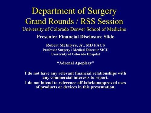
Department <strong>of</strong> Surgery<br />
Grand Rounds / RSS Session<br />
<strong>University</strong> <strong>of</strong> <strong>Colorado</strong> <strong>Denver</strong> School <strong>of</strong> Medicine<br />
Presenter Financial Disclosure Slide<br />
Robert McIntyre, Jr., MD FACS<br />
Pr<strong>of</strong>essor Surgery / Medical Director SICU<br />
<strong>University</strong> <strong>of</strong> <strong>Colorado</strong> Hospital<br />
“<strong>Adrenal</strong> <strong>Apoplexy</strong>”<br />
I do not have any relevant financial relationships with<br />
any commercial interests to report.<br />
I do not intend to reference <strong>of</strong>f-label/unapproved uses<br />
<strong>of</strong> products or devices in this presentation.
“<strong>Adrenal</strong> <strong>Apoplexy</strong>”<br />
• Dictionary.com<br />
• <strong>Apoplexy</strong><br />
– A stroke<br />
– A sudden usually marked loss <strong>of</strong> bodily<br />
function due to rupture or occlusion <strong>of</strong> a blood<br />
vessel<br />
– A hemorrhage into an organ or tissue<br />
Glomerulosa aldosterone (salt)<br />
Fasciculata cortisol, deoxycorticosterone (sugar)<br />
Reticularis androgens (sex)
Case History 1:<br />
• A 55 year old male<br />
has vague abdminal<br />
pain. He has a 10 year<br />
history <strong>of</strong> HTN. A CTS<br />
shows a 4 cm adrenal<br />
mass.
Question 1: The next step in<br />
management will include<br />
• A) FNA biopsy<br />
• B) Serum calcium and PTH levels<br />
• C) Urinary 5-hydroxyindoleacedic acid<br />
• D) Wait 6 months and repeat CTS<br />
• E) 1 mg Dexamethasone suppression test
Question 1: The next step in<br />
management will include<br />
• A) FNA biopsy<br />
• B) Serum calcium and PTH levels<br />
• C) Urinary 5-hydroxyindoleacedic acid<br />
• D) Wait 6 months and repeat CTS<br />
• E) 1 mg Dexamethasone suppression test
<strong>Adrenal</strong> Incidentaloma<br />
• Definition: otherwise unsuspected adrenal<br />
mass on imaging<br />
– Excludes patients imaged as work up for<br />
cancer<br />
• Up to 8.7% autopsy series
<strong>Adrenal</strong> Incidentaloma<br />
3 questions<br />
• Is the tumor hormonally active?<br />
• Does the tumor have the radiographic<br />
characteristics <strong>of</strong> a malignancy?<br />
• Does the patient have a history <strong>of</strong> cancer?
<strong>Adrenal</strong> Incidentaloma<br />
Differential<br />
• Nonfunctional: 80%<br />
• Hypercortisolism: 5% Subclinical Cushing’s<br />
Syndrome (SCS)<br />
• Aldosteronism: 1%<br />
• Pheochromocytoma: 5%<br />
• <strong>Adrenal</strong> Cortical Cancer 5%<br />
• Other<br />
– Ganglioneuromas, Myelolipomas, Benign cysts<br />
• Metastasis: 2.5%
Case History 4<br />
• 51 year old female has HTN, weight gain,<br />
diabetes, and easy bruising. She has a BP<br />
<strong>of</strong> 154/95, weighs 250 lbs., and a<br />
characteristic buffalo hump. The diagnosis<br />
<strong>of</strong> Cushing’s syndrome is made.
Question 4: Which <strong>of</strong> the following<br />
statements is true<br />
• A) Ectopic ACTH secretion is most commonly<br />
due to medullary thyroid cancer<br />
• B) Cushing’s disease is the most common cause<br />
<strong>of</strong> endogenous Cushing’s syndrome<br />
• C) An elevated 24 hour urinary cortisol is<br />
diagnostic for Cushing’s disease<br />
• D) The most accurate test to rule out exogenous<br />
Cushing’s syndrome is a C-peptide level<br />
• E) An adrenal adenoma is the most common<br />
cause <strong>of</strong> ACTH dependent Cushing’s syndrome
<strong>Adrenal</strong> Incidentaloma<br />
Screening<br />
• Hypercortisolism<br />
– 1 mg Dexamethasone suppression test<br />
• AM serum cortisol > 5.0 mcg/dl<br />
• Suppression < 1.8 mcg/dl (best NPV)<br />
– Low ACTH, DHEAS<br />
– Second confirmatory test<br />
• Evening salivary cortisol, 2 day low dose<br />
dexamethasone suppresion test, 24 hour urine<br />
cortisol<br />
Zeiger et al Endocrine Practice 2009
<strong>Adrenal</strong> Incidentaloma<br />
Screening<br />
• Aldosteronism (if hypertensive)<br />
– PAC/PRA > 30, PAC > 20 ng/dl<br />
– Confirm with lack <strong>of</strong> suppression with salt loading<br />
• Pheochromocytoma<br />
– Plasma free metanephrine and normetanephrine<br />
(higher false +)<br />
• > 3-4 times normal<br />
– 24 hour urine metanephrines (higher false -)<br />
• > 1800 mcg<br />
– Always before needle biopsy<br />
– Always before resection<br />
– RET, VHL, Succinate dehydrogenase genes<br />
Zeiger et al Endocrine Practice 2009
<strong>Adrenal</strong> Incidentaloma<br />
Radiologic Imaging<br />
• Density measurement on non contrast<br />
CTS or in phase and out <strong>of</strong> phase MRI<br />
– Intracellular lipid<br />
– < 10 HU<br />
• Contrast washout kinetics on CTS<br />
– Immediate after contrast<br />
– 10-15 delay<br />
– > 50% washout<br />
• Size, homogeneity, borders, metastasis<br />
Zeiger et al Endocrine Practice 2009
<strong>Adrenal</strong> Incidentaloma<br />
Follow up<br />
• Imaging<br />
– 3-6 months<br />
– Yearly 1-2 years<br />
• Hormone evaluation<br />
– Annually up to 5 years<br />
Follow up (years) 1 2 5<br />
Imaging growth 6 14 29<br />
Hormonally active 17 29 47<br />
Zeiger et al Endocrine Practice 2009
Subclinical Cushing’s Syndrome<br />
• Natural history is poorly known<br />
• Progression to overt Cushing’s rare<br />
• Long term morbidity and mortality data do<br />
not exist<br />
• No long term data for medical<br />
management<br />
• Controversial: young patient with recent<br />
onset or worse co-morbidity
Cushing’s Syndrome<br />
• Exogenous Steroids<br />
• ACTH Dependent (80%)<br />
– Pituitary adenoma (Cushing’s disease) 70%<br />
– Ectopic ACTH 15%<br />
– Ectopic CRH Rare<br />
• ACTH Independent (20%)<br />
– Cortisol producing adenoma 10%<br />
– Cortisol producing adrenal carcinoma 5%<br />
– Bilateral adrenal hyperplasia Rare<br />
– Macronodular hyperplasia Rare
Hypercortisolism<br />
Perioperative Management<br />
• High risk DVT<br />
• Suppressed HPA axis<br />
– Glucocorticoid replacement 6-18 months
Case History 3<br />
• 44 year old female reports pounding<br />
headaches, flushing, and hypertension for<br />
12 months. She has elevated urinary<br />
catecholamines and metanephrines. There<br />
is no family history <strong>of</strong> endocrinopathy.<br />
CTS shows a 5 cm adrenal mass.
Question 3: You advise which<br />
management:<br />
• A) Bilateral adrenal vein sampling<br />
• B) Preoperative MIBG scan<br />
• C) Open adrenalectomy due to concern for<br />
malignancy<br />
• D) Alpha and beta blockade for 3 months<br />
• E) Alpha blockade then laparoscopic<br />
adrenalectomy
Pheochromocytoma<br />
Perioperative Management<br />
• α blockade<br />
– Phenoxybenzamine, Doxazosin<br />
– Orthostatic hypotension, tachycardia, nasal<br />
congestion<br />
• Metyrosine<br />
• β blockade<br />
– Tachycardia, extrasystoles, arrhythmias<br />
• Calcium channel blockers<br />
• Liberal fluid and salt intake
Pheochromocytoma<br />
Intra-operative Management<br />
• A line<br />
• CVP<br />
• Nitroprusside, nicardapine, phentolamine<br />
• Esmolol<br />
• Fluid and adrenergic agents
Risk Factors for Hemodynamic Instability<br />
during Surgery for Pheochromocytoma<br />
Pretreatment α blockade (PXB or DOX)<br />
48 <strong>of</strong> 73 (66%) MAP < 100 mm Hg<br />
39 <strong>of</strong> 48 (81%) BP < 130/85 mm Hg<br />
25 <strong>of</strong> 73 (34%) MAP >100 mm Hg<br />
Bruynzeel et al JCEM 2010
Risk Factors for Hemodynamic Instability<br />
during Surgery for Pheochromocytoma<br />
Intraoperative time SBP above 160 mm Hg<br />
plasma norepinephrine levels (r = 0.23; P < 0.05),<br />
tumor diameter (r = 0.36; P < 0.01),<br />
postural BP fall (r = 0.30; P < 0.05).<br />
Bruynzeel et al JCEM 2010
Risk Factors for Hemodynamic Instability<br />
during Surgery for Pheochromocytoma<br />
• Postoperative MAP was significantly higher PXB vs DOX<br />
(P < 0.01).<br />
• No relation between the PXB or DOX dosage and<br />
intraoperative BP fluctuations or postoperative<br />
hypotension.<br />
• The doses <strong>of</strong> esmolol (25 patients) were significantly<br />
higher in the PXB group compared with the DOX group<br />
(314.5 mg, 25.0–5520; vs. 95.0 mg, 0.06–2500 mg; P <<br />
0.05).<br />
• Other vasoactive drugs as phenylephrine (n = 15),<br />
nitroglycerine (n = 24), NE (n = 36), and phentolamine (n<br />
= 28) did not differ between both groups.<br />
Bruynzeel et al JCEM 2010
Pheochromocytoma<br />
Postoperative Management<br />
• Pathology difficult<br />
• Malignancy<br />
– Invasion, metastasis<br />
• Annual follow up<br />
– 16% recurrence at 10 years<br />
• Malignant<br />
• Right<br />
• Extra-adrenal<br />
Amar et al JCEM 2005
Primary Hyperaldosteronism<br />
• Drug resistant hypertension<br />
• Hypokalemia<br />
– Spontaneous (< 3.5 mEq/L)<br />
– Severe diuretic induced (
Primary Hyperaldosteronism<br />
Screening, Confirmation<br />
• HTN<br />
• Aldosterone to Renin Ratio (ARR):<br />
– PAC (ng/dl) to PRA (ng.ml)<br />
• ARR > 30 combined with PAC > 20<br />
• Morning, out <strong>of</strong> bed 2 hours seated for 15 minutes<br />
• Lack <strong>of</strong> suppression<br />
– High salt diet<br />
– Saline suppression
Primary Hyperaldosteronism<br />
Imaging and Localization<br />
• CT imaging<br />
• Young patient,
Primary Hyperaldosteronism<br />
<strong>Adrenal</strong> Vein Sampling<br />
• Success right vein catheterization survey:<br />
74%, referral center: 95%<br />
• Continuous cosyntropin stimulation<br />
• Corrected aldo/cortisol ratios one side to<br />
the other 4:1: unilateral disease<br />
• CTS alone:<br />
– 21% inappropriate excluded from surgery<br />
– 25% inappropriate adrenalectomy<br />
Young et al Surgery 2004; Nwariaku Arch Surg 2006
Primary Hyperaldosteronism<br />
Therapy<br />
• Medical: spironolactone, eplerenone<br />
• Unilateral adrenalectomy<br />
– 100% cure hypokalemia<br />
– 90% significant improvement in HTN<br />
– 30-60% cure HTN<br />
• Young, women, shorter duration <strong>of</strong> HTN,<br />
less drugs, normal Cr, no family history <strong>of</strong><br />
HTN<br />
Sawka Ann Int Med 2001; Celen et al Arch<br />
Surg 1996; Ferris et al Br J Med 1975
Hyperaldosteronism<br />
Surgical Therapy- Outcome<br />
Variable Normal BP Persistent HTN<br />
Sex 0.003<br />
Male 47% 53%<br />
Female 76.5% 23.5%<br />
Age 0.028<br />
< 40 88% 12%<br />
40-49 64.5% 35.5%<br />
> 50 57% 43%<br />
Family Hx HTN NS<br />
K NS<br />
Duration HTN (mo) 85.5 140.7 0.007<br />
BP at Dx 180/107 200/118 0.05<br />
Cr Cl 76 65 0.05<br />
Pathology NS<br />
Size (mm) 18 17 NS<br />
Obara Surgery et al 1992
Case History 2<br />
• 58 year old male had a PET-CTS which shows a<br />
3 cm FDG avid mass in the left adrenal gland.<br />
He has a history <strong>of</strong> melanoma on the left leg and<br />
had excision and groin lymph node dissection.
Question 2: The next best course<br />
<strong>of</strong> action is which <strong>of</strong> the following:<br />
• A) Repeat CTS in 3 months<br />
• B) Proceed to laparoscopic<br />
adrenalectomy<br />
• C) 24 hour urine metanephrines<br />
• D) FNA biopsy to determine if it is<br />
recurrent melanoma<br />
• E) Open adrenalectomy
Adrenocortical Carcinoma<br />
• Determinants <strong>of</strong> survival<br />
– Stage at presentation<br />
– Curative resection
Adrenocortical Carcinoma<br />
Presentation<br />
• Paucity <strong>of</strong> symptoms<br />
• Abd pressure or mass<br />
• 2/3 hormonally active<br />
– Cushings<br />
– Virilization
Adrenocortical Carcinoma<br />
Imaging<br />
• Radiographic phenotype<br />
– < 5% <strong>of</strong> incidentalomas<br />
– 2% lesions < 4 cm, 6% 4.1-6 cm<br />
– 25% > 6 cm<br />
– > 10 HU on noncontrast CTS (most > 30 HU)<br />
– < 50% washout on delayed contrast CTS<br />
– Irregular borders, heterogeneous, metastatic<br />
Barzon et al Eur J Endocrinol 2003; Angeli Horm Res 2000;
Adrenocortical Carcinoma<br />
Biochemical Evaluation<br />
• Detailed hormone evaluation<br />
• Steroid precursors<br />
– DHEA-S<br />
• Identify tumor marker
Incidentaloma:Metastatic Disease<br />
• 2.1-2.5% prevalence <strong>of</strong> metastatic disease<br />
among incidentalomas<br />
• Occult malignancy: adrenal mass at presentation<br />
5.8%<br />
• FNAB: only in patients with history <strong>of</strong> cancers<br />
(particularly lung, breast, kidney),<br />
– no signs <strong>of</strong> other metastases,<br />
– a heterogeneous mass with a high unenhanced<br />
attenuation value (>20HU)<br />
– after exclusion <strong>of</strong> pheochromocytoma.<br />
• PET CTS<br />
Schteingart et al Endocrine Related Cancer 2005;<br />
Barzon et al Eur J Endocrinol 2003;<br />
Lee et al Surgery 1998
Adrenocortical Carcinoma<br />
Operative Management<br />
• Open resection<br />
– En bloc<br />
• Laparoscopy<br />
– Local and peritoneal dissemination
Adrenocortical Carcinoma<br />
Medical Therapy<br />
• Adjuvant treatment<br />
Controversial<br />
– Mitotane (inhibit<br />
adrenocortical steroid<br />
biosynthesis )<br />
• Complete resection with poor<br />
prognostic features<br />
• Prolong disease free survival<br />
• No effect on overall survival<br />
• Start early<br />
• Toxicity<br />
Huang and Fojo JCEM 2008; Terzolo et al NEJM 2007
Adrenocortical Carcinoma<br />
Medical Therapy<br />
• Metastatic disease: PET CTS<br />
– Radiation<br />
• Symptomatic local recurrence or bone, brain, other mets<br />
– RFA: option but value is not proven<br />
– Chemotherapy<br />
• The evidence regarding efficacy <strong>of</strong> first-line therapy is very<br />
limited (level C). Possible protocols are:<br />
• Etoposide+doxorubicin+cisplatin+mitotane<br />
• Streptozotocin+mitotane<br />
• Mitotane alone or platin+etoposide+mitotane<br />
Schteingart et al Endocrine Related Cancer 2005