COLLEGIO DI DIREZIONE - Azienda Ospedaliera di Parma
COLLEGIO DI DIREZIONE - Azienda Ospedaliera di Parma
COLLEGIO DI DIREZIONE - Azienda Ospedaliera di Parma
Create successful ePaper yourself
Turn your PDF publications into a flip-book with our unique Google optimized e-Paper software.
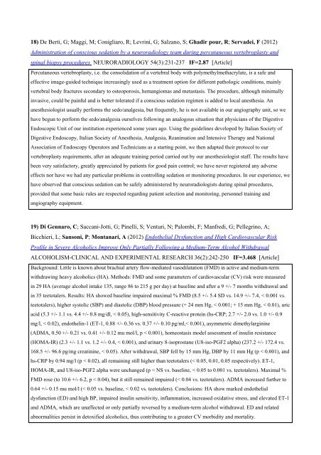
18) De Berti, G; Maggi, M; Conigliaro, R; Levrini, G; Salzano, S; Gha<strong>di</strong>r pour, R; Servadei, F (2012)<br />
Administration of conscious sedation by a neurora<strong>di</strong>ology team during percutaneous vertebroplasty and<br />
spinal biopsy procedures NEURORA<strong>DI</strong>OLOGY 54(3):231-237 IF=2.87 [Article]<br />
Percutaneous vertebroplasty, i.e. the consolidation of a vertebral body with polymethylmethacrylate, is a safe and<br />
effective image-guided technique increasingly used as a treatment option for <strong>di</strong>fferent pathologic con<strong>di</strong>tions, mainly<br />
vertebral body fractures secondary to osteoporosis, hemangiomas and metastasis. The procedure, although minimally<br />
invasive, could be painful and is better tolerated if a conscious sedation regimen is added to local anesthesia. An<br />
anesthesiologist usually performs the sedo/analgesia, but frequently, he is not available in our angiography unit, so we<br />
have begun to perform the sedo/analgesia ourselves following an analogous situation that physicians of the Digestive<br />
Endoscopic Unit of our institution experienced some years ago. Using the guidelines developed by Italian Society of<br />
Digestive Endoscopy, Italian Society of Anesthesia, Analgesia, Reanimation and Intensive Therapy and National<br />
Association of Endoscopy Operators and Technicians as a starting point, we then adapted their protocol to our<br />
vertebroplasty requirements, after an adequate training period carried out by our anesthesiologist staff. The results have<br />
been very satisfactory, greatly appreciated by patients for good pain control; we have never registered any adverse<br />
effects nor have we had any particular problems in controlling sedation or monitoring procedures. In our experience, we<br />
have observed that conscious sedation can be safely administered by neurora<strong>di</strong>ologists during spinal procedures,<br />
provided that some basic rules are respected regar<strong>di</strong>ng patient selection and monitoring, personnel training and<br />
angiography equipment.<br />
19) Di Gennaro, C; Saccani-Jotti, G; Pinelli, S; Venturi, N; Palombi, F; Manfre<strong>di</strong>, G; Pellegrino, A;<br />
Bicchieri, L; Sansoni, P; Montanari, A (2012) Endothelial Dysfunction and High Car<strong>di</strong>ovascular Risk<br />
Profile in Severe Alcoholics Improve Only Partially Following a Me<strong>di</strong>um-Term Alcohol Withdrawal<br />
ALCOHOLISM-CLINICAL AND EXPERIMENTAL RESEARCH 36(2):242-250 IF=3.468 [Article]<br />
Background: Little is known about brachial artery flow-me<strong>di</strong>ated vaso<strong>di</strong>latation (FMD) in active and me<strong>di</strong>um-term<br />
withdrawing heavy alcoholics (HA). Methods: FMD and some parameters of car<strong>di</strong>ovascular (CV) risk were measured<br />
in 29 HA (average alcohol intake 135, range 86 to 215 g per day) at baseline and after a 9 +/- 7 months withdrawal and<br />
in 35 teetotalers. Results: HA showed baseline impaired maximal % FMD (8.5 +/- 5.4 SD vs. 14.9 +/- 7.4, < 0.001 vs.<br />
teetotalers), higher systolic (SBP) and <strong>di</strong>astolic (DBP) blood pressure (+ 24 mm Hg, < 0.001; + 15 mm Hg, < 0.01), uric<br />
acid (5.3 +/- 1.1 vs. 4.4 +/- 0.8 mg/dl, < 0.05), high-sensitivity C-reactive protein (hs-CRP; 2.7 +/- 2.0 vs. 1.0 +/- 0.9<br />
mg/l, < 0.02), endothelin-1 (ET-1, 0.88 +/- 0.36 vs. 0.37 +/- 0.10 pg/ml,< 0.001), asymmetric <strong>di</strong>methylarginine<br />
(ADMA, 0.50 +/- 0.21 vs. 0.41 +/- 0.12 mu mol/l, p < 0.001), homeostasis model assessment of insulin resistance<br />
(HOMA-IR) (2.3 +/- 1.1 vs. 1.2 +/- 0.4, < 0.001), and urinary 8-isoprostane (U8-iso-PGF2 alpha) (237.2 +/- 172.4 vs.<br />
168.5 +/- 96.6 pg/mg creatinine, < 0.05). After withdrawal, SBP fell by 15 mm Hg, DBP by 11 mm Hg (p < 0.001), and<br />
hs-CRP by 0.94 mg/l (p < 0.02), all remaining still higher than teetotalers (< 0.05, 0.01, 0.05 respectively). ET-1,<br />
HOMA-IR, and U8-iso-PGF2 alpha were unchanged (p = NS vs. baseline, < 0.05 to 0.001 vs. teetotalers). Maximal %<br />
FMD rose (to 10.6 +/- 6.2, p < 0.04), but it still remained impaired (< 0.04 vs. teetotalers). ADMA increased further to<br />
0.64 +/- 0.15 mu mol/l (< 0.05 vs. baseline, < 0.02 vs. teetotalers). Conclusions: HA show marked endothelial<br />
dysfunction (ED) and high BP, impaired insulin sensitivity, inflammation, increased oxidative stress, and elevated ET-1<br />
and ADMA, which are unaffected or only partially reversed by a me<strong>di</strong>um-term alcohol withdrawal. ED and related<br />
abnormalities persist in detoxified alcoholics, thus contributing to a greater CV morbi<strong>di</strong>ty and mortality.


