RGB and HSV colour models in colour identification of digital ... - Ecet
RGB and HSV colour models in colour identification of digital ... - Ecet
RGB and HSV colour models in colour identification of digital ... - Ecet

You also want an ePaper? Increase the reach of your titles
YUMPU automatically turns print PDFs into web optimized ePapers that Google loves.
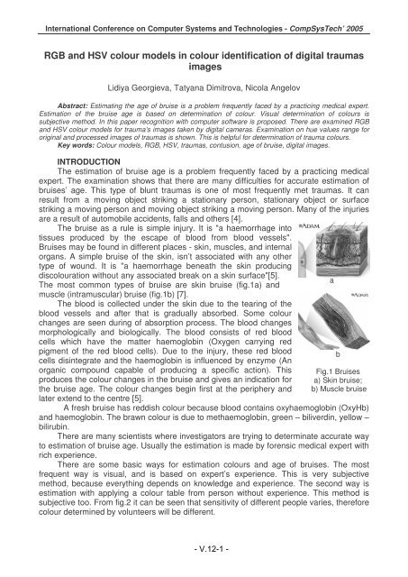
International Conference on Computer Systems <strong>and</strong> Technologies - CompSysTech’ 2005<br />
<strong>RGB</strong> <strong>and</strong> <strong>HSV</strong> <strong>colour</strong> <strong>models</strong> <strong>in</strong> <strong>colour</strong> <strong>identification</strong> <strong>of</strong> <strong>digital</strong> traumas<br />
images<br />
Lidiya Georgieva, Tatyana Dimitrova, Nicola Angelov<br />
Abstract: Estimat<strong>in</strong>g the age <strong>of</strong> bruise is a problem frequently faced by a practic<strong>in</strong>g medical expert.<br />
Estimation <strong>of</strong> the bruise age is based on determ<strong>in</strong>ation <strong>of</strong> <strong>colour</strong>. Visual determ<strong>in</strong>ation <strong>of</strong> <strong>colour</strong>s is<br />
subjective method. In this paper recognition with computer s<strong>of</strong>tware is proposed. There are exam<strong>in</strong>ed <strong>RGB</strong><br />
<strong>and</strong> <strong>HSV</strong> <strong>colour</strong> <strong>models</strong> for trauma’s images taken by <strong>digital</strong> cameras. Exam<strong>in</strong>ation on hue values range for<br />
orig<strong>in</strong>al <strong>and</strong> processed images <strong>of</strong> traumas is shown. This is helpful for determ<strong>in</strong>ation <strong>of</strong> trauma <strong>colour</strong>s.<br />
Key words: Colour <strong>models</strong>, <strong>RGB</strong>, <strong>HSV</strong>, traumas, contusion, age <strong>of</strong> bruise, <strong>digital</strong> images.<br />
INTRODUCTION<br />
The estimation <strong>of</strong> bruise age is a problem frequently faced by a practic<strong>in</strong>g medical<br />
expert. The exam<strong>in</strong>ation shows that there are many difficulties for accurate estimation <strong>of</strong><br />
bruises’ age. This type <strong>of</strong> blunt traumas is one <strong>of</strong> most frequently met traumas. It can<br />
result from a mov<strong>in</strong>g object strik<strong>in</strong>g a stationary person, stationary object or surface<br />
strik<strong>in</strong>g a mov<strong>in</strong>g person <strong>and</strong> mov<strong>in</strong>g object strik<strong>in</strong>g a mov<strong>in</strong>g person. Many <strong>of</strong> the <strong>in</strong>juries<br />
are a result <strong>of</strong> automobile accidents, falls <strong>and</strong> others [4].<br />
The bruise as a rule is simple <strong>in</strong>jury. It is "a haemorrhage <strong>in</strong>to<br />
tissues produced by the escape <strong>of</strong> blood from blood vessels".<br />
Bruises may be found <strong>in</strong> different places - sk<strong>in</strong>, muscles, <strong>and</strong> <strong>in</strong>ternal<br />
organs. A simple bruise <strong>of</strong> the sk<strong>in</strong>, isn’t associated with any other<br />
type <strong>of</strong> wound. It is "a haemorrhage beneath the sk<strong>in</strong> produc<strong>in</strong>g<br />
dis<strong>colour</strong>ation without any associated break on a sk<strong>in</strong> surface"[5].<br />
The most common types <strong>of</strong> bruise are sk<strong>in</strong> bruise (fig.1a) <strong>and</strong><br />
muscle (<strong>in</strong>tramuscular) bruise (fig.1b) [7].<br />
The blood is collected under the sk<strong>in</strong> due to the tear<strong>in</strong>g <strong>of</strong> the<br />
blood vessels <strong>and</strong> after that is gradually absorbed. Some <strong>colour</strong><br />
changes are seen dur<strong>in</strong>g <strong>of</strong> absorption process. The blood changes<br />
morphologically <strong>and</strong> biologically. The blood consists <strong>of</strong> red blood<br />
cells which have the matter haemoglob<strong>in</strong> (Oxygen carry<strong>in</strong>g red<br />
pigment <strong>of</strong> the red blood cells). Due to the <strong>in</strong>jury, these red blood<br />
cells dis<strong>in</strong>tegrate <strong>and</strong> the haemoglob<strong>in</strong> is <strong>in</strong>fluenced by enzyme (An<br />
organic compound capable <strong>of</strong> produc<strong>in</strong>g a specific action). This<br />
produces the <strong>colour</strong> changes <strong>in</strong> the bruise <strong>and</strong> gives an <strong>in</strong>dication for<br />
the bruise age. The <strong>colour</strong> changes beg<strong>in</strong> first at the periphery <strong>and</strong><br />
later extend to the centre [5].<br />
A fresh bruise has reddish <strong>colour</strong> because blood conta<strong>in</strong>s oxyhaemoglob<strong>in</strong> (OxyHb)<br />
<strong>and</strong> haemoglob<strong>in</strong>. The brawn <strong>colour</strong> is due to methaemoglob<strong>in</strong>, green – biliverd<strong>in</strong>, yellow –<br />
bilirub<strong>in</strong>.<br />
There are many scientists where <strong>in</strong>vestigators are try<strong>in</strong>g to determ<strong>in</strong>ate accurate way<br />
to estimation <strong>of</strong> bruise age. Usually the estimation is made by forensic medical expert with<br />
rich experience.<br />
There are some basic ways for estimation <strong>colour</strong>s <strong>and</strong> age <strong>of</strong> bruises. The most<br />
frequent way is visual, <strong>and</strong> is based on expert’s experience. This is very subjective<br />
method, because everyth<strong>in</strong>g depends on knowledge <strong>and</strong> experience. The second way is<br />
estimation with apply<strong>in</strong>g a <strong>colour</strong> table from person without experience. This method is<br />
subjective too. From fig.2 it can be seen that sensitivity <strong>of</strong> different people varies, therefore<br />
<strong>colour</strong> determ<strong>in</strong>ed by volunteers will be different.<br />
- V.12-1 -<br />
a<br />
b<br />
Fig.1 Bruises<br />
a) Sk<strong>in</strong> bruise;<br />
b) Muscle bruise
International Conference on Computer Systems <strong>and</strong> Technologies - CompSysTech’ 2005<br />
Ultraviolet photographs are used <strong>in</strong> forensic practice too, but it is used only as a pro<strong>of</strong><br />
for presence <strong>of</strong> traumas. With such photographs may give evidence that a person had<br />
contusion, but can not def<strong>in</strong>e when this contusion has happened.<br />
Comparatively new ways are based on the use <strong>of</strong> a spectrophotometer or <strong>digital</strong><br />
camera.<br />
Sensitivity<br />
human3<br />
human2<br />
Solid state<br />
human1<br />
Wavelength<br />
Fig.2. Sensitivity <strong>of</strong> some persons<br />
The priority <strong>of</strong> us<strong>in</strong>g <strong>digital</strong> camera is a possibility for computer image process<strong>in</strong>g. A<br />
most objective estimation <strong>of</strong> <strong>colour</strong>s can be made with the help <strong>of</strong> a computer <strong>and</strong><br />
s<strong>of</strong>tware products. Many different <strong>colour</strong> <strong>models</strong> exist <strong>in</strong> computer graphics. Each model<br />
uses 3D coord<strong>in</strong>ate system to identify uniquely <strong>in</strong>dividual <strong>colour</strong>s. In this work two <strong>colour</strong><br />
<strong>models</strong> are chosen - <strong>RGB</strong> <strong>and</strong> <strong>HSV</strong> <strong>colour</strong> <strong>models</strong>.<br />
WORK WITH COLOUR MODELS<br />
When talk<strong>in</strong>g about <strong>colour</strong>s, we have to keep <strong>in</strong> m<strong>in</strong>d<br />
that <strong>colour</strong> can be realized <strong>in</strong> different ways. The <strong>colour</strong><br />
<strong>models</strong> are based on a physical, physiological <strong>and</strong> technical<br />
aspect [3].<br />
<strong>RGB</strong> (Red, Green, Blue) <strong>colour</strong> model (Fig.3)<br />
To see how a <strong>RGB</strong> model works, we first have to look at<br />
the technical aspect <strong>of</strong> <strong>colour</strong>. The human eye perceives<br />
<strong>colour</strong> reaction through a mix <strong>of</strong> red, green <strong>and</strong> blue signals.<br />
It was a thought that each <strong>colour</strong> can be mixed with only<br />
these three primary <strong>colour</strong>s.<br />
<strong>HSV</strong> (Hue, Saturation, Value ) <strong>colour</strong> model (Fig.4)<br />
The <strong>HSV</strong> <strong>colour</strong> model, also called HSB (Hue,<br />
Saturation, Brightness), def<strong>in</strong>es a <strong>colour</strong> space <strong>in</strong> terms <strong>of</strong><br />
three constituent components:<br />
- Hue is the <strong>colour</strong> type (such as red, magenta, blue,<br />
cyan, green or yellow). Hue ranges from 0-360 deg.<br />
- Saturation refers to the <strong>in</strong>tensity <strong>of</strong> specific hue.<br />
Saturation ranges are from 0 to 100%. In this work saturation<br />
is present<strong>in</strong>g <strong>in</strong> range 0-255.<br />
- Value refers to the brightness <strong>of</strong> the <strong>colour</strong>.<br />
Saturation ranges are from 0 to 100%. Value ranges are<br />
from 0-100%. In this work saturation <strong>and</strong> value are<br />
present<strong>in</strong>g <strong>in</strong> range 0-255.<br />
A typical image <strong>of</strong> the bruise<br />
It is possible to see a typical view <strong>of</strong> the bruise on the fig.5<br />
<strong>and</strong> recognize some <strong>colour</strong>s - red, yellow, green <strong>and</strong> violet.<br />
Fig.5. Orig<strong>in</strong>al image - V.12-2 Fig.6. -<br />
Processed image<br />
Fig.3. <strong>RGB</strong> <strong>colour</strong> model<br />
Fig.4. <strong>HSV</strong> <strong>colour</strong> model
International Conference on Computer Systems <strong>and</strong> Technologies - CompSysTech’ 2005<br />
The image is processed <strong>and</strong> the result is shown on fig.6. Process<strong>in</strong>g <strong>in</strong>clude follow<br />
steps. First the operator has chosen a rectangular area near the bruise. The area was<br />
without chang<strong>in</strong>g <strong>of</strong> natural <strong>colour</strong> <strong>of</strong> the sk<strong>in</strong>. After that the area is analyzed for<br />
determ<strong>in</strong><strong>in</strong>g the <strong>of</strong>ten met values <strong>of</strong> the tree components - red, green <strong>and</strong> blue for the<br />
healthy sk<strong>in</strong>. The next step <strong>of</strong> this process<strong>in</strong>g procedure is the extraction from the <strong>colour</strong> <strong>of</strong><br />
pixels <strong>in</strong> the image the <strong>of</strong>ten met <strong>RGB</strong> components.<br />
The 3D <strong>models</strong> <strong>of</strong> the distribution <strong>of</strong> all components’ values <strong>of</strong> each pixel <strong>in</strong> the two<br />
images - orig<strong>in</strong>al <strong>and</strong> processed can be seen <strong>in</strong> fig.7.<br />
Fig.7. 3D <strong>models</strong> - <strong>RGB</strong> <strong>and</strong> <strong>HSV</strong> for the <strong>colour</strong> distribution for the two images<br />
a) orig<strong>in</strong>al image <strong>in</strong> <strong>RGB</strong>; b) orig<strong>in</strong>al image <strong>in</strong> <strong>HSV</strong>; c) processed image <strong>in</strong> <strong>RGB</strong>;<br />
d) processed image <strong>in</strong> <strong>HSV</strong><br />
The follow<strong>in</strong>g conclusions can be made for the graphs <strong>in</strong> fig.7:<br />
The <strong>RGB</strong> model is difficult for def<strong>in</strong><strong>in</strong>g a diapason <strong>in</strong> which the <strong>colour</strong> values make<br />
changes especially for the shade <strong>of</strong> concrete <strong>colour</strong>. The receiver set is situated on the<br />
cube diagonal (fig.7a, c);<br />
The <strong>HSV</strong> model gives a set, <strong>in</strong> which the boundaries for the hue component can<br />
easily be def<strong>in</strong>ed;<br />
In the orig<strong>in</strong>al image (fig.5) some <strong>colour</strong>s are visible - red, green, yellow <strong>and</strong><br />
violet. If we present the values <strong>of</strong> the particular <strong>RGB</strong> components <strong>in</strong> the 3D graph (fig.7a) it<br />
is difficult to recognize <strong>colour</strong>s <strong>in</strong> the image us<strong>in</strong>g only this graph. After the real image<br />
process<strong>in</strong>g from <strong>RGB</strong> <strong>in</strong> <strong>HSV</strong> model the new graph (fig.7b) shows a presence only <strong>of</strong> red<br />
areas - the set has hue <strong>in</strong> the <strong>in</strong>terval 0-50. It is the reddish <strong>colour</strong> <strong>in</strong> <strong>HSV</strong> hexagone;<br />
After the process<strong>in</strong>g <strong>of</strong> the orig<strong>in</strong>al image the result (fig.6) clearly shows different<br />
<strong>colour</strong>s. The apply<strong>in</strong>g <strong>of</strong> the <strong>RGB</strong> model made the same troubles. The image converted <strong>in</strong><br />
<strong>HSV</strong> model allows settl<strong>in</strong>g the other <strong>colour</strong>s besides the red. Hue has values out <strong>of</strong> the red<br />
diapason <strong>in</strong> the HVS hexagone.<br />
Separate different <strong>colour</strong> segments from <strong>digital</strong> images <strong>of</strong> traumas<br />
The <strong>colour</strong> recognition <strong>of</strong> the bruise <strong>and</strong> its surround<strong>in</strong>g <strong>in</strong> the moment <strong>of</strong> evaluation<br />
has very important mean<strong>in</strong>g for correct bruise ag<strong>in</strong>g. It is difficult to def<strong>in</strong>e real presence <strong>of</strong><br />
clear <strong>colour</strong>s <strong>and</strong> these which are received by mixture <strong>of</strong> some <strong>of</strong> them. The concept for<br />
the particular <strong>colour</strong> <strong>of</strong> different persons varies. Therefore def<strong>in</strong><strong>in</strong>g <strong>of</strong> separate trauma<br />
<strong>colour</strong>s by one objective evaluator (which is exactly personal computer) is useful <strong>in</strong><br />
<strong>in</strong>vestigations <strong>of</strong> determ<strong>in</strong>ation <strong>of</strong> the traumas age.<br />
The <strong>colour</strong>s, accord<strong>in</strong>g to expert op<strong>in</strong>ion <strong>in</strong> separate papers (research def<strong>in</strong>ition <strong>of</strong><br />
bruise age), which appear from the moment <strong>of</strong> trauma receiv<strong>in</strong>g to full recovery are - red,<br />
blue, green, yellow <strong>and</strong> violet. There were made analysis <strong>of</strong> large number <strong>of</strong> <strong>digital</strong> images<br />
<strong>of</strong> this type <strong>of</strong> trauma. From them there were cut 25 areas, which present different shades<br />
<strong>of</strong> each <strong>colour</strong> mentioned above. These areas were processed by extraction from the<br />
<strong>colour</strong> <strong>of</strong> pixels <strong>in</strong> the image <strong>of</strong> <strong>of</strong>ten met <strong>RGB</strong> components for healthy sk<strong>in</strong>. The orig<strong>in</strong>al<br />
<strong>and</strong> processed images were present <strong>in</strong> <strong>HSV</strong> <strong>colour</strong> model.<br />
- V.12-3 -
International Conference on Computer Systems <strong>and</strong> Technologies - CompSysTech’ 2005<br />
As it is known, <strong>in</strong> this model hue is the attribute that corresponds to the object’s red,<br />
orange, yellow, green, blue or violet. This attribute is <strong>of</strong>ten related to the hue circle, which<br />
has been recognized by artist, <strong>colour</strong> technologist, <strong>and</strong> decorators for years [1]. Exactly<br />
therefore there were made analysis <strong>of</strong> value <strong>of</strong> this component from <strong>HSV</strong> <strong>colour</strong> model.<br />
The results are show <strong>in</strong> diagram (Fig.7) for each <strong>colour</strong>.<br />
- Red <strong>colour</strong> - is one <strong>of</strong> the most frequent <strong>colour</strong> <strong>in</strong> traumas. There are cute 25<br />
segments, which are def<strong>in</strong>ed as reddish <strong>colour</strong>. On the next figure (Fig.8) the distribution<br />
<strong>of</strong> hue values for orig<strong>in</strong>al reddish areas is shown (fig.8a) <strong>and</strong> for processed areas (fig.8b).<br />
a b<br />
Fig.8 Hue distribution for reddish segments: a) orig<strong>in</strong>al image; b) processed image<br />
- Yellow <strong>colour</strong> – accord<strong>in</strong>g to more publications this <strong>colour</strong> is most significant for<br />
the determ<strong>in</strong>ation <strong>of</strong> bruise age. It is known that this <strong>colour</strong> is present <strong>in</strong> trauma, which has<br />
more than 18 hours <strong>of</strong> age, but the opposite is not true. That is to say that <strong>in</strong> trauma, which<br />
are more than 18 hours <strong>of</strong> age this <strong>colour</strong> may be present, but it may also not.<br />
a b<br />
Fig.9 Hue distribution for willowish segments: a) orig<strong>in</strong>al image; b) processed image<br />
- Green <strong>colour</strong> – it is very difficult to differentiate whether this is clearly green <strong>colour</strong><br />
or mixture from yellow <strong>and</strong> blue.<br />
a b<br />
Fig.10 Hue distribution for greenish segments: a) orig<strong>in</strong>al image; b) processed image<br />
- V.12-4 -
International Conference on Computer Systems <strong>and</strong> Technologies - CompSysTech’ 2005<br />
- Blue <strong>colour</strong><br />
a b<br />
Fig.11 Hue distribution for bluish segments: a) orig<strong>in</strong>al image; b) processed image<br />
- Violet <strong>colour</strong><br />
a b<br />
Fig.12 Hue distribution for violet segments: a) orig<strong>in</strong>al image; b) processed image<br />
Discussion <strong>of</strong> achieved results<br />
Accord<strong>in</strong>g to the research <strong>in</strong> this work the follow<strong>in</strong>g conclusions can be made:<br />
red <strong>colour</strong>:<br />
Work<strong>in</strong>g with orig<strong>in</strong>al <strong>digital</strong> images hue takes values from reddish diapason (30-340).<br />
On the other h<strong>and</strong> the same reddish areas after their process<strong>in</strong>g the diapason <strong>of</strong> hue<br />
values is already more large (20-300), which present regions for red <strong>and</strong> magenta <strong>colour</strong>s.<br />
yellow <strong>colour</strong>:<br />
Work<strong>in</strong>g with orig<strong>in</strong>al <strong>digital</strong> images hue takes values approximately <strong>in</strong> the diapason<br />
from 10 to 45, which corresponds to orange <strong>colour</strong>. Besides the same yellowish areas<br />
after their process<strong>in</strong>g the diapason <strong>of</strong> hue values is already larger <strong>and</strong> corresponds to the<br />
diapason from red to green <strong>colour</strong> – where different shade <strong>of</strong> yellow <strong>colour</strong> can be exactly<br />
met.<br />
green <strong>colour</strong>:<br />
Work<strong>in</strong>g with orig<strong>in</strong>al <strong>digital</strong> images hue takes values approximately <strong>in</strong> diapason from<br />
0 to 240, which corresponds to red, yellow, green, cyan <strong>and</strong> blue <strong>colour</strong>s. Besides the<br />
same greenish areas after their process<strong>in</strong>g the diapason <strong>of</strong> hue values is already 20-220<br />
<strong>and</strong> corresponds to the diapason from orange to cyan-blue <strong>colour</strong> – where different shade<br />
<strong>of</strong> green <strong>colour</strong> can be exactly met.<br />
blue <strong>colour</strong>:<br />
- V.12-5 -
International Conference on Computer Systems <strong>and</strong> Technologies - CompSysTech’ 2005<br />
Despite the fact that the areas before extract<strong>in</strong>g shows as bluish, after process<strong>in</strong>g<br />
they do not show as so bluish becauses they are surrounded by healthy <strong>colour</strong>ed sk<strong>in</strong>. For<br />
this reason the value <strong>of</strong> Hue almost varies <strong>in</strong> the whole diapason <strong>of</strong> ranges (0-360°) which<br />
<strong>in</strong>cludes all k<strong>in</strong>d <strong>of</strong> <strong>colour</strong>s – Fig.11. Only <strong>in</strong> the <strong>in</strong>terval from 60 to 140° (where the <strong>colour</strong>s<br />
green-cyan, green <strong>and</strong> green-yellow are situated) hue does not receive values. Work<strong>in</strong>g<br />
with the same bluish areas after their process<strong>in</strong>g, the diapason <strong>of</strong> hue value is already<br />
more limited (120-320).The centre <strong>of</strong> the blue regions is exactly <strong>in</strong> the <strong>colour</strong>s circle. The<br />
widen<strong>in</strong>g <strong>of</strong> this diapason to the areas <strong>of</strong> green <strong>and</strong> magenta <strong>colour</strong>s is more probably due<br />
to the troubles <strong>of</strong> separat<strong>in</strong>g the blue-green <strong>colour</strong> from the blue-violet <strong>colour</strong>.<br />
violet <strong>colour</strong>:<br />
Work<strong>in</strong>g with orig<strong>in</strong>al <strong>digital</strong> images hue takes values approximately <strong>in</strong> diapason from<br />
0 to 30 <strong>and</strong> from 260 to 360, which corresponds to yellow, red, magenta <strong>and</strong> blue <strong>colour</strong>s.<br />
Beside that the same violetish areas after their process<strong>in</strong>g the diapason <strong>of</strong> hue values is<br />
already approximately from 0 to 20 <strong>and</strong> from 180 to 360 <strong>and</strong> corresponds to the diapason<br />
from red, magenta, blue, cyan <strong>colour</strong>s – where different shade <strong>of</strong> violet’s <strong>colour</strong> can be<br />
exactly met.<br />
CONCLUSIONS AND FUTURE WORK<br />
As a result from the <strong>in</strong>vestigation we can make the conclusions that the image<br />
process<strong>in</strong>g with extract<strong>in</strong>g the <strong>of</strong>ten met values <strong>of</strong> the <strong>colour</strong> components is useful for<br />
determ<strong>in</strong>ation <strong>of</strong> trauma <strong>colour</strong>s <strong>and</strong> farther segmentation <strong>of</strong> <strong>colour</strong>s <strong>in</strong> the different <strong>digital</strong><br />
trauma images. The use <strong>of</strong> <strong>RGB</strong> <strong>colour</strong> model let to difficult def<strong>in</strong>ition <strong>of</strong> the boundaries <strong>of</strong><br />
set <strong>of</strong> values for the variety shade <strong>of</strong> concrete <strong>colour</strong>. <strong>HSV</strong> <strong>colour</strong> model is useful for the<br />
def<strong>in</strong>ition <strong>of</strong> exist<strong>in</strong>g trauma <strong>colour</strong>s.<br />
REFERENCES<br />
[1] Harold R., An Introduction to Appearance Analysis, SS Number 84 A repr<strong>in</strong>t from<br />
GATFWorld, the magaz<strong>in</strong>e <strong>of</strong> the Graphic Arts Technical Foundation, 2001.<br />
[2] Maguire S, M K Mann, J Sibert, A Kemp, Can you age bruises accurately <strong>in</strong> children? A<br />
systematic review, Arch Dis Child 2005;90:187–189.<br />
[4] Moore J., Module 12 – Trauma, Parkl<strong>and</strong> College, Natural Sciences Department<br />
Champaign, IL, http://virtual.parkl<strong>and</strong>.edu/bio225/outl<strong>in</strong>e.htm<br />
[5] Saxena J.P.pr<strong>of</strong>, Medicolegal Expert cum Toxicologist <strong>and</strong> Advocate, Medico-Legal<br />
Significance Of Bruise, http://www.legalservice<strong>in</strong>dia.com/medicolegal/bruise.htm<br />
[6] Vogeley Ev., MD, JD; Mary Clyde Pierce, MD; G<strong>in</strong>a Bertocci, PhD, PE, Arch Pediatr<br />
Adolesc Med/ vol 156, Mar,2002.<br />
[7] http://www.adam.com<br />
ABOUT THE AUTHORS<br />
Assoc.Pr<strong>of</strong>. Lidia Georgieva, PhD, Department <strong>of</strong> Computer Systems, University <strong>of</strong><br />
Rousse, Phone: +359 82 888 380, e-mail: lgeorgieva@ecs.ru.acad.bg.<br />
Tatyana Velikova Dimitrova PhD student, Department <strong>of</strong> Computer Systems,<br />
University <strong>of</strong> Rousse, e-mail: tdimitrova@ecs.ru.acad.bg<br />
Assoc.Pr<strong>of</strong>. Nicola Angelov, Department <strong>of</strong> Automatics, Information <strong>and</strong> Control<br />
Techniques, University <strong>of</strong> Rousse, Phone: +359 82 888 678, e-mail: Angel<strong>of</strong>@ru.acad.bg<br />
- V.12-6 -