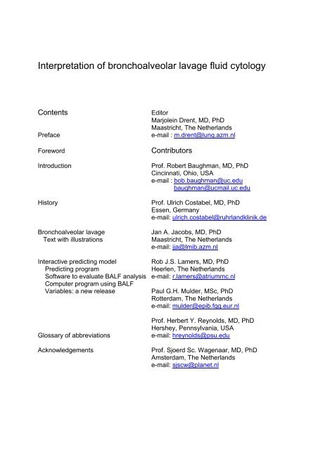
Interpretation of bronchoalveolar lavage fluid cytology - ILD care
Interpretation of bronchoalveolar lavage fluid cytology - ILD care
Interpretation of bronchoalveolar lavage fluid cytology - ILD care

You also want an ePaper? Increase the reach of your titles
YUMPU automatically turns print PDFs into web optimized ePapers that Google loves.
<strong>Interpretation</strong> <strong>of</strong> <strong>bronchoalveolar</strong> <strong>lavage</strong> <strong>fluid</strong> <strong>cytology</strong><br />
Contents Editor<br />
Marjolein Drent, MD, PhD<br />
Maastricht, The Netherlands<br />
Preface e-mail : m.drent@lung.azm.nl<br />
Foreword Contributors<br />
Introduction Pr<strong>of</strong>. Robert Baughman, MD, PhD<br />
Cincinnati, Ohio, USA<br />
e-mail : bob.baughman@uc.edu<br />
baughman@ucmail.uc.edu<br />
History Pr<strong>of</strong>. Ulrich Costabel, MD, PhD<br />
Essen, Germany<br />
e-mail: ulrich.costabel@ruhrlandklinik.de<br />
Bronchoalveolar <strong>lavage</strong> Jan A. Jacobs, MD, PhD<br />
Text with illustrations Maastricht, The Netherlands<br />
e-mail: jja@lmib.azm.nl<br />
Interactive predicting model Rob J.S. Lamers, MD, PhD<br />
Predicting program Heerlen, The Netherlands<br />
S<strong>of</strong>tware to evaluate BALF analysis e-mail: r.lamers@atriummc.nl<br />
Computer program using BALF<br />
Variables: a new release Paul G.H. Mulder, MSc, PhD<br />
Rotterdam, The Netherlands<br />
e-mail: mulder@epib.fgg.eur.nl<br />
Pr<strong>of</strong>. Herbert Y. Reynolds, MD, PhD<br />
Hershey, Pennsylvania, USA<br />
Glossary <strong>of</strong> abbreviations e-mail: hreynolds@psu.edu<br />
Acknowledgements Pr<strong>of</strong>. Sjoerd Sc. Wagenaar, MD, PhD<br />
Amsterdam, The Netherlands<br />
e-mail: sjscw@planet.nl
Preface<br />
<strong>Interpretation</strong> <strong>of</strong> BALF <strong>cytology</strong><br />
Bronchoalveolar <strong>lavage</strong> (BAL) explores large areas <strong>of</strong> the alveolar compartment providing cells as<br />
well as non-cellular constituents from the lower respiratory tract. It opens a window to the lung.<br />
Alterations in BAL <strong>fluid</strong> and cells reflect pathological changes in the lung parenchyma. The BAL<br />
procedure was developed as a research tool. Meanwhile its usefulness, also for clinical applications,<br />
has been appreciated worldwide in diagnostic work-up <strong>of</strong> infectious and non-infectious interstitial lung<br />
diseases. Moreover, BAL has several advantages over biopsy procedures. It is a safe, easily<br />
performed, minimally invasive, and well tolerated procedure. In this respect, when the clinician<br />
decides that a BAL might be helpful to provide diagnostic material, it is mandatory to consider the<br />
provided information obtained from BAL <strong>fluid</strong> analysis <strong>care</strong>fully and to have reliable diagnostic criteria.<br />
Therefore, the interpretation <strong>of</strong> BAL <strong>fluid</strong> <strong>cytology</strong> has to be standardized to improve the diagnostic<br />
power.<br />
It is with this background that the concept for this CD-rom was developed. It is aimed mainly at<br />
clinicians who are having to deal with the diagnostic problems <strong>of</strong> patients with diffuse interstitial lung<br />
diseases.The introductory sections summarize the history <strong>of</strong> BAL. Furthermore, the importance <strong>of</strong><br />
standardization <strong>of</strong> handling BAL samples will be discussed. Additionally, BAL <strong>fluid</strong> <strong>cytology</strong> features<br />
are presented and the interpretation <strong>of</strong> the BAL <strong>fluid</strong> cell differentials is discussed.<br />
Recently, a validated computer program based on logistic regression analysis using BAL <strong>fluid</strong><br />
analysis results to distinguish between the three most common interstitial lung diseases: sarcoidosis,<br />
idiopathic pulmonary fibrosis, extrinsic allergic alveolitis or drug-induced pneumonitis was developed.<br />
One <strong>of</strong> the limitations <strong>of</strong> this program was that it was not useful in discriminating between infectious<br />
disorders and non-infectious disorders.<br />
An updated windows 2000 version <strong>of</strong> this validated computer program - thought to improve the<br />
diagnostic power <strong>of</strong> BAL <strong>fluid</strong> analysis - is presented.<br />
It is hoped that this CD-rom will be a source <strong>of</strong> reference in the work-up and interpretation <strong>of</strong> BAL <strong>fluid</strong><br />
<strong>cytology</strong> to everybody involved in the management <strong>of</strong> patients suffering from diffuse interstitial lung<br />
diseases or with suspected pneumonia.<br />
Although details hidden in BAL <strong>fluid</strong> may add useful information about a patient’s disorder, the results<br />
should be considered in the context <strong>of</strong> other information from conventional investigative methods and<br />
the individual’s unique history. To establish the diagnosis a thorough history is essential as it may<br />
identify a potential aetiological factor.<br />
Marjolein Drent
Foreword<br />
<strong>Interpretation</strong> <strong>of</strong> BALF <strong>cytology</strong><br />
Bronchoalveolar <strong>lavage</strong> (BAL) has become a widely applied diagnostic tool in pulmonary medicine.<br />
This holds true for both infectious and non-infectious infiltrative and immunological lung diseases.<br />
Barriers which tried to restrict the use <strong>of</strong> BAL to research application and to put down its clinical value<br />
have finally been overcome: In two recently published international statements (ATS, ERS, also<br />
WASOG) on the major interstitial lung diseases, BAL was considered to be helpful in strengthening<br />
the diagnosis in a sarcoidosis patient without biopsy; BAL and/or transbronchial biopsy were<br />
considered as a requirement to exclude other diseases in a patient with idiopathic pulmonary<br />
fibrosis/UIP who does not undergo surgical biopsy (one <strong>of</strong> the four major criteria for making a clinical<br />
diagnosis <strong>of</strong> the disease).<br />
BAL studies should not be limited to counting the cell differentials only. At least as important as<br />
looking at cell differentials is to observe the morphological appearances <strong>of</strong> cells and particles.<br />
Examples are the different morphology in extrinsic allergic alveolitis (foamy macrophages,<br />
heterogeneous macrophage size, presence <strong>of</strong> plasma cells) versus that <strong>of</strong> sarcoidosis (more<br />
monomorphous appearance <strong>of</strong> macrophages, less activated lymphocytes), the presence <strong>of</strong> malignant<br />
cells, the characteristic features <strong>of</strong> alveolar proteinosis, or dust particles such as asbestos bodies, and<br />
other features.<br />
Also, it is important to consider BAL cell differentials not in isolation but in the context <strong>of</strong> the clinical<br />
setting and the radiological, particularly the HR-CT appearance <strong>of</strong> the disease. For example, if the CT<br />
scan shows a patchy ground glass pattern, BAL may be able to reveal that this patient suffers from<br />
extrinsic allergic alveolitis (high lymphocyte count), or a smoking related respiratory bronchiolitis/<br />
interstitial lung disease (high smoker’s macrophage count and normal cell differential), or alveolar<br />
haemorrhage (high count <strong>of</strong> haemosiderin laden macrophages).<br />
This CD-rom interpretation <strong>of</strong> BAL <strong>fluid</strong> <strong>cytology</strong> is exactly using such an approach. The work<br />
provides a detailed insight into the specific morphologic features <strong>of</strong> BAL and the interpretation <strong>of</strong> the<br />
BAL cell differentials and also presents illustrative cases with their clinical pr<strong>of</strong>ile and characteristic<br />
imaging findings.<br />
We hope that this work will find a warm appreciation amongst chest physicians, pathologists,<br />
laboratory technicians, in short, amongst everybody involved in the work-up and interpretation <strong>of</strong> BAL<br />
<strong>fluid</strong> <strong>cytology</strong>.<br />
Pr<strong>of</strong>. Ulrich Costabel, MD, FCCP<br />
Essen, March 2001
<strong>Interpretation</strong> <strong>of</strong> BALF <strong>cytology</strong><br />
Introduction: Importance <strong>of</strong> standardization <strong>of</strong> handling BAL<br />
<strong>fluid</strong> samples<br />
Pr<strong>of</strong>. Robert P. Baughman, MD<br />
University <strong>of</strong> Cincinnati Medical Center<br />
Holmes, Room 1001<br />
Eden Avenue and Albert Sabin Way<br />
Cincinnati, Ohio 45267-0565<br />
e-mail: baughmrp@ucmail.uc.edu<br />
The concept <strong>of</strong> <strong>bronchoalveolar</strong> <strong>lavage</strong> (BAL) is not new. Bronchial <strong>lavage</strong> was a technique performed during rigid<br />
bronchoscopy, especially for obtaining samples in patients with tuberculosis. To sample the alveolar space,<br />
FINLEY described the use <strong>of</strong> the Matras catheter (used to perform bronchograms without a bronchoscope) to instill<br />
and immediately aspirate <strong>fluid</strong> [1]. However, the use <strong>of</strong> a flexible bronchoscope to obtain BAL <strong>fluid</strong> (BALF) samples<br />
was popularized by Dr. HERBERT REYNOLDS [2]. The technique has been widely used for both research and<br />
diagnostic indications.<br />
Technical aspects<br />
The technical aspects <strong>of</strong> the <strong>lavage</strong> procedure can not be ignored. Some <strong>of</strong> the controversy about interpreting<br />
<strong>lavage</strong> results can be traced to different centers having different results because <strong>of</strong> variations in the technique. As<br />
in any test, the details are important. This work deals with the cellular aspects <strong>of</strong> BAL.. In order to interpret the<br />
cellular information, one should be sure that the information has been obtained in a reliable manner. I will review<br />
some <strong>of</strong> the difficulties which have been described with various aspects <strong>of</strong> <strong>lavage</strong>.<br />
One <strong>of</strong> the difficulties inherent with BAL is the variability <strong>of</strong> the sampling [3]. The <strong>lavage</strong> process provides a<br />
washing <strong>of</strong> the alveolar space. In the process <strong>of</strong> washing the space, the amount <strong>of</strong> cells and <strong>fluid</strong> returned will vary.<br />
This variability may be on the basis <strong>of</strong> the underlying disease. Other factors also affect the variability: the amount <strong>of</strong><br />
<strong>fluid</strong> instilled [4,5], the amount <strong>of</strong> pressure used to aspirate the <strong>fluid</strong>, the method <strong>of</strong> collecting the aspirated <strong>fluid</strong> [6],<br />
and the handling <strong>of</strong> the aspirated <strong>fluid</strong>. In addition, different parts <strong>of</strong> the lung may have different BALF findings [7,8].<br />
For Pneumocystic carinii, <strong>lavage</strong> in the upper lobe may contain far more organism than <strong>lavage</strong> from the middle or<br />
lower lobe [9]. Some <strong>of</strong> the differences are due to operator differences, some are due to the conditions when the<br />
procedure is done.<br />
To deal with the variability <strong>of</strong> <strong>lavage</strong>, several groups have proposed using markers [10]. These include external<br />
markers, such as methylene blue [11], or internal markers, such as urea [12]. Unfortunately, none <strong>of</strong> these markers<br />
is perfect. This is because the alveolar space is not a water tight vessel. During the process <strong>of</strong> <strong>lavage</strong>, <strong>fluid</strong> and<br />
proteins are crossing from the lung <strong>fluid</strong> into the cells and blood stream. Also, internal proteins efflux into the lung<br />
<strong>fluid</strong> [3]. Because <strong>of</strong> this variability <strong>of</strong> <strong>lavage</strong>, a recent recommendations <strong>of</strong> an European Respiratory Society Task<br />
Force was to report acellular content <strong>of</strong> BALF per ml <strong>of</strong> aspirated <strong>fluid</strong> [13].<br />
Standardization <strong>of</strong> other aspects <strong>of</strong> BAL have been attempted. In the United States, KING et al. demonstrated that<br />
BAL interpretation could be improved by using a standard approach across several centers [14]. However, those<br />
recommendations have not been widely followed. The European Respiratory Society (ERS) had made a series <strong>of</strong><br />
recommendations on performing BAL [15,16]. In a recent report, the ERS has also included recommendations<br />
regarding the reporting <strong>of</strong> BALF analysis results. These recommendations are summarized in Table 1 [13].<br />
Introduction. R.P. Baughman. 2001. 1
Table 1. - Causes <strong>of</strong> variability and recommendations for obtaining and processing BALF*.<br />
Source <strong>of</strong> variability Recommendation<br />
Disease process itself Specify underlying disease<br />
<strong>Interpretation</strong> <strong>of</strong> BALF <strong>cytology</strong><br />
Dwell time to aspirate <strong>fluid</strong> Keep to a minimum and specify dwell time, especially if<br />
prolonged<br />
Suction pressure during aspiration Keep to a minimum (25-100 mm Hg)<br />
Physician doing <strong>lavage</strong> procedure Specify<br />
The handling <strong>of</strong> <strong>lavage</strong> <strong>fluid</strong> (e.g. filtered versus<br />
not filtered, concentrated)<br />
State which technique<br />
Volume <strong>of</strong> instilled <strong>fluid</strong> Use at least >100 mL instilled volume and report volume<br />
instilled (ERS Task Force recommended 200-240 mL [16])<br />
Number <strong>of</strong> aliquots Specify and standardize (ERS Task Force recommended<br />
four [16])<br />
Position <strong>of</strong> patient Specify<br />
Area which is <strong>lavage</strong>d (e.g. one versus two<br />
lobes, middle lobe versus lower lobe, other)<br />
Specify<br />
Variability <strong>of</strong> return <strong>of</strong> <strong>lavage</strong> <strong>fluid</strong> Report volume and percentage <strong>of</strong> <strong>fluid</strong> recovered<br />
Established minimum percent recovery<br />
Reporting measurements <strong>of</strong> acellular<br />
components<br />
Handling <strong>of</strong> BALF, including if handle the first<br />
aliquot separately from rest<br />
Standardly report values per mL <strong>of</strong> BALF recovered (as well<br />
as any other special approaches)<br />
Specify<br />
* Modified from European Respiratory Society Task Force report [13].<br />
Bronchoalveolar <strong>lavage</strong> <strong>fluid</strong> <strong>cytology</strong><br />
The <strong>cytology</strong> <strong>of</strong> BALF is an analysis <strong>of</strong> the cellular components <strong>of</strong> the specimen. The most abundant cells retrieved<br />
by BAL are the inflammatory cells which line the alveolar space. These include the macrophage, lymphocyte, and<br />
neutrophil. The alveolar macrophage is the most common cell in the BALF. In a normal subject, it represents more<br />
than 80% <strong>of</strong> the cells retrieved. The lymphocyte represents approximately ten percent <strong>of</strong> the cells retrieved. In the<br />
healthy smoker, up to five percent neutrophils may be seen. In the healthy nonsmoker, neutrophils are rarely seen<br />
in the BALF [14,15].<br />
The normal values for the percentage <strong>of</strong> lymphocytes and macrophages varies between laboratories. This is in<br />
part because the preparation <strong>of</strong> the <strong>of</strong> the slide can lead to a variation <strong>of</strong> proportion <strong>of</strong> lymphocytes. This includes<br />
the use <strong>of</strong> a cytocentrifuge versus membrane to prepare the slide [17]. For the cytocentrifuge, the speed <strong>of</strong> the<br />
centrifuge, the area <strong>of</strong> the slide, and the number <strong>of</strong> cells counted have all been shown to lead to differences in<br />
counts [18]. In addition, the cells can be sometimes difficult to distinguish using the standard Giemsa based stain.<br />
In active inflammatory diseases, such as sarcoidosis, monocytes recruited into the lung can look like activated<br />
lymphocytes. Nonspecific esterase is another way to identify macrophages, and flow cytometry with a panlymphocyte<br />
marker can count lymphocytes [19]. However, these are time consuming stains and usually not<br />
necessary. The variation in cellular differential seems to be greater between laboratories than within the same<br />
laboratory [14]. However, there are no standards which have been used to certify laboratories.<br />
Diagnostic value <strong>of</strong> <strong>bronchoalveolar</strong> <strong>lavage</strong><br />
Cytologic examination has also been used to detect malignancy. This technique is fairly standard and criteria for<br />
malignancy used for other bronchial samples can be readily applied to BALF samples [20]. For some conditions,<br />
such as lymphangitic spread <strong>of</strong> breast cancer, BAL has been quite useful in providing a diagnostic sample [21,22].<br />
For patients with <strong>bronchoalveolar</strong> cell carcinoma, BAL may have the highest diagnostic yield [20].<br />
BAL has become an accepted technique for diagnosis <strong>of</strong> infection [23,24]. This includes opportunistic infections,<br />
such as P. carinii [25]. The diagnosis <strong>of</strong> P. carinii usually requires direct visualization <strong>of</strong> the organism, since it does<br />
Introduction. R.P. Baughman. 2001. 2
<strong>Interpretation</strong> <strong>of</strong> BALF <strong>cytology</strong><br />
not grow in culture. Several stains have proved useful in detecting P. carinii. The standard stain for detecting P.<br />
carinii has been a silver stain, such as the Methenamine stain [26]. There are more sensitive stains such as the<br />
immun<strong>of</strong>luorescent stains [27,28] and even polymerase chain reaction has been employed [29]. These more<br />
sensitive, and expensive techniques have not proved necessary for the evaluation <strong>of</strong> most immunosuppressed<br />
patients when examining BALF. They are usually reserved for specimens with a lower number <strong>of</strong> organisms, such<br />
as induced sputum and bronchial wash [27-29]. The Wright-Giemsa and similar stains are used for defining<br />
cellular morphology. A modified Wright-Giemsa can be done within five minutes. It has been shown to detect P.<br />
carinii in the BALF <strong>of</strong> over 70% <strong>of</strong> HIV infected patients with P. carinii pneumonia [30]. It is not as sensitive in non-<br />
HIV infected patients, who have a smaller number <strong>of</strong> organisms [30]. The technique <strong>of</strong> BAL does have an affect on<br />
the yield for P. carinii. The volume <strong>of</strong> instilled <strong>fluid</strong> does not appear to be crucial, since the number <strong>of</strong> P. carinii<br />
organisms is relatively constant during sequential BAL, with increasing volumes <strong>of</strong> BALF instilled [31]. On the other<br />
hand, the area in which BAL is performed has proved important [32]. Multiple <strong>lavage</strong>s or <strong>lavage</strong> directed to the<br />
upper lobe or area <strong>of</strong> most diseases has enhanced the yield <strong>of</strong> BALF for diagnosing P. carinii pneumonia [8,9].<br />
Beyond the organism itself, the cellular differential count has clinical information. Increased neutrophils or<br />
eosinophils are associated with a worse prognosis [33].<br />
Bacterial infections may also be diagnosed by BAL. These include unusual bacterial infections such as Legionella<br />
[34]. BAL has also been used to diagnose routine bacterial infections. For the diagnosis <strong>of</strong> routine infections, one<br />
needs to perform semi-quantitative cultures <strong>of</strong> the BALF [35]. The rationale behind semi-quantitative cultures was<br />
to separate colonization from deep seated infection. Semi-quantitative cultures have proved useful for evaluating<br />
BALF samples for both ventilated [36] and non-ventilated patients [37]. In addition to the standardized BAL<br />
procedure, it is important to culture the <strong>fluid</strong> in a standard manner. Recommendations have been made for the<br />
technique <strong>of</strong> BALF cultures [38]. The examination <strong>of</strong> the cells in bacterial pneumonia usually demonstrates a<br />
marked increase in neutrophils. This is not specific. The use <strong>of</strong> Gram stain to identify bacteria was shown to be<br />
useful in diagnosing pneumonia [35]. Subsequently, CHASTRE proposed counting the number <strong>of</strong> cells with<br />
intracellular organisms (ICO) [39]. The diagnostic importance <strong>of</strong> cells with ICO is still unclear [36].<br />
In conclusion, the analysis <strong>of</strong> cells obtained by BAL can be quite valuable clinically. To improve the<br />
information, one should educate themselves on what they are seeing. This CD-ROM provides excellent<br />
examples <strong>of</strong> the types <strong>of</strong> cells one can see and how to integrate the BAL findings with the clinical<br />
presentation.<br />
The cellular analysis <strong>of</strong> the BALF relies on an accurate counting <strong>of</strong> the various cells involved. Care should be<br />
taken to identify the cells, and differential counting should be done consistently. Although universal<br />
standards are not yet available, it seems obvious that each institution should be sure that the various<br />
readers <strong>of</strong> BALF samples have agreement about their BALF differential counts [19]. Using a standard<br />
technique for performing the BAL and handling the sample will also reduce variability.<br />
References<br />
1. Finley T N, Swenson E W, Curran WS, et al. Bronchopulmonary <strong>lavage</strong> in normal subjects and patients with<br />
obstructive lung disease. Ann Intern Med 1967; 66: 651-658. [no abstract available]<br />
2. Reynolds HY, Newball HH. Analysis <strong>of</strong> proteins and respiratory cells obtained from human lungs by bronchial<br />
<strong>lavage</strong>. J Lab Clin Med 1974; 84: 559-573. [no abstract available]<br />
3. Baughman RP. The uncertainties <strong>of</strong> <strong>bronchoalveolar</strong> <strong>lavage</strong>. Eur Respir J 1997; 10: 1940-1942. [no abstract<br />
available]<br />
4. Dohn MN, Baughman RP. Effect <strong>of</strong> changing instilled volume for <strong>bronchoalveolar</strong> <strong>lavage</strong> in patients with<br />
interstitial lung disease. Am Rev Respir Dis 1985; 132: 390-392. [abstract]<br />
5. Rennard SI, Ghafouri M, Thompson AB, et al. Fractional processing <strong>of</strong> sequential <strong>bronchoalveolar</strong> <strong>lavage</strong> to<br />
separate bronchial and alveolar samples. Am Rev Respir Dis 1990; 141: 208-217. [abstract]<br />
6. Thompson AB, Robbins RA, Ghafouri MA, Linder J, Rennard SI. Bronchoalveolar <strong>lavage</strong> <strong>fluid</strong> processing. Effect<br />
<strong>of</strong> membrane filtration on neutrophil recovery. Acta Cytol 1989; 33: 544-549. [abstract]<br />
7. Garcia JG, Wolven RG, Garcia PL, Keogh BA. Assessment <strong>of</strong> interlobar variation <strong>of</strong> <strong>bronchoalveolar</strong> <strong>lavage</strong><br />
cellular differentials in interstitial lung diseases. Am Rev Respir Dis 1986; 133: 444-449. [abstract]<br />
8. Levine S J, Kennedy D, Shelhamer JH, et al. Diagnosis <strong>of</strong> Pneumocystis carinii pneumonia by multiple lobe,<br />
site-directed <strong>bronchoalveolar</strong> <strong>lavage</strong> with immun<strong>of</strong>luorescent monoclonal antibody staining in human<br />
immunodeficiency virus- infected patients receiving aerosolized pentamidine chemoprophylaxis. Am Rev<br />
Introduction. R.P. Baughman. 2001. 3
<strong>Interpretation</strong> <strong>of</strong> BALF <strong>cytology</strong><br />
Respir Dis 1992; 146: 838-843. [abstract]<br />
9. Baughman RP, Dohn MN, Shipley R, Buchsbaum JA, Frame PT. Increased Pneumocystis carinii recovery from<br />
the upper lobes in Pneumocystis pneumonia. The effect <strong>of</strong> aerosol pentamidine prophylaxis. Chest 1993; 103:<br />
426-432.<br />
10. Ward C, Effros RM, Walters EH. Assessment <strong>of</strong> epithelial lining <strong>fluid</strong> during <strong>bronchoalveolar</strong> <strong>lavage</strong>. Eur Respir<br />
Rev 1999; 9: 66,32-37. [no abstract available]<br />
11. Abernathy RS. Childhood sarcoidosis in Arkansas. South Med J 1985; 78: 435-439. [abstract]<br />
12. Rennard SI, Basset G, Lecossier D, O'Donnell K, Martin P, Crystal RG. Estimation <strong>of</strong> volume <strong>of</strong> epithelial lining<br />
<strong>fluid</strong> recovered by <strong>lavage</strong> using urea as marker <strong>of</strong> dilution. J Appl Physiol 1986; 60: 532-538. [abstract]<br />
13. Haslam PL, Baughman RP. Guidelines for measurement <strong>of</strong> acellular components and recommendations for<br />
standardization <strong>of</strong> bronchoaveolar <strong>lavage</strong> (BAL). Report <strong>of</strong> the European Respiratopry Society Task Force. Eur<br />
Respir Rev 1999. [PDF-file]<br />
14. The BAL Cooperative Group Steering Committee. Bronchoalveolar <strong>lavage</strong> constituents in healthy individuals,<br />
idiopathic pulmonary fibrosis, and selected comparison groups. Am Rev Respir Dis 1990; 141: S169-S202.<br />
[abstract]<br />
15. Klech H, Hutter C, Costabel U. Clinical guidelines and indications for <strong>bronchoalveolar</strong> <strong>lavage</strong> (BAL). Eur Respir<br />
Rev 1992; 2: 47-127. [no abstract available]<br />
16. Klech H, Pohl W. Technical recommendations and guidelines for <strong>bronchoalveolar</strong> <strong>lavage</strong> (BAL). Eur Respir J<br />
1989; 2: 561-585. [abstract]<br />
17. Saltini C, Hance AJ, Ferrans VJ, et al. Accurate quantification <strong>of</strong> cells recovered by <strong>bronchoalveolar</strong> <strong>lavage</strong>. Am<br />
Rev Respir Dis 1984; 130: 650-658. [abstract]<br />
18.De Brauwer EIGB, Drent M, Mulder PGH, Bruggeman CA, Wagenaar SjSc, Jacobs JA. Differential cell analysis <strong>of</strong><br />
cytocentrifuged <strong>bronchoalveolar</strong> <strong>lavage</strong> <strong>fluid</strong> samples affected by the area counted. Analyt Quant Cytol Histol<br />
2000; 22: 143-149. [abstract]<br />
19. Baughman RP, Stroh<strong>of</strong>er S, Kim CK. Variation <strong>of</strong> differential cell counts <strong>of</strong> <strong>bronchoalveolar</strong> <strong>lavage</strong> <strong>fluid</strong>. Arch<br />
Pathol Lab Med 1986; 110: 341-343. [no abstract available]<br />
20. Rennard SI. Bronchoalveolar <strong>lavage</strong> in the diagnosis <strong>of</strong> cancer. Lung 1990; 168 Suppl: 1035-1040. [abstract]<br />
21. Radio SJ, Rennard SI, Kessinger A, Vaughan WP, Linder J. Breast carcinoma in <strong>bronchoalveolar</strong> <strong>lavage</strong>. A<br />
cytologic and immunocytochemical study. Arch Pathol Lab Med 1989; 113: 333-336. [no abstract available]<br />
22. Lower EE, Baughman RP. Pulmonary lymphangitic metastasis from breast cancer. Lymphocytic alveolitis is<br />
associated with favorable prognosis. Chest 1992; 102: 1113-1117. [abstract]<br />
23. Stover DE., Zaman MB, Hajdu SI, et al. Role <strong>of</strong> <strong>bronchoalveolar</strong> <strong>lavage</strong> in the diagnosis <strong>of</strong> diffuse pulmonary<br />
infiltrates in the immunosuppressed host. Ann Intern Med 1984; 101: 1-7. [abstract]<br />
24. Baughman RP. The lung in the immunocompromised patient: infectious complications part 1. Respiration<br />
1999; 66: 2-11. [abstract]<br />
25. Golden JA, Hollsander H, Stulbarg MS, Gamsu G. Bronchoalveolar <strong>lavage</strong> as the exclusive diagnostic modality<br />
for Pneumocystis carinii pneumonia: a prospective study among patients with acquired immunodeficiency<br />
syndrome. Chest 1986; 90: 18-22. [abstract]<br />
26. Baughman RP. Current methods <strong>of</strong> diagnosis. in Pneumocystis carinii pneumonia edited by Walzer, P. D.<br />
Marcel Decker, Inc. New York 381-401, 1994. [no abstract available]<br />
27. Kovacs JA, Ng VL, Masur H, et al. Diagnosis <strong>of</strong> Pneumocystis carinii pneumonia: improved detection in sputum<br />
with use <strong>of</strong> monoclonal antibodies. N Engl J Med 1988; 318: 589-593. [abstract]<br />
28. Baughman RP, Stroh<strong>of</strong>er SS, Clinton BA, Nickol AD, Frame PT. The use <strong>of</strong> an indirect fluorescent antibody test<br />
for detecting Pneumocystis carinii. Arch Pathol Lab Med 1989; 113: 1062-1065. [abstract]<br />
29. Leigh TR, Wakefield AE, Peters SE, Hopkin JM, Collins JV. Comparison <strong>of</strong> DNA amplification and<br />
immun<strong>of</strong>luorescence for detecting Pneumocystis carinii in patients receiving immunosuppressive therapy.<br />
Transplantation 1992; 54: 468-470. [abstract]<br />
30. Tollerud DJ, Wesseler TA, Kim CK, Baughman RP. Use <strong>of</strong> a rapid differential stain for identifying Pneumocystis<br />
carinii in <strong>bronchoalveolar</strong> <strong>lavage</strong> <strong>fluid</strong>. Diagnostic efficacy in patients with AIDS. Chest 1989; 95: 494-497.<br />
[abstract]<br />
31. Baughman RP, Stroh<strong>of</strong>er S, Colangelo G, Frame PT. Semiquantitative technique for estimating Pneumocystis<br />
carinii burden in the lung. J Clin Microbiol 1990; 28: 1425-1427. [abstract]<br />
32. Jules-Elysee KM, Stover DE, Zaman MB, Bernard EM, White DA. Aerosolized pentamidine: effect on diagnosis<br />
and presentation <strong>of</strong> Pneumocystis carinii pneumonia. Ann Intern Med 1990; 112: 750-757. [abstract]<br />
33. Mason GR, Hashimoto CH, Dickman PS, Foutty LF, Cobb CJ. Prognostic implications <strong>of</strong> <strong>bronchoalveolar</strong><br />
<strong>lavage</strong> neutrophilia in patients with Pneumocystis carinii pneumonia and AIDS. Am Rev Respir Dis 1989; 139:<br />
1336-1342. [abstract]<br />
Introduction. R.P. Baughman. 2001. 4
<strong>Interpretation</strong> <strong>of</strong> BALF <strong>cytology</strong><br />
34. Kohorst WR, Schonfield SA, Macklin JE, et al. Rapid diagnosis <strong>of</strong> legionnaires disease by <strong>bronchoalveolar</strong><br />
<strong>lavage</strong>. Chest 1983; 84: 186-190. [abstract]<br />
35. Thorpe JE, Baughman RP, Frame PT, Wesseler TA, Staneck JL. Bronchoalveolar <strong>lavage</strong> for diagnosing acute<br />
bacterial pneumonia. J Infect Dis 1987; 155: 855-861. [abstract]<br />
36. Torres A. el Ebiary M. Bronchoscopic <strong>bronchoalveolar</strong> <strong>lavage</strong> for the diagnosis <strong>of</strong> ventilator-associated<br />
pneumonia. Chest 2000; 117: 198S-202S. [abstract]<br />
37.Cantral DE, Tape TG, Reed EC, Spurzem JR., Rennard SI, Thompson AB. Quantitative culture <strong>of</strong><br />
<strong>bronchoalveolar</strong> <strong>lavage</strong> <strong>fluid</strong> for the diagnosis <strong>of</strong> bacterial pneumonia. Am J Med 1993; 95: 601-607. [abstract]<br />
38. Baselski VS, el Torky M, Coalson JJ, Griffin JP. The standardization <strong>of</strong> criteria for processing and interpreting<br />
laboratory specimens in patients with suspected ventilator- associated pneumonia. Chest 1992; 102: 571S-<br />
579S. [no abstract available]<br />
39.Chastre J, Fagon JY, Soler P, et al. Diagnosis <strong>of</strong> nosocomial bacterial pneumonia in intubated patients<br />
undergoing ventilation: comparison <strong>of</strong> the usefulness <strong>of</strong> <strong>bronchoalveolar</strong> <strong>lavage</strong> and the protected specimen<br />
brush. Am J Med 1988; 85: 499-506. [abstract]<br />
Introduction. R.P. Baughman. 2001. 5
<strong>Interpretation</strong> <strong>of</strong> BALF <strong>cytology</strong><br />
History: Visualization <strong>of</strong> the cells in BALF has directed<br />
clinical lung research<br />
Pr<strong>of</strong>. Herbert Y. Reynolds, M.D., J. Lloyd Huck Pr<strong>of</strong>essor <strong>of</strong> Medicine, Chair,<br />
Department <strong>of</strong> Medicine, Milton S. Hershey Medical Center,<br />
Hershey, Pennsylvania<br />
Sampling the lower respiratory tract by <strong>bronchoalveolar</strong> <strong>lavage</strong> (BAL) to retrieve detachable airway and alveolar<br />
cells and to collect non-cellular soluble proteins and other substances has been invaluable for stimulating<br />
research <strong>of</strong> the normal lung and <strong>of</strong> the diseased lung in patient-volunteer participants [1]. This has been<br />
performed much more easily and safely since the availability <strong>of</strong> fiberoptic bronchoscopy [2]. Although BAL analysis<br />
has contributed to the scientific understanding <strong>of</strong> the healthy and disease-afflicted lung, it has had helpful clinical<br />
applications as well [3-11]. Material on the present CD-ROM, an update <strong>of</strong> prior work published by Dr. DRENT and<br />
colleagues [12] about the use <strong>of</strong> BAL analysis in the diagnosis <strong>of</strong> interstitial lung diseases (<strong>ILD</strong>) has been<br />
extended to further differentiate BAL cellular findings in infectious illnesses from other <strong>ILD</strong>. It is a pleasure to <strong>of</strong>fer<br />
a historical perspective about obtaining lung cells, when the technique <strong>of</strong> fiberoptic bronchoscopy was just<br />
becoming widely utilized about 30 years ago [13-15], and how this stimulated me and my clinical colleagues to<br />
investigate the normal respiratory tract and the immunopathology associated with several forms <strong>of</strong> <strong>ILD</strong>.<br />
A good picture may be worth a thousand descriptive words about the appearance <strong>of</strong> lung cells, and more<br />
importantly what they may have been doing in situ - behaving and doing their usually thorough and competent job<br />
<strong>of</strong> respiratory host defense [16,17], or causing mischief and creating or perpetuating illness [18]? Looks, however,<br />
may be deceiving, as activated-appearing cells may be responding appropriately by secreting cytokines,<br />
processing antigens, and stimulating immune reactions, or attracting inflammatory back-up cells [19]. A picture<br />
must be extrapolated to a reasonable clinical setting and a feasible patient diagnosis.<br />
The ease and safety <strong>of</strong> fiberoptic bronchoscopy, coupled with BAL for retrieval <strong>of</strong> airway-alveolar cells that can be<br />
detached and washed out <strong>of</strong> the distal airways or <strong>lavage</strong>d selectively from the bronchial surface (or brushed <strong>of</strong>f),<br />
and also combined with tissue biopsies (transbronchial and endobronchial) - all together allow for a<br />
comprehensive, direct, interior view and sampling <strong>of</strong> the respiratory tract. This has provided tremendous insight<br />
into the cause and immunopathogenesis <strong>of</strong> many airway and parenchymal respiratory diseases and has<br />
facilitated patient diagnosis. The capability <strong>of</strong> using normal volunteer subjects for bronchoscopy and BAL has been<br />
<strong>of</strong> great help in establishing normal values. Thus, pulmonologists can have direct visualization out into the small<br />
conducting airways [20], perform BAL and obtain tissue from the lower respiratory tract, a combination that is<br />
unique and not readily available from most other organ systems, except perhaps the urogenital and<br />
gastrointestinal tracts and large skeletal joints (knee). This direct interior ‘view’ is also different from those<br />
achieved with radiographic and scan imaging.<br />
In my clinical research experience, four distinct and memorable instances <strong>of</strong> an initial view <strong>of</strong> BAL retrieved lung<br />
cells were especially impressionable. Each stimulated further investigation about why certain cells were grouped<br />
or associated together: 1) normal human alveolar macrophages (AM) and the few lymphocytes that accompanied<br />
them in BAL <strong>fluid</strong> (BALF); 2) rosetted lymphocytes stuck to the surface <strong>of</strong> alveolar macrophages from a patient with<br />
pulmonary sarcoidosis,3) the foamy appearance <strong>of</strong> alveolar macrophages that were almost engulfed by the<br />
numerous surrounding lymphocytes obtained from a patient with hypersensitivity pneumonitis, and 4) the<br />
presence <strong>of</strong> polymorphonuclear neutrophils (PMNs) and eosinophils mixed among AM in BALF from patients with<br />
idiopathic pulmonary fibrosis (IPF). These ‘sightings’ occurred over a brief period, after we began to do BAL on<br />
normal human volunteers [21] and patient participants with forms <strong>of</strong> interstitial lung diseases [22,23]. For me, the<br />
observation <strong>of</strong> lung cells has always been a two-fold process - initially looking at a wet cell preparation [21] taken<br />
from the freshly centrifuged BAL <strong>fluid</strong>, revealing viable cells and the glitter <strong>of</strong> intracellular inclusions or granules,<br />
and then counting the traditionally stained cell preparation. Personally, I have always felt more confident <strong>of</strong> my wet<br />
prep differential cell count for cell enumeration than the cytocentrifuge or filter preparation <strong>of</strong> stained cells.<br />
Certainly nothing can give the excitement or thrill <strong>of</strong> seeing the living cells and feeling their energy and interaction.<br />
Living lung cellular biology is the only description that comes to mind and pen.<br />
History. H.Y. Reynolds. 2001. 1
Normal alveolar macrophages and respiratory lymphocytes<br />
<strong>Interpretation</strong> <strong>of</strong> BALF <strong>cytology</strong><br />
Following <strong>lavage</strong> recovery <strong>of</strong> rabbit macrophages [24] for in vitro study, research interest peaked to obtain human<br />
lung cells initially from normal volunteer participants through a variety <strong>of</strong> endobronchially placed catheters<br />
(reviewed Ref. 1) and then with fiberoptic bronchoscopy [25,26]. Among the population <strong>of</strong> respiratory cells from<br />
normal young non-smokers, most <strong>of</strong> the cells were alveolar macrophages but about 15% were lymphocytes plus a<br />
few PMNs or other inflammatory cells [21]. Considerable interest was focused on the AM as a phagocytic cell, and<br />
we too were eager to study its immune receptors [27] and antibody-mediated uptake <strong>of</strong> opsonized bacteria [28].<br />
Alveolar macrophages seemed to exist in various sizes as recovered in normal BAL, suggesting subpopulations <strong>of</strong><br />
them with perhaps different functions or representing various stages <strong>of</strong> development [29,30]. The intent was to find<br />
ways to manipulate lung humoral immune responses that might improve the efficiency <strong>of</strong> host defenses to cope<br />
with microbes entering the lower respiratory tract.<br />
Overlooked were a very small population <strong>of</strong> other macrophages now identified as dendritic cells (also Langerhans’<br />
cells) [31], the more efficient antigen presenting cell so effective in initiating cellular immune responses in the lung<br />
[32-34] or increased by smoking [35,36] and associated with certain diseases [37-39].<br />
Others [4,40-42] were attracted to the lymphoid cells initially, and information obtained about cellular immunity<br />
stimulated by lymphocytes became important in many diseases such as sarcoidosis, hypersensitivity<br />
pneumonitis, occupational diseases and respiratory infections such as HIV.<br />
Sarcoidosis: adherence <strong>of</strong> lymphocytes to alveolar macrophages<br />
In a fresh wet mount BAL cell preparation, viewed under phase contrast microscopy, from a patient with active<br />
sarcoidosis [3], the appearance <strong>of</strong> lymphocytes stuck to the surface <strong>of</strong> AMs is striking; the cells adhere and do not<br />
fall <strong>of</strong>f and are not phagocytosed by the AM. A cytospin stained cell preparation makes these rosettes easy to<br />
notice. Occasionally, a spontaneous rosette can be found among BAL cells from a normal smoker or non-smoker,<br />
and YEAGER and colleagues [43] found that 1.8-2.1% <strong>of</strong> AM had one or more adherent lymphocytes. In their group <strong>of</strong><br />
14 sarcoidosis patients, macrophage rosettes involved 8-11% <strong>of</strong> the AMs. We found 4 <strong>of</strong> 17 BAL specimens from<br />
sarcoidosis patients had lymphocyte-macrophage rosettes [44]. The implication <strong>of</strong> these rosettes is still<br />
spectulative, but HUNNINGHAKE and colleagues [5, 45,46] found that the lymphocytosis in BALF from many patients<br />
with sarcoidosis was comprised mainly T-cells which were activated. T-cells were important in creating the<br />
alveolits that probably proceeded granuloma formation. Lung T-cells release a pertinent cytokine in the cellular<br />
activation mechanism(s) <strong>of</strong> sarcoidosis [47]. Certainly, T-cell immune mechanisms have become the research<br />
focus <strong>of</strong> many investigators.<br />
Lung cells in hypersensitivity pneumonitis<br />
This has seemed to be the perfect respiratory disease to study with distinct episodic clinical features, <strong>of</strong>ten a<br />
defined antigen that stimulates specific humoral responses (precipitating antibodies), striking parenchymal lung<br />
involvement that is at first acute but can become chronic and persist (<strong>of</strong>ten establishing host tolerance when<br />
repeated antigen exposure continues), and decidedly immunologic in nature (but not allergic) [48-50]. Performing<br />
BAL analysis on the first patients we encountered [22] was fascinating - all the ingredients were present in BALF<br />
between the alveolar cells and the <strong>lavage</strong> supernatant <strong>fluid</strong> (and contrasted with the patient’s serum) to fit the<br />
immunologic response together.<br />
The patient’s clinical history usually gave good clues to the kind <strong>of</strong> antigen(s) encountered in his/her environment<br />
or workplace. A specific precipitating antigen-antibody reaction could be found in serum and occasionally in BAL<br />
<strong>fluid</strong> (for 3 <strong>of</strong> the initial 7 patients studied [23]), which was in the IgG fraction in BALF. Although IgM levels were<br />
detected in BALF, no precipitating antibody activity was found. IgE and two complement components, C 4 and C 6,<br />
were in normal amounts in BALF. The other surprise was the composition <strong>of</strong> the BAL cells. Lymphocytes<br />
predominated in the cellular count, a mean count <strong>of</strong> about 62%, and the majority were T-lymphocytes (mean 70%),<br />
and about 6% were B-cells (assayed with sheep erythrocyte forming rosettes). Alveolar macrophages, reciprocally<br />
reduced in proportion to the high percentage <strong>of</strong> lymphocytes, were about 29% <strong>of</strong> the total <strong>lavage</strong> recovered cells.<br />
Within the macrophage population, larger than usual size macrophages were seen with vaculolated and opaque<br />
appearing cytoplasm and few granules; these were considered ‘foamy’ or engorged macrophages [51].<br />
History. H.Y. Reynolds. 2001. 2
<strong>Interpretation</strong> <strong>of</strong> BALF <strong>cytology</strong><br />
Importantly, when subjects with hypersensitivity pneumonitis (9 <strong>of</strong> 10 had pigeon or bird fancier’s lung disease)<br />
were challenged with inhaled diluted pigeon serum by FOURNIER and colleagues [52] and re-evaluated by BAL 24<br />
hours after inhalation and again five or eight days later, the cellular findings in BALF were quite different from the<br />
baseline <strong>lavage</strong> results, obtained five days prior to challenge. For the ten HP patients, the baseline lymphocyte<br />
percentage was on average 60% and PMN 8.3% (range 0-31%); 24 hours after challenge the total cells recovered<br />
in BALF doubled with the lymphocyte percentage constituting about 38%, but PMNs now accounted for an average<br />
<strong>of</strong> 41%. Within five to eight days, counts returned to baseline for the six subjects who underwent a third <strong>lavage</strong><br />
study. This acute inflammatory cell response <strong>of</strong> PMNs after antigen challenge seemed like an Arthus response or<br />
Type 3 reaction [48,49].<br />
It was important that LEATHERMAN and colleagues [53] more precisely determined the BAL T-cell phenotypes in<br />
hypersensitivity pneumonitis as largely CD8 cells (T-suppressor/cytotoxic cells) in contrast to the high percentage<br />
<strong>of</strong> CD4 T-cells found in sarcoidosis. Subsequently, others [54] found that patients with an insidious onset <strong>of</strong><br />
hypersensitivity pneumonitis and development <strong>of</strong> pulmonary fibrosis could have a relative increase in CD4 Tlymphocytes<br />
in BALF versus a non-fibrosis group with increased CD8 T-cells, raising the possibility that CD8Tcells<br />
might have a protective effect on developing fibrosis. This is relevant to the long term effects <strong>of</strong> continued<br />
antigen exposure (continued farming or pigeon contact) that has been thoroughly studied by CORMIER and<br />
colleagues in Quebec, Canada.<br />
Asymptomatic dairy farmers with serum precipitins to Micropolyspora faeni underwent BAL [55] for cellular and<br />
immunoglobulin analysis. Six <strong>of</strong> ten farmers had an increased number <strong>of</strong> cells in BALF and a high percentage <strong>of</strong><br />
lymphocytes (mean 52.5%); also, four <strong>of</strong> the six had positive precipitins in BALF. Thus, normal farmers may have<br />
BAL findings consistent with an immunologic alveolitis. When these asymptomatic farmers were re-evaluated two<br />
years later [56], and then six or seven years later [57], 33 <strong>of</strong> 43 original farmers were re-evaluated but this did not<br />
include another BAL. Only five <strong>of</strong> the 33 were no longer farmers by pr<strong>of</strong>ession. However, original <strong>lavage</strong> findings <strong>of</strong><br />
an elevated percentage <strong>of</strong> BAL lymphocytes did not predict later who would develop functional lung deterioration or<br />
extrinsic allergic alveolitis. Therefore, the respiratory system <strong>of</strong> many persons can modulate the impact <strong>of</strong> repeated<br />
and continued foreign aerosol antigen exposure by developing a subclinical cellular immune response that may<br />
not progress to overt illness, thus their lungs can coexist or become tolerant to an environmental<br />
antigen/substance. In other subjects this exposure can create symptomatic lung disease. Allergy in the typical<br />
fashion does not develop either. Much can still be investigated about this occupational illness <strong>of</strong> defined cause<br />
(<strong>of</strong>ten) that provides a window into the subtle mechanisms <strong>of</strong> respiratory immune responsiveness.<br />
Idiopathic pulmonary fibrosis: a mixed inflammatory cell response in BALF<br />
Considerable clinical and research interest has existed for forms <strong>of</strong> diffuse interstitial pneumonia causing fibrosis,<br />
acutely or more chronic in development. This was probably rekindled with a presentation by Drs. LOUIS HAMMAN and<br />
ARNOLD R. RICH [58] <strong>of</strong> four cases <strong>of</strong> fulminating fibrosis seen and studied at the Johns Hopkins Hospital between<br />
1931-35. Later, several prominent pulmonary clinicians and pathologists [59,60,61] reported on series <strong>of</strong> patients<br />
that elucidated the clinical course, histopathology and treatment <strong>of</strong> <strong>ILD</strong>. In this context, a task force, directed by Dr.<br />
CLAUDE LENFANT, to develop a lung research program for the National Heart and Lung Institute <strong>of</strong> the National<br />
Institutes <strong>of</strong> Health, published in 1972 [62], recommended the study <strong>of</strong> diffuse fibrosis <strong>of</strong> unknown causes and<br />
acquired connective tissue diseases to identify etiologic agents, and assess other risk factors such as genetic<br />
constitution and altered immunologic mechanisms. With this mandate the Pulmonary Branch’s research agenda,<br />
directed by the Branch Chief, Dr. RONALD G. CRYSTAL, began a concerted effort to investigate interstitial lung<br />
diseases. A patient protocol was developed by Dr. CRYSTAL and Dr. JACK D. FULMER, co-principal investigators, in<br />
late 1974 and patients were enrolled soon after. This project on diffuse fibrotic lung disease was a correlative<br />
study <strong>of</strong> the etiology, pathogenesis, pathophysiology and therapy <strong>of</strong> these patients. Patient participants would<br />
receive a fiberoptic bronchoscopy with BAL performed by the Pulmonary Branch Staff physicians; BALF cellular and<br />
protein analysis were done by Dr. REYNOLDS, a collaborating investigator located nearby in the Laboratory <strong>of</strong><br />
Clinical Investigation, National Institute <strong>of</strong> Allergy and Infectious Diseases at NIH. The protocol’s enrollment <strong>of</strong><br />
patients moved quickly, and the interesting BALF analyses were reported less than a year later at an NIH<br />
Combined Clinical Staff Conference, November 13, 1975 [22] and published later in more detail [23]. The first<br />
decade’s work on <strong>ILD</strong> was best reviewed by Dr.CRYSTAL and colleagues [63] as a medical progress report in the<br />
History. H.Y. Reynolds. 2001. 3
New England Journal <strong>of</strong> Medicine.<br />
<strong>Interpretation</strong> <strong>of</strong> BALF <strong>cytology</strong><br />
In contrast to normals including smokers [21], the cellular differentials from the IPF patient participants [23] were<br />
quite different - notably PMNs (mean percentage 33 in stained differential count) and eosinophils (mean 2.7%)<br />
were increased from seven IPF patients not receiving corticosteroid therapy. For 12 patients on therapy, the PMN<br />
percentage was less (mean 14%), but still higher than normal controls (< 3%), and the eosinophils were still<br />
increased (mean 3.2%).Cellular counts <strong>of</strong> lymphocytes (mean 12.1%) in untreated and treated patients (mean<br />
7.7%) were within the range <strong>of</strong> normals. The macrophages which comprised the remaining cells were not<br />
remarkable in appearance.<br />
The surprise about IPF BAL cells was an apparent mixed alveolar inflammatory reaction <strong>of</strong> PMNs and <strong>of</strong> some<br />
eosinophils. Both <strong>of</strong> these cell types subsequently received research attention. HASLAM with colleagues [64]<br />
analyzed BAL cells from patients (n=36) with lone cryptogenic fibrosing alveolitis [60], which is quite similar to IPF,<br />
and other patients (n=15) with associated connective tissue diseases. They found for lone CFA subjects that the<br />
mean BALF PMN percentage was 13% and the eosinophil percentage was about 6% (over 2.4% for 19/36<br />
patients). Other studies (65,66) continued to analyze these cells and correlate them with response to treatment<br />
and prognosis (reviewed in [1]).The neutrophilic alveolitis characteristic <strong>of</strong> many patients with IPF also stimulated<br />
research for chemotactic stimuli that might attract PMNs into the lungs [67]. Subsequent identification <strong>of</strong> a<br />
proinflammatory cytokine, interleukin-8, derived from alveolar macrophages is involved in the process [68,69].<br />
In conclusion, identification <strong>of</strong> cells in BAL obtained from normal people and those with a variety <strong>of</strong> lung<br />
diseases has helped define the milieu <strong>of</strong> the lower respiratory tract which has promoted research <strong>of</strong> the<br />
specific function(s) and interactions that occur to maintain health and what changes in the composition and<br />
activity <strong>of</strong> cells that occurs with illness. Although much has been described, more is still left to uncover and<br />
learn to prevent development <strong>of</strong> illness.<br />
References<br />
1. Reynolds HY. Use <strong>of</strong> <strong>bronchoalveolar</strong> <strong>lavage</strong> in humans - past necessity and future imperative. Lung 2000;<br />
178:271-293. [no abstract available]<br />
2. Reynolds HY. State <strong>of</strong> art. Bronchoalveolar <strong>lavage</strong>. Am Rev Respir Dis 1987; 135:250-263. [abstract]<br />
3. Weinberger SE, Kelman JA, Elson NA, et al. Bronchoalveolar <strong>lavage</strong> in interstitial lung disease. Ann Intern Med<br />
1978; 89:459-466. [no abstract available]<br />
4. Hunninghake GW, Gadek JE, Kawanami O, et al. Inflammatory and immune processes in the human lung in<br />
health and disease: evaluation by <strong>bronchoalveolar</strong> <strong>lavage</strong>. Am J Pathol 1979; 97:149-206. [no abstract<br />
available]<br />
5. Daniele RP, Elias JA, Epstein PE, Rossman MD. Bronchoalveolar <strong>lavage</strong>: role in the pathogenesis, diagnosis,<br />
and management <strong>of</strong> interstitial lung disease. Ann Intern Med 1985; 102:93-108. [abstract]<br />
6. Turner-Warwick M, Haslam PL. The value <strong>of</strong> serial <strong>bronchoalveolar</strong> <strong>lavage</strong>s in assessing the clinical progress<br />
<strong>of</strong> patients with cryptogenic fibrosing alveolits. Am Rev Respir Dis 1987; 135:26-34. [no abstract available]<br />
7. Stoller JK, Rankin JA, Reynolds HY. The impact <strong>of</strong> <strong>bronchoalveolar</strong> <strong>lavage</strong> cell analysis on clinicians’ diagnostic<br />
reasoning about interstitial lung disease. Chest 1987; 92:839-843. [no abstract available]<br />
8. Official American Thoracic Society Statement Adopted by the ATS Board <strong>of</strong> Directors. Clinical role <strong>of</strong><br />
<strong>bronchoalveolar</strong> <strong>lavage</strong> in adults with pulmonary disease. Am Rev Respir Dis 1990; 142:481-486. [no abstract<br />
available]<br />
9. Klech H, Hutter C. Clinical guidelines and indications for <strong>bronchoalveolar</strong> <strong>lavage</strong> (BAL): report <strong>of</strong> the European<br />
Society <strong>of</strong> Pneumology Task Force on BAL. Eur Respir J 1990; 3:937-974. [abstract]<br />
10. Technical recommendations and guidelines for <strong>bronchoalveolar</strong> <strong>lavage</strong> (BAL) - report <strong>of</strong> the European<br />
Society <strong>of</strong> Pneumonology Task Group on BAL (eds. Klech H, Pohl W) Eur Respir J 1989; 2:561-585.<br />
[abstract]<br />
11. Haslam PL, Baughman RP. Report <strong>of</strong> ERS Task Force: guidelines for measurement <strong>of</strong> acellular components<br />
and standardization <strong>of</strong> BAL. Eur Respir J 1999; 14:245-248. [abstract ]<br />
12. Drent M, van Nierop MAMF, Gerritsen FA, et al. A computer program using BALF-analysis results as a<br />
diagnostic tool in interstitial lung diseases. Am J Respir Crit Care Med 1996; 153:736-741. [abstract] [full paper]<br />
13. Ikeda S. Flexible bronch<strong>of</strong>iberscope. Ann Otol Rhinol Laryngol 1970; 79:916-919. [no abstract available]<br />
History. H.Y. Reynolds. 2001. 4
<strong>Interpretation</strong> <strong>of</strong> BALF <strong>cytology</strong><br />
14. Ikeda S, Yanai N, Ishikawa S. Flexible bronch<strong>of</strong>iberscope. Keio J Med 1968; 17:1-16. [no abstract available]<br />
15. Sackner MA, Wanner A, Landa J. Applications <strong>of</strong> bronch<strong>of</strong>iberoscopy. Chest 1972; 62 (suppl):70-78. [no abstract<br />
available]<br />
16. Reynolds HY. Immunologic system in the respiratory tract. Physiol Rev 1991; 71:1117-1133.<br />
17. Reynolds HY. Integrated host defense against infections. in The Lung: Scientific Foundations, 2nd Edition<br />
(eds. Crystal RC, West JB, Weibel ER, Barnes PJ) 1997, Lippincott-Raven Publishers, Philadelphia, pp 2353-<br />
2365. [no abstract available]<br />
18. Reynolds HY.Lung inflammation: normal host defense or a complication <strong>of</strong> some diseases? Ann Rev Med<br />
1987; 38:295-323. [abstract]<br />
19. Sibille Y, Reynolds HY. State <strong>of</strong> the art. Macrophages and polymorphonuclear neutrophils in lung defense and<br />
injury. Am Rev Respir Dis 1990; 141:471-501. [abstract]<br />
20. Tanaka M, Takizawa H, Satoh M, et al. Assessment <strong>of</strong> an ultrathin bronchoscope that allows cytodiagnosis <strong>of</strong><br />
small airways. Chest 1994; 106:1443-1447. [no abstract available]<br />
21. Reynolds HY, Newball HH. Analysis <strong>of</strong> proteins and respiratory cells obtained from human lungs by bronchial<br />
<strong>lavage</strong>. J Lab Clin Med 1974; 84:559-573. [no abstract available]<br />
22. Crystal RG, Fulmer JD, Roberts WC, et al. Idiopathic pulmonary fibrosis. Clinical, histologic, radiographic,<br />
physiologic, scintigraphic, cytologic, and biochemical aspects. Ann Intern Med 1976; 85:769-788. [no abstract<br />
available]<br />
23. Reynolds HY, Fulmer JD, Kazmierowski JA, et al. Analysis <strong>of</strong> cellular and protein content <strong>of</strong> broncho-alveolar<br />
<strong>lavage</strong> <strong>fluid</strong> from patients with idiopathic pulmonary fibrosis and chronic hypersensitivity pneumonitis. J Clin<br />
Invest 1977; 59:165-175. [no abstract available]<br />
24. Myrvik QN, Leake ES, Fariss B. Lysozyme content <strong>of</strong> alveolar and periotoneal macrophages from the rabbit. J<br />
Immunol 1961; 86:133-136. [no abstract available]<br />
25. Cantrell ET, Warr GA, Busbee DL, Martin RR. Induction <strong>of</strong> aryl hydrocarbon hydroxylase in human pulmonary<br />
alveolar macrophages by cigarette smoking. J Clin Invest 1973; 52:1881-1884. [no abstract available]<br />
26. Yeager H, Zimmer SM, Schwartz SL. Pinocytosis by human alveolar macrophages: comparison <strong>of</strong> smokers and<br />
nonsmokers. J Clin Invest 1974; 54:247-251. [no abstract available]<br />
27. Reynolds HY, Atkinson JP, Newball HH, Frank MM. Receptors for immunoglobulin and complement on human<br />
alveolar macrophages. J Immunol 1975; 114:1813-1819. [no abstract available]<br />
28. Reynolds HY, Kazmierowski JA, Newball HH. Specificity <strong>of</strong> opsonic antibodies to enhance phagocytosis <strong>of</strong><br />
Pseudomonas aeruginosa by human alveolar macrophages. J Clin Invest 1975; 56:376-385. [no abstract<br />
available]<br />
29. Sandron D, Reynolds HY, Laval AM, et al. Human alveolar macrophage subpopulations isolated on<br />
discontinuous albumin gradients: cytological and functional heterogeneity. Eur J Respir Dis 1986; 68:177-185.<br />
[no abstract available]<br />
30. Sandron D, Reynolds HY, Venet A, et al. Human alveolar macrophage subpopulations isolated on<br />
discontinuous albumin gradients: functional data in normals and sarcoid patients. Eur J Respir Dis 1986;<br />
69:226-234. [no abstract available]<br />
31. Steinman RM, Cohn ZA. Identification <strong>of</strong> a novel cell type in peripheral lymphoid organs <strong>of</strong> mice. I. Morphology,<br />
quantitation, tissue distribution. J Exp Med 1973; 137:1142-1162. [no abstract available]<br />
32. Weissler JC, Lyon CR, Lipscomb MF, Toews GB. Human pulmonary macrophages: functional comparison <strong>of</strong><br />
cells obtained from whole lung and by <strong>bronchoalveolar</strong> <strong>lavage</strong>. Am Rev Respir Dis 1986; 133:473-477. [no<br />
abstract available]<br />
33. Sertl K, Takemura T, Tschachler E, et al. Dendritic cells with antigen-presenting capability reside in airway<br />
epithelium, lung parenchyma, and visceral pleura. J Exp Med 1986; 163:436-451. [no abstract available]<br />
34. van Haarst JMW, Hoogsteden HC, de Wit HJ, et al. Dendritic cells and their precursors isolated from human<br />
<strong>bronchoalveolar</strong> <strong>lavage</strong>: immunocytologic and functional properties. Am J Respir Cell Mol Biol 1994; 11:344-<br />
350. [no abstract available]<br />
35. Casolaro MA, Bernaudin J-F, Saltini C, Ferrans VJ, Crystal RG. Accumulation <strong>of</strong> Langerhans’ cells on the<br />
epithelial surface <strong>of</strong> the lower respiratory tract in normal subjects in association with cigarette smoking. Am<br />
Rev Respir Dis 1988; 137:406-411. [no abstract available]<br />
36. Soler P, Moreau A, Basset F, Hance AJ. Cigarette smoking-induced changes in the number and differentiated<br />
state <strong>of</strong> pulmonary dendritic cells/Langerhans’ cells. Am Rev Respir Dis 1989; 139:1112-1117. [no abstract<br />
available]<br />
37. Kawanami O, Basset F, Ferrans VJ, et al. Pulmonary Langerhans’ cells in patients with fibrotic lung disorders.<br />
History. H.Y. Reynolds. 2001. 5
<strong>Interpretation</strong> <strong>of</strong> BALF <strong>cytology</strong><br />
Lab Invest 1981; 44:227-233. [no abstract available]<br />
38. Tazi A, Soler P, Hance AJ. Adult pulmonary Langerhans’ cell histiocytosis. Thorax 2000; 55:405-416. [no<br />
abstract available]<br />
39. Reynolds HY. Advances in understanding pulmonary host defense mechanisms: dendritic cell function and<br />
immunomodulation. in Current Opinion in Pulmonary Medicine 2000, Lippincott Williams & Wilkins,<br />
Philadelphia, 6:209-216. [no abstract available]<br />
40. Daniele RP, Altose MD, Salisbury BG, Rowlands DT Jr. Characterization <strong>of</strong> lymphocyte subpopulations in<br />
normal human lungs. Chest 1975; 67:52S-53S. [no abstract available]<br />
41. Daniele RP, Altose MD, Rowland DR Jr. Immunocomponent cells from the lower respiratory tract <strong>of</strong> normal<br />
human lungs. J Clin Invest 1975; 59:986-996. [no abstract available]<br />
42. Warr GA, Martin RR, Holleman CL, Criswell BS. Classification <strong>of</strong> bronchial lymphocytes from nonsmokers and<br />
smokers. Am Rev Respir Dis 1976; 113:96-100. [no abstract available]<br />
43. Yeager H Jr, Williams MC, Beekman JF. Sarcoidosis: analysis <strong>of</strong> cells obtained by bronchial <strong>lavage</strong>. Amer Rev<br />
Respir Dis 1977; 116:951-955. [no abstract available]<br />
44. Reynolds HY. The importance <strong>of</strong> lymphocytes in pulmonary health and disease. Lung 1978; 155:225-242. [no<br />
abstract available]<br />
45. Hunninghake GW, Kawanami O, Ferrans VJ, et al. Characterization <strong>of</strong> the inflammatory and immune effector<br />
cells in the lung parenchyma <strong>of</strong> patients with interstitial lung disease. Am Rev Respir Dis 1981; 123:407-412.<br />
[no abstract available]<br />
46. Crystal RG, Roberts WC, Hunninghake GW, et al. Pulmonary sarcoidosis: a disease characterized and<br />
perpetuated by activated lung T-lymphocytes. Ann Intern Med 1981; 94:73-94. [no abstract available]<br />
47. Pinkston P, Bitterman PB, Crystal RG. Spontaneous release <strong>of</strong> interleukin-2 by lung T lymphocytes in active<br />
pulmonary sarcoidosis. N Engl J Med 1983; 308:793-800. [no abstract available]<br />
48. Schlueter DP. Response <strong>of</strong> the lung to inhaled antigens. Am J Med 1974; 57:476-489. [no abstract available]<br />
49. Reynolds HY. Hypersensitivity pneumonitis. Clinics in Chest Medicine 1982, W.B. Saunders, Inc., Philadelphia,<br />
Vol. 3, pp 503-519. [no abstract available]<br />
50. Reynolds HY. Hypersensitivity pneumonitis: correlation <strong>of</strong> cellular and immunologic changes with clinical<br />
phases <strong>of</strong> disease. Lung 1988; 166:189-208. [no abstract available]<br />
51. Fink JN. Hypersensitivity pneumonitis. in Immunologic and Infectious Reactions in the Lung (eds. Kirkpatrick<br />
CH, Reynolds HY; Exec. Ed.: Lenfant C) 1976, Marcel Dekker, Inc., New York, pp 229-241. [no abstract available]<br />
52. Fournier E, Tonnel AB, Gosset P, et al. Early neutrophil alveolitis after antigen inhalation in hypersensitivity<br />
pneumonitis. Chest 1985; 88:563-566. [no abstract available]<br />
53. Leatherman JW, Michael AF, Schwartz BA, Hoidal JR. Lung T cells in hypersensitivity pneumonitis. Ann Intern<br />
Med 1984; 100:390-392. [no abstract available]<br />
54. Murayama J, Yoshizawa Y, Ohtsuka M, Hasegawa S. Lung fibrosis in hypersensitivity pneumonitis. Association<br />
with CD4+ but not CD8+ cell dominant alveolitis and insidious onset. Chest 1993; 104:38-43. [no abstract<br />
available]<br />
55. Solal-Céigny PH, Laviolette M, Hébert J, Cormier Y. Immune reactions in the lungs <strong>of</strong> asymptomatic dairy<br />
farmers. Am Rev Respir Dis, 1982; 126:964-967. [no abstract available]<br />
56. Cormier Y, Bélanger J, Beaudoin J, et al. Abnormal <strong>bronchoalveolar</strong> <strong>lavage</strong> in asymptomatic farmers - study <strong>of</strong><br />
lymphocytes. Am Rev Respir Dis 1984; 130:1046-1049. [no abstract available]<br />
57. Gariépy L, Cormier Y, Laviolette M, Tardif André. Predictive value <strong>of</strong> <strong>bronchoalveolar</strong> <strong>lavage</strong> cells and serum<br />
preciptins in asymptomatic dairy farmers. Am Rev Respir Dis 1989; 140:1386-1389. [no abstract available]<br />
58. Hammon L, Rich AR. Fulminating diffuse interstitial fibrosis <strong>of</strong> the lungs. Trans Am Clinical Climatol Assoc<br />
1935; 51:154-177. [no abstract available]<br />
59. Liebow AA, Steer A, Billingsley JG. Desquamative interstitial pneumonia. Am J Med 1965; 39:369-404. [no<br />
abstract available]<br />
60. Scadding JG, Hinson KFW. Diffuse fibrosing alveolitis (diffuse interstitial fibrosis <strong>of</strong> the lungs) - correlation <strong>of</strong><br />
histology at biopsy with prognosis. Thorax 1967; 22:291-304. [no abstract available]<br />
61. Carrington CB, Gaensler EA, Coutu RE, et al. Natural history and treated course <strong>of</strong> usual and desquamative<br />
interstitial pneumonia. N Engl J Med 1978; 298:801-809. [no abstract available]<br />
62. Respiratory Diseases - Task Force Report on Problems, Research Approaches and Needs (Chair - C. Lenfant)<br />
Panel Report 5:73-83 (Chair H.A. Dickie), Bethesda, MD. DHEW publication # (NIH) 73-432, US Government<br />
Printing Office, Washington, DC 20402. [no abstract available]<br />
63. Crystal RG, Bitterman PB, Rennard SI, et al. Interstitial lung diseases <strong>of</strong> unknown cause Part I, II. N Engl J Med<br />
History. H.Y. Reynolds. 2001. 6
<strong>Interpretation</strong> <strong>of</strong> BALF <strong>cytology</strong><br />
1984; 310:154-166, 235-244. [no abstract available]<br />
64. Haslam PL, Turton CWG, Lukoszek A, et al. Bronchoalveolar <strong>lavage</strong> <strong>fluid</strong> cell counts in cryptogenic fibrosing<br />
alveolitis and their relation to therapy. Thorax 1980; 35:328-339. [no abstract available]<br />
65. Haslam PL. Bronchoalveolar <strong>lavage</strong>. Semin Respir Med 1984; 6:55-70. [no abstract available]<br />
66. Watters LC, Schwarz MI, Cherniak RM, et al. Idiopathic pulmonary fibrosis. Pretreatment <strong>bronchoalveolar</strong> <strong>lavage</strong><br />
cellular constituents and their relationships with lung histiopathology and clinical response to therapy. Am Rev<br />
Respir Dis 1987; 135:696-704. [no abstract available]<br />
67. Hunninghake GW, Gadek JE, Lawley TJ, Crystal RG. Mechanisms <strong>of</strong> neutrophil accumulation in the lungs <strong>of</strong><br />
patients with idiopathic pulmonary fibrosis. J Clin Invest 1981; 68:259-269. [no abstract available]<br />
68. Lynch JP, Standiford TJ, Rolfe MW, et al. Neutrophilic alveolitis in idiopathic pulmonary fibrosis. The role <strong>of</strong><br />
interleukin-8. Am Rev Respir Dis 1992; 145:1433-1439. [abstract]<br />
69. Southcott AM, Jones KP, Li D, et al.Interleukin-8. Differential expression in lone fibrosing alveolitis and systemic<br />
sclerosis. Am J Respir Crit Care Med 1995; 151:1604-1612. [no abstract available]<br />
History. H.Y. Reynolds. 2001. 7
Bronchoalveolar <strong>lavage</strong><br />
Marjolein Drent<br />
Jan A. Jacobs<br />
Sjoerd Sc. Wagenaar<br />
<strong>Interpretation</strong> <strong>of</strong> BALF <strong>cytology</strong><br />
Summary<br />
Bronchoalveolar <strong>lavage</strong> readily explores large areas <strong>of</strong> the alveolar compartment. After the introduction as a<br />
research tool, <strong>bronchoalveolar</strong> <strong>lavage</strong> has been appreciated extensively for clinical applications in the field <strong>of</strong><br />
opportunistic infections and interstitial lung diseases. It is considered as a safe, minimally invasive procedure,<br />
associated with virtually no morbidity. In selected cases, <strong>bronchoalveolar</strong> <strong>lavage</strong> is useful for establishing or ruling<br />
out a diagnosis with only a low risk <strong>of</strong> in correct diagnosis. In contrast, the role <strong>of</strong> <strong>bronchoalveolar</strong> <strong>lavage</strong> in the<br />
management and prediction <strong>of</strong> the prognosis <strong>of</strong> a certain disorder is, so far, rather controversial. The potential<br />
practical value <strong>of</strong> BAL in the diagnosis <strong>of</strong> diffuse interstitial lung diseases will be discussed.<br />
Eur Respir Mon 2000;14: 63-78.<br />
Introduction<br />
Diffuse interstitial lung disease (D<strong>ILD</strong>) poses a significant challenge for the clinician because the aetiology is <strong>of</strong>ten<br />
unknown. To establish the diagnosis, a thorough history is essential as it may identify a potential aetiological factor<br />
(e.g. drug reaction, subtle or prolonged environmental and/or occupational exposures). Pulmonary diseases have<br />
traditionally been evaluated by laboratory tests, lung function tests, imaging procedures and tissue biopsies [1-3].<br />
Bronchoalveolar <strong>lavage</strong> (BAL) represents an additional tool in assessing the health status <strong>of</strong> the lung. BAL is a<br />
procedure in which the <strong>bronchoalveolar</strong> region <strong>of</strong> the respiratory tract is <strong>lavage</strong>d or washed with an isotonic salt<br />
solution. It is a method for sampling cells and solutes from a large area deep within the tissue <strong>of</strong> the lung. BAL has<br />
emerged to be useful both in fundamental research and for clinical purposes [4-9]. Clinically, this procedure was first<br />
used at Yale in 1922 as a therapeutic tool, e.g. in the management <strong>of</strong> phosgene poisoning and as a means <strong>of</strong><br />
removing abundant secretions [4]. Gradually, BAL became more widely applied in the treatment <strong>of</strong> various<br />
pulmonary disorders, such as cystic fibrosis, alveolar microlithiasis, alveolar proteinosis and lipoid pneumonia [4-10].<br />
The application <strong>of</strong> BAL for diagnostic purposes has significantly improved the diagnostic work-up <strong>of</strong> D<strong>ILD</strong> [4-14]. It<br />
is broadly indicated in patients with diffuse chest radiograph abnormalities, whatever the suspected cause. The<br />
underlying disorders may be <strong>of</strong> infectious, noninfectious immunological, malignant, environmental or occupational<br />
aetiology. Even in conditions in which <strong>lavage</strong> is not diagnostic, the results may be inconsistent with the suspected<br />
diagnosis, and then focus attention on more appropriate, further investigations. For example, even a normal <strong>lavage</strong><br />
may be useful to exclude some disorders with high probability.<br />
Furthermore, BAL has the potential to be useful in the assessment <strong>of</strong> disease activity, in determining prognosis [15],<br />
and in guiding therapy. These are, however, the most critically discussed topics in the field <strong>of</strong> BAL [9, 11, 15-18].<br />
Bronchoalveolar <strong>lavage</strong> procedure<br />
The BAL technique is not completely standardized. Details <strong>of</strong> the different steps <strong>of</strong> the procedure used may vary a<br />
great deal between laboratories. Sometimes these differences give rise to problems when comparing data. Attempts<br />
have been made to set up a frame-work for the different steps <strong>of</strong> the procedure, such as the amount and<br />
temperature <strong>of</strong> <strong>fluid</strong> injected, the number <strong>of</strong> aliquots used, the “dwelling time” and the aspiration pressure. Guidelines<br />
and recommendations for using a standardized approach regarding the procedure as well as processing the material<br />
have been published [5, 7, 8, 13, 19].<br />
In general, after local anaesthesia <strong>of</strong> the larynx and bronchial tree with lidocaine to control cough, BAL can be<br />
performed through a bronchoscope transnasally, transorally, or through an in-place tube. After a complete inspection<br />
<strong>of</strong> the airways, a fibreoptic bronchoscope is gently impacted, or “wedged”, into a segmental or subsegmental<br />
bronchus [7, 13]. From an anatomical point <strong>of</strong> view, either the middle lobe or lingula is most convenient to access,<br />
and, therefore routinely used [13, 19]. From these lobes, 20% or more <strong>fluid</strong> and cells are recovered compared to<br />
the lower lobes. The basic goal is to ensure that the injected <strong>fluid</strong> reaches the appropriate pathological area. In<br />
addition, the aspirate has to be a representative sample containing solutes and cells obtained from the lower<br />
respiratory tract, reflecting the pathophysiological process <strong>of</strong> the disease. In general, results obtained at one site<br />
are representative for the whole lung especially, in D<strong>ILD</strong>, <strong>lavage</strong>s <strong>of</strong> separate segments yielded similar results.<br />
However, in localized disease such as inflammatory infiltrates and malignant lesions, <strong>lavage</strong> at different sites may<br />
provide different results [13]. In these localized disorders, it is recommended that the area <strong>of</strong> greatest abnormality,<br />
demonstrated on the chest radiograph or high resolution computed tomography (HRCT) scan, be chosen as an<br />
appropriate segment for BAL [13].<br />
The most commonly used <strong>lavage</strong> solution is sterile isotonic saline, prewarmed to body temperature. The <strong>fluid</strong> is<br />
instilled into the subsegment through the biopsy channel <strong>of</strong> the bronchoscope and, subsequently, immediately<br />
aspirated and recovered. Aspiration should be performed by applying gentle suction. Suction which is too forceful<br />
can cause collapse <strong>of</strong> the distal airways or trauma <strong>of</strong> the airway mucosa, and so alter the BAL <strong>fluid</strong> (BALF) pr<strong>of</strong>ile<br />
Bronchoalveolar <strong>lavage</strong>. Drent et al. Eur Respir Mon 2000. 1
<strong>Interpretation</strong> <strong>of</strong> BALF <strong>cytology</strong><br />
[7, 13]. The major technical variation in the BAL procedure is the <strong>fluid</strong> volume used. Usually, the total volume infused<br />
ranges from 100-300 mL using multiple aliquots <strong>of</strong> 20-50 mL. Reports by various groups performing <strong>lavage</strong> with<br />
different amounts <strong>of</strong> saline suggest that, at least in healthy individuals, the information about cell types obtained in<br />
volumes ranging from 100-250 mL are comparable [8].<br />
Side-effects <strong>of</strong> <strong>bronchoalveolar</strong> <strong>lavage</strong><br />
BAL is a safe, noninvasive and generally well-tolerated procedure [7, 9, 13]. Only one report was found describing<br />
a patient who died with a picture consistent with acute pulmonary oedema and septic shock following BAL [20]. The<br />
authors hypothesized that the fulminate deterioration in this patient was caused by a massive release <strong>of</strong><br />
inflammatory mediators, resulting in hypotension, pulmonary oedema and, finally, multiple organ failure.<br />
Table 1. - Possible consequences and side effects <strong>of</strong> <strong>bronchoalveolar</strong> <strong>lavage</strong> (BAL)<br />
Side-effect Occurrence<br />
Fever 10%-30%, some hours after BAL<br />
Inflammatory response Transient increase in the lower respiratory tract neutrophils, resolving<br />
within 72 h<br />
Bronchospasm Rarely in normoreactive, more frequent in hyperreactive patients<br />
Wheezing In hyperreactive patients up to 1-2 weeks<br />
Crackles Within 24 h over dependent areas<br />
Lung function decrement Transient decrease <strong>of</strong> FEV1, VC, PEF, Pa,O2<br />
Transient rise <strong>of</strong> Pa,CO2 in patients with COPD<br />
Chest radiograph abnormalities Radiographic infiltrates 30 min following BAL (90%) <strong>of</strong> which the vast<br />
majority are resolved within 24 h<br />
Epithelial integrity change No effect on lung epithelial permeability 24 hours after BAL<br />
Transient decrease <strong>of</strong> ciliary beat frequency<br />
Bleeding Not significant<br />
Heart rhythm irregularity
<strong>Interpretation</strong> <strong>of</strong> BALF <strong>cytology</strong><br />
Diagnostic applications <strong>of</strong> BALF analysis in patients with diffuse lung disease<br />
The diagnostic evaluation <strong>of</strong> patients with D<strong>ILD</strong> is a clinical challenge. Thorough clinical assessment should be<br />
considered as the key diagnostic procedure as a specific diagnosis, even with lung biopsy, is dependent on clinical<br />
information. To establish or confirm the diagnosis in patients with D<strong>ILD</strong> many ancillary noninvasive procedures are<br />
required [1]. Although histologically confirmation is preferable and tissue specimen are sometimes mandatory for<br />
a definitive diagnosis, BALF analysis <strong>of</strong>fers the opportunity to establish the diagnosis in selected cases. If analysis<br />
<strong>of</strong> BALF is diagnostic or highly suggestive <strong>of</strong> the underlying disease process with only a low risk <strong>of</strong> a false diagnosis,<br />
it obviates more invasive procedures such as biopsy procedures [1, 2, 9, 14]. In the appropriate clinical setting, the<br />
diagnosis <strong>of</strong> alveolar proteinosis, pulmonary histocytosis X, extrinsic allergic alveolitis or hypersensitivity pneumonitis<br />
and drug-induced pneumonitis, eosinophilic pneumonia, pulmonary haemosiderosis, sarcoidosis and infections may<br />
be established with appropriate analysis <strong>of</strong> BALF [8, 25]. Together with a thorough clinical evaluation BALF analysis<br />
may be the key to diagnosis making lung biopsy unnecessary in most cases. However, if despite this thorough<br />
clinical evaluation the diagnosis remains unclear, a biopsy should be considered as the final diagnostic step [1, 2].<br />
Prospective studies are needed to address the cost effectiveness <strong>of</strong> clinical assessment, routine high resolution<br />
computed tomography (HRCT), BAL, transbronchial biopsy, surgical lung biopsy (open versus thoracoscopic) as<br />
diagnostic procedures and their effect on the clinical outcome [1].<br />
Cellular BALF characteristics as valuable adjunct to diagnosis<br />
Table 2. – Normal values <strong>of</strong> the cellular <strong>bronchoalveolar</strong> <strong>lavage</strong> pr<strong>of</strong>ile<br />
Nonsmokers Smokers<br />
Yield<br />
mL 138±89(110-170) 128±15(100-150)<br />
percentage<br />
Total cell count<br />
59.3±3.4(55-85) 57.8±2.3(50-75)<br />
cells x10 4 .mL -1<br />
Alveolar macrophages<br />
10.3±1.6(10.1-18.2) 33.2±3.9(25-60)<br />
cells x10 4 .mL -1<br />
9.3±1.4(9.0-10.5) 27.9±3.2(25-42)<br />
percentage<br />
Lymphocytes<br />
89.8± 0.7(83-95) 94.5± 0.6(90-95)<br />
cells x10 4 .mL -1<br />
0.8±0.1(0.7-1.5) 0.8±0.3(0.6-1.5)<br />
percentage<br />
Polymorphonuclear neutrophils<br />
8.4±0.7(7.5-12.5) 3.8 ±0.5 (2.5-12.5)<br />
cells x10 4 .mL -1<br />
0.10±0.03(0.05-0.25) 0.31±0.90(0.25-0.95)<br />
percentage<br />
Eosinophils<br />
1.3±0.2(1.0-2.0) 1.2±0.2(1.0-2.5)<br />
cells x10 4 .mL -1<br />
0.03±0.05(0.02-0.08) 0.14±0.03(0.10-0.35)<br />
percentage<br />
Mast Cells<br />
0.44±0.11(0.21-0.52) 0.43±0.08(0.30-0.80)<br />
cells x10 4 .mL -1<br />
0.01±0.01(0.01-0.02) 0.16±0.06(0.02-0.09)<br />
percentage 0.09±0.03(0.10-0.35) 0.43±0.08(0.02-1.01)<br />
CD4 + /CD8 + -ratio 2.6±0.2(2.2-2.8) 1.5±0.1(0.7-1.8)<br />
Data are expressed as mean values±SEM with range in parentheses.<br />
Normally, BALF samples, obtained from healthy nonsmoking controls, contain 80-90% alveolar macrophages (AMs),<br />
5-15% lymphocytes, 1-3% polymorphonuclear neutrophils (PMNs),
Table 3. - Characteristics <strong>of</strong> the cellular BALF pr<strong>of</strong>ile in the most common (diffuse) lung diseases.<br />
<strong>Interpretation</strong> <strong>of</strong> BALF <strong>cytology</strong><br />
CD4/CD8<br />
AMs Lym PMNs Eos PC MC ratio<br />
Noninfectious diseases<br />
Sarcoidosis + = =/+ 0 =/+ +/=/-<br />
Extrinsic allergic alveolitis 'Foamy' aspect ++ + =/+ 1/0 ++ -/=<br />
Drug-induced pneumonitis 'Foamy' aspect ++ + + 1/0 ++ -/=<br />
Idiopathic pulmonary fibrosis + +/++ + 0 + =<br />
BOOP 'Foamy' aspect + + + 0/1 =/+ -<br />
Eosinophilic pneumonia + = ++ 1/0 =/+ -<br />
Alveolar proteinosis 'Foamy' aspect + = = 0 ? +/=<br />
Connective-tissue disorders + =/+ =/+ 0 =/+ +/=/-<br />
Pneumoconiosis Inclusion particles + + =/+ 0 =/+ +/=/-<br />
Diffuse alveolar haemorrhage Fe-staining:+++ =/+ + =/+ 0 ? =<br />
ARDS Fe-staining:+ + ++ + 0 =/+ -/=<br />
Malignancies<br />
Bronchus carcinoma = = = 0 = =<br />
Lymphangitis carcinomatosa + +/= +/= 0/1 +/= -/=<br />
Hematologic malignancies + + + 0/1 + -/=<br />
Infectious diseases<br />
Bacterial Intracellular bacteria = ++ + 0 ? =<br />
Viral Inclusion bodies + + + 0 ? -/=<br />
Tuberculosis + = =/+ 0 + =<br />
HIV infection (AIDS) Inclusion bodies + + +/= 0 ? -<br />
(viral infections)<br />
?=not known. +, ++: increased;=: normal; -: decreased; 0: not present; 1 present; AMs: alveolar macrophages;<br />
Lym: lymphocytes; PMNs: polymorphonuclear neutrophils; Eos: eosinophils; PC: plasma cells; MC: mast cells;<br />
BOOP: bronchiolitis obliterans with organizing pneumonia; ARDS: adult respiratory distress syndrome.<br />
Specific diseases<br />
Sarcoidosis<br />
Among other clinical features, sarcoidosis is characterized by a predominant increase <strong>of</strong> the absolute and relative<br />
number <strong>of</strong> lymphocytes and an increase <strong>of</strong> the CD4/CD8 ratio in BALF [14-18, 26]. However, other D<strong>ILD</strong> (table 3),<br />
as well as other extra thoracic granulomatous diseases, such as Crohn’s disease and primary biliary cirrhosis may<br />
demonstrate a lymphocyte alveolitis similar to sarcoidosis. Furthermore, diseases with an increased number <strong>of</strong><br />
lymphocytes in BALF can be further differentiated into those with an elevated, normal, or decreased CD4/CD8 ratio<br />
(table 3). However, neither the number <strong>of</strong> lymphocytes nor the CD4/CD8 ratio in BALF are specific features <strong>of</strong> any<br />
pulmonary disorder [9]. Recently, in a sarcoidosis patient population 18% <strong>of</strong> the cases were identified with a<br />
CD4/CD8 ratio in BALF below 2.0, whereas in a population <strong>of</strong> patients suffering from extrinsic allergic alveolitis 12%<br />
<strong>of</strong> the cases had a ratio above 3.5 [27], indicating that this ratio has limited diagnostic power. In line with this,<br />
KANTROW et al. [28] reported that the CD4/CD8 ratio in sarcoidosis appeared to be highly variable.<br />
In contrast, differentiation <strong>of</strong> cells present in BALF, e.g. AMs, lymphocytes, PMNs and eosinophils was found to be<br />
much more disease specific [14, 29]. The clinical significance <strong>of</strong> BALF cell pr<strong>of</strong>iles depends on the extent to which<br />
they reflect the underlying pathological process within the lung. A number <strong>of</strong> selected variables provided from BALF<br />
analysis appeared to be helpful in predicting the diagnosis <strong>of</strong> an arbitrary patient suspected <strong>of</strong> having one <strong>of</strong> the<br />
three most common D<strong>ILD</strong> (table 4) [14].<br />
Patient one suffered from dry cough, fatigue and chest pain. The chest radiograph demonstrated diffuse interstitial<br />
infiltration <strong>of</strong> both lungs. The cellular BALF analysis results are summarized in table 4. Not a predominant cell type<br />
present in BALF appeared to be important, but a combination <strong>of</strong> features, an elevated total cell count, predominantly<br />
lymphocytes, together with a nearly normal percentage <strong>of</strong> eosinophils and PMNs and the lack <strong>of</strong> plasma cells, made<br />
it possible to distinguish the most likely diagnosis sarcoidosis from extrinsic allergic alveolitis and idiopathic<br />
pulmonary fibrosis. Ultimately, the diagnosis was histological confirmed. Therefore, <strong>care</strong>ful evaluation <strong>of</strong> the cellular<br />
pr<strong>of</strong>ile, together with an extensive evaluation <strong>of</strong> clinical and radiological features, allows prediction <strong>of</strong> the underlying<br />
disorder with a high sensitivity and specificity [14]. These three disorders, sarcoidosis, extrinsic allergic alveolitis or<br />
hypersensitivity pneumonitis, associated either with a drug reaction or inhalation <strong>of</strong> antigens causing a host<br />
response, and idiopathic pulmonary fibrosis, may display similarities in clinical presentation, but demonstrate a<br />
different cellular pr<strong>of</strong>ile in BALF (table 4) [14].<br />
Extrinsic allergic alveolitis or hypersensitivity pneumonitis<br />
The suspicion <strong>of</strong> a certain diagnosis such as hypersensitivity pneumonitis is generally raised by the elicitation <strong>of</strong><br />
Bronchoalveolar <strong>lavage</strong>. Drent et al. Eur Respir Mon 2000. 4
<strong>Interpretation</strong> <strong>of</strong> BALF <strong>cytology</strong><br />
a history <strong>of</strong> exposure <strong>of</strong> respirable antigens, such as mammalian and avian proteins, the use <strong>of</strong> certain drugs [30-34],<br />
and by altered environmental factors. The presence <strong>of</strong> plasma cells together with “foamy macrophages” and an<br />
increase <strong>of</strong> the number <strong>of</strong> lymphocytes in BALF is very suggestive <strong>of</strong> the diagnosis extrinsic allergic alveolitis or<br />
drug-induced hypersensitivity pneumonitis [35-39]. In patient 2 (table 4) the total cell count was increased,<br />
predominantly the lymphocytes, but also the PMNs and eosinophils were found to be high. Furthermore, some<br />
plasma cells were found. This BALF pr<strong>of</strong>ile, together with the knowledge that the patient kept parrots, and a<br />
reticulonodular pattern demonstrated on a HRCT scan made the diagnosis extrinsic allergic alveolitis, highly likely<br />
[14, 35]. Moreover, after avoidance <strong>of</strong> exposure to the parrots the patient’s clinical condition improved and a control<br />
HRCT scan showed none <strong>of</strong> the earlier reported abnormalities anymore. In patient three a rather similar pr<strong>of</strong>ile was<br />
found. This patient was know to have rheumatoid arthritis and had recently been treated with gold injections.<br />
Thereafter, she developed a drug-induced pneumonitis induced by gold [39]. After the treatment with gold was<br />
discontinued and a short period (two weeks) <strong>of</strong> corticosteroid treatment was carried out, the clinical condition as well<br />
as functional and radiographic features <strong>of</strong> this patient improved dramatically. Other diseases with presence <strong>of</strong><br />
plasma cells in BALF include bronchiolitis obliterans with organizing pneumonia (BOOP), chronic eosinophilic<br />
pneumonia (CEP), Legionella pneumonia and malignant non-Hodgkin’s lymphoma [35, 37].<br />
Table 4. - Summary <strong>of</strong> BALF characteristics <strong>of</strong> some patients suffering from various diffuse interstitial lung diseases.<br />
Patient<br />
1 2 3 4 5 6<br />
Age, yrs 30 45 48 51 30 58<br />
Sex male female female male female male<br />
Smoking no no no no no no<br />
Yield, mL 128 75 120 80 50 75<br />
TCC x 10 4 .mL -1<br />
29 110 100 20 64 22<br />
Alveolar macrophages, % 65.8 18.2 19.6 65.7 43.2 6.1<br />
Lymphocytes, % 33.2 61.6 51.0 14.8 43.2 19.9<br />
PMNs, % 0.6 12.8 22.2 12.4 4.2 84.0<br />
Eosinophils, % 0.2 6.2 7.0 6.8 42.8 0<br />
Mast cells, % 0.2 1.0 0.2 0.3 0.4 0<br />
Plasma cells, % 0 0.2 0 0 0 0<br />
CD4/CD8 ratio 1.2 1.8 1.9 2.8 0.8 2.2<br />
Culture negative negative negative negative negative negative<br />
Most likely diagnosis Sar EAA DP IPF AEP SwS<br />
(99.9%)* (99.6%)* (98.1%)* (94.3%)* (nd) (nd)<br />
TCC: total cell count; PMNs: polymorphonuclear neutrophils; Sar: sarcoidosis; EAA:extrinsic allergic alveolitis induced by parrots;<br />
DP: drug-induced pneumonitis induced by gold in a patient with rheumatoid arthritis; AEP: acute eosinophilic pneumonia induced<br />
by minocycline; SwS: Sweet’s syndrome with pulmonary involvement associated with myelodysplasia [46]; nd: not done; *most likely<br />
diagnosis with likelihood ratio as predicted with a computer model.<br />
Idiopathic pulmonary fibrosis<br />
Idiopathic pulmonary fibrosis or cryptogenic pulmonary fibrosis has no pathognomic clinical, biochemical or<br />
pathologic features. This entity is currently diagnosed by exclusion <strong>of</strong> other specific entities resembling idiopathic<br />
pulmonary fibrosis. In line with this, the <strong>lavage</strong> pr<strong>of</strong>ile alone is nonspecific in idiopathic pulmonary fibrosis [12, 14].<br />
However, the cellular BALF pr<strong>of</strong>ile appeared to be quite different from the BALF pr<strong>of</strong>ile assessed from patients<br />
suffering from disorders with similar clinical presentation, e.g. sarcoidosis or hypersensitivity pneumonitis (table 4,<br />
patient 4) [14]. An increase in the number <strong>of</strong> PMNs in 70-90% <strong>of</strong> the cases, together with an increase in the number<br />
<strong>of</strong> eosinophils in 40-60%, and, additionally, an increase in lymphocytes in 10-20% was reported [11, 12, 14]. An<br />
increase in lymphocytes alone is quite rare in idiopathic pulmonary fibrosis, and other diseases should then<br />
thoroughly be excluded. Although BALF <strong>cytology</strong> is nonspecific, it can be very helpful in certain clinical<br />
circumstances. For example, a patient with slowly progressive D<strong>ILD</strong>, finger clubbing, and subpleural fibrosis with<br />
honeycombing found on HRCT.<br />
Other diffuse interstitial lung diseases<br />
A pronounced increase <strong>of</strong> the number <strong>of</strong> neutrophils is also found in other lung diseases, e.g. acute respiratory<br />
distress syndrome (ARDS) [40], bacterial pneumonia [25, 41], subacute extrinsic allergic alveolitis [8, 38], collagen<br />
vascular diseases [8, 42], connective-tissue disorders [8, 43] or idiopathic bronchiolitis obliterans [43, 44] (table 3).<br />
Furthermore, myeloproliferative disorders associated with Sweet’s syndrome may involve extra cutaneous sites<br />
including the lung (table 4) [46, 47].<br />
A predominant eosinophilia is indicative <strong>of</strong> an eosinophilic pneumonia, the Churg-Strauss syndrome, allergic<br />
bronchopulmonary aspergillosis, or a drug-induced eosinophilic lung reaction (tables 3,4) [8, 11, 48]. Using<br />
monoclonal antibody techniques, pulmonary histiocytosis X can be diagnosed in the appropriate clinical setting<br />
Bronchoalveolar <strong>lavage</strong>. Drent et al. Eur Respir Mon 2000. 5
<strong>Interpretation</strong> <strong>of</strong> BALF <strong>cytology</strong><br />
(CD1+ Langerhans cells >4% in 50% <strong>of</strong> patients) [8, 11].<br />
Mast cells or basophils are found to be increased in BALF obtained from patients suffering from extrinsic allergic<br />
alveolitis (hypersensitivity pneumonitis), especially, from those patients suffering from the acute form [37, 49]. In this<br />
respect, an involvement <strong>of</strong> mast cells in the antigen-dependent effector phase <strong>of</strong> delayed hypersensitivity responses<br />
has been implicated. Furthermore, increases in BALF mast cells have been reported in tuberculosis, malignant<br />
lymphomas and idiopathic pulmonary fibrosis as well as asthma, and, although to a much lesser extent, in<br />
sarcoidosis [50].<br />
Identifying characteristic cells, which normally are not present in BALF specimen, may be helpful in confirming a<br />
suspected diagnosis, such as malignant cells, e.g. Reed-Sternberg cells in BALF <strong>of</strong> a patient with a pulmonary<br />
localization <strong>of</strong> Hodgkin’s disease [29] or lymphoplasmocytoid cells in BALF obtained from a patient suffering from<br />
pulmonary infiltration associated with myelodysplasia [46].<br />
Increasing numbers <strong>of</strong> patients who are immunocompromised, either by HIV-infection or by receiving<br />
immunosuppressive treatment for malignancy or organ transplantation, are prone to develop pulmonary infections,<br />
including Pneumocystis carinii pneumonia, cytomegalovirus (CMV), fungal pneumonia, and mycobacterial disease.<br />
In this setting, <strong>lavage</strong> has probably achieved the most potential practical value in identifying such infections and<br />
differentiating them from alveolar haemorrhage, pulmonary involvement by the underlying malignancy [8], and druginduced<br />
pneumonitis [11,30-34]. The sensitivity <strong>of</strong> BALF analysis in the diagnosis <strong>of</strong> bacterial infections ranges from<br />
60-90%; in mycobacterial, fungal, and most viral infections from 70-80%; and in P. carinii pneumonia 95% and<br />
higher [9].<br />
In pulmonary alveolar proteinosis, BALF analysis may obviate the need <strong>of</strong> biopsy in almost all cases. The gross<br />
appearance <strong>of</strong> the <strong>fluid</strong> is milky and turbid. Light microscopy reveals acellular oval bodies; few and “foamy”<br />
macrophages; and a dirty background due to large amounts <strong>of</strong> amorphous debris [7, 8, 11]. Foamy macrophages<br />
or ‘lipid-laden’ alveolar macrophages might also be identified in BALF samples obtained from patients suffering from<br />
hypersensitivity pneumonitis [36, 38], drug-induced pneumonitis [30-35] or lipoid pneumonia either caused by<br />
injection or inhalation <strong>of</strong> oil substrates [8, 51, 52].<br />
Diffuse alveolar haemorrhage (DAH) or the alveolar haemorrhage syndromes, associated with disorders such as<br />
Goodpasture’s syndrome, Wegener’s granulomatosis and other vasculitides, idiopathic pulmonary haemosiderosis,<br />
collagen vascular diseases and drug reactions, can be established by BAL, even if the bleeding is occult, by<br />
identifying numerous haemosiderin-laden macrophages [7, 8]. Diffuse alveolar damage (DAD), a pattern <strong>of</strong> injury<br />
in acute D<strong>ILD</strong>, particularly in patients with adult respiratory distress syndrome (ARDS) and those who are<br />
immunosuppressed, also is associated with alveolar haemorrhage syndromes, drugs [53, 54] and toxic inhalants<br />
[8]. In patients with fresh bleeding episodes, free red blood cells can be found in BALF as well as fragments <strong>of</strong> red<br />
blood cells in the cytoplasm <strong>of</strong> AMs [8]. As many syndromes may cause DAH other clinical and laboratory features<br />
are required to establish the cause <strong>of</strong> the bleeding [8].<br />
Noncellular constituents <strong>of</strong> BALF<br />
Analysis <strong>of</strong> the soluble components <strong>of</strong> BALF has gained increasing attention [19]. However, it is impossible to<br />
evaluate completely the vast repertoire and clinical application <strong>of</strong> this potent group <strong>of</strong> effector molecules. Soluble<br />
BALF components may originate from various sources including passive transudation, active transport, and local<br />
production [7, 19]. The various inflammatory cells discussed above, which mediate their responses by releasing or<br />
generating chemical compounds or by recruiting other cells to release or activate additional inflammatory mediators,<br />
account for the local production. The number <strong>of</strong> cytokines and other biological mediators detected and quantified<br />
in the lower respiratory tract continues to increase, but, so far, none has achieved clinical usefulness. The majority<br />
<strong>of</strong> the noncellular components include proteins, such as cytokines and immunoglobulins, surfactant-like lipids and<br />
phospholipids, complement factors and enzymes [54]. The quantitative expression <strong>of</strong> noncellular <strong>lavage</strong> constituents<br />
is hampered by the lack <strong>of</strong> satisfactory reference standards to correct for the variable and unpredictable dilutional<br />
effects <strong>of</strong> the epithelial lining <strong>fluid</strong> during the procedure.<br />
Attempts have been made to evaluate the clinical relevance <strong>of</strong> all these solutes. It is assumed that the most reliable<br />
BALF solute data are derived from investigating large molecular weight substances (which are unlikely to move<br />
acutely from plasma to BALF) and using comparative ratios <strong>of</strong> one with another. In extrinsic allergic alveolitis, the<br />
immunoglobulin (Ig) levels, as well as the ratios <strong>of</strong> IgA, IgM, and IgG to albumin, appeared to be increased in BALF<br />
compared to plasma. Local synthesis <strong>of</strong> Igs by plasma cells has been suggested to account for these changes [11,<br />
38]. Identification <strong>of</strong> paraproteins in BALF samples may be <strong>of</strong> additional value to distinguish between a pulmonary<br />
localization <strong>of</strong> malignant lymphomas and other pulmonary disorders [46].<br />
For screening the degree <strong>of</strong> pulmonary damage, the analysis <strong>of</strong> BALF can be limited to a relatively small number<br />
<strong>of</strong> parameters [55, 56]. An important and well known marker <strong>of</strong> an inflammatory response in the <strong>bronchoalveolar</strong><br />
region is the number <strong>of</strong> PMNs present in BALF. Furthermore, assessing cellular enzymes - such as lactate<br />
dehydrogenase (LDH), alkaline phosphatase - appeared to be <strong>of</strong> additional value indicating which cells are involved<br />
in the pathological process [55]. An increase <strong>of</strong> LDH levels in BALF appeared to be associated with AMs or PMNs<br />
involvement [57, 58]. Recently, the LDH isoenzyme pattern was found to be more or less cell specific. A low<br />
LDH3/LDH5 ratio was found to be related to AMs, whereas a high ratio was related to PMNs [57, 58]. An increased<br />
alkaline phosphatase activity in BALF has been associated with increased secretory activity <strong>of</strong> cells normally not<br />
Bronchoalveolar <strong>lavage</strong>. Drent et al. Eur Respir Mon 2000. 6
<strong>Interpretation</strong> <strong>of</strong> BALF <strong>cytology</strong><br />
present in BALF, e.g. alveolar type II cells [59-61]. An accurate view <strong>of</strong> the patient at work or in the milieu where<br />
respiratory symptoms occur can provide important clues. Dust particles in AMs or elevated asbestos body counts<br />
in BALF and/or the presence <strong>of</strong> birefringent material or inclusion bodies [62], point towards dust or fibre exposure<br />
that may cause illness [8, 62]. Additionally, inhalation challenge with the <strong>of</strong>fending substance in a controlled setting<br />
to reproduce symptoms, can confirm and/or modify the presumable association <strong>of</strong> the respiratory symptoms with<br />
the exposure.<br />
Conclusion<br />
Bronchoalveolar <strong>lavage</strong>, an easily performed and well tolerated procedure, is able to provide cellular<br />
contents, cellular products, and proteins from the lower respiratory tract. When applied according to<br />
standardized protocols, and, considered in the context <strong>of</strong> other information from conventional ancillary<br />
diagnostic tests, BAL appears to be useful in the diagnosis <strong>of</strong> diffuse lung disease. In this respect, BAL<br />
has the advantage <strong>of</strong> avoiding more invasive diagnostic procedures, such as tissue biopsies, in selected<br />
cases. It is likely that BAL will continue to serve as an important procedure for clinical purposes, as well<br />
as a method that facilitates the understanding <strong>of</strong> pathogenesis in processes causing diffuse lung disease.<br />
References<br />
1. Raghu G. Interstitial lung disease: a diagnostic approach. Are CT scan and lung biopsy indicated in every patient? Am J Respir<br />
Crit Care Med 1995; 151: 909-914. [no abstract available]<br />
2. du Bois RM. Diffuse lung disease: a view for the future. Sarcoidosis Vasc Diffuse Lung Dis 1997; 14: 23-30. [no abstract<br />
available]<br />
3. Reynolds HY. Diagnosis and management strategies for diffuse interstitial lung disease. Chest 1998; 113: 192-202. [abstract]<br />
4. Gee JB, Fick RB. Bronchoalveolar <strong>lavage</strong>. Thorax 1980; 35: 1-8. [no abstract available]<br />
5. Reynolds HY. Bronchoalveolar <strong>lavage</strong>. Am Rev Respir Dis 1987; 135: 250-263. [abstract]<br />
6. Walters EH, Gardiner PV. Bronchoalveolar <strong>lavage</strong> as a research tool. Thorax 1991; 46: 613-618. [no abstract available]<br />
7. Baughman RP. Bronchoalveolar <strong>lavage</strong>, 1st ed., St. Louis, Mobsy Year Book, 1992. [no abstract available]<br />
8. Klech H, Hutter C, Costabel U, eds. Clinical guidelines and indications for <strong>bronchoalveolar</strong> <strong>lavage</strong>: report <strong>of</strong> the European Society<br />
<strong>of</strong> Pneumonology Task Group on BAL. Eur Respir Rev 1992; 2: 47-127. [no abstract available]<br />
9. Costabel U. CD4/CD8 ratios in <strong>bronchoalveolar</strong> <strong>lavage</strong> <strong>fluid</strong>: <strong>of</strong> value for diagnosis sarcoidosis? Eur Respir J 1997; 10: 2699-<br />
2700. [no abstract available]<br />
10. Chang HY, Chen CW, Chen CY, et al. Successful treatment <strong>of</strong> diffuse lipoid pneumonitis with whole lung <strong>lavage</strong>. Thorax 1993;<br />
48: 947-948. [abstract]<br />
11. Daniele RP, Elias JA, Epstein PE, Rossman MD. Bronchoalveolar <strong>lavage</strong>: role in the pathogenesis, diagnosis, and management<br />
<strong>of</strong> interstitial lung disease. Ann Intern Med 1985; 102: 93-108. [abstract]<br />
12. The BAL Cooperative Group Steering Committee. Bronchoalveolar <strong>lavage</strong> constituents in healthy individuals, idiopathic<br />
pulmonary fibrosis, and selected comparison groups. Am Rev Respir Dis 141; 1990: 169-202. [abstract]<br />
13. Klech H, Pohl W. Technical recommendations and guidelines for <strong>bronchoalveolar</strong> <strong>lavage</strong> (BAL). Eur Respir J 1989; 2: 561-585.<br />
[abstract]<br />
14. Drent M, Nierop van MAMF, Gerritsen FA, et al. Computer program using BALF analysis results as diagnostic tool in interstitial<br />
lung diseases. Am J Respir Crit Care Med 1996; 153: 736-741. [abstract] [full paper]<br />
15. Drent M, Jacobs JA, Vries de J,et al. Does the cellular <strong>bronchoalveolar</strong> <strong>lavage</strong> <strong>fluid</strong> pr<strong>of</strong>ile reflect the severity <strong>of</strong> sarcoidosis?<br />
Eur Respir J 1999; 13: 1338-1344. [abstract] [full text PDF-file]<br />
16. Ward K, O'Conner C, Odlum C, Fitzgerald XM. Prognostic value <strong>of</strong> <strong>bronchoalveolar</strong> <strong>lavage</strong> in sarcoidosis: the critical influence<br />
<strong>of</strong> disease presentation. Thorax 1989; 44: 6-12. [abstract]<br />
17. Verstraeten A, Demedts M, Verwilghen J, et al. Predictive value <strong>of</strong> <strong>bronchoalveolar</strong> <strong>lavage</strong> in pulmonary sarcoidosis. Chest 1990;<br />
98: 560-567. [abstract]<br />
18. Fireman E, Vardinon N, Burke, et al. Predictive value <strong>of</strong> response to treatment <strong>of</strong> T-lymphocyte subpopulations in idiopathic<br />
pulmonary fibrosis. Eur Respir J 1998; 11: 706-711. [abstract]<br />
19. Baughman RP, Haslam PL, eds. Guidelines for measurement <strong>of</strong> acellular components and recommendations for standardisation<br />
<strong>of</strong> <strong>bronchoalveolar</strong> <strong>lavage</strong> (BAL). Eur Respir Rev 1999; 9: 66. [abstract]<br />
20. Fijter de JW, Hoeven van der JG, Eggelmeijer F, Meinders AE. Sepsis syndrome and death after <strong>bronchoalveolar</strong> <strong>lavage</strong>. Chest<br />
1993: 104: 1296-1297. [no abstract available]<br />
21. Djamin RS, Drent M, Scheurs AJM, et al. Diagnosis <strong>of</strong> Pneumocystis carinii pneumonia in HIV-positive patients. Bronchoalveolar<br />
<strong>lavage</strong> versus bronchial brushing. Acta Cytol 1998; 42:933-938. [abstract]<br />
22. Marquette CH, Copin M-C, Wallet F, et al. Diagnostic tests for pneumonia in ventilated patients: prospective evaluation <strong>of</strong><br />
diagnostic accuracy using histology as a diagnostic gold standard. Am J Respir Crit Care Med 1995; 151: 1878-1888. [abstract]<br />
23. De Brauwer EIGB, Jacobs JA, Nieman F, et al. Test characteristics <strong>of</strong> Acridine Orange, Gram and May-Grünwald Giemsa stains<br />
for enumeration <strong>of</strong> intracellular organisms in <strong>bronchoalveolar</strong> <strong>lavage</strong> <strong>fluid</strong>. J Clin Microbiol 1999; 37: 427-497. [abstract] [full<br />
paper]<br />
24. Gerbeaux P, Ledoray V, Boussuges A, et al. Diagnosis <strong>of</strong> nosocomial pneumonia in mechanically ventilated patients:<br />
repeatability <strong>of</strong> the <strong>bronchoalveolar</strong> <strong>lavage</strong>. Am J Respir Crit Care Med 1998; 157: 76-80. [abstract]<br />
25. Jacobs JA, Brauwer de EIGB, Ramsay G, et al. Detection <strong>of</strong> non-infectious conditions mimicking pneumonia in the intensive <strong>care</strong><br />
setting: usefulness <strong>of</strong> BAL <strong>fluid</strong> <strong>cytology</strong>. Respir Med 1999; 93: 571-578. [abstract]<br />
Bronchoalveolar <strong>lavage</strong>. Drent et al. Eur Respir Mon 2000. 7
<strong>Interpretation</strong> <strong>of</strong> BALF <strong>cytology</strong><br />
26. Costabel U, Zaiss AW, Guzman J. Sensitivity and specificity <strong>of</strong> BAL findings in sarcoidosis. Sarcoidosis 1992; 9: Suppl. 1, 211-<br />
214. [no abstract available]<br />
27. Drent M, Grutters JC, Mulder PGH, et al. Is the different T helper cell activity in sarcoidosis and extrinsic allergic alveolitis also<br />
reflected by the cellular <strong>bronchoalveolar</strong> <strong>lavage</strong> <strong>fluid</strong> pr<strong>of</strong>ile? Sarcoidosis Vasc Diffuse Lung Dis 1997; 14: 31-38. [abstract]<br />
28. Kantrow SP, Meyer KC, Kidd P, Raghu G. The CD4/CD8 ratio in BAL <strong>fluid</strong> is highly variable in sarcoidosis. Eur Respir J 1997;<br />
10: 2716-2721. [no abstract available]<br />
29. Drent M, Wagenaar SjSc, Mulder PGH, et al. Bronchoalveolar <strong>lavage</strong> <strong>fluid</strong> pr<strong>of</strong>iles in sarcoidosis, tuberculosis, non-Hodgkin's<br />
and Hodgkin's disease: an evaluation <strong>of</strong> differences. Chest 1994; 105: 514-520. [abstract]<br />
30. Israel-Biet D, Labrune S, Huchon GJ. Drug-induced lung disease: 1990 review. Eur Respir J 1991; 4: 465-478. [abstract]<br />
31. Racine EC, Meyers JL, Swanson SJ, Pisang RJ. Drug-induced pulmonary disease. An Update. Chest 1992; 102: 239-250. [no<br />
abstract available]<br />
32. Schnabel A, Richter C, Bauerfeind S, Gross WL. Bronchoalveolar <strong>lavage</strong> cell pr<strong>of</strong>ile in methotrexate induced pneumonitis.<br />
Thorax 1997; 52: 377-379. [abstract]<br />
33. Salaffi F, Manganelli P, Carotti M, et al. Methotrexate-induced pneumonitis in patients with rheumatoid arthritis and psoriatic<br />
arthritis: report <strong>of</strong> five cases and review <strong>of</strong> the literature. Clin Rheumatol 1997; 16: 296-304. [abstract]<br />
34. Jesserun GAJ, Crijns HJGM. Amiodarone pulmonary toxicity. Dose and duration <strong>of</strong> treatment are not the only determinants <strong>of</strong><br />
toxicity. BMJ 1997; 314: 619-620. [abstract]<br />
35. Drent M, Velzen-Blad van H, Diamant M, et al. Differential diagnostic value <strong>of</strong> plasma cells in <strong>bronchoalveolar</strong> <strong>lavage</strong> <strong>fluid</strong>. Chest<br />
1993; 103: 1720-1724. [abstract]<br />
36. Costabel U. The alveolitis <strong>of</strong> hypersensitivity pneumonitis. Eur Respir J 1988; 1: 5-9. [abstract]<br />
37. Drent M, Velzen-Blad van H, Diamant M, et al. Bronchoalveolar <strong>lavage</strong> in extrinsic allergic alveolitis: effect <strong>of</strong> time elapsed since<br />
antigen exposure. Eur Respir J 1993;6: 1276-1281. [abstract]<br />
38. Drent M, Wagenaar SjSc, Velzen-Blad van H, et al. Relationship between plasma cell levels and pr<strong>of</strong>ile <strong>of</strong> <strong>bronchoalveolar</strong><br />
<strong>lavage</strong> <strong>fluid</strong> in patients with subacute extrinsic allergic alveolitis. Thorax 1993; 48:835-839. [abstract]<br />
39. Tomioka H, King TE. Gold-induced pulmonary disease: clinical features, outcome, and differentiation from rheumatoid lung<br />
disease. Am J Respir Crit Care Med 1997; 155:1011-1020. [abstract]<br />
40. Chollet-Martin S, Gatecel C, Kermarrec N, et al. Alveolar neutrophil functions and cytokine levels in patients with the adult<br />
respiratory distress syndrome during nitric oxide inhalation. Am J Respir Crit Care Med 1996; 153: 985-990. [abstract]<br />
41. Kirtland SH, Corley DE, Winterbauer RH, et al. The diagnosis <strong>of</strong> ventilator-associated pneumonia. A comparison <strong>of</strong> histologic,<br />
microbiologic, and clinical criteria. Chest 1997; 112: 445-457. [abstract]<br />
42. Nagai S, Izumi T. Immunopathology <strong>of</strong> collagen vascular disease. Curr Opin Pulm Med 1997; 3: 356-360. [abstract]<br />
43. Gabbay E, Tarala R, Will R, et al. Interstitial lung disease in recent onset rheumatoid artritis. Am J Respir Crit Care Med 1997;<br />
156: 528-535. [abstract]<br />
44. Ichikawa Y, Koga H, Tanaka M, et al. Neutrophilia in <strong>bronchoalveolar</strong> <strong>lavage</strong> <strong>fluid</strong> <strong>of</strong> diffuse panbronchiolitis. Chest 1990; 98:<br />
917-923. [abstract]<br />
45. Nagai S, Kitaichi M, Itoh H, et al. Idiopathic nonspecific interstitial pneumonia/ fibrosis: comparison with idiopathic pulmonary<br />
fibrosis and BOOP. Eur Respir J 1998; 12: 1010-1019. [abstract]<br />
46. Drent M, Peters FPJ, Jacobs JA, et al. Pulmonary infiltration associated with myelodysplasia. Ann Oncol 1997; 8: 905-909.<br />
[abstract]<br />
47. Thurnheer R, Stammberger U, Hailemariam S, Russi EW. Bronchial manifestation <strong>of</strong> acute febrile neutrophilic dermatosis<br />
(Sweet’s syndrome). Eur Respir J 1998; 11: 978-980. [abstract]<br />
48. Allen JA, Davis WB, Pacht ER. Diagnostic significance <strong>of</strong> increased <strong>bronchoalveolar</strong> <strong>lavage</strong> <strong>fluid</strong> eosinophils. Am Rev Respir<br />
Dis 1990; 142: 642-647. [abstract]<br />
49. Haslam PL, Dewar A, Butchers P, et al. Mast cells, atypical lymphocytes, and neutrophils in <strong>bronchoalveolar</strong> <strong>lavage</strong> in extrinsic<br />
allergic alveolitis. Comparison with other interstitial lung diseases. Am Rev Respir Dis 1987; 135: 35-47. [abstract]<br />
50. Pesci A, Bertorelli G, Gabrielli M, Oliveri D. Mast cells in fibrotic lung disorders. Chest 1993; 103: 989-996. [abstract]<br />
51. Drent M, Cobben NAM, Henderson RF, et al. BAL <strong>fluid</strong> LDH activity and LDH isoenzyme pattern in lipoid pneumonia caused<br />
by an intravenous injection <strong>of</strong> lamp oil. Eur Respir J 1996; 9: 2416-2418. [abstract]<br />
52. Nagata N, Kawajiri T, Hayashi T, et al. Interstitial pneumonitis and fibrosis associated with the inhalation <strong>of</strong> hair spray.<br />
Respiration 1997; 64: 310-312. [abstract]<br />
53. Wilschut FA, Cobben NAM, Thunnissen FBJM, et al. Recurrent respiratory distress associated with carbamazepine overdose.<br />
Eur Respir J 1997; 10: 2163-2165. [abstract]<br />
54. Pittet JF, Mackersie RC, Martin TR, Matthay MA. Biological markers <strong>of</strong> acute lung injury: prognostic and pathogenic significance.<br />
Am J Respir Crit Care Med 1997; 155: 1187-1205. [no abstract available]<br />
55. Drent M, Cobben NAM, Henderson RF, et al. Usefulness <strong>of</strong> lactate dehydrogenase and its isoenzymes as indicators <strong>of</strong> lung<br />
damage and inflammation. Eur Respir J 1996; 9: 1736-1742. [abstract]<br />
56. Henderson RF, Driscoll KE, Harkema JR, et al. A comparison <strong>of</strong> the inflammatory response <strong>of</strong> the lung to inhaled versus instilled<br />
particles in F344 rats. Fundam Appl Toxocol 1995; 24: 183-197. [abstract]<br />
57. Cobben NAM, Drent M, Jacobs JA, et al. Relationship between enzymatic markers <strong>of</strong> pulmonary cell damage and cellular pr<strong>of</strong>ile:<br />
a study in <strong>bronchoalveolar</strong> <strong>lavage</strong> <strong>fluid</strong>. Exp Lung Res 1999; 25: 99-111. [abstract]<br />
58. Cobben NAM, Jacobs JA, Dieijen-Visser van MP, et al. Diagnostic value <strong>of</strong> BALF cellular pr<strong>of</strong>ile and enzymes in pulmonary<br />
disorders. Eur Respir J 1999;14: 496-502. [abstract] [full paper]<br />
59. Capelli A, Lusuardi M, Cerutti CG, Donner CF. Lung alkaline phosphatase as a marker <strong>of</strong> fibrosis in chronic interstitial disorders.<br />
Am J Respir Crit Care Med 1997; 155: 249-253. [abstract]<br />
60. Janssen YMW, Marsh JP, Absher MP, et al. Expression <strong>of</strong> antioxidant enzymes in rat lungs after inhalation <strong>of</strong> asbestos or silica.<br />
J Biol Chem 1992; 267: 10625-30. [abstract]<br />
61. Henderson RF, Scott GG, Waide JJ. Source <strong>of</strong> alkaline phosphatase activity in epithelial lining <strong>fluid</strong> <strong>of</strong> normal and injured F344<br />
rat lungs. Toxicol Appl Pharmacol 1995; 134:170-174. [abstract]<br />
62. Drent M, Bomans PHH, van Suylen RJ, et al. The association <strong>of</strong> man-made mineral fibre exposure and sarcoidlike granulomas.<br />
Respir Med 2000; 94: 815-820. [abstract]<br />
Bronchoalveolar <strong>lavage</strong>. Drent et al. Eur Respir Mon 2000. 8
<strong>Interpretation</strong> <strong>of</strong> BALF <strong>cytology</strong><br />
A computer program using BALF analysis results as a<br />
diagnostic tool in interstitial lung diseases<br />
Marjolein Drent<br />
Maarten A.M.F. van Nierop<br />
Frank A.Gerritsen<br />
Emiel F.M. Wouters<br />
Paul G.H. Mulder<br />
Abstract<br />
Background-Recently, we showed that it is possible to distinguish between three common interstitial lung diseases<br />
(<strong>ILD</strong>) with similarities in clinical presentation by using a number <strong>of</strong> selected variables derived from <strong>bronchoalveolar</strong><br />
<strong>lavage</strong> <strong>fluid</strong> (BALF) analysis. The aim <strong>of</strong> this study was to develop a more general discriminant model, based on<br />
polychotomous logistic regression analysis.<br />
Study design-The 277 patients involved in the study belonged to diagnostic groups with sarcoidosis (n=193),<br />
extrinsic allergic alveolitis (EAA; n=39), and idiopathic pulmonary fibrosis (IPF; n=45). The diagnosis had been<br />
established independently <strong>of</strong> the BALF-analysis results. Results-The variables used to discriminate among these<br />
patient groups were the yield <strong>of</strong> recovered BALF, total cell count, and percentages <strong>of</strong> alveolar macrophages,<br />
lymphocytes, neutrophils, and eosinophils. In order to test the predictive power <strong>of</strong> the logistic model, we used 128<br />
patients having sarcoidosis (n=91), EAA (n=5), or IPF (n=32) from another hospital. In this test set the agreement <strong>of</strong><br />
predicted with actual diagnostic-group membership was the same as in the learning set in which the logistic model<br />
was fitted: 94.5% <strong>of</strong> the cases were correctly classified.<br />
Conclusion-A validated computer program based on the polychotomous logistic regression model can be used to<br />
predict the diagnosis for an arbitrary patient with information provided by BALF-analysis, and is thought to be <strong>of</strong><br />
diagnostic value in patients suspected <strong>of</strong> having <strong>ILD</strong>.<br />
Am J Respir Crit Care Med 1996; 153: 736-741.<br />
Introduction<br />
Bronchoalveolar <strong>lavage</strong> (BAL) has the advantage <strong>of</strong> having a high sensitivity for the diagnosis and evaluation <strong>of</strong> a<br />
great variety <strong>of</strong> inflammatory processes in the lung, particularly interstitial lung diseases (<strong>ILD</strong>) [1-4]. Common<br />
clinical, radiographic, and pathophysiologic features form the basis for collectively referring to this complex group <strong>of</strong><br />
disorders as <strong>ILD</strong>. Despite a thorough history and physical examination, blood and serologic tests, imaging<br />
procedures and physiologic tests, a specific diagnosis most <strong>of</strong>ten requires the use <strong>of</strong> specialized procedures such<br />
as fiberoptic bronchoscopy with BAL, transbronchial biopsy, and thoracoscopic or surgical lung biopsy [2]. Since<br />
BAL, a safe and minimally invasive procedure, enables cellular and noncellular components from small airways and<br />
alveolar spaces to be obtained and examined, it may replace more invasive diagnostic procedures in the evaluation<br />
<strong>of</strong> <strong>ILD</strong>. In addition, many studies employing BAL have resulted in major advances in understanding <strong>of</strong> the cell<br />
biology <strong>of</strong> the lung and <strong>of</strong> the pathogenesis <strong>of</strong> many pulmonary diseases [5-7]. Usually, <strong>ILD</strong> show an alveolitis characterized<br />
by an accumulation <strong>of</strong> inflammatory and immune effector cells within the interstitium and the alveolar<br />
spaces. The type <strong>of</strong> inflammatory cells in the lower respiratory tract may vary among the various <strong>ILD</strong>, and helps to<br />
confirm the diagnosis [1,3,6,8]. Excess activated, proliferating T-lymphocytes in the BAL <strong>fluid</strong> (BALF) are associated<br />
with granulomatous diseases such as sarcoidosis and hypersensitivity reactions such as extrinsic allergic alveolitis<br />
(EAA) [9-11]. However, the composition <strong>of</strong> lymphocyte subpopulations in BALF differs in these disorders. EAA is<br />
mainly characterized by a low CD4 + /CD8 + (helper:suppressor) ratio [9,10]. In contrast, a high CD4 + /CD8 + ratio is frequently<br />
found in sarcoidosis [1,6]. Also, in EAA, a mild increase in polymorphonuclear neutrophils (PMNs) and mast<br />
cells can be found shortly after the inhalation <strong>of</strong> antigen [8,12,13]. Moreover, the presence <strong>of</strong> plasma cells in BALF<br />
samples is highly suggestive <strong>of</strong> EAA [14]. Despite random increases in BALF lymphocytes, PMNs, and eosinophils<br />
in about two-thirds <strong>of</strong> IPF patients, there are no typical cellular BALF features <strong>of</strong> this disease [1,15,16].<br />
The predominant inflammatory cells obtained by BAL therefore provide a useful indication <strong>of</strong> the nature <strong>of</strong> the<br />
S<strong>of</strong>tware to evaluate BALF analysis. Drent et al. Am J Respir Crit Care Med 1996.<br />
1
<strong>Interpretation</strong> <strong>of</strong> BALF <strong>cytology</strong><br />
underlying disease, particularly when the diagnosis is not clear from other investigations. Moreover, because biopsy<br />
specimens cannot always be obtained, it is sometimes necessary to do without histologic confirmation. Therefore,<br />
BAL appears to be a useful adjunct to clinical findings and findings in other investigations. However, until now, there<br />
has been no standarized consensus about features <strong>of</strong> the cellular composition <strong>of</strong> BALF in the respective pulmonary<br />
disorders.<br />
In a previous study [1], we showed that it is possible to distinguish between the three most common kinds <strong>of</strong> <strong>ILD</strong> in<br />
case with similarities in clinical presentation (i.e., sarcoidosis, EAA and IPF, which collectively constitute about 90%<br />
<strong>of</strong> cases <strong>of</strong> <strong>ILD</strong>). Besides the yield <strong>of</strong> recovered BALF, selected cellular variables derived from BAL analysis that<br />
could be used to discriminate among the three diagnostic groups were the total cell count and the percentages <strong>of</strong><br />
alveolar macrophages, lymphocytes, PMNs, and eosinophils.<br />
The purpose <strong>of</strong> this study was try to standardize the diagnostic workup <strong>of</strong> patients with <strong>ILD</strong> using the results <strong>of</strong> BALF<br />
analysis, with sarcoidosis, EAA, and IPF as examples.<br />
We especially wanted to show how improvements can be made in the approach to analyzing BALF results, and to<br />
achieve greater efficiency in interpreting these results, so as to make BALF analysis more universal and<br />
independent <strong>of</strong> individual investigator interpretation and therefore, more valuable in patients with <strong>ILD</strong>. In view <strong>of</strong> this,<br />
we developed a computer program based on polychotomous logistic regression analysis. In order to evaluate the<br />
predictive power <strong>of</strong> this program, we tested it in a patient population with <strong>ILD</strong> from another hospital.<br />
Methods<br />
Patients<br />
Retrospectively, the initial BALF specimens <strong>of</strong> patients with sarcoidosis, EAA and IPF (n=277) were selected from<br />
all BALF analyses (n=2,008) performed during the 10-yr period, from 1980 to 1990 in the Sint Antonius Hospital,<br />
Nieuwegein, the Netherlands. This earlier study included consecutive patients with sarcoidosis (n=193) at the time<br />
<strong>of</strong> diagnosis. The patient group consisted <strong>of</strong> patients with disease detected on a routine chest radiograph (n=37),<br />
patients with respiratory and general constitutional symptoms (n=110), and patients with erythema nodosum and/or<br />
arthralgia and hilar lymphadenopathy (i.e., Löfgren's syndrome; n=46). All patients presented with Stage I or II<br />
disease by chest radiography; none presented with Stage III disease. The diagnosis was histologically proven by<br />
biopsy <strong>of</strong> mediastinal lymph nodes, transbronchial biopsy, open-lung biopsy, or liver biopsy. The BAL used in the<br />
present study was done when the sarcoidosis patients were admitted to the hospital to establish the diagnosis <strong>of</strong><br />
their disease and before corticosteroids were given.<br />
The diagnosis <strong>of</strong> EAA was based on clinical information, chest radiography, pulmonary function tests, the presence<br />
<strong>of</strong> precipitins in peripheral blood, and the disappearance <strong>of</strong> symptoms after avoidance <strong>of</strong> the causative antigen or, in<br />
some cases, after a short course <strong>of</strong> treatment with corticosteroids. All EAA patients (n=39) had had recent contact<br />
with the causative antigen, but not within the last 48 h before BAL.<br />
Patients with IPF (n=45) commonly presented with an onset <strong>of</strong> breathlessness with exercise and a nonproductive<br />
cough, and sometimes with constitutional symptoms. The diagnosis IPF was based on clinical information, chest<br />
radiography, pulmonary function tests ( i.e., a decreased lung compliance and diffusion capacity for carbon<br />
monoxide; and hypoxemia, especially with exercise, without hypercapnia). Additionally, IPF was histologically<br />
proven by open-lung biopsy. The demonstrated alveolitis was characterized by an infiltration <strong>of</strong> mononuclear cells,<br />
interstitial pneumonitis, and/or derangement <strong>of</strong> parenchymal structures (i.e., fibrosis). No patient received corticosteroids<br />
or other medications, either at the time <strong>of</strong> <strong>lavage</strong> or before. The control group consisted <strong>of</strong> 30 healthy<br />
volunteers, without any history <strong>of</strong> pulmonary disease, who had normal chest radiographs and lung function tests.<br />
S<strong>of</strong>tware to evaluate BALF analysis. Drent et al. Am J Respir Crit Care Med 1996.<br />
2
Table 1. - Characteristics <strong>of</strong> the study subjects.<br />
Groups n m* Age Female Male NSm Sm<br />
<strong>Interpretation</strong> <strong>of</strong> BALF <strong>cytology</strong><br />
Controls 30 0 33 (21-55) 15 15 15 15 (14.9±8.8)<br />
Learning set<br />
Sar 193 3 35 (18-79) 96 94 145 45 (15.7±7.8)<br />
EAA 39 1 50 (23-78) 14 24 34 4 (12.8±6.9)<br />
IPF 45 1 60 (30-79) 16 28 27 17 (18.0±5.1)<br />
Test set<br />
Sar 91 0 37 (17-77) 55 36 79 12 (15.7±6.1)<br />
EAA 5 0 52 (40-66) 1 4 3 2 (20.0±5.0)<br />
IPF 32 0 66 (50-79) 6 26 21 11 (17.4±8.1)<br />
Definition <strong>of</strong> abbreviations: m=missings, *missing at least one discriminating variable and thus not used in analysis;<br />
Mean with range in parentheses; NSm=Nonsmokers; Sm=Smokers; Number <strong>of</strong> smokers, mean number <strong>of</strong> cigarettes a<br />
day ± standard deviation in parentheses; Sar=sarcoidosis; EAA=extrinsic allergic alveolitis; IPF=idiopathic pulmonary fibrosis.<br />
In order to test the predictive power <strong>of</strong> the computer program based on the logistic model fitted in the learning set<br />
described previously, we used a set <strong>of</strong> 128 patients from another hospital (i.e., the Rijnstate Hospital, Arnhem, the<br />
Netherlands) who had sarcoidosis, EAA, or IPF. The diagnostic procedures used for this test population were<br />
comparable with those for the population that we used to develop the computer program (the learning set). In<br />
summary, the diagnoses <strong>of</strong> sarcoidosis and IPF were proven histologically by biopsy, and the diagnosis <strong>of</strong> EAA was<br />
confirmed by clinical information, chest radiography, pulmonary function testing, the presence <strong>of</strong> precipitins in<br />
peripheral blood, and the disappearance <strong>of</strong> the symptoms after avoidance <strong>of</strong> the causative antigen, or, in some<br />
cases, after a short course <strong>of</strong> treatment with corticosteroids. Table 1 lists the characteristics <strong>of</strong> the control subjects<br />
and patient groups studies. The study was approved by the ethical committees <strong>of</strong> both participating hospitals.<br />
Bronchoalveolar <strong>lavage</strong><br />
As reported previously [1], BAL was performed during fiberoptic bronchoscopy. Because the BAL procedure was<br />
the same in both hospitals, the results were fully comparable. The patients were premedicated with atropine and<br />
sometimes diazepam or codeine, and given local anaesthesia <strong>of</strong> the larynx and bronchial tree (tetracaine 0.5%).<br />
BAL was performed by standardized washing <strong>of</strong> the middle lobe with four 50-mL aliquots <strong>of</strong> sterile saline (0.9%<br />
NaCl) at 37 o C. Peripheral blood samples were taken simultaneously.<br />
Recovered BALF was kept on ice in a siliconized specimen trap, and was separated from cellular components by<br />
centrifugation (5 min, at 350 x g). Supernatants were directly stored at -70 o C after an additional centrifugation step<br />
(10 min, at 1,000 x g). Cells were washed twice, counted, and suspended in minimal essential medium (MEM; Gibco,<br />
Grand Island, NY) supplemented with 1% bovine serum albumin (BSA; Organon Teknika, Boxtel, the Netherlands).<br />
Preparations <strong>of</strong> cell suspensions were made in a cytocentrifuge (Shandon, Runcorn, UK). Cytospin slides <strong>of</strong><br />
BALF cells were stained with May-Grünwald-Giemsa (MGG; Merck, Darmstadt, Germany) for cell differentiation. At<br />
least, 1,000 cells were counted.<br />
Statistical methods<br />
In order to distinguish the three diagnostic groups from one another, a polychotomous logistic regression analysis<br />
[17,18] was done on the learning set, according to the following procedure: Each <strong>of</strong> the 277 patients in the learning<br />
set belonged to one and only one diagnostic group. Five <strong>of</strong> the cases (three sarcoidosis patients, one EAA patient,<br />
and one IPF patient) had at least one missing discriminating variable, and 272 cases were therefore used for the<br />
analysis. Of these, 190 patients belonged to the sarcoidosis group, 38 to the EAA group, and 44 to the IPF group<br />
(Table 1). Hence, an arbitrary patient within the total learning set had a probability <strong>of</strong> 190/272=0.70, 38/272=0.14<br />
S<strong>of</strong>tware to evaluate BALF analysis. Drent et al. Am J Respir Crit Care Med 1996.<br />
3
<strong>Interpretation</strong> <strong>of</strong> BALF <strong>cytology</strong><br />
and 44/272=0.16, respectively, <strong>of</strong> belonging to each <strong>of</strong> the three diagnostic groups. These probabilities, which add<br />
up to 1 (as they should), are called "prior probabilities". If a set <strong>of</strong> predefined characteristics (so-called "explanatory<br />
variables") <strong>of</strong> a patient is known, these characteristics can be involved in varying the prior probabilities for that<br />
particular patients. For example, the probabilities for a patient who is a smoker may differ from the aforementioned<br />
probabilities, and also from the probabilities <strong>of</strong> a nonsmoker. The latter probabilities, which can be calculated if we<br />
know the smoking status <strong>of</strong> a patient, are the so-called "posterior probabilities". Polychotomous logistic regression<br />
analysis is a statistical technique that can be used to calculate these posterior probabilities from the prior<br />
probabilities and from the patient characteristics. If there are only two diagnostic groups, the analysis is simply<br />
called a logistic regression analysis, without the adjective "polychotomous".<br />
By means <strong>of</strong> polychotomous logistic regression analysis, an allocation rule can be derived according to which an<br />
arbitrary patient is allocated to one and only one <strong>of</strong> J disjoint diagnostic groups, j = 1,...,J, on the basis <strong>of</strong> p<br />
explanatory variables X1,X2,...,Xp, that contain some information about the diagnostic group to which the patient<br />
belongs. This information is assembled in J - 1 linear predictor scores Yj , which are defined as linear combinations<br />
<strong>of</strong> the explanatory variables:<br />
Yj =ßojß1X1+...ßpj xp , j=1,....,J-1 (1)<br />
Hence, there are J - 1 sets <strong>of</strong> unknown coefficients {ß0j ,ß1j ,...ßpj} to be estimated. For this purpose a polychotomous<br />
logistic regression model is postulated for the probability pj , defined as the probability that a patient with scores y1<br />
,...,yJ-1 belongs to a particular diagnostic category j, as follows:<br />
For j = 1,…, J – 1,<br />
with ∑ jp j = 1.<br />
p<br />
p<br />
j<br />
exp<br />
= 1<br />
1+<br />
exp<br />
∑ − J<br />
j=<br />
1<br />
∑ −<br />
J<br />
= J 1<br />
1+<br />
j=<br />
1<br />
The coefficients {ß0j ,ß1j ,...ßpj} in the linear predictor scores yj (j=1, ...,J-1) can be estimated by means <strong>of</strong> the<br />
maximum likelihood technique, using standard statistical s<strong>of</strong>tware (e.g., the polychotomous logistic regression [PR]<br />
module <strong>of</strong> the Biomedical Package [BMDP]. After estimating these coefficients, the probability pj can be calculated<br />
for each patient with the foregoing formula, using that patient's values <strong>of</strong> the explanatory variables, x1,...,xp. The<br />
value <strong>of</strong> j for which pj is maximum for a patient is defined as the predicted group to which the patient belongs. When<br />
the ß coefficients are estimated from the total number, n, <strong>of</strong> patients, with nj patients per diagnostic category j, then<br />
an<br />
additive part in the estimated β ∧<br />
0 j coefficients is ln(nj /nJ ):<br />
β ∧<br />
( y j )<br />
( y )<br />
1<br />
exp<br />
j<br />
( y )<br />
j<br />
0 j =áj + ln(nj /nJ), j =1,...,J-1, (4)<br />
S<strong>of</strong>tware to evaluate BALF analysis. Drent et al. Am J Respir Crit Care Med 1996.<br />
(2)<br />
(3)<br />
4
<strong>Interpretation</strong> <strong>of</strong> BALF <strong>cytology</strong><br />
which represents the only coefficients influenced by the relative sizes <strong>of</strong> the diagnostic groups. For predicting the<br />
group memberships <strong>of</strong> patients in a population whose diagnostic group have other relative sizes (e.g., Nj /NJ0),<br />
then β ∧<br />
0 j coefficients have to be adapted as follows:<br />
β ∧<br />
0 j(new)= β ∧<br />
0 j - ln(nj /nJ ) + ln(Nj /NJ ) (5)<br />
In our application with three diagnostic groups (J = 3), we had two linear predictors, <strong>of</strong> which the estimated ßcoefficients<br />
and standard errors are presented in Table 2.<br />
Table 2.- Estimated coefficients and standard errors (SE) <strong>of</strong> the polychotomous logistic regression model.<br />
Explanatory variable F1 SE F2 SE<br />
Constant 11.68 4.26 16.19 5.51<br />
Age yr -0.2072 0.05 -0.1406 0.05<br />
Sex (female) 0.7863 0.82 0.9682 1.01<br />
Smoking (yes) 2.389 2.65 -0.9572 3.15<br />
Smoking x (age 2 ) -0.4481x10 -3<br />
0.82x10 -3<br />
0.8495x10 -3<br />
0.11x10 -2<br />
BALF<br />
Yield(out/in)x100 0.0672 0.04 -0.0310 0.04<br />
Cells x10 4 .mL -1<br />
-0.0764 0.02 -0.0120 0.02<br />
AMs %; squared -0.2913x10 -3<br />
0.41x10 -3<br />
-0.2009x10 -2<br />
0.82x10 -3<br />
PMNs %; squared 0.5536x10 -3<br />
0.55x10 -3<br />
-0.2613x10 -2<br />
0.22x10 -2<br />
Lym %; squared 0.1465x10 -2<br />
0.13x10 -2<br />
0.6466x10 -3 0.15x10 -2<br />
Eos %; squared -0.1986 0.0623 -0.0109 0.0116<br />
Definition <strong>of</strong> abbreviations: F1=function 1; F2=function 2; AMs=alveolar macrophages;<br />
PMNs=polymorphonuclear neutrophils; Lym=lymphocytes; Eos=eosinophils.<br />
After the logistic regression analysis was done, a computer program was created according to the regression<br />
model. This program was tested by predicting <strong>ILD</strong>, using the population mentioned earlier, that had one <strong>of</strong> the three<br />
<strong>ILD</strong> from another hospital, that we investigated.<br />
Results<br />
Table 3 contains the results <strong>of</strong> BALF-cell analyses <strong>of</strong> the control subjects and patients with sarcoidosis, EAA, or IPF.<br />
Since statistically significant differences were found among nonsmokers (NSm) and smokers (Sm) within the<br />
studied groups, these data are shown separately. The data are presented as mean values ± SEM.<br />
The classification results for both the learning set and the test set are listed in Table 4. These results were obtained<br />
by applying the allocation rule as described earlier. A polychotomous logistic regression analysis was performed on<br />
the data for the <strong>ILD</strong> patients. In this group <strong>of</strong> 277 patients (the learning set), the computer prediction resulted in a<br />
different outcome (i.e., the highest likelihood for 24 patients <strong>of</strong> a particular diagnosis) than did physicians' judgment<br />
based on anamnesis and various tests (mainly biopsy; see Table 4).<br />
The rate <strong>of</strong> accuracy <strong>of</strong> classification was independent <strong>of</strong> the type <strong>of</strong> <strong>ILD</strong>, and no patient was excluded because <strong>of</strong><br />
outlying parameters. Moreover, when the outcome <strong>of</strong> the program differed from the physician's judgment, the<br />
program expressed doubt, and in most cases calculated a substantial percentage, although not the highest, toward<br />
the correct diagnosis. Thereafter, in order to test the predictive power <strong>of</strong> the model, the patient population from the<br />
other hospital in the study was used. In the latter group <strong>of</strong> 128 patients (test set), the computer prediction gave a<br />
different outcome for seven patients than did physicians' judgment (Table 4).<br />
S<strong>of</strong>tware to evaluate BALF analysis. Drent et al. Am J Respir Crit Care Med 1996.<br />
5
<strong>Interpretation</strong> <strong>of</strong> BALF <strong>cytology</strong><br />
The main finding in those cases not accurately categorized by the polychotomous logistic regression analysis (i.e.,<br />
"diagnostic misses" was a significantly different yield <strong>of</strong> the BALF from that <strong>of</strong> the prespective populations in 16 <strong>of</strong><br />
the 24 misclassified cases in the learning set and five <strong>of</strong> the seven misclassified cases in the test set (data not<br />
shown).<br />
Table 3.-BALF variables from the study subjects used in the polychotomous regression model*.<br />
Study group Yield TCCx10 6<br />
AMs PMNs Lym Eos<br />
C (NSm) 65.4±2.2 13.3±1.5 87.0±1.9 1.6±0.4 11.0±1.3 0.34±0.1<br />
C (Sm) 63.2±2.8 30.8±4.9 91.6±2.2 1.2±0.4 6.8±2.4 0.41±0.1<br />
Sar (NSm) 55.6±1.0 21.6±1.8 63.2±1.5 1.7±0.4 34.3±1.5 0.55±0.1<br />
Sar § (NSm) 55.9±1.0 22.4±1.4 58.2±2.6 1.8±0.3 39.1±2.5 0.79±0.1<br />
Sar(Sm) 56.6±1.6 23.0±1.8 74.9±2.5 1.2±0.2 23.0±1.8 0.72±0.2<br />
Sar § (Sm) 56.9±3.1 25.4±4.7 78.8±4.2 1.2±0.3 19.5±4.6 0.51±0.9<br />
EAA(NSm) 46.4±1.8 41.2±5.0 38.2±1.5 4.3±0.6 58.1±2.6 2.61±0.5<br />
EAA § (NSm) 45.8±5.5 33.6±6.2 44.7±5.3 1.1±0.2 51.5±6.1 2.70±2.3<br />
EAA(Sm) 43.4±4.6 50.3±20.6 57.3±5.6 5.8±1.2 40.8± 7.5 3.33±2.3<br />
EAA § (Sm) 51.6±11.6 30.4±10.7 35.7±7.8 7.7±4.6 53.6±14.1 2.51±1.2<br />
IPF(NSm) 42.6±2.4 20.9±5.0 67.8±3.7 11.7±1.9 13.8±3.0 6.44±1.8<br />
IPF § (NSm) 49.1±1.7 22.7±3.4 72.2±5.1 7.4 ±1.4 11.8±2.1 8.51±3.4<br />
IPF(Sm) 46.1±4.2 29.7±6.2 63.3±8.5 18.9±8.9 10.2±2.7 7.60±2.8<br />
IPF § (Sm) 48.4±2.7 46.2±5.8 76.0±6.2 11.5±5.2 5.1±1.2 7.13±2.9<br />
Definition <strong>of</strong> abbreviations: TCC=total cell count; AMs=alveolar macrophages; PMNs=polymorphonuclear neutrophils;<br />
Lym=lymphocytes; Eos=eosinophils; C=control subjects; Sar=sarcoidosis; EAA=extrinsic allergic alveolitis;<br />
IPF=idiopathic pulmonary fibrosis; NSm=nonsmokers; Sm=smokers.<br />
* Data <strong>of</strong> the studied populations are expressed as mean ± SEM. Percentages <strong>of</strong> the total cell count (TCC).<br />
Learning set. § Test set.<br />
Table 4.- Classification results for patients with interstitial lung diseases.<br />
Predicted Group Membership (n) Percentage<br />
Actual Group Sar EAA IPF Total SPEC DE PV +<br />
Learning set<br />
Sar 180 4 6 190 85.4 94.7 93.8 87.5<br />
EAA 6 31 1 38 97.9 81.6 86.1 97.0<br />
IPF 6 1 37 44 96.9 84.1 84.1 96.9<br />
Total 192 36 44 272<br />
Test set<br />
Sar 85 1 5 91 97.3 93.4 98.8 85.7<br />
EAA 1 4 0 5 99.2 80.0 80.0 99.2<br />
IPF 0 0 32 32 94.8 100 86.5 100<br />
Total 86 5 37 128<br />
Definition <strong>of</strong> abbreviations: Sar=sarcoidosis; EAA=extrinsic allergic alveolitis; IPF=idiopathic pulmonary fibrosis;<br />
SPEC=specificity; DE=diagnostic effectiveness (i.e., sensitivity);<br />
PV + =positive predicted value; PV - =negative predicted value.<br />
S<strong>of</strong>tware to evaluate BALF analysis. Drent et al. Am J Respir Crit Care Med 1996.<br />
PV -<br />
6
<strong>Interpretation</strong> <strong>of</strong> BALF <strong>cytology</strong><br />
Learning set<br />
The three respective prior probabilities that an arbitrary patient with IDL (without using any additional information)<br />
actually belonged to a diagnostic group were, for sarcoidosis 100(190/272)=69.9%, for EAA 100(38/272)=14.0%,<br />
and for IPF 100(44/272)=16.2%. The percentage <strong>of</strong> patients correctly classified among all patients with a given<br />
actual diagnosis, called the "diagnostic effectiveness" [19] or sensitivity, was 100 ([180+31+37]/272)=91.2. The<br />
diagnostic effectiveness for sarcoidosis was 100(180/190)= 94.7%,for EAA 100(31/38)=81.6%, and IPF 100(-<br />
37/44)= 84.1% (Table 4). The predicted value <strong>of</strong> a classification can be calculated as the probability that a patient<br />
actually belongs in the predicted group. For the classification <strong>of</strong> sarcoidosis, the predicted value (PV + ) was<br />
100(180/192)=93.8%, and for the classification <strong>of</strong> EAA and IPF, the corresponding values were 100(31/36)= 86.1%<br />
and 100(37/44)=84.1%, respectively.<br />
The specificity, or probability <strong>of</strong> nonsarcoidosis in the group without sarcoidosis was 100([31+1+1<br />
+37]/[38+44])=85.4%, for non-EAA it was 100x([180+6+6+37]/[190+44])=97.9%, and for non-IPF is was<br />
100([180+4+6+31]/[190+38])=96.9%.<br />
The predicted value <strong>of</strong> the negative result (PV - ) for the group with nonsarcoidosis was 100([31+1+1+37]/ [36+ 44])=<br />
87.5%, for non-EAA it was 100([180+6+6+37]/[192+44])=97.0%, and for non-IPF it was 100([180+<br />
4+6+31]/[192+36])=96.9% (see Table 4).<br />
Test set<br />
In order to validate the computer program in a patient population independent <strong>of</strong> the learning population, a<br />
population having the selected <strong>ILD</strong> from another hospital was used as a test set. The diagnoses <strong>of</strong> sarcoidosis,<br />
EAA, and IPF had been established by the diagnostic procedures described earlier, independently <strong>of</strong> the BALFanalysis<br />
results.<br />
The three respective prior probabilities that an arbitrary patient (without using any additional information) actually<br />
belonged to a diagnostic group were, for sarcoidosis 100(91/128)=71.1%, for EAA 100(5/128)=3.9%, and for IPF<br />
100(32/128)=25.0%.<br />
The percentage <strong>of</strong> patients correctly classified among all patients in the test set with a given actual diagnosis, or<br />
diagnostic effectiveness, was 100([85+4+32]/ 128)=94.5. The diagnostic effectiveness for sarcoidosis was<br />
100(85/91)= 93.4%, for EAA 100(4/5)=80.0%, and for IPF 100 (32/32)=100% (Table 4). For the diagnosis <strong>of</strong><br />
sarcoidosis, the predicted value (PV + ) was 100(85/86)= 98.8%, and for the diagnoses <strong>of</strong> EAA and IPF, these values<br />
were 100(4/5)= 80.0% and 100(32/37)=86.5%, respectively.<br />
The specificity, or the probability <strong>of</strong> the prediction <strong>of</strong> nonsarcoidosis in the group without sarcoidosis, was<br />
100([4+0+0+32]/[5+32])=97.3%, for non-EAA it was 100([85+5+0+32]/[91+ 32])=99.2%, and for non-IPF 100([85<br />
+1+1+4]/[91+5])=94.8%. The predicted value <strong>of</strong> the negative result (PV - ) for the group with nonsarcoidosis was<br />
100([4+ 0+0+32]/[5+37])=85.7%, for non-EAA it was 100([85+5+0+32]/[86+37])= 99.2%, and for non-IPF it was<br />
100([85+1+ 1+4] /[86+5])=100% (Table 4).<br />
Discussion<br />
The present study illustrates that the polychotomous logistic regression model (computer program) developed in the<br />
study can be used to predict the most likely <strong>of</strong> the three common diagnoses within <strong>ILD</strong> (i.e., sarcoidosis, EAA, and<br />
IPF), given a patient's cellular BALF-analysis results. The model provided a correct prediction in 91.2% <strong>of</strong> the<br />
learning population, and <strong>of</strong> 94.5% in the respective independent test set used to validate the computer program,<br />
irrespective <strong>of</strong> any other clinical information. In most cases the program predicted the correct diagnosis with great<br />
sensitivity as well as specificity. Moreover, when the outcome <strong>of</strong> the program differed from the physician's judgment,<br />
it calculated a substantial percentage although not the highest, <strong>of</strong> the correct diagnosis. As far as we know, this is<br />
the first validated computer program to be a practical diagnostic guide when interpreting BALF-analysis results <strong>of</strong><br />
patients with <strong>ILD</strong>. However, the analysis developed in the present study includes only BALF features having to do<br />
with the presentation <strong>of</strong> the disease; the computer program based on the analysis cannot provide the prognosis for<br />
such patients or predict their response <strong>of</strong> therapy. Furthermore, the program differentiates only the three types <strong>of</strong><br />
<strong>ILD</strong>, that were studied, and not among other pulmonary disorders. Disorders with a similar clinical presentation<br />
requiring rapid diagnosis and a totally different management approach, such as malignancies and infections, should<br />
particularly be distinguished from one another. However, such disorders have specific diagnostic features, such as<br />
tumour cells or pathogenic microorganisms, and their diagnosis is not based on inflammatory cell differentiation per<br />
S<strong>of</strong>tware to evaluate BALF analysis. Drent et al. Am J Respir Crit Care Med 1996.<br />
7
<strong>Interpretation</strong> <strong>of</strong> BALF <strong>cytology</strong><br />
se. In view <strong>of</strong> this, the analysis <strong>of</strong> BALF should also focus on the presence <strong>of</strong> malignant cells [20], and BALF should<br />
be evaluated by microbiologists as well.<br />
Many factors, related to the BAL procedure and the population being studied are thought to greatly influence both<br />
the yield and the pr<strong>of</strong>ile <strong>of</strong> BALF. In order to make the procedure as comparable as possible for various pulmonary<br />
disorders, we used a standard BAL procedure. The model is adjusted for yield because the recovery is related to<br />
the BAL procedure and to pulmonary function, among other factors. Previously, many investigators reported that<br />
smoking was a very important confounding factor in BALF analysis [1,3,21-23]. It has been suggested that smoking<br />
modifies the inflammatory reaction in the lungs, especially the cellular immunoregulation, and may therefore<br />
influence the composition <strong>of</strong> BALF [21,22]. To this end, we added the smoking status <strong>of</strong> a patient to the model as a<br />
personal characteristic. In using the model, it is therefore <strong>of</strong> great importance to have correct information about the<br />
smoking history <strong>of</strong> a patient and the patient's environmental exposure.<br />
In this study, all patients who were treated with corticosteroids, which are known immunoregulators [24-26], as well<br />
as with any other drugs, that might influence the cellular pr<strong>of</strong>ile <strong>of</strong> the BALF, such as immunosuppressive agents,<br />
were excluded. When testing the computer program based on the model, we found that the use <strong>of</strong> corticosteroids at<br />
the time <strong>of</strong> or shortly before BAL was performed was associated with changes in the BALF pr<strong>of</strong>ile and therefore<br />
rendered unreliable the prediction made by the program. We suggest, as have others [26], that one <strong>of</strong> the most<br />
important effects <strong>of</strong> corticosteroids is their influence on eosinophils. Corticosteroids lead to decreased numbers <strong>of</strong><br />
eosinophils [25] and downregulation <strong>of</strong> the function <strong>of</strong> eosinophils (adherence as well as chemotaxis) [26].<br />
Moreover, many other effects <strong>of</strong> corticosteroids, such as a downregulation <strong>of</strong> the function <strong>of</strong> alveolar marcophages<br />
(AMs) and changes in the BALF pr<strong>of</strong>ile have also been reported [25]. Therefore, when interpreting BALF analysis<br />
results such as diagnostic indicators, the importance <strong>of</strong> knowing whether the patient uses any "confounding" drug<br />
should be clear.<br />
Interestingly, including only the BALF cell pr<strong>of</strong>ile in our model led to a correctly predicted diagnosis in a high<br />
percentage <strong>of</strong> cases. With regard to characteristic changes in the cellular composition <strong>of</strong> BALF in the selected <strong>ILD</strong>,<br />
our results confirm those described by others, showing high absolute and relative numbers <strong>of</strong> lymphocytes in<br />
sarcoidosis and EAA [6,10,13], high numbers <strong>of</strong> plasma cells and mast cells in EAA [8,9], and an increased number<br />
<strong>of</strong> PMNs and eosinophils in IPF [3,15,16], in both the learning and test populations. Since practically no BALF<br />
plasma cells were found in sarcoidosis or IPF, both <strong>of</strong> which, as stated earlier, may be difficult to differentiate from<br />
EAA, the presence <strong>of</strong> plasma cells in BALF is highly suggestive <strong>of</strong> EAA. Because they were an almost "perfect"<br />
discriminator, we could not include plasma cells in the logistic regression, and they were not incorporated in our<br />
analysis.<br />
WINTERBAUER and coworkers [6] found that a high CD4 + /CD8 + ratio and a low percentage <strong>of</strong> PMNs and eosinophils<br />
in BALF had a positive predictive value in distinguishing sarcoidosis from nonsarcoidosis. Despite this, the inclusion<br />
<strong>of</strong> the BALF CD4 + /CD8 + ratio in our analysis did not result in a better prediction. Furthermore, almost 20% <strong>of</strong> the<br />
patients with sarcoidosis demonstrated a decreased CD4 + /CD8 + ratio, and 10% <strong>of</strong> the patients with EAA had a<br />
CD4 + /CD8 + ratio greater than 4 (data not shown). In these cases, therefore, the CD4 + /CD8 + ratio was not a reliable<br />
discriminator <strong>of</strong> sarcoidosis from EAA. Moreover, inclusion <strong>of</strong> the BALF yield and cellular variables in these<br />
particular only revealed a very high percentage <strong>of</strong> cases that were already correctly predicted. Consequently, an<br />
incorrect prediction based on interpretation <strong>of</strong> the CD4 + /CD8 + ratio in BALF was avoided. This suggests that the<br />
computer program demonstrated in the present study also has a high predictive power among more or less<br />
"atypical" BALF-analysis results.<br />
In conclusion, the validated polychotomous logistic regression model provides a very reliable prediction <strong>of</strong><br />
the correct diagnosis for an arbitrary patient with interstitial lung diseases, given information obtained<br />
from BALF analysis. We therefore suggest that the computer developed from the model may improve the<br />
diagnostic efficiency <strong>of</strong> BALF analysis in patients suspected <strong>of</strong> having one <strong>of</strong> having one <strong>of</strong> the three<br />
most common types <strong>of</strong> interstitial lung diseases.<br />
S<strong>of</strong>tware to evaluate BALF analysis. Drent et al. Am J Respir Crit Care Med 1996.<br />
8
References<br />
<strong>Interpretation</strong> <strong>of</strong> BALF <strong>cytology</strong><br />
1. Drent M, Mulder PGH, Wagenaar SjSc, et al. Differences in BAL <strong>fluid</strong> variables in interstitial lung diseases<br />
evaluated by discriminant analysis. Eur Respir J 1993; 6:803-810. [abstract]<br />
2. Bois du RM. Diffuse lung disease: an approach to management. Br Med J 1994 309:175-179. [no abstract<br />
available]<br />
3. The BAL Cooperative Group Steering Committee. Bronchoalveolar <strong>lavage</strong> constituents in healthy individuals,<br />
idiopathic pulmonary fibrosis, and selected comparison groups. Am Rev Respir Dis 1990;141:169-202.<br />
[abstract]<br />
4. Klech H. 4th International conference on <strong>bronchoalveolar</strong> <strong>lavage</strong>, organized by the European BAL task group.<br />
Eur Respir J 1994;7:1381-1383. [no abstract available]<br />
5. Trentin L, Marcer G, Chilosi M, et al. Longitudinal study <strong>of</strong> alveolitis in hypersensitivity pneumonitis patients: an<br />
immunological evaluation. J Allergy Clin Immunol 1988; 82:577-585. [no abstract available]<br />
6. Winterbauer RH, Lammert J, Selland M, et al. Bronchoalveolar <strong>lavage</strong> cell populations in the diagnosis <strong>of</strong><br />
sarcoidosis. Chest 1993; 104:352-361. [abstract]<br />
7. Agustini C, Chilosi M, Zambello R, Trentin L, Semenzato G. Pulmonary immune cells in health and disease:<br />
lymphocytes. Eur Respir J 1993; 6:1378-1401. [abstract]<br />
8. Laviolette M, Cormier Y, Loiseau A, et al. Bronchoalveolar mast cells in normal farmers and subjects with farmer's<br />
lung. Diagnostic, prognostic, and physiologic significance. Am Rev Respir Dis 1991;144:855-860.<br />
[abstract]<br />
9. Costabel, U. 1988. The alveolitis <strong>of</strong> hypersensitivity pneumonitis. Eur Respir J 1:5-9. [abstract]<br />
10. Milburn HJ. Lymphocyte subsets in hypersensitivity pneumonitis. Eur Respir J 1992 ; 5:5-7. [no abstract<br />
available]<br />
11. Drent M, Velzen-Blad van H, Diamant M, Hoogsteden HC, Bosch van den JMM. Relationship between disease<br />
presentation <strong>of</strong> sarcoidosis and T lymphocyte pr<strong>of</strong>ile: a study in <strong>bronchoalveolar</strong> <strong>lavage</strong> <strong>fluid</strong>. Chest<br />
1993;104:795-800. [abstract]<br />
12. Drent M, Velzen-Blad van H, Diamant M, et al. Bronchoalveolar <strong>lavage</strong> in extrinsic allergic alveolitis: effect <strong>of</strong><br />
time elapsed since antigen exposure. Eur Respir J 1993; 6:1276-1281. [abstract]<br />
13. Reynolds SP, Edwards JH, Jones KP, Davies BH. Inhalation challenge in pigeon breeder's disease: BAL <strong>fluid</strong><br />
changes after 6 hours. Eur Respir J 1993 ; 6:467-476. [abstract]<br />
14. Drent M, Velzen-Blad van H, Diamant M, et al. Differential diagnostic value <strong>of</strong> plasma cells in <strong>bronchoalveolar</strong><br />
<strong>lavage</strong> <strong>fluid</strong>. Chest 1993;103:1720-1724. [abstract]<br />
15. Ozaki T, Hayashi H, Tani K, et al. Neutrophil factors in the respiratory tract <strong>of</strong> patients with chronic diseases or<br />
idiopathic pulmonary fibrosis. Am Rev Respir Dis 1992; 42:321-331. [abstract]<br />
16. Lynch JP, Stanford TJ, Rolfe MW, Kunkel SL, Strieter RM. Neutrophilic alveolitis in idiopathic pulmonary fibrosis.<br />
The role <strong>of</strong> interleukin-8. Am Rev Respir Dis 1992;145:1433-1439. [abstract]<br />
17. McCullagh P. Regression models for ordinal data. J Royal Statist Soc B 1980; 42:109-142. [no abstract<br />
available]<br />
18. Koch G, Edwards S. Clinical efficacy trials with categorical data. In C.E. Peace, editor. Biopharmaceutical<br />
statistics for drug development. Marcel Dekker, New York. 1988. [no abstract available]<br />
19. Plomteux G. Multivariate analysis <strong>of</strong> an enzymic pr<strong>of</strong>ile for the differential diagnosis <strong>of</strong> viral hepatitis. Clin Chem<br />
1980; 26/13:1897-1899. [no abstract available]<br />
20. Drent M, Wagenaar SjSc, Mulder PGH, et al. Bronchoalveolar <strong>lavage</strong> <strong>fluid</strong> pr<strong>of</strong>iles in sarcoidosis, tuberculosis,<br />
non-Hodgkin's and Hodgkin's disease: an evaluation <strong>of</strong> differences. Chest 1994; 105:514-520. [abstract]<br />
21. Valberg PA, Jensen WA, Rose RM. Cell organelle motions in <strong>bronchoalveolar</strong> <strong>lavage</strong> macrophages from<br />
smokers and nonsmokers. Am Rev Respir Dis 1990; 141:1272-1279. [abstract]<br />
22. Costabel U, Bross KJ, Reuter C, Ruhle K-H, Matthys H. Alterations in immunoregulatory T-cell subsets in<br />
cigarette smokers. A phenotypic analysis <strong>of</strong> <strong>bronchoalveolar</strong> <strong>lavage</strong> and blood monocytes. Chest 1986; 90:39-<br />
44. [abstract]<br />
23. Hughes DA, Haslam PL. Effect <strong>of</strong> smoking on the lipid composition <strong>of</strong> lung lining <strong>fluid</strong> and the relationship<br />
between immunostimulating lipids, inflammatory cells and foamy macrophages in extrinsic allergic alveolitis. Eur<br />
Respir J 1990 ; 3:1128-1139. [abstract]<br />
24. Bois du RM. Corticosteroids in sarcoidosis: friend or foe? Eur Respir J 1994 ; 7:1203-1209. [no abstract<br />
S<strong>of</strong>tware to evaluate BALF analysis. Drent et al. Am J Respir Crit Care Med 1996.<br />
9
<strong>Interpretation</strong> <strong>of</strong> BALF <strong>cytology</strong><br />
available]<br />
25. Hunninghake GW, Gilbert S, Pueringer R, et al. C. Outcome <strong>of</strong> treatment for sarcoidosis. Am J Respir Crit Care<br />
Med 1994;149:893-898. [abstract]<br />
26.Allen JN, Davies WB. Eosinophilic lung diseases. Am J Respir Crit Care Med 1994; 150:1423-1438. [no abstract<br />
available]<br />
S<strong>of</strong>tware to evaluate BALF analysis. Drent et al. Am J Respir Crit Care Med 1996. 10
<strong>Interpretation</strong> <strong>of</strong> BALF <strong>cytology</strong><br />
Computer program supporting the diagnostic accuracy <strong>of</strong><br />
cellular BALF analysis: a new release<br />
Marjolein Drent<br />
Jan A. Jacobs<br />
Nicolle A.M. Cobben<br />
Ulrich Costabel<br />
Emiel F.M. Wouters<br />
Paul G.H. Mulder<br />
Abstract<br />
Background - Recently, we developed a validated computer program based on polychotomous logistic regression<br />
analysis using <strong>bronchoalveolar</strong> <strong>lavage</strong> <strong>fluid</strong> (BALF) results to distinguish between the three most common<br />
interstitial lung diseases (<strong>ILD</strong>): sarcoidosis, idiopathic pulmonary fibrosis (IPF) and extrinsic allergic alveolitis (EAA)<br />
or drug-induced pneumonitis. One <strong>of</strong> the limitations <strong>of</strong> this program was that it was not useful in discriminating<br />
between infectious disorders and non-infectious disorders.<br />
Study design - Therefore, we added BALF samples obtained from patients with a confirmed bacterial pulmonary<br />
infection based on culture results > 10 4 cfu.ml -1 (Group I: n=31) to the study population mentioned above (Group II:<br />
n=272).<br />
Results - Notably, just one variable, i.e. the percentage polymorphonuclear neutrophils, allowed to distinguish<br />
between infectious and non-infectious disorders. The agreement <strong>of</strong> predicted with the actual diagnostic group<br />
membership was 99.67% (Group I and Group II). Additionally, 91.2% <strong>of</strong> the cases with <strong>ILD</strong> were correctly classified.<br />
Conclusion - This updated windows version 2000 <strong>of</strong> the validated computer program provides a very reliable<br />
prediction <strong>of</strong> the correct diagnosis for an arbitrary patient with suspected pneumonia or with <strong>ILD</strong> given information<br />
obtained from BALF analys is results, and is thought to improve the diagnostic power <strong>of</strong> BALF analysis.<br />
Respir Med 2001: 95: 781-786.<br />
Introduction<br />
Diffuse interstitial lung disease (D<strong>ILD</strong>) poses a significant challenge for the clinician because the aetiology is <strong>of</strong>ten<br />
unknown [1,2]. To establish the diagnosis a thorough history is essential as it may identify a potential etiologic<br />
factor. Many diagnostic procedures are useful, in particular a high-resolution computed tomography (HRCT) scan<br />
and a <strong>bronchoalveolar</strong> <strong>lavage</strong> (BAL) [1]. A BAL readily explores large areas <strong>of</strong> the alveolar compartment providing<br />
cells, as well as noncellular constituents from the lower respiratory tract. Therefore, BAL represents an additional<br />
tool in assessing diseases involving the lung parenchyma. After the introduction as a research tool, BAL has been<br />
appreciated extensively for clinical applications [3,4]. When applied according to standardized protocols, and,<br />
considered in the context <strong>of</strong> other information from conventional ancillary diagnostic tests, BAL appears to be useful<br />
in the diagnosis <strong>of</strong> D<strong>ILD</strong>, pneumonia, especially opportunistic infections, and, sometimes malignancies with<br />
pulmonary localisation [3-7]. Careful analysis <strong>of</strong> the cellular BAL <strong>fluid</strong> (BALF) pr<strong>of</strong>ile, together with a thorough<br />
evaluation <strong>of</strong> clinical and radiological features, allows prediction <strong>of</strong> the underlying disorder or ruling out a diagnosis<br />
with a high sensitivity and specificity [4]. In this respect, it has the advantage <strong>of</strong> avoiding more invasive diagnostic<br />
procedures, such as tissue biopsies.<br />
If the clinician decides that a BAL might be helpful to provide diagnostic material it is mandatory to have reliable<br />
diagnostic criteria. Therefore, the interpretation <strong>of</strong> cellular BALF analysis results has to be standardized to improve<br />
the diagnostic power. Previously, we studied cellular BALF data from a large cohort <strong>of</strong> patients suffering from either<br />
sarcoidosis, extrinsic allergic alveolitis (EAA) or idiopathic pulmonary fibrosis (IPF). A logistic regression equation<br />
was constructed, using several BALF parameters as variables to provide the most likely diagnosis [8,9]. The<br />
diagnosis had been established independently <strong>of</strong> the BALF-analysis results. The variables used to discriminate<br />
among these patient groups were the yield <strong>of</strong> recovered BALF, total cell count, and percentages <strong>of</strong> alveolar<br />
macrophages, lymphocytes, neutrophils, and eosinophils. This equation appeared to be accurate in 91.2% <strong>of</strong> the<br />
cases. To date, inclusion <strong>of</strong> the BALF CD4/CD8 ratio in the analysis did not result in a better prediction. However,<br />
one <strong>of</strong> the limitations <strong>of</strong> this logistic regression equation was - among others - that it was not useful to distinguish<br />
disorders <strong>of</strong> infectious aetiology and non-infectious aetiology [8,9]. Furthermore, JACOBS et al. demonstrated the<br />
value <strong>of</strong> BALF cytological findings for the diagnosis <strong>of</strong> non-infectious conditions in patients with suspected<br />
pneumonia [7]. In line with this, COBBEN et al. found that only one cellular variable, the number <strong>of</strong> polymorphonuclear<br />
Computer program using BALF variables: a new release. Drent at al. Respir Med 2001. 1
<strong>Interpretation</strong> <strong>of</strong> BALF <strong>cytology</strong><br />
neutrophils (PMNs), appeared to distinguish between infectious and non-infectious disorders [10]. Moreover, the<br />
first DOS edition <strong>of</strong> the computer model [9] appeared not to be millennium pro<strong>of</strong>.<br />
Therefore, the aim <strong>of</strong> this study was to provide an updated clinical useful computer program (a millennium pro<strong>of</strong><br />
windows version) to distinguish the three most common above mentioned <strong>ILD</strong>, as well as to distinguish those noninfectious<br />
disorders from bacterial infectious pulmonary disorders using just one simple s<strong>of</strong>tware program.<br />
Material and methods<br />
General experimental design<br />
Group I consists <strong>of</strong> the initial BALF specimens <strong>of</strong> patients with a confirmed nosocomial bacterial pulmonary<br />
infection (n=33) selected from BALF analyses performed during a 2-yr period in the University Hospital Maastricht,<br />
the Netherlands as previously reported [10]. All these patients were suspected <strong>of</strong> having pneumonia as defined by<br />
clinical and radiological criteria with new, persistent or progressive infiltrate on the chest radiograph [11].The<br />
positive culture results <strong>of</strong> the BALF samples obtained from group I were: Haemophilus influenzae (n=7),<br />
Staphylococcus aureus (n=6), Pseudomonas aeruginosa (n=6), Escherichia coli (n=2), Proteus mirabilis (n=2),<br />
Streptococcus pneumoniae (n=1), Klebsiella pneumoniae (n=1), Klebsiella oxytoca (n=1), Citrobacter diversus<br />
(n=1), Pseudomonas aeruginosa and Proteus mirabilis (n=1), Staphylococcus aureus and Haemophilus influenzae<br />
(n=1), Streptococcus pneumoniae and Neisseria meningitidis (n=1), Escherichia coli and Klebsiella oxytoca (n=1),<br />
Escherichia coli and Haemophilus influenzae (n=1), Proteus mirabilis and Haemophilus influenzae (n=1).<br />
Group II consists <strong>of</strong> the initial BALF specimens obtained from patients with sarcoidosis, EAA or IPF (n=272)<br />
selected from BALF analyses performed in a 10-yr period at the Sint Antonius Hospital, Nieuwegein, the<br />
Netherlands [8]. As previously reported [8,9], the diagnosis sarcoidosis was histological proven The diagnosis EAA<br />
was based on clinical information, chest radiography, pulmonary function tests, the presence <strong>of</strong> precipitines in<br />
peripheral blood and disappearance <strong>of</strong> symptoms after avoidance <strong>of</strong> the causative antigen or, in some cases, after<br />
a short treatment with corticosteroids. The diagnosis IPF was histological proven. The alveolitis demonstrated an<br />
infiltration <strong>of</strong> mononuclear cells, interstitial pneumonitis and/or derangement <strong>of</strong> parenchymal structures, i.e. fibrosis<br />
[8,9].<br />
Measurements<br />
BAL was performed during fibreoptic bronchoscopy. The procedure was reported previously [8-10]. Briefly, BAL was<br />
performed by standardized washing <strong>of</strong> the involved lobe (Group I) or the middle lobe or lingula (Group II) with four<br />
aliquots <strong>of</strong> 50 ml sterile saline (0.9 % NaCl) at 37 o C after premedication (0.5 mg atropine intramuscular and<br />
sometimes 5-10 mg diazepam orally), and local anaesthesia <strong>of</strong> the larynx and bronchial tree (lidocaine 0.5%). In<br />
Group I, if the patient was intubated, the bronchoscope was introduced through the tube.<br />
Upon arrival in the laboratory, the recovered volume <strong>of</strong> the BALF was recorded. The first fraction (bronchial fraction)<br />
was discarded and the remaining fractions were pooled. After mixing, the BALF was split into two portions. Portion<br />
one was immediately sent to the department <strong>of</strong> clinical chemistry and portion two was used for cytologic and<br />
microbiological analysis. The total cell count was performed in a Füchs-Rosenthal haemocytometer chamber.<br />
Cytocentrifugation was done with the Shandon Cytospin 3 apparatus (Shandon Scientific Ltd. Astmoor, England),<br />
using the following conditions; speed 650 rpm, time: 10 min, and acceleration rate: low. In order to obtain<br />
monolayer preparations, the number <strong>of</strong> drops per preparation was adjusted according to the total cell count. The<br />
preparations were air dried and subsequently stained according to the May-Grünwald Giemsa (MGG) and Gram<br />
staining methods. At least 500 nucleated cells were counted. In Group II infection was excluded based on culture<br />
results <strong>of</strong> BALF. In Group I the number <strong>of</strong> cells containing intracellular organisms was expressed as a percentage<br />
<strong>of</strong> all nucleated cells counted [12]. BALF samples containing excessive amounts <strong>of</strong> red blood cells, squamous<br />
epithelial and/or ciliated cells, background debris or damaged nucleated cells were excluded from analysis, as well<br />
as BALF samples with Pneumocystis carinii cysts or Mycobacterium tuberculosis. Quantitative bacterial cultures<br />
were performed on appropriate media incubated both aerobically and anaerobically. Mycobacterial and fungal<br />
cultures were performed on all BALF samples. Of infectious aetiology, BALF samples were defined as those<br />
samples with a quantitative culture yielding 10 4 colony forming units cfu.mL -1 .<br />
Statistical evaluation<br />
Logistic regression analysis was used to discriminate Group I (n=31; subjects with a pulmonary infection [10]) from<br />
Group II (n=272; subjects with one <strong>of</strong> the three most common <strong>ILD</strong> mentioned above [8,9]). In an earlier study a<br />
logistic regression model for distinguishing the three common <strong>ILD</strong> from each other (sarcoidosis, IPF, and EAA)<br />
was developed, using mainly variables derived from BALF. This previously developed logistic regression model<br />
had also been tested (validated) in another data set [9].<br />
Rather than considering Group I as a fourth disease and redeveloping and revalidating a new logistic regression<br />
model for simultaneously distinguishing four lung diseases from each other, a stepwise strategy is chosen. In the<br />
first step a logistic regression model (to be developed in this study) will be used for distinguishing BALF samples<br />
obtained from patients with a pulmonary infection (Group I) from BALF <strong>of</strong> patients with <strong>ILD</strong> <strong>of</strong> non-infectious origin<br />
Computer program using BALF variables: a new release. Drent at al. Respir Med 2001. 2
<strong>Interpretation</strong> <strong>of</strong> BALF <strong>cytology</strong><br />
(Group II). If the distinction turns out to be in favour <strong>of</strong> non-infectious lung disease, in the second step, the already<br />
validated logistic regression model can be used for distinguishing between the three non-infectious <strong>ILD</strong>:<br />
sarcoidosis, IPF and EAA [9].<br />
Results<br />
The characteristics and BALF analysis results <strong>of</strong> the Group I (10) and Group II [8,9] were reported previously. These<br />
data are summarized in table 1. In contrast to what was found in the subpopulations <strong>of</strong> Group II, in Group I no<br />
significant difference was demonstrated between smokers and non-smokers [10].<br />
Table 1.-BALFvariables from the studied subpopulations used in the polychotomous regression model<br />
Study groups (n)<br />
Sar (Nsm) (145)<br />
Sar (Sm) (45)<br />
EEA (Nsm) (34)<br />
EAA (Sm) (4)<br />
IPF (Nsm) (27)<br />
IPF (Sm) (17)<br />
Infectious (31)<br />
(Nsm+Sm)<br />
Yield,% TCCx10 4 .mL -1 AMs*<br />
55.6±1.0<br />
(20.0-85.0)<br />
56.6±1.6<br />
(22.5-82.5)<br />
46.4±1.8<br />
(25.0-67.5)<br />
43.4±4.6<br />
(31.0-52.5)<br />
42.6±2.4<br />
(12.5-65.0)<br />
46.1±4.2<br />
(30.0-70.0)<br />
28.9±5.1<br />
(7.5-67.5)<br />
18.9±10.0<br />
(3.6-63.0)<br />
31.5±23.9<br />
(6.4-167.1)<br />
41.7±25.6<br />
(3.4-110.9)<br />
54.6±34.6<br />
(30.0-105.7)<br />
24.2±29.5<br />
(5.5-168.8)<br />
51.9±25.2<br />
(5.5-168.8)<br />
329.1±84.8<br />
(7-2432)<br />
63.2±1.5<br />
(18.1-98.0)<br />
74.9±2.5<br />
(33.0-98.8)<br />
38.2±1.5<br />
(5.2-75.1)<br />
57.3±5.6<br />
(41.1-72.8)<br />
67.8±3.7<br />
(44.8-91.7)<br />
63.3± 8.5<br />
(8.1-88.8)<br />
7.0±1.3<br />
(0-25.0 )<br />
PMNs* Lym* Eos*<br />
1.7± 0.4<br />
(0-64.0)<br />
1.2± 0.2<br />
(0-4.8)<br />
4.3±0.6<br />
(0.2-12.4)<br />
5.8±1.2<br />
(2.6-8.0)<br />
11.7±1.9<br />
(0.9-39.5)<br />
18.9± 8.9<br />
(0.2-89.6)<br />
90.5±1.5<br />
(68.8-99.8)<br />
34.5±1.5<br />
(0.9-80.2)<br />
23.0±1.8<br />
(0-67.0)<br />
58.1±2.6<br />
(14.6-83.2)<br />
40.8±7.5<br />
(19.8-53.9)<br />
13.8±3.0<br />
(0.9-43.6)<br />
10.2±2.7<br />
(0.1-20.0)<br />
1.9±0.5<br />
(0-10.8)<br />
0.55±0.1<br />
(0-5.0)<br />
0.7±0.2<br />
(0-5.1)<br />
2.6±0.5<br />
(0-14.3)<br />
3.3±2.3<br />
(0.5-10.3)<br />
6.4 ±1.8<br />
(0-38.6)<br />
7.6±2.8<br />
(0.3-48.2)<br />
0.04±0.3<br />
(0-0.8)<br />
Data are expressed as mean ± standard error <strong>of</strong> the mean (SEM) and range in parentheses. Definition <strong>of</strong> abbreviations:<br />
n=number <strong>of</strong> cases; TCC=total cell count; AMs=alveolar macrophages; PMNs= polymorphonuclear neutrophils;<br />
Lym=lymphocytes; Eos=eosinophils; Sar=sarcoidosis; EAA=extrinsic allergic alveolitis; IPF=idiopathic pulmonary fibrosis;<br />
NSm=nonsmokers; Sm=smokers. *percentages <strong>of</strong> the total cell count<br />
he explanatory variables derived from BALF used in this study for distinguishing pulmonary diseases <strong>of</strong> infectious<br />
and non-infectious origin were: yield or recovery ((out/in) x 100% <strong>of</strong> the BALF), total cell count (TCC) x 10 4 .mL -1 , the<br />
percentages <strong>of</strong> eosinophils, polymorphonuclear neutrophils (PMNs), lymphocytes and alveolar macrophages,<br />
respectively. These variables were non-missing for 31 subjects with a pulmonary disorder <strong>of</strong> a bacterial infectious<br />
origin (Group I) and 272 subjects with non-infectious <strong>ILD</strong> (Group II). Univariate testing (using the Mann-Whitney test)<br />
showed significant differences between both groups <strong>of</strong> patients for each <strong>of</strong> these variables separately (p
<strong>Interpretation</strong> <strong>of</strong> BALF <strong>cytology</strong><br />
Discussion<br />
This study demonstrated that the cellular pr<strong>of</strong>ile <strong>of</strong> BALF samples <strong>of</strong> bacterial infectious aetiology appeared to be<br />
significantly different from samples <strong>of</strong> non-infectious aetiology. Notably, just one variable, e.g. the percentage <strong>of</strong><br />
PMNs, allowed to distinguish between bacterial infections and non-infectious disorders. The agreement <strong>of</strong><br />
predicted with the actual diagnostic group membership was 99.67% for this distinction. The previous equation [8]<br />
has not changed at all, but a preliminary analysis using a single cut-<strong>of</strong>f value for the percentage PMNs (65%) was<br />
performed to rule out infection (before the multivariate equation was applied to diagnose the type <strong>of</strong> <strong>ILD</strong>). The<br />
reason for having chosen this conservative and theoretically sub-optimal strategy is that not all variables used for<br />
discrimination within Group II were available in all cases <strong>of</strong> Group I (e.g., smoking). Considering Group I a fourth<br />
disease would then have invalidated the prediction rules used for discriminating the three diseases within Group II.<br />
Another reason is that with only two centres there is the risk that the discriminatory power <strong>of</strong> distinguishing Group I<br />
from Group II is confounded by a potential accuracy bias between the laboratories <strong>of</strong> both centres. It would not be<br />
right to sacrifice the earlier well-validated model in Group II for this. Additionally, 91.2% <strong>of</strong> the cases within the noninfectious<br />
group were correctly classified with respect to their actual <strong>ILD</strong> diagnosis (sarcoidosis, EAA or IPF) [8,9].<br />
Note that not a predominant cell type present in BALF, e.g. the percentage <strong>of</strong> lymphocytes nor the CD4/CD8 ratio,<br />
but a combination <strong>of</strong> features, an elevated total cell count, predominantly lymphocytes together with a nearly normal<br />
percentage <strong>of</strong> eosinophils and PMNs and the lack <strong>of</strong> plasma cells appeared to be important to distinguish the most<br />
likely diagnosis sarcoidosis in a certain case from an infectious aetiology, EAA and IPF, respectively.<br />
In addition to their defensive role, PMNs have been implicated more recently in injurious processes associated with<br />
both acute and chronic pulmonary diseases [10,13]. In the normal lung, PMNs are commonly absent. However, in<br />
certain conditions, PMNs can accumulate within the lung structures [10,13,14]. Until now little attention has been<br />
paid to the usefulness <strong>of</strong> this particular cell in distinguishing BALF samples <strong>of</strong> infectious aetiology from noninfectious<br />
aetiology. Previously KIRTLAND et al. [15] found that less than 50% PMNs in BALF had a 100% negative<br />
predictive value for pneumonia. MARQUETTE et al. [16] also found increased PMNs in patients with pneumonia<br />
(87±13%) in comparison to patients without pneumonia (49±32%). In line with this, an increased number <strong>of</strong> PMNs<br />
was found in BALF samples <strong>of</strong> infectious aetiology (Group I) compared to the group <strong>of</strong> non-infectious aetiology<br />
(Group II)[10]. The percentage <strong>of</strong> PMNs demonstrated a high specificity in this study. However, we realize that one <strong>of</strong><br />
the important limitations <strong>of</strong> the present analysis is that it not allows to distinguish other pulmonary disorders with a<br />
neutrophil excess in BALF such as acute respiratory distress syndrome (ARDS) [17-20], acute interstitial<br />
pneumonia (AIP) [21], severe IPF, nor Sweet’s syndrome with pulmonary infiltration associated with myelodysplasia<br />
[6] from active bacterial pulmonary infections. Exclusion <strong>of</strong> infectious pneumonias is <strong>of</strong> great clinical importance as<br />
these disorders require a different therapeutic approach. Together with the clinical history and other investigations,<br />
identifying numerous haemosiderin-laden macrophages may be indicative <strong>of</strong> acute lung injury diffuse alveolar<br />
damage (DAD), associated with alveolar haemorrhage syndromes [3,4,7], underlying systemic diseases (e.g.<br />
connective tissue disease and vasculitis), toxic inhalants, and drugs that may cause DAD [21]). Future studies are<br />
required to evaluate whether certain BALF features are helpful in establishing the correct diagnosis [7,22,23].<br />
Furthermore, the cellular pr<strong>of</strong>ile <strong>of</strong> BALF obtained from patients suffering from other infections including viral<br />
infections, Pneumocystis carinii and Mycobacterium tuberculosis should be evaluated as well.<br />
To achieve the highest diagnostic effort, BALF analysis should include appropriate cellular analysis (easily<br />
discernible on MGG stained cytocentrifuged preparations), additional Perls’ stain for heamosiderin visualisation or<br />
iron staining as well as microbiological evaluation. To date, in an appropriate clinical setting, <strong>care</strong>ful BALF analysis<br />
may contribute to the diagnosis <strong>of</strong> various <strong>ILD</strong>, including alveolar proteinosis, pulmonary histiocytosis X, EAA, druginduced<br />
pneumonitis, diffuse alveolar damage, eosinophilic pneumonia, BOOP, IPF and sarcoidosis [4,9,24,25].<br />
Furthermore, in critical ill patients the presence <strong>of</strong> more than one cause <strong>of</strong> the pulmonary damage, such as an<br />
infection in combination with a drug-induced pneumonitis should always be considered and an extensive review <strong>of</strong><br />
the clinical record is mandatory to allow reliable interpretation <strong>of</strong> BALF analysis results [7].<br />
As mentioned above, one <strong>of</strong> the limitations <strong>of</strong> the presented computer program is that it only includes the three<br />
most frequent <strong>ILD</strong>, i.e. sarcoidosis, IPF and EAA, and bacterial pneumonia. One should be aware <strong>of</strong> this limitation,<br />
and, therefore always look <strong>care</strong>fully at the BALF <strong>cytology</strong>. How the diagnostic accuracy <strong>of</strong> the program would be, if<br />
other causes <strong>of</strong> <strong>ILD</strong> would also be included, such as acute lung injury, bronchiolitis obliterans with organizing<br />
pneumonia (BOOP), or eosinophilic lung diseases as well as viral infections, is unknown at present and has to be<br />
explored. To date, the cellular BALF pr<strong>of</strong>ile <strong>of</strong> a drug-induced hypersensitivity reaction appeared to be similar to the<br />
EAA pr<strong>of</strong>ile (data not shown). Moreover, a high number <strong>of</strong> eosinophils (more than 25%) in BALF makes an<br />
eosinophilic lung disease reasonable. The presence <strong>of</strong> Reed Sternberg cells makes a lymphoma highly likely<br />
[4,24]. A milky aspect <strong>of</strong> the BALF justified the suspicion <strong>of</strong> alveolar proteinosis [4]. Furthermore, the presence <strong>of</strong><br />
Computer program using BALF variables: a new release. Drent at al. Respir Med 2001. 4
<strong>Interpretation</strong> <strong>of</strong> BALF <strong>cytology</strong><br />
numerous iron-laden macrophages points to causes <strong>of</strong> disorders associated with DAD. Additional iron staining<br />
may be helpful to distinguish disorders with DAD form other <strong>ILD</strong>, e.g. pulmonary vascular disorders such as M.<br />
Wegener from sarcoidosis as both may present with a lymphocytosis in BALF [26]. Moreover, the value <strong>of</strong> BALF<br />
<strong>cytology</strong> findings for the diagnosis <strong>of</strong> non-infectious conditions in ICU patients with suspected pneumonia was<br />
demonstrated [7]. The medical history is extremely important to identify the possible cause <strong>of</strong> the pulmonary<br />
damage in those cases. In line with this, one should always interpreted the BALF data in the context <strong>of</strong> other<br />
relevant information such as the occupational history <strong>of</strong> the patient, as well as other clinical parameters, such as<br />
the presence <strong>of</strong> precipitines, an elevated serum ACE level, HRCT features etc. Therefore, we want to emphasize the<br />
importance <strong>of</strong> <strong>care</strong>ful assessment <strong>of</strong> the all cytological BALF characteristics in order to improve the diagnostic<br />
accuracy in patients with diffuse lung damage. The computer model should be used with caution considering the<br />
limited number <strong>of</strong> diseases included in the model. However, the included <strong>ILD</strong> i.e. sarcoidosis, IPF and EAA cover<br />
for about 80-90 percent <strong>of</strong> all <strong>ILD</strong>.<br />
This study only focussed on BALF data obtained in the diagnostic work-up <strong>of</strong> patients suffering from diffuse lung<br />
diseases. Although the accuracy <strong>of</strong> high resolution computed tomography (HRCT) in the differential diagnosis <strong>of</strong><br />
these patients has been widely appreciated [1,2,27-29], no HRCT data were included in this model. It would be very<br />
helpful if a HRCT score could be used to classify a certain disorder. However, the attempts made to evaluate the<br />
usefulness <strong>of</strong> HRCT in characterize disease were mainly descriptive [27]. Moreover, the appearances on HRCT<br />
were correlated with disease activity in fibrosing alveolitis [29-31], acute farmer’s lung [32], sarcoidosis [33, 34] and<br />
Wegener’s granulomatosis [26]. It would be <strong>of</strong> great interest to develop a valuable diagnostic quantitative HRCT<br />
score which discriminates between certain <strong>ILD</strong>. Additionally, a prospective study should address the effect <strong>of</strong><br />
including this score in a logistic regression model upon the diagnostic probabilities <strong>of</strong> the BALF data presented in<br />
this study. Including more clinical data in the model may further improve the diagnostic accuracy <strong>of</strong> patients<br />
suffering from diffuse lung diseases.<br />
In conclusion, when applied according to standardized protocols, and considered in the context <strong>of</strong> other<br />
information from conventional ancillary diagnostic tests, such as a HRCT scan, BAL appears to be useful in<br />
the diagnosis <strong>of</strong> diffuse lung diseases. The updated computer program provides a reliable prediction <strong>of</strong> the<br />
diagnosis <strong>of</strong> a bacterial pneumonia or one <strong>of</strong> the three most frequent interstitial lung diseases, and is thought<br />
to improve the diagnostic power <strong>of</strong> BALF analysis in conjunction with other important diagnostic procedures.<br />
References<br />
1. Raghu G. Interstitial lung disease: a diagnostic approach. Are CT scan and lung biopsy indicated in every<br />
patient? Am J Respir Crit Care Med 1995; 151: 909-914. [no abstract available]<br />
2. Bois du RM. Diffuse lung disease: a view for future. Sarcoidosis Vasc Diffuse Lung Dis 1997; 14: 23-30. [no<br />
abstract available]<br />
3. Klech H, Hutter C, Costabel U, eds. Clinical guidelines and indications for <strong>bronchoalveolar</strong> <strong>lavage</strong>: report <strong>of</strong> the<br />
European Society <strong>of</strong> Pneumonology Task Group on BAL. Eur Respir Rev 1992; 2: 47-127. [no abstract available]<br />
4. Drent M, Jacobs JA, Wagenaar SjSc. Methodology for approach to diagnosis <strong>of</strong> diffuse interstitial lung disease<br />
(D<strong>ILD</strong>). Bronchoalveolar <strong>lavage</strong>. Eur Respir Mon 2000; 14: 63-78. [full paper]<br />
5. Nagai S, Kitaichi M, Itoh H, et al. Idiopathic nonspecific interstitial pneumonia/fibrosis: comparison with<br />
idiopathic pulmonary fibrosis and BOOP. Eur Respir J 1998; 12: 1010-1011. [abstract]<br />
6. Drent M, Peters FPJ, Jacobs JA, Maassen van de Brink KI, Wagenaar SjSc, Wouters EFM. Pulmonary infiltration<br />
associated with myelodysplasia. Ann Oncol 1997; 8: 905-909. [abstract]<br />
7. Jacobs JA, Brauwer de EIGB, Ramsay G, et al. Detection <strong>of</strong> non-infectious conditions mimicking pneumonia in<br />
the intensive <strong>care</strong> setting: usefulness <strong>of</strong> BAL <strong>fluid</strong> <strong>cytology</strong>. Respir Med 1999; 93: 571-578. [abstract]<br />
8. Drent M, Mulder PGH, Wagenaar SjSc, Hoogsteden HC, Velzen-Blad van H, Bosch van den JMM. Differences in<br />
BAL <strong>fluid</strong> variables in interstitial lung diseases evaluated by discriminant analysis. Eur Respir J 1993; 6: 803-<br />
810.[abstract]<br />
9. Drent M, Nierop van MAMF, Gerritsen FA, Wouters EFM, Mulder PGH. Computer program using BALF analysis<br />
results as diagnostic tool in interstitial lung diseases. Am J Respir Crit Care Med 1996; 153: 736-741. [full<br />
paper]<br />
10. Cobben NAM, Jacobs JA, Dieijen-Visser van MP, Mulder PGH, Wouters EFM, Drent M. Diagnostic value <strong>of</strong> BALF<br />
cellular pr<strong>of</strong>ile and enzymes in infectious pulmonary disorders. Eur Respir J 1999; 14: 496-502. [abstract]<br />
11. Bonten MJM, Bergmans DCJJ, Stobberingh EE, et al. Implementation <strong>of</strong> bronchoscopic techniques in the<br />
diagnosis <strong>of</strong> ventilator-associated pneumonia to reduce antibiotic use. Am J Respir Crit Care Med 1997; 156:<br />
1920-1924. [abstract]<br />
Computer program using BALF variables: a new release. Drent at al. Respir Med 2001. 5
<strong>Interpretation</strong> <strong>of</strong> BALF <strong>cytology</strong><br />
12. Brauwer de EIGB, Jacobs JA, Nieman F, Bruggeman CA, Drent M. Test characteristics <strong>of</strong> acridine orange, Gram<br />
and May-Grünwald-Giemsa Stains for enumeration <strong>of</strong> intracellular organisms in <strong>bronchoalveolar</strong> <strong>lavage</strong> <strong>fluid</strong>. J<br />
Clin Microbiol 1999; 37: 427-429. [abstract]<br />
13. Sibille S, Reynolds HY. State <strong>of</strong> the art: Macrophages and polymorphonuclear neutrophils in lung defence and<br />
injury. Am Rev Respir Dis 1990; 141: 471-501. [abstract]<br />
14. Costabel U. Bronchoalveolar <strong>lavage</strong> characteristics <strong>of</strong> the bronchiolar diseases. In: Epler GR, ed. Diseases <strong>of</strong><br />
the bronchioles. New York: Raven Press, Ltd. 1994; 59-76. [no abstract available]<br />
15. Kirtland SH, Corley DE, Winterbauer RH, et al. The diagnosis <strong>of</strong> ventilator associated pneumonia. Chest 1997;<br />
112: 445-457. [abstract]<br />
16. Marquette CH, Copin MC, Wallet F, et al. Diagnostic test for pneumonia in ventilated patients: prospective<br />
evaluation <strong>of</strong> diagnostic accuracy using histology as a diagnostic gold standard. Am J Respir Crit Care Med<br />
1995; 151: 1878-1888. [abstract]<br />
17. Chastre J, Trouillet JL, Vuagnat A, et al. Nosocomial pneumonia in patients with acute respiratory distress<br />
syndrome. Am J Crit Care Med 1998; 157: 1165-1172. [abstract]<br />
18. Meduri GU. The role <strong>of</strong> the host defense response in the progression and outcome <strong>of</strong> ARDS: pathophysiological<br />
correlations and response to glucocorticoid treatment. Eur Respir J 1996; 9: 2650-2670. [abstract]<br />
19. Steinberg KP, Milberg JA, Martin TR, Maunders RJ, Cockrill BA, Hudson LD. Evolution <strong>of</strong> <strong>bronchoalveolar</strong> cell<br />
populations in the acute respiratory distress syndrome. Am J Respir Crit Care Med 1994; 150: 113-122.<br />
[abstract]<br />
20. Marshall R, Bellingan G, Laurent G. The acute respiratory distress syndrome: fibrosis in the fast lane. Thorax<br />
1998; 53: 815-817. [no abstract available]<br />
21. Bouros D, Nicholson AC, Polychronopoulos, Bois du RM. Acute interstitail pneumonia. Eur Respir J 2000 ; 15:<br />
412-418. [no abstract available]<br />
22. Drent M, Velzen-Blad van H, Diamant M, et al. Differential diagnostic value <strong>of</strong> plasma cells in <strong>bronchoalveolar</strong><br />
<strong>lavage</strong> <strong>fluid</strong>. Chest 1993; 103: 1720-1724. [abstract]<br />
23. Cobben NAM, Drent M, Jacobs JA, Schmitz MPJ, Mulder PGH, Henderson RF, Wouters EFM, Dieijen-Visser van<br />
MP. Relationship between enzymatic markers <strong>of</strong> pulmonary cell damage and cellular pr<strong>of</strong>ile: a study in<br />
<strong>bronchoalveolar</strong> <strong>lavage</strong> <strong>fluid</strong>. Exp Lung Res 1999; 25: 99-111. [abstract]<br />
24. Drent M, Wagenaar SjSc, Mulder PGH, Velzen-Blad van H, Diamant M, Bosch van den JMM. Bronchoalveolar<br />
<strong>lavage</strong> <strong>fluid</strong> pr<strong>of</strong>iles in sarcoidosis, tuberculosis, non-Hodgkin's and Hodgkin's disease: an evaluation <strong>of</strong><br />
differences. Chest 1994; 105: 514-520. [abstract]<br />
25. Costabel U, Teschler H, Guzman J. Bronchiolitis obliterans organizing pneumonia (BOOP): the cytological and<br />
immunocytological pr<strong>of</strong>ile <strong>of</strong> <strong>bronchoalveolar</strong> <strong>lavage</strong>. Eur Respir J 1992; 5: 791-797. [abstract]<br />
26. Schnabel A, Reuter M, Gloeckner K, Müller-Quernheim J, Gross WL. Bronchoalveolar <strong>lavage</strong> cell pr<strong>of</strong>iles in<br />
Wegener’s granulomatosis. Respir Med 1999; 93: 498-506. [no abstract available]<br />
27. Lynch DA. Imaging <strong>of</strong> diffuse infiltrative lung disease. Eur Respir Mon 2000; 14: 29-54. [no abstract available]<br />
28. Swensen S, Augenbaugh G, Meyers J. Diffuse lung disease: Diagnostic accuracy <strong>of</strong> CT in patients undergoing<br />
surgical biopsy <strong>of</strong> the lung. Radiology 1997; 205: 229-234. [no abstract available]<br />
29. Raghu G, Mageto YN, Lockhart D, et al. The accuracy <strong>of</strong> the clinical diagnosis <strong>of</strong> new-onset idiopathic pulmonary<br />
fibrosis and other interstitial lung disease. A prospective study. Chest 1999; 116: 1168-74. [no abstract<br />
available]<br />
30. Wells AU, Hansell DM, Rubens B, et al. The predictive value <strong>of</strong> appearance on thin-section computed<br />
tomography in fibrosing alveolitis. Am Rev Respir Dis 1993; 148: 1076-82. [no abstract available]<br />
31. Tung KT, Wells AU, Rubens MB, et al. Accuracy <strong>of</strong> the typical computed tomographic appearance <strong>of</strong> fibrosing<br />
alveolitis. Thorax 1993; 48: 334-338. [no abstract available]<br />
32. Cormier Y, Brown M, Worthy S, Racine G, Müller NL. High-resolution computed tomographic characteristics in<br />
acute farmer’s lung and its follow-up. Eur Respir J 2000; 16: 56-60. [no abstract available]<br />
33. Oberstein A, von Aitzewitz H, Schweden F, Müller-Quernheim J. Non invasive evaluation <strong>of</strong> the inflammatory<br />
activity in sarcoidosis with high-resolution computed tomography. Sarcoidosis Vasc Diffuse Lung Dis 1997 ; 14:<br />
65-72. [no abstract available]<br />
34. Drent M, Jacobs JA, Lamers RJS, Liem IH, Wouters EFM. Does the cellular <strong>bronchoalveolar</strong> <strong>lavage</strong> <strong>fluid</strong> pr<strong>of</strong>ile<br />
reflect the severity <strong>of</strong> sarcoidosis? Eur Respir J 1999; 13: 1338-1344. [abstract] [full paper]<br />
Computer program using BALF variables: a new release. Drent at al. Respir Med 2001. 6