6. mending a broken heart - Stem Cell Information
6. mending a broken heart - Stem Cell Information
6. mending a broken heart - Stem Cell Information

Create successful ePaper yourself
Turn your PDF publications into a flip-book with our unique Google optimized e-Paper software.
<strong>6.</strong> MenDing a Broken Heart:<br />
steM <strong>Cell</strong>s anD CarDiaC repair<br />
Charles A. Goldthwaite, Jr., Ph.D.<br />
Heart Failure:<br />
tHe Disease anD its Causes<br />
Cardiovascular disease (CVD), which includes<br />
hypertension, coronary <strong>heart</strong> disease (CHD),<br />
stroke, and congestive <strong>heart</strong> failure (CHF), has<br />
ranked as the number one cause of death in the United<br />
States every year since 1900 except 1918, when the<br />
nation struggled with an influenza epidemic. 1 In 2002,<br />
CVD claimed roughly as many lives as cancer, chronic<br />
lower respiratory diseases, accidents, diabetes mellitus,<br />
influenza, and pneumonia combined. According to<br />
data from the 1999-2002 National Health and<br />
Nutrition Examination Survey (NHANES), CVD caused<br />
approximately 1.4 million deaths (38.0 percent of all<br />
deaths) in the U.S. in 2002. Nearly 2600 Americans die<br />
of CVD each day, roughly one death every 34 seconds.<br />
Moreover, within a year of diagnosis, one in five<br />
patients with CHF will die. CVD also creates a growing<br />
economic burden; the total health care cost of CVD<br />
in 2005 was estimated at $393.5 billion dollars.<br />
Given the aging of the U.S. population and the<br />
relatively dramatic recent increases in the prevalence<br />
of cardiovascular risk factors such as obesity and type<br />
2 diabetes, 2,3 CVD will continue to be a significant<br />
health concern well into the 21st century. However,<br />
improvements in the acute treatment of <strong>heart</strong> attacks<br />
and an increasing arsenal of drugs have facilitated<br />
survival. In the U.S. alone, an estimated 7.1 million<br />
people have survived a <strong>heart</strong> attack, while 4.9 million<br />
live with CHF. 1 These trends suggest an unmet need for<br />
therapies to regenerate or repair damaged cardiac tissue.<br />
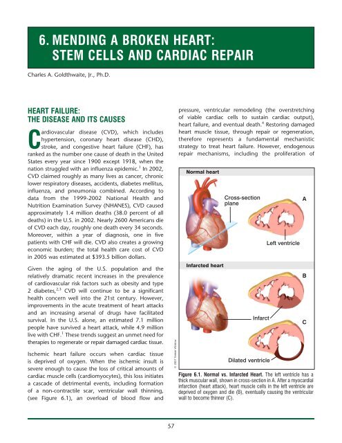
Ischemic <strong>heart</strong> failure occurs when cardiac tissue<br />
is deprived of oxygen. When the ischemic insult is<br />
severe enough to cause the loss of critical amounts of<br />
cardiac muscle cells (cardiomyocytes), this loss initiates<br />
a cascade of detrimental events, including formation<br />
of a non-contractile scar, ventricular wall thinning,<br />
(see Figure <strong>6.</strong>1), an overload of blood flow and<br />
57<br />
© 2007 Terese Winslow<br />
pressure, ventricular remodeling (the overstretching<br />
of viable cardiac cells to sustain cardiac output),<br />
<strong>heart</strong> failure, and eventual death. 4 Restoring damaged<br />
<strong>heart</strong> muscle tissue, through repair or regeneration,<br />
therefore represents a fundamental mechanistic<br />
strategy to treat <strong>heart</strong> failure. However, endogenous<br />
repair mechanisms, including the proliferation of<br />
Normal <strong>heart</strong><br />
Infarcted <strong>heart</strong><br />
Cross-section<br />
plane<br />
Infarct<br />
Dilated ventricle<br />
Left ventricle<br />
Figure <strong>6.</strong>1. normal vs. infarcted Heart. The left ventricle has a<br />
thick muscular wall, shown in cross-section in A. After a myocardial<br />
infarction (<strong>heart</strong> attack), <strong>heart</strong> muscle cells in the left ventricle are<br />
deprived of oxygen and die (B), eventually causing the ventricular<br />
wall to become thinner (C).<br />
A<br />
B<br />
C
Mending a Broken Heart: <strong>Stem</strong> <strong>Cell</strong>s and Cardiac Repair<br />
cardiomyocytes under conditions of severe blood<br />
vessel stress or vessel formation and tissue generation<br />
via the migration of bone-marrow-derived stem cells<br />
to the site of damage, are in themselves insufficient<br />
to restore lost <strong>heart</strong> muscle tissue (myocardium) or<br />
cardiac function. 5 Current pharmacologic interventions<br />
for <strong>heart</strong> disease, including beta-blockers, diuretics,<br />
and angiotensin-converting enzyme (ACE) inhibitors,<br />
and surgical treatment options, such as changing the<br />
shape of the left ventricle and implanting assistive<br />
devices such as pacemakers or defibrillators, do not<br />
restore function to damaged tissue. Moreover, while<br />
implantation of mechanical ventricular assist devices<br />
can provide long-term improvement in <strong>heart</strong> function,<br />
complications such as infection and blood clots remain<br />
problematic. 6 Although <strong>heart</strong> transplantation offers<br />
a viable option to replace damaged myocardium in<br />
selected individuals, organ availability and transplant<br />
rejection complications limit the widespread practical<br />
use of this approach.<br />
The difficulty in regenerating damaged myocardial<br />
tissue has led researchers to explore the application<br />
of embryonic and adult-derived stem cells for cardiac<br />
repair. A number of stem cell types, including embryonic<br />
stem (ES) cells, cardiac stem cells that naturally reside<br />
within the <strong>heart</strong>, myoblasts (muscle stem cells), adult<br />
bone marrow-derived cells, mesenchymal cells (bone<br />
marrow-derived cells that give rise to tissues such as<br />
muscle, bone, tendons, ligaments, and adipose tissue),<br />
endothelial progenitor cells (cells that give rise to the<br />
endothelium, the interior lining of blood vessels), and<br />
umbilical cord blood cells, have been investigated to<br />
varying extents as possible sources for regenerating<br />
damaged myocardium. All have been tested in mouse<br />
or rat models, and some have been tested in large<br />
animal models such as pigs. Preliminary clinical data<br />
for many of these cell types have also been gathered in<br />
selected patient populations.<br />
However, clinical trials to date using stem cells to<br />
repair damaged cardiac tissue vary in terms of the<br />
condition being treated, the method of cell delivery,<br />
and the primary outcome measured by the study,<br />
thus hampering direct comparisons between trials. 7<br />
Some patients who have received stem cells for<br />
myocardial repair have reduced cardiac blood flow<br />
(myocardial ischemia), while others have more<br />
pronounced congestive <strong>heart</strong> failure and still others<br />
are recovering from <strong>heart</strong> attacks. In some cases, the<br />
patient’s underlying condition influences the way that<br />
58<br />
the stem cells are delivered to his/her <strong>heart</strong> (see the<br />
section, “Methods of <strong>Cell</strong> Delivery” for details). Even<br />
among patients undergoing comparable procedures,<br />
the clinical study design can affect the reporting of<br />
results. Some studies have focused on safety issues<br />
and adverse effects of the transplantation procedures;<br />
others have assessed improvements in ventricular<br />
function or the delivery of arterial blood. Furthermore,<br />
no published trial has directly compared two or more<br />
stem cell types, and the transplanted cells may be<br />
autologous (i.e., derived from the person on whom<br />
they are used) or allogeneic (i.e., originating from<br />
another person) in origin. Finally, most of these trials<br />
use unlabeled cells, making it difficult for investigators<br />
to follow the cells’ course through the body after<br />
transplantation (see the section “Considerations for<br />
Using These <strong>Stem</strong> <strong>Cell</strong>s in the Clinical Setting” at the<br />
end of this article for more details).<br />
Despite the relative infancy of this field, initial results<br />
from the application of stem cells to restore cardiac<br />
function have been promising. This article will review<br />
the research supporting each of the aforementioned<br />
cell types as potential source materials for myocardial<br />
regeneration and will conclude with a discussion of<br />
general issues that relate to their clinical application.<br />
MeCHanisMs oF aCtion<br />
In 2001, Menasche, et.al. described the successful<br />
implantation of autologous skeletal myoblasts (cells<br />
that divide to repair and/or increase the size of<br />
voluntary muscles) into the post-infarction scar of<br />
a patient with severe ischemic <strong>heart</strong> failure who<br />
was undergoing coronary artery bypass surgery. 8<br />
Following the procedure, the researchers used imaging<br />
techniques to observe the <strong>heart</strong>’s muscular wall and to<br />
assess its ability to beat. When they examined patients<br />
5 months after treatment, they concluded that treated<br />
<strong>heart</strong>s pumped blood more efficiently and seemed to<br />
demonstrate improved tissue health. This case study<br />
suggested that stem cells may represent a viable<br />
resource for treating ischemic <strong>heart</strong> failure, spawning<br />
several dozen clinical studies of stem cell therapy for<br />
cardiac repair (see Boyle, et.al. 7 for a complete list)<br />
and inspiring the development of Phase I and Phase II<br />
clinical trials. These trials have revealed the complexity<br />
of using stem cells for cardiac repair, and considerations<br />
for using stem cells in the clinical setting are discussed<br />
in a subsequent section of this report.
The mechanism by which stem cells promote cardiac<br />
repair remains controversial, and it is likely that the cells<br />
regenerate myocardium through several pathways.<br />
Initially, scientists believed that transplanted cells<br />
differentiated into cardiac cells, blood vessels, or other<br />
cells damaged by CVD. 9-11 However, this model has<br />
been recently supplanted by the idea that transplanted<br />
stem cells release growth factors and other molecules<br />
that promote blood vessel formation (angiogenesis)<br />
or stimulate “resident” cardiac stem cells to repair<br />
damage. 12-14 Additional mechanisms for stem-cell<br />
mediated <strong>heart</strong> repair, including strengthening of the<br />
post-infarct scar 15 and the fusion of donor cells with<br />
host cardiomyocytes, 16 have also been proposed.<br />
MetHoDs oF <strong>Cell</strong> Delivery<br />
Regardless of which mechanism(s) will ultimately prove<br />
to be the most significant in stem-cell mediated<br />
cardiac repair, cells must be successfully delivered to<br />
the site of injury to maximize the restored function.<br />
In preliminary clinical studies, researchers have used<br />
several approaches to deliver stem cells. Common<br />
approaches include intravenous injection and direct<br />
infusion into the coronary arteries. These methods<br />
can be used in patients whose blood flow has been<br />
restored to their <strong>heart</strong>s after a <strong>heart</strong> attack, provided<br />
that they do not have additional cardiac dysfunction<br />
12, 17<br />
that results in total occlusion or poor arterial flow.<br />
Of these two methods, intracoronary infusion offers<br />
the advantage of directed local delivery, thereby<br />
increasing the number of cells that reach the target<br />
tissue relative to the number that will home to the<br />
<strong>heart</strong> once they have been placed in the circulation.<br />
However, these strategies may be of limited benefit<br />
to those who have poor circulation, and stem cells<br />
are often injected directly into the ventricular wall of<br />
these patients. This endomyocardial injection may be<br />
carried out either via a catheter or during open-<strong>heart</strong><br />
surgery. 18<br />
To determine the ideal site to inject stem cells, doctors<br />
use mapping or direct visualization to identify the<br />
locations of scars and viable cardiac tissue. Despite<br />
improvements in delivery efficiency, however, the<br />
success of these methods remains limited by the<br />
death of the transplanted cells; as many as 90% of<br />
transplanted cells die shortly after implantation as a<br />
result of physical stress, myocardial inflammation, and<br />
myocardial hypoxia. 4 Timing of delivery may slow the<br />
Mending a Broken Heart: <strong>Stem</strong> <strong>Cell</strong>s and Cardiac Repair<br />
59<br />
rate of deterioration of tissue function, although this<br />
issue remains a hurdle for therapeutic approaches.<br />
types oF steM <strong>Cell</strong>s investigateD<br />
to regenerate DaMageD<br />
MyoCarDial tissue<br />
Embryonic and adult stem cells have been investigated<br />
to regenerate damaged myocardial tissue in animal<br />
models and in a limited number of clinical studies. A<br />
brief review of work to date and specific considerations<br />
for the application of various cell types will be discussed<br />
in the following sections.<br />
Embryonic <strong>Stem</strong> (ES) <strong>Cell</strong>s<br />
Because ES cells are pluripotent, they can potentially<br />
give rise to the variety of cell types that are instrumental<br />
in regenerating damaged myocardium, including<br />
cardiomyocytes, endothelial cells, and smooth muscle<br />
cells. To this end, mouse and human ES cells have been<br />
shown to differentiate spontaneously to form endothelial<br />
and smooth muscle cells in vitro 19 and in vivo, 20,21 and<br />
human ES cells differentiate into myocytes with the<br />
structural and functional properties of cardiomyocytes. 22-<br />
24 Moreover, ES cells that were transplanted into<br />
ischemically-injured myocardium in rats differentiated<br />
into normal myocardial cells that remained viable for<br />
up to four months, 25 suggesting that these cells may be<br />
candidates for regenerative therapy in humans.<br />
However, several key hurdles must be overcome before<br />
human ES cells can be used for clinical applications.<br />
Foremost, ethical issues related to embryo access<br />
currently limit the avenues of investigation. In addition,<br />
human ES cells must go through rigorous testing and<br />
purification procedures before the cells can be used<br />
as sources to regenerate tissue. First, researchers must<br />
verify that their putative ES cells are pluripotent. To<br />
prove that they have established a human ES cell line,<br />
researchers inject the cells into immunocompromised<br />
mice; i.e., mice that have a dysfunctional immune<br />
system. Because the injected cells cannot be destroyed<br />
by the mouse’s immune system, they survive and<br />
proliferate. Under these conditions, pluripotent cells<br />
will form a teratoma, a multi-layered, benign tumor<br />
that contains cells derived from all three embryonic<br />
germ layers. Teratoma formation indicates that the<br />
stem cells have the capacity to give rise to all cell types<br />
in the body.
Mending a Broken Heart: <strong>Stem</strong> <strong>Cell</strong>s and Cardiac Repair<br />
The pluripotency of ES cells can complicate their<br />
clinical application. While undifferentiated ES cells may<br />
possibly serve as sources of specific cell populations<br />
used in myocardial repair, it is essential that tight<br />
quality control be maintained with respect to the<br />
differentiated cells. Any differentiated cells that would<br />
be used to regenerate <strong>heart</strong> tissue must be purified<br />
before transplantation can be considered. If injected<br />
regenerative cells are accidentally contaminated with<br />
undifferentiated ES cells, a tumor could possibly form<br />
as a result of the cell transplant. 4 However, purification<br />
methodologies continue to improve; one recent report<br />
describes a method to identify and select cardiomyocytes<br />
during human ES cell differentiation that may<br />
make these cells a viable option in the future. 26<br />
This concern illustrates the scientific challenges that<br />
accompany the use of all human stem cells, whether<br />
derived from embryonic or adult tissues. Predictable<br />
control of cell proliferation and differentiation requires<br />
additional basic research on the molecular and genetic<br />
signals that regulate cell division and specialization.<br />
Furthermore, long-term cell stability must be well<br />
understood before human ES-derived cells can be used<br />
in regenerative medicine. The propensity for genetic<br />
mutation in the human ES cells must be determined, and<br />
the survival of differentiated, ES-derived cells following<br />
transplantation must be assessed. Furthermore, once<br />
cells have been transplanted, undesirable interactions<br />
between the host tissue and the injected cells must<br />
be minimized. <strong>Cell</strong>s or tissues derived from ES cells<br />
that are currently available for use in humans are not<br />
tissue-matched to patients and thus would require<br />
immunosuppression to limit immune rejection. 18<br />
Skeletal Myoblasts<br />
While skeletal myoblasts (SMs) are committed progenitors<br />
of skeletal muscle cells, their autologous origin,<br />
high proliferative potential, commitment to a myogenic<br />
lineage, and resistance to ischemia promoted their use<br />
as the first stem cell type to be explored extensively<br />
for cardiac application. Studies in rats and humans<br />
have demonstrated that these cells can repopulate<br />
scar tissue and improve left ventricular function<br />
following transplantation. 27 However, SM-derived<br />
cardiomyocytes do not function in complete concert<br />
with native myocardium. The expression of two key<br />
proteins involved in electromechanical cell integration,<br />
N-cadherin and connexin 43, are downregulated in<br />
vivo, 28 and the engrafted cells develop a contractile<br />
60<br />
activity phenotype that appears to be unaffected by<br />
neighboring cardiomyocytes. 29<br />
To date, the safety and feasibility of transplanting SM<br />
cells have been explored in a series of small studies<br />
enrolling a collective total of nearly 100 patients. Most<br />
of these procedures were carried out during open-<strong>heart</strong><br />
surgery, although a couple of studies have investigated<br />
direct myocardial injection and transcoronary<br />
administration. Sustained ventricular tachycardia, a<br />
life-threatening arrhythmia and unexpected side-effect,<br />
occurred in early implantation studies, possibly resulting<br />
from the lack of electrical coupling between SM-derived<br />
cardiomyocytes and native tissue. 30,31 Changes in preimplantation<br />
protocols have minimized the occurrence<br />
of arrhythmias in conjunction with the use of SM cells,<br />
and Phase II studies of skeletal myoblast therapy are<br />
presently underway.<br />
Human Adult Bone-Marrow Derived <strong>Cell</strong>s<br />
In 2001, Jackson, et.al. demonstrated that cardiomyocytes<br />
and endothelial cells could be regenerated in a<br />
mouse <strong>heart</strong> attack model through the introduction<br />
of adult mouse bone marrow-derived stem cells. 9 That<br />
same year, Orlic and colleagues showed that direct<br />
injection of mouse bone marrow-derived cells into the<br />
damaged ventricular wall following an induced <strong>heart</strong><br />
attack led to the formation of new cardiomyocytes,<br />
vascular endothelium, and smooth muscle cells. 11 Nine<br />
days after transplanting the stem cells, the newlyformed<br />
myocardium occupied nearly 70 percent of<br />
the damaged portion of the ventricle, and survival<br />
rates were greater in mice that received these cells<br />
than in those that did not. While several subsequent<br />
studies have questioned whether these cells actually<br />
differentiate into cardiomyocytes, 32,33 the evidence to<br />
support their ability to prevent remodeling has been<br />
demonstrated in many laboratories. 7<br />
Based on these findings, researchers have investigated<br />
the potential of human adult bone marrow as a source of<br />
stem cells for cardiac repair. Adult bone marrow contains<br />
several stem cell populations, including hematopoietic<br />
stem cells (which differentiate into all of the cellular<br />
components of blood), endothelial progenitor cells, and<br />
mesenchymal stem cells; successful application of these<br />
cells usually necessitates isolating a particular cell type<br />
on the basis of its’ unique cell-surface receptors. In the<br />
past three years, the transplantation of bone marrow<br />
mononuclear cells (BMMNCs), a mixed population of<br />
blood and cells that includes stem and progenitor cells,
has been explored in more patients and clinical studies<br />
of cardiac repair than any other type of stem cell.7<br />
The results from clinical studies of BMMNC transplantation<br />
have been promising but mixed. However, it should be<br />
noted that these studies have been conducted under<br />
a variety of conditions, thereby hampering direct<br />
comparison. The cells have been delivered via open<strong>heart</strong><br />
surgery and endomyocardial and intracoronary<br />
catheterization. Several studies, including the Bone<br />
Marrow Transfer to Enhance ST-Elevation Infarct<br />
Regeneration (BOOST) and the Transplantation of<br />
Progenitor <strong>Cell</strong>s and Regeneration Enhancement in<br />
Acute Myocardial Infarction (TOPCARE-AMI) trials,<br />
have shown that intracoronary infusion of BMMNCs<br />
following a <strong>heart</strong> attack significantly improves the left<br />
ventricular (LV) ejection fraction, or the volume of blood<br />
pumped out of the left ventricle with each <strong>heart</strong>beat. 34-<br />
36 However, other studies have indicated either no<br />
improvement in LV ejection fraction upon treatment 37 or<br />
an increased LV ejection fraction in the control group. 38<br />
An early study that used endomyocardial injection<br />
to enhance targeted delivery indicated a significant<br />
improvement in overall LV function. 39 Discrepancies<br />
such as these may reflect differences in cell preparation<br />
protocols or baseline patient statistics. As larger trials<br />
are developed, these issues can be explored more<br />
systematically.<br />
Mesenchymal (Bone Marrow Stromal) <strong>Cell</strong>s<br />
Mesenchymal stem cells (MSCs) are precursors<br />
of non-hematopoietic tissues (e.g., muscle, bone,<br />
tendons, ligaments, adipose tissue, and fibroblasts)<br />
that are obtained relatively easily from autologous bone<br />
marrow. They remain multipotent following expansion<br />
in vitro, exhibit relatively low immunogenicity,<br />
and can be frozen easily. While these properties<br />
make the cells amenable to preparation and delivery<br />
protocols, scientists can also culture them under<br />
special conditions to differentiate them into cells that<br />
resemble cardiac myocytes. This property enables their<br />
application to cardiac regeneration. MSCs differentiate<br />
into endothelial cells when cultured with vascular<br />
endothelial growth factor 40 and cardiomyogenic (CMG)<br />
cells when treated with the DNA-demethylating<br />
agent, 5-azacytidine.41 More important, however,<br />
is the observation that MSCs can differentiate into<br />
cardiomyocytes and endothelial cells in vivo when<br />
transplanted to the <strong>heart</strong> following myocardial infarct<br />
(MI) or non-injury in pig, mouse, or rat models. 42-45<br />
Mending a Broken Heart: <strong>Stem</strong> <strong>Cell</strong>s and Cardiac Repair<br />
61<br />
Additionally, the ability of MSCs to restore functionality<br />
may be enhanced by the simultaneous transplantation<br />
of other stem cell types. 43<br />
Several animal model studies have shown that treatment<br />
with MSCs significantly increases myocardial<br />
function and capillary formation. 5,41 One advantage<br />
of using these cells in human studies is their low<br />
immunogenicity; allogeneic MSCs injected into<br />
infarcted myocardium in a pig model regenerated myocardium<br />
and reduced infarct size without evidence of<br />
rejection. 46 A randomized clinical trial implanting MSCs<br />
after MI has demonstrated significant improvement in<br />
global and regional LV function, 47 and clinical trials<br />
are currently underway to investigate the application<br />
of allogeneic and autologous MSCs for acute MI and<br />
myocardial ischemia, respectively.<br />
Resident Cardiac <strong>Stem</strong> <strong>Cell</strong>s<br />
Recent evidence suggests that the <strong>heart</strong> contains a<br />
small population of endogenous stem cells that most<br />
likely facilitate minor repair and turnover-mediated<br />
cell replacement. 7 These cells have been isolated and<br />
characterized in mouse, rat, and human tissues. 48,49<br />
The cells can be harvested in limited quantity from<br />
human endomyocardial biopsy specimens 50 and can<br />
be injected into the site of infarction to promote<br />
cardiomyocyte formation and improvements in systolic<br />
function. 49 Separation and expansion ex vivo over<br />
a period of weeks are necessary to obtain sufficient<br />
quantities of these cells for experimental purposes.<br />
However, their potential as a convenient resource for<br />
autologous stem cell therapy has led the National<br />
Heart, Lung, and Blood Institute to fund forthcoming<br />
clinical trials that will explore the use of cardiac stem<br />
cells for myocardial regeneration.<br />
Endothelial Progenitor <strong>Cell</strong>s<br />
The endothelium is a layer of specialized cells that<br />
lines the interior surface of all blood vessels (including<br />
the <strong>heart</strong>). This layer provides an interface between<br />
circulating blood and the vessel wall. Endothelial<br />
progenitor cells (EPCs) are bone marrow-derived stem<br />
cells that are recruited into the peripheral blood in<br />
response to tissue ischemia. 4 EPCs are precursor cells<br />
that express some cell-surface markers characteristic<br />
of mature endothelium and some of hematopoietic<br />
cells. 19,51-53 EPCs home in on ischemic areas, where<br />
they differentiate into new blood vessels; following<br />
a <strong>heart</strong> attack, intravenously injected EPCs home
Mending a Broken Heart: <strong>Stem</strong> <strong>Cell</strong>s and Cardiac Repair<br />
to the damaged region within 48 hours. 12 The new<br />
vascularization induced by these cells prevents cardiomyocyte<br />
apoptosis (programmed cell death) and LV<br />
remodeling, thereby preserving ventricular function. 13<br />
However, no change has been observed in non-infarcted<br />
regions upon EPC administration. Clinical trials are<br />
currently underway to assess EPC therapy for growing<br />
new blood vessels and regenerating myocardium.<br />
Other <strong>Cell</strong>s: Umbilical Cord Blood <strong>Stem</strong> <strong>Cell</strong>s,<br />
Fibroblasts, and Peripheral Blood CD34 + <strong>Cell</strong>s<br />
Several other cell populations, including umbilical<br />
cord blood (UCB) stem cells, fibroblasts (cells that<br />
synthesize the extracellular matrix of connective tissues),<br />
and peripheral blood CD34 + cells, have potential<br />
therapeutic uses for regenerating cardiac tissue.<br />
Although these cell types have not been investigated<br />
in clinical trials of <strong>heart</strong> disease, preliminary studies in<br />
animal models indicate several potential applications<br />
in humans.<br />
Umbilical cord blood contains enriched populations of<br />
hematopoietic stem cells and mesencyhmal precursor<br />
cells relative to the quantities present in adult blood or<br />
bone marrow. 54,55 When injected intravenously into the<br />
tail vein in a mouse model of MI, human mononuclear<br />
UCB cells formed new blood vessels in the infarcted<br />
<strong>heart</strong>. 56 A human DNA assay was used to determine the<br />
migration pattern of the cells after injection; although<br />
they homed only to injured areas within the <strong>heart</strong>, they<br />
were also detected in the marrow, spleen, and liver.<br />
When injected directly into the infarcted area in a rat<br />
model of MI, human mononuclear UCB cells improved<br />
ventricular function. 57 Staining for CD34 and other<br />
markers found on the cell surface of hematopoietic<br />
stem cells indicated that some of the cells survived in<br />
the myocardium. Results similar to these have been<br />
observed following the injection of human unrestricted<br />
somatic stem cells from UCB into a pig MI model. 58<br />
Adult peripheral blood CD34 + cells offer the advantage<br />
of being obtained relatively easily from autologous<br />
sources. 59 Although some studies using a mouse model<br />
of MI claim that these cells can transdifferentiate into<br />
cardiomyocytes, endothelial cells, and smooth muscle<br />
cells at the site of tissue injury, 60 this conclusion is<br />
highly contested. Recent studies that involve the<br />
direct injection of blood-borne or bone marrowderived<br />
hematopoietic stem cells into the infarcted<br />
region of a mouse model of MI found no evidence of<br />
myocardial regeneration following injection of either<br />
62<br />
cell type. 33 Instead, these hematopoietic stem cells<br />
followed traditional differentiation patterns into blood<br />
cells within the microenvironment of the injured <strong>heart</strong>.<br />
Whether these cells will ultimately find application in<br />
myocardial regeneration remains to be determined.<br />
Autologous fibroblasts offer a different strategy to<br />
combat myocardial damage by replacing scar tissue<br />
with a more elastic, muscle-like tissue and inhibiting<br />
host matrix degradation. 4 The cells may be manipulated<br />
to express muscle-specific transcription factors that<br />
promote their differentiation into myotubes such as<br />
those derived from skeletal myoblasts. 61 One month<br />
after these cells were implanted into the post-infarction<br />
scar in a rat model of MI, they occupied a large portion<br />
of the scar but were not functionally integrated. 61<br />
Although the effects on ventricular function were not<br />
evaluated in this study, authors noted that modified<br />
autologous fibroblasts may ultimately prove useful in<br />
elderly patients who have a limited population of autologous<br />
skeletal myoblasts or bone marrow stem cells.<br />
ConsiDerations For using tHese<br />
steM <strong>Cell</strong>s in tHe CliniCal setting<br />
As these examples indicate, many types of stem<br />
cells have been applied to regenerate damaged<br />
myocardium. In select applications, stem cells have<br />
demonstrated sufficient promise to warrant further<br />
exploration in large-scale, controlled clinical trials.<br />
However, the current breadth of application of these<br />
cells has made it difficult to compare and contextualize<br />
the results generated by the various trials. Most<br />
studies published to date have enrolled fewer than<br />
25 patients, and the studies vary in terms of cell types<br />
and preparations used, methods of delivery, patient<br />
populations, and trial outcomes. However, the mixed<br />
results that have been observed in these studies do not<br />
necessarily argue against using stem cells for cardiac<br />
repair. Rather, preliminary results illuminate the many<br />
gaps in understanding of the mechanisms by which<br />
these cells regenerate myocardial tissue and argue<br />
for improved characterization of cell preparations and<br />
delivery methods to support clinical applications.<br />
Future clinical trials that use stem cells for myocardial<br />
repair must address two concerns that accompany<br />
the delivery of these cells: 1) safety and 2) tracking<br />
the cells to their ultimate destination(s). Although<br />
stem cells appear to be relatively safe in the majority<br />
of recipients to date, an increased frequency of non
sustained ventricular tachycardia, an arrhythmia, has<br />
been reported in conjunction with the use of skeletal<br />
myoblasts. 30,62-64 While this proarrhythmic effect occurs<br />
relatively early after cell delivery and does not appear<br />
to be permanent, its presence highlights the need for<br />
careful safety monitoring when these cells are used.<br />
Additionally, animal models have demonstrated that<br />
stem cells rapidly diffuse from the <strong>heart</strong> to other<br />
organs (e.g., lungs, kidneys, liver, spleen) within a<br />
few hours of transplantation, 65,66 an effect observed<br />
regardless of whether the cells are injected locally<br />
into the myocardium. This migration may or may not<br />
cause side-effects in patients; however, it remains a<br />
concern related to the delivery of stem cells in humans.<br />
(Note: Techniques to label stem cells for tracking<br />
purposes and to assess their safety are discussed in<br />
more detail in other articles in this publication).<br />
In addition to safety and tracking, several logistical issues<br />
must also be addressed before stem cells can be used<br />
routinely in the clinic. While cell tracking methodologies<br />
allow researchers to determine migration patterns, the<br />
stem cells must target their desired destination(s) and be<br />
retained there for a sufficient amount of time to achieve<br />
benefit. To facilitate targeting and enable clinical use,<br />
stem cells must be delivered easily and efficiently to<br />
their sites of application. Finally, the ease by which the<br />
cells can be obtained and the cost of cell preparation<br />
will also influence their transition to the clinic.<br />
ConClusions<br />
The evidence to date suggests that stem cells<br />
hold promise as a therapy to regenerate damaged<br />
myocardium. Given the worldwide prevalence of<br />
cardiac dysfunction and the limited availability of<br />
tissue for cardiac transplantation, stem cells could<br />
ultimately fulfill a large-scale unmet clinical need and<br />
improve the quality of life for millions of people with<br />
CVD. However, the use of these cells in this setting is<br />
currently in its infancy — much remains to be learned<br />
about the mechanisms by which stem cells repair and<br />
regenerate myocardium, the optimal cell types and<br />
modes of their delivery, and the safety issues that<br />
will accompany their use. As the results of large-scale<br />
clinical trials become available, researchers will begin<br />
to identify ways to standardize and optimize the use of<br />
these cells, thereby providing clinicians with powerful<br />
tools to mend a <strong>broken</strong> <strong>heart</strong>.<br />
Mending a Broken Heart: <strong>Stem</strong> <strong>Cell</strong>s and Cardiac Repair<br />
63<br />
reFerenCes<br />
1. American Heart Association. Heart Disease and Stroke<br />
Statistics–2005. Dallas: American Heart Association; 2005.<br />
2. Mokdad AH, Ford ES, Bowman BA, et al. Prevalence of<br />
obesity, diabetes, and obesity-related health-risk factors,<br />
2001. JAMA. 2003;289:76-79.<br />
3. Flegal KM, Carroll MD, Ogden CL, Johnson CL. Prevalence<br />
and trends in obesity among US adults, 1999-2000. JAMA.<br />
2002;288:1723-1727.<br />
4. Rosenstrauch D, Poglajen G, Zidar N, Gregoric ID. <strong>Stem</strong><br />
cell therapy for ischemic <strong>heart</strong> failure. Tex Heart Ist J.<br />
2005;32:339-347.<br />
5. Mangi AA, Noiseux N, Kong D, et al. Mesenchymal<br />
stem cells modified with Akt prevent remodeling and<br />
restore performance of infarcted <strong>heart</strong>s. Nat Med.<br />
2003;9:1195-1201.<br />
<strong>6.</strong> Lietz K, Long JW, Kfoury AG, et al. Outcomes of left<br />
ventricular assist device implantation as destination therapy<br />
in the post-REMATCH era: implications for patient selection.<br />
Circulation. 2007;116:497-505.<br />
7. Boyle AJ, Schulman SP, Hare JM. Is stem cell therapy ready<br />
for patients? <strong>Stem</strong> cell therapy for cardiac repair. Circulation.<br />
2006;114:339-352.<br />
8. Menasche P, Hagege AA, Scorsin M, et al. Myoblast<br />
transplantation for <strong>heart</strong> failure. Lancet. 2001;357:279-280.<br />
9. Jackson KA, Majka SM, Wang H, et al. Regeneration of<br />
ischemic cardiac muscle and vascular endothelium by adult<br />
stem cells. J Clin Invest. 2001;107:1395-1402.<br />
10. Condorelli G, Borello U, De Angelis L, et al. Cardiomyocytes<br />
induce endothelial cells to transdifferentiate into cardiac<br />
muscle: implications for myocardium regeneration. Proc<br />
Natl Acad Sci USA. 2001;98:10733-10738.<br />
11. Orlic D, Kajstura J, Chimenti S, et al. Bone marrow cells<br />
regenerate infarcted myocardium. Nature. 2001;<br />
410:701-705.<br />
12. Kocher AA, Schuster MD, Szaboles MJ, et al.<br />
Neovascularization of ischemic myocardium by human<br />
bone-marrow derived angioblasts prevents cardiomyocyte<br />
apoptosis, reduces remodelling and improves cardiac<br />
function. Nat Med. 2001;7:430-43<strong>6.</strong><br />
13. Schuster MD, Kocher AA, Seki T, et al. Myocardial<br />
neovascularization by bone marrow angioblasts results<br />
in cardiomyocyte regeneration. Am J Physiol Heart Circ<br />
Physiol. 2004;287:H525-H532.<br />
14. Gnecchi M, He H, Liang OD, et al. Paracrine action<br />
accounts for marked protection of ischemic <strong>heart</strong><br />
by Akt-modified mesenchymal stem cells. Nat Med.<br />
2005;11:367-368.<br />
15. Fujii T, Yau TM, Weisel RD, et al. <strong>Cell</strong> transplantation to<br />
prevent <strong>heart</strong> failure: a comparison of cell types. Ann Thorac<br />
Surg. 2003;76:2062-2070.
Mending a Broken Heart: <strong>Stem</strong> <strong>Cell</strong>s and Cardiac Repair<br />
1<strong>6.</strong> Nygren JM, Jovinge S, Breitbach M, et al. Bone marrowderived<br />
hematopoietic cells generate cardiomyocytes at low<br />
frequency through cell fusion, but not transdifferentiation.<br />
Nat Med. 2004;10:494-501.<br />
17. Strauer BE, Brehm M, Zeus T, et al. Repair of infarcted<br />
myocardium by autologous intracoronary mononuclear<br />
bone marrow cell transplantation in humans. Circulation.<br />
2002;106:1913-1918.<br />
18. Oettgen P. Cardiac stem cell therapy: need for optimization<br />
of efficacy and safety monitoring. Circulation.<br />
2006;114:353-358.<br />
19. Vittet D, Prandidni MH, Berthier R, et al. Embryonic stem<br />
cells differentiate in vitro to endothelial cells through<br />
successive maturation steps. Blood. 1996;88:3424-3431.<br />
20. Marchetti S, Gimond C, Iljin K, et al. Endothelial cells<br />
genetically selected from differentiating mouse embryonic<br />
stem cells incorporate at sites of neovascularization in vivo.<br />
J <strong>Cell</strong> Sci. 2002;115:2075-2085.<br />
21. Yamashita J, Itoh H, Hirashima M, et al. Flk1-positive<br />
cells derived from embryonic stem cells serve as vascular<br />
progenitors. Nature. 2000;408:92-9<strong>6.</strong><br />
22. Kehat I, Kenyagin-Karsenti D, Snir M, et al. Human<br />
embryonic stem cells can differentiate into myocytes with<br />
structural and functional properties of cardiomyocytes.<br />
J Clin Invest. 2001;108:407-414.<br />
23. Kehat I, Gepstein A, Spira A, Itskovitz-Eldor J, Gepstein L.<br />
High-resolution electrophysiological assessment of human<br />
embryonic stem cell-derived cardiomyocytes: a novel<br />
in vitro model for the study of conduction. Circ Res.<br />
2002;91:659-661.<br />
24. Westfall MV, Pasyk KA, Yule DI, Samuelson LC, Metzger JM.<br />
Ultrastructure and cell-cell coupling of cardiac myocytes<br />
differentiating in embryonic stem cell cultures. <strong>Cell</strong> Motil<br />
Cytoskel. 1998;36:43-54.<br />
25. Min JY, Yang Y, Converso KL, et al. Transplantation<br />
of embryonic stem cells improves cardiac function in<br />
postinfarcted rats. J Appl Physiol. 2002;92:288-29<strong>6.</strong><br />
2<strong>6.</strong> Huber I, Itzhaki I, Caspi O, et al. Identification and selection<br />
of cardiomyocytes during human embryonic stem cell<br />
differentiation. FASEB J. 2007;21:2551-2563.<br />
27. Dowell JD, Rubart M, Pasumarthi KB, Soonpaa MH, Field<br />
LJ. Myocyte and myogenic stem cell transplantation in the<br />
<strong>heart</strong>. Cardiovasc Res. 2003;58:336-350.<br />
28. Reinecke H, MacDonald GH, Hauschka SD, Murry<br />
CE. Electromechanical coupling between skeletal and<br />
cardiac muscle. Implications for infarct repair. J <strong>Cell</strong> Biol.<br />
2000;149:731-740.<br />
29. Leobon B, Garcin I, Menasche P, Vilquin JT, Audinat E,<br />
Charpak S. Myoblasts transplanted into rat infarcted<br />
myocardium are functionally isolated from their host.<br />
Proc Natl Acad Sci USA. 2003;100:7808-7811.<br />
64<br />
30. Menasche P, Hagege AA, Vilquin J-T, et al. Autologous<br />
skeletal myoblast transplantation for severe postinfarction<br />
left ventricular dysfunction. J Am Coll Cardiol.<br />
2003;41:1078-1083.<br />
31. Siminiak T, Kalawski R, Fiszer D, et al. Autologous skeletal<br />
myoblast transplantation for the treatment of postinfarction<br />
myocardial injury: phase I clinical study with 12<br />
months of follow-up. Am Heart J. 2004;148:531-537.<br />
32. Murry CE, Soonpaa MH, Reinecke H, et al. Haematopoietic<br />
stem cells do not transdifferentiate into cardiac myocytes<br />
in myocardial infarcts. Nature. 2004;428:664-668.<br />
33. Balsam LB, Wagers AJ, Christensen JL, Kofidis T, Weissman<br />
IL, Robbins RC. Haematopoietic stem cells adopt mature<br />
haematopoietic fates in ischaemic myocardium. Nature.<br />
2004;428:668-673.<br />
34. Assmus B, Schachinger V, Teupe C, et al. Transplantation<br />
of Progenitor <strong>Cell</strong>s and Regeneration Enhancement in<br />
Acute Myocardial Infarction (TOPCARE-AMI). Circulation.<br />
2002;106:3009-3017.<br />
35. Schachinger V, Assmus B, Britten MB, et al. Transplantation<br />
of progenitor cells and regeneration enhancement in<br />
acute myocardial infarction: final one-year results of the<br />
TOPCARE-AMI Trial. J Am Coll Cardiol. 2004;44:1690.<br />
3<strong>6.</strong> Wollert KC, Meyer GP, Lotz J, et al. Intra-coronary<br />
autologous bone-marrow cell transfer after myocardial<br />
infarction: the BOOST randomised controlled clinical trial.<br />
Lancet. 2004;364:141-148.<br />
37. Janssens S, Dubois C, Bogaert J, et al. Autologous bone<br />
marrow-derived stem-cell transfer in patients with<br />
ST-segment elevation myocardial infarction: double-blind,<br />
randomised, controlled trial. Lancet. 2005;367:113-121.<br />
38. Cleland JG, Freemantle N, Coletta AP, Clark AL. Clinical<br />
trials update from the American Heart Association:<br />
REPAIR-AMI, ASTAMI, JELIS, MEGA, REVIVE-II, SURVIVE,<br />
and PROACTIVE. Eur J Heart Fail. 2006;8:105-110.<br />
39. Perin EC, Dohmann HFR, Borojevic R, et al. Transendocardial,<br />
autologous bone marrow cell transplantation<br />
for severe, chronic ischemic <strong>heart</strong> failure. Circulation.<br />
2003;107:2294-2302.<br />
40. MacKenzie TC, Flake AW. Human mesenchymal stem cells:<br />
insights from a surrogate in vivo assay system. <strong>Cell</strong>s Tissues<br />
Organs. 2002;171:90-95.<br />
41. Davani S, Marandin A, Mersin N, et al. Mesenchymal<br />
progenitor cells differentiate into an endothelial phenotype,<br />
enhance vascular density, and improve <strong>heart</strong> function<br />
in a rat cellular cardiomyoplasty model. Circulation.<br />
2003;108(suppl II):II-253-II-258.<br />
42. Makino S, Fukuda K, Miyoshi S, et al. Cardiomyocytes<br />
can be generated from marrow stromal cells in vitro.<br />
J Clin Invest. 1999;103:697-705.<br />
43. Min J-Y, Sullivan MF, Yang Y, et al. Significant<br />
improvement of <strong>heart</strong> function by cotransplantation of<br />
human mesenchymal stem cells and fetal cardiomyocytes in<br />
postinfarcted pigs. Ann Thorac Surg. 2002;74:1568-1575.
44. Shake JG, Gruber PJ, Baumgartner WA, et al. Mesenchymal<br />
stem cell implantation in a swine myocardial infarct model:<br />
engraftment and functional effects. Ann Thorac Surg.<br />
2002;73:1919-192<strong>6.</strong><br />
45. Toma C, Pittenger MF, Cahill KS, Byrne BJ, Kessler<br />
PD. Human mesenchymal stem cells differentiate to a<br />
cardiomyocyte phenotype in the adult murine <strong>heart</strong>.<br />
Circulation. 2002;105:93-98.<br />
4<strong>6.</strong> Amado LC, Saliaris AP, Schuleri KH, et al. Cardiac repair<br />
with intramyocardial injection of allogeneic mesenchymal<br />
stem cells after myocardial infarction. Proc Natl Acad Sci<br />
USA. 2005;102:11474-11479.<br />
47. Chen S-I, Fang W-W, Ye F, et al. Effect on left ventricular<br />
function of intracoronary transplantation of autologous<br />
bone marrow mesenchymal stem cells in patients with<br />
acute myocardial infarction. Am J Cardiol. 2004;94:92-95.<br />
48. Beltrami AP, Barlucchi L, Torella D, et al. Adult cardiac stem<br />
cells are multipotent and support myocardial regeneration.<br />
<strong>Cell</strong>. 2003;114:763-77<strong>6.</strong><br />
49. Messina E, De Angelis L, Frati G, et al. Isolation and<br />
expansion of adult cardiac stem cells from human and<br />
murine <strong>heart</strong>. Circ Res. 2004;95:911-921.<br />
50. Smith RR, Barile L, Cho HC, et al. Unique phenotype<br />
of cardiospheres derived from human endomyocardial<br />
biopsies. Circulation. 2005;112(suppl II):II-334.<br />
51. Eichmann A, Corbel C, Nataf V, Vaigot P, Breant C, Le<br />
Dourain NM. Ligand-dependent development of the<br />
endothelial and hematopoietic lineages from embryonic<br />
mesodermal cells expressing vascular endothelial growth<br />
factor receptor 2. Proc Natl Acad Sci USA. 1997;<br />
94:5141-514<strong>6.</strong><br />
52. Sato TN, Quin Y, Kozak CA, Audus KL. Tie-1 and Tie-2<br />
define another class of putative receptor tyrosine kinase<br />
genes expressed in early embryonic vascular system.<br />
Proc Natl Acad Sci USA. 1993;90:9355-9358.<br />
53. Suri C, Jones PF, Patan S, et al. Requisite role of<br />
angiopoietin-1, a ligand for the TIE2 receptor, during<br />
embryonic angiogenesis. <strong>Cell</strong>. 1996;87:1171-1180.<br />
54. Erices A, Conget P, Minguell JJ. Mesenchymal progenitor<br />
cells in human umbilical cord blood. Br J Haematol.<br />
2000;109:235-242.<br />
55. Mayani H, Lansdorp PM. Biology of human umbilical cord<br />
blood-derived hematopoietic stem/progenitor cells. <strong>Stem</strong><br />
<strong>Cell</strong>s. 1998;16:153-165.<br />
Mending a Broken Heart: <strong>Stem</strong> <strong>Cell</strong>s and Cardiac Repair<br />
65<br />
5<strong>6.</strong> Ma N, Stamm C, Kaminski A, et al. Human cord blood<br />
cells induce angiogenesis following myocardial infarction<br />
in NOD/scid-mice. Cardiovasc Res. 2005;66:45-54.<br />
57. Hirata Y, Sata M, Motomura N, et al. Human umbilical<br />
cord blood cells improve cardiac function after myocardial<br />
infarction. Biochem Biophys Res Commun. 2005;<br />
327:609-614.<br />
58. Kim B-O, Tian H, Prasongsukarn K, et al. <strong>Cell</strong> transplantation<br />
improves ventricular function after a myocardial infarction:<br />
a preclinical study of human unrestricted somatic stem<br />
cells in a porcine model. Circulation. 2005;<br />
112 (suppl I):I-96-I-104.<br />
59. Korbling M, Katz RL, Khanna A, et al. Hepatocytes and<br />
epithelial cells of donor origin in recipients of peripheralblood<br />
stem cells. N Engl J Med. 2002;346:738-74<strong>6.</strong><br />
60. Yeh ET, Zhang S, Wu HD, Korbling M, Willerson JT, Estrov<br />
Z. Transdifferentiation of human peripheral blood CD34 + -<br />
enriched cell population into cardiomyocytes, endothelial<br />
cells, and smooth muscle cells in vivo. Circulation.<br />
2003;108:2070-2073.<br />
61. Etzion S, Barbash IM, Feinberg MS, et al. <strong>Cell</strong>ular<br />
cardiomyoplasty of cardiac fibroblasts by adenoviral delivery<br />
of MyoD ex vivo: an unlimited source of cells for myocardial<br />
repair. Circulation. 2002;106(12 Suppl 1):I125-I130.<br />
62. Smits PC, van Genus RJ, Poldermans D, et al. Catheterbased<br />
intramyocardial injection of autologous skeletal<br />
myoblasts as a primary treatment of ischemic <strong>heart</strong> failure:<br />
clinical experience with six-month follow-up. J Am Coll<br />
Cardiol. 2003;42:2063-2069.<br />
63. Dib N, McCarthy P, Campbell A, et al. Feasibility and safety<br />
of autologous myoblast transplantation in patients with<br />
ischemic cardiomyopathy. <strong>Cell</strong> Transplant. 2005;14:11-19.<br />
64. Pagani FD, DerSimonian H, Zawadzka A, et al. Autologous<br />
skeletal myoblasts transplanted to ischemia-damaged<br />
myocardium in humans: histological analysis of cell survival<br />
and differentiation. J Am Coll Cardiol. 2003;41:879-888.<br />
65. Dow J, Simkhovich BZ, Kedes L, Kloner RA. Washout<br />
of transplanted cells from the <strong>heart</strong>: a potential new<br />
hurdle for cell transplantation therapy. Cardiovasc Res.<br />
2005;67:301-307.<br />
6<strong>6.</strong> Muller-Ehmsen J, Whittaker P, Kloner RA, et al. Survival<br />
and development of neonatal rat cardiomyocytes<br />
transplanted into adult myocardium. J Mol <strong>Cell</strong> Cardiol.<br />
2002;34:107-11<strong>6.</strong>