Pan-Axonal Neurofilament Marker (SMI 312 ... - Eurogentec
Pan-Axonal Neurofilament Marker (SMI 312 ... - Eurogentec
Pan-Axonal Neurofilament Marker (SMI 312 ... - Eurogentec

Create successful ePaper yourself
Turn your PDF publications into a flip-book with our unique Google optimized e-Paper software.
Description:<br />
Monoclonal Antibody against <strong>Pan</strong>-<strong>Axonal</strong><br />
<strong>Neurofilament</strong> <strong>Marker</strong><br />
Intended Use:<br />
**Research Use Only (RUO)**<br />
This product is sold for laboratory<br />
research use only, not for human or invivo<br />
use.<br />
Form:<br />
Ascites Fluid<br />
Clone:<br />
<strong>SMI</strong>-<strong>312</strong><br />
Host:<br />
Mouse<br />
IsoType:<br />
IgG1 Cocktail<br />
Species Reactivity:<br />
Mammalian<br />
<strong>Pan</strong>-<strong>Axonal</strong> <strong>Neurofilament</strong> <strong>Marker</strong> (<strong>SMI</strong><br />
<strong>312</strong>) Monoclonal Antibody<br />
Catalog Number: <strong>SMI</strong>-<strong>312</strong>R<br />
Available Size: 0.1 mL, 0.5 mL<br />
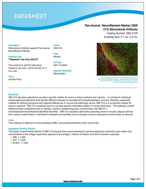
Immunostaining of the <strong>SMI</strong> <strong>312</strong> antibody (green) on rat<br />
brain (8um horizontal section). Other structures visualized<br />
with anti-GFAP (red, PRB-571C) and Hoechst. Photo<br />
courtesy of Molecular Expressions.<br />
Specificity:<br />
<strong>SMI</strong> <strong>312</strong> has been selected to provide a specific marker for axons in tissue sections and cultures. In contrast to individual<br />
antiphosphoneurofilaments that identify different subsets of neurofilament phosphoepitopes, and are, therefore, especially<br />
suitable for defining functional and regional differences in normal and pathologic axons, <strong>SMI</strong> <strong>312</strong> is a convenient marker for<br />
axons in general. <strong>SMI</strong> <strong>312</strong> visualizes axons in an area-specific maturation pattern in human fetal brain. The antibody cocktail<br />
defines nuclear borderlines and is, thereby, useful in establishing early connectivities with <strong>SMI</strong> 311,<br />
antinonphosphoneurofilament-identified dendrites. <strong>SMI</strong> <strong>312</strong> visualizes aberrantly sprouting axons in neuritic plaques derived<br />
from cortico-cortical fibers in Alzheimer's disease and identifies loss of synaptic circuitry proposed to be the basis of memory<br />
Uses:<br />
This antibody is effective in immunoblotting (WB), immunohistochemistry (IHC) and ELISA.<br />
Suggested Working Dilution:<br />
The extent of permissible dilution of <strong>SMI</strong> <strong>312</strong> beyond those recommended for general application depends upon nature and<br />
concentration of the antigen examined, species of the antigen, method of fixation and kind of section examined.<br />
• WB: 1:1,000<br />
• IHC: 1:1,000<br />
• ELISA: 1:1,000<br />
www.covance.com<br />
Page 1 of 2
Storage:<br />
Store at -20°C. Upon initial thawing, apportion into working<br />
aliquots and store at -20°C. Avoid repeated freeze-thaw<br />
cycles to prevent denaturing the antibody. For long-term<br />
storage, keep the antibody at -80°C.<br />
References:<br />
Bussière T, Bard F, Barbour R, Grajeda H, Guido T, Khan K, Schenk D,<br />
Games D, Seubert P, Buttini M. Morphological characterization of<br />
Thioflavin-S-Positive amyloid plaques in transgenic Alzheimer mice and<br />
effect of passive Abeta immunotherapy on their clearance. Am J Pathol<br />
165:987-995, 2004.<br />
Chung RS, Vickers JC, Chuah MI, West AK. Metallothionein-IIA promotes<br />
initial neurite elongation and postinjury reactive neurite growth and<br />
facilitates healing after focal cortical brain injury. J Neurosci 23:3336-<br />
3342, 2003.<br />
Soifer D, Nicoletti V, Cabane K, Mack K, Poulos B: Expression of<br />
neurofilament protein NF-H in L cells. J. Neurosc Res 30:63, 1991.<br />
Pestronk A, Watson DF, Yuan CM: <strong>Neurofilament</strong> phosphorylation in<br />
motor axons with high and low growth potential. J Neurochem 54:977,<br />
1990.<br />
Sternberger LA, Harwell LW, Sternberger NH: Neurotypy: Regional<br />
individuality in rat brain detected by mmunocytochemistry with<br />
monoclonal antibodies. Proc Natl Acad Sci USA 79:1326-30, 1982.<br />
Warranty/Conditions:<br />
Covance products may not be resold or modified for resale without prior<br />
written approval.All sales are subject to Covance Antibody Products<br />
Terms and Conditions of Sale.<br />
Datasheet Revision Date:<br />
03/13/2011<br />
Antibody Products:<br />
Email:<br />
+1.800.922.2226<br />
+1.510.594.3800<br />
ab.products@covance.com<br />
<strong>Pan</strong>-<strong>Axonal</strong> <strong>Neurofilament</strong> <strong>Marker</strong> (<strong>SMI</strong> <strong>312</strong>) Monoclonal Antibody<br />
Catalog Number: <strong>SMI</strong>-<strong>312</strong>R<br />
www.covance.com<br />
Related Products:<br />
• <strong>Neurofilament</strong> H Phosphorylated (<strong>SMI</strong> 34) Monoclonal<br />
Antibody, Catalog Number: <strong>SMI</strong>-34R<br />
• Normal Goat Antibody, Catalog Number: <strong>SMI</strong>-6020C<br />
• <strong>Neurofilament</strong>s, Hypophosphorylated (<strong>SMI</strong> 35) Monoclonal<br />
Antibody, Catalog Number: <strong>SMI</strong>-35R<br />
• <strong>Neurofilament</strong>s, Hypophosphorylated (<strong>SMI</strong> 35) Monoclonal<br />
Antibody, Catalog Number: <strong>SMI</strong>-35R<br />
• <strong>Neurofilament</strong> H Phosphorylated (<strong>SMI</strong> 310) Monoclonal<br />
Antibody, Catalog Number: <strong>SMI</strong>-310R<br />
• Mouse ClonoPAP®, Catalog Number: <strong>SMI</strong>-4050L<br />
• <strong>Neurofilament</strong>s, Phosphorylated (<strong>SMI</strong> 31) Monoclonal Antibody,<br />
Catalog Number: <strong>SMI</strong>-31R<br />
• Ultra Streptavidin (USA) Horseradish peroxidase (HRP) 50<br />
Test, Multi-Species, DAB Detection Kit (SIGNET), Catalog<br />
Number: SIG-32250<br />
• Ultra Streptavidin (USA) Horseradish peroxidase (HRP) 50<br />
Test, Multi-Species, AEC Detection Kit (SIGNET), Catalog<br />
Number: SIG-32248<br />
Immunostaining of the <strong>SMI</strong> <strong>312</strong> antibody (green) on rat<br />
brain (8um horizontal section). Other structures visualized<br />
with anti-GFAP (red, PRB-571C) and Hoechst. Photo<br />
courtesy of Molecular Expressions.<br />
Page 2 of 2