The use of quantitative light-induced fluorescence (QLF) to ... - Library
The use of quantitative light-induced fluorescence (QLF) to ... - Library
The use of quantitative light-induced fluorescence (QLF) to ... - Library

You also want an ePaper? Increase the reach of your titles
YUMPU automatically turns print PDFs into web optimized ePapers that Google loves.
2 JOURNAL OF FORENSIC SCIENCES<br />
quire cleaned, dry teeth and may, therefore, be impractical within<br />
the forensic environment.<br />
A new method for detecting composite res<strong>to</strong>rations is described<br />
here.<br />
Quantitative Light-<strong>induced</strong> <strong>fluorescence</strong> (<strong>QLF</strong>)<br />
Quantitative <strong>light</strong> <strong>induced</strong> <strong>fluorescence</strong> (<strong>QLF</strong>) is a new technique<br />
for the detection <strong>of</strong> very early mineral changes (early decay)<br />
in enamel (9). <strong>The</strong> <strong>QLF</strong> system is based upon the au<strong>to</strong>-<strong>fluorescence</strong><br />
<strong>of</strong> human enamel and that this <strong>fluorescence</strong> will decrease with decreased<br />
mineral content (10). Early caries are, therefore, seen as<br />
dark areas and these can be quantified and longitudinally moni<strong>to</strong>red.<br />
<strong>The</strong> <strong>QLF</strong> device (Inspek<strong>to</strong>r Research Systems, BV, <strong>The</strong><br />
Netherlands, www.inspek<strong>to</strong>r.nl) increases the contrast between<br />
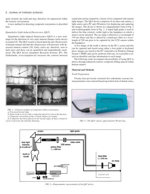
FIG. 1—Clinical examples <strong>of</strong> composite (white) res<strong>to</strong>rations:<br />
a) Fractured incisal edge.<br />
b) Same <strong>to</strong>oth as in (a) with a composite placed <strong>to</strong> res<strong>to</strong>re the fracture.<br />
c) Composite res<strong>to</strong>ration <strong>of</strong> the occlusal surface <strong>of</strong> a molar.<br />
d) Composite has been placed on the mesial edges <strong>of</strong> these central incisors<br />
<strong>to</strong> decrease the mid-line diastema.<br />
sound and carious enamel by a fac<strong>to</strong>r <strong>of</strong> ten compared with natural<br />
<strong>light</strong> images. <strong>The</strong> <strong>QLF</strong> device comprises <strong>of</strong> an intra-oral camera, a<br />
<strong>light</strong> source and a PC unit (Windows) for displaying and capturing<br />
the images. <strong>The</strong> device is shown in a diagrammatic form in Fig. 2<br />
and in pho<strong>to</strong>graphic form in Fig. 3. A liquid <strong>light</strong> guide is <strong>use</strong>d <strong>to</strong><br />
deliver the blue colored, visible <strong>light</strong> <strong>to</strong> the handpiece <strong>to</strong> which a<br />
mirror can be attached. <strong>The</strong> arc lamp is filtered <strong>to</strong> a wavelength <strong>of</strong><br />
370 nm (blue) and this is altered by a band-pass filter <strong>to</strong> a wavelength<br />
<strong>of</strong> 520 nm prior <strong>to</strong> be captured by the CCD camera within<br />
the handpiece.<br />
A live image <strong>of</strong> the <strong>to</strong>oth is shown on the PC’s screen and this<br />
can be captured and s<strong>to</strong>red using either a foot pedal or keyboard<br />
press. Images are s<strong>to</strong>red <strong>to</strong> the PC’s hard drive in Windows bitmap<br />
format (*.BMP) and can be archived with ease. It is possible <strong>to</strong> attach<br />
an identifier and date information <strong>to</strong> each image.<br />
<strong>The</strong> following study investigates the possibility <strong>of</strong> using <strong>QLF</strong> <strong>to</strong><br />
detect, through enhanced contrast, composite fillings placed within<br />
human enamel.<br />
Material and Methods<br />
Tooth Preparation<br />
FIG. 2—Diagrammatic representation <strong>of</strong> the <strong>QLF</strong> device.<br />
Twenty-four previously extracted (for orthodontic reasons) human<br />
premolars were selected based upon their lack <strong>of</strong> dental caries,<br />
FIG. 3—<strong>The</strong> <strong>QLF</strong> camera, approximately 90 mm long.