Nucleated RBCs—Significance in the Peripheral Blood Film
Nucleated RBCs—Significance in the Peripheral Blood Film
Nucleated RBCs—Significance in the Peripheral Blood Film

Create successful ePaper yourself
Turn your PDF publications into a flip-book with our unique Google optimized e-Paper software.
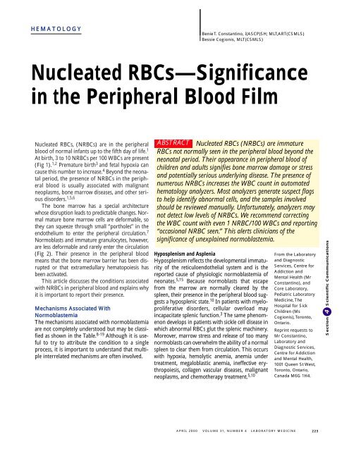
H E M A T O L O G Y<br />
<strong>Nucleated</strong> RBCs, (NRBCs) are <strong>in</strong> <strong>the</strong> peripheral<br />
blood of normal <strong>in</strong>fants up to <strong>the</strong> fifth day of life. 1<br />
At birth, 3 to 10 NRBCs per 100 WBCs are present<br />
(Fig 1). 1,2 Premature birth 3 and fetal hypoxia can<br />
cause this number to <strong>in</strong>crease. 4 Beyond <strong>the</strong> neonatal<br />
period, <strong>the</strong> presence of NRBCs <strong>in</strong> <strong>the</strong> peripheral<br />
blood is usually associated with malignant<br />
neoplasms, bone marrow diseases, and o<strong>the</strong>r serious<br />
disorders. 1,5,6<br />
The bone marrow has a special architecture<br />
whose disruption leads to predictable changes. Normal<br />
mature bone marrow cells are deformable, so<br />
<strong>the</strong>y can squeeze through small “portholes” <strong>in</strong> <strong>the</strong><br />
endo<strong>the</strong>lium to enter <strong>the</strong> peripheral circulation. 7<br />
Normoblasts and immature granulocytes, however,<br />
are less deformable and rarely enter <strong>the</strong> circulation<br />
(Fig 2). Their presence <strong>in</strong> <strong>the</strong> peripheral blood<br />
means that <strong>the</strong> bone marrow barrier has been disrupted<br />
or that extramedullary hematopoiesis has<br />
been activated.<br />
This article discusses <strong>the</strong> conditions associated<br />
with NRBCs <strong>in</strong> peripheral blood and expla<strong>in</strong>s why<br />
it is important to report <strong>the</strong>ir presence.<br />
Mechanisms Associated With<br />
Normoblastemia<br />
The mechanisms associated with normoblastemia<br />
are not completely understood but may be classified<br />
as shown <strong>in</strong> <strong>the</strong> Table. 8-19 Although it is useful<br />
to try to attribute <strong>the</strong> condition to a s<strong>in</strong>gle<br />
process, it is important to understand that multiple<br />
<strong>in</strong>terrelated mechanisms are often <strong>in</strong>volved.<br />
Benie T. Constant<strong>in</strong>o, I(ASCP)SH; MLT,ART(CSMLS)<br />
Bessie Cogionis, MLT(CSMLS)<br />
<strong>Nucleated</strong> <strong>RBCs—Significance</strong><br />
<strong>in</strong> <strong>the</strong> <strong>Peripheral</strong> <strong>Blood</strong> <strong>Film</strong><br />
ABSTRACT<br />
<strong>Nucleated</strong> RBCs (NRBCs) are immature<br />
RBCs not normally seen <strong>in</strong> <strong>the</strong> peripheral blood beyond <strong>the</strong><br />
neonatal period. Their appearance <strong>in</strong> peripheral blood of<br />
children and adults signifies bone marrow damage or stress<br />
and potentially serious underly<strong>in</strong>g disease. The presence of<br />
numerous NRBCs <strong>in</strong>creases <strong>the</strong> WBC count <strong>in</strong> automated<br />
hematology analyzers. Most analyzers generate suspect flags<br />
to help identify abnormal cells, and <strong>the</strong> samples <strong>in</strong>volved<br />
should be reviewed manually. Unfortunately, analyzers may<br />
not detect low levels of NRBCs. We recommend correct<strong>in</strong>g<br />
<strong>the</strong> WBC count with even 1 NRBC/100 WBCs and report<strong>in</strong>g<br />
“occasional NRBC seen.” This alerts cl<strong>in</strong>icians of <strong>the</strong><br />
significance of unexpla<strong>in</strong>ed normoblastemia.<br />
Hyposplenism and Asplenia<br />
Hyposplenism reflects <strong>the</strong> developmental immaturity<br />
of <strong>the</strong> reticuloendo<strong>the</strong>lial system and is <strong>the</strong><br />
reported cause of physiologic normoblastemia of<br />
neonates. 5,15 Because normoblasts that escape<br />
from <strong>the</strong> marrow are normally cleared by <strong>the</strong><br />
spleen, <strong>the</strong>ir presence <strong>in</strong> <strong>the</strong> peripheral blood suggests<br />
a hyposplenic state. 10 In patients with myeloproliferative<br />
disorders, cellular overload may<br />
<strong>in</strong>capacitate splenic function. 3 The same phenomenon<br />
develops <strong>in</strong> patients with sickle cell disease <strong>in</strong><br />
which abnormal RBCs glut <strong>the</strong> splenic mach<strong>in</strong>ery.<br />
Moreover, marrow stress and release of too many<br />
normoblasts can overwhelm <strong>the</strong> ability of a normal<br />
spleen to clear <strong>the</strong>m from circulation. This occurs<br />
with hypoxia, hemolytic anemia, anemia under<br />
treatment, megaloblastic anemia, <strong>in</strong>effective erythropoiesis,<br />
collagen vascular diseases, malignant<br />
neoplasms, and chemo<strong>the</strong>rapy treatment. 5,10<br />
From <strong>the</strong> Laboratory<br />
and Diagnostic<br />
Services, Centre for<br />
Addiction and<br />
Mental Health (Mr<br />
Constant<strong>in</strong>o), and<br />
Core Laboratory,<br />
Pediatric Laboratory<br />
Medic<strong>in</strong>e, The<br />
Hospital for Sick<br />
Children (Ms<br />
Cogionis), Toronto,<br />
Ontario.<br />
Repr<strong>in</strong>t requests to<br />
Mr Constant<strong>in</strong>o,<br />
Laboratory and<br />
Diagnostic Services,<br />
Centre for Addiction<br />
and Mental Health,<br />
1001 Queen St West,<br />
Toronto, Ontario,<br />
Canada M6G 1H4.<br />
APRIL 2000 VOLUME 31, NUMBER 4 LABORATORY MEDICINE 2 2 3<br />
Section 4 Scientific Communications
Fig 1. Wright-sta<strong>in</strong>ed peripheral blood smear from a normal newborn show<strong>in</strong>g<br />
polychromasia, a nucleated RBC, and a neutrophil. Cells are crowded toge<strong>the</strong>r<br />
due to <strong>the</strong> high RBC count (3 1,000). From Maedel L, Sommer S. <strong>Blood</strong> Cell<br />
Morphology: Morphologic Changes <strong>in</strong> Erythrocytes, 4. Chicago, IL: ASCP Press;<br />
1993: Fig 4-31.<br />
Fig 2. Wright-sta<strong>in</strong>ed<br />
bone marrow smear<br />
show<strong>in</strong>g stages of<br />
erythroid maturation:<br />
basophilic<br />
normoblast,<br />
polychromatophilic<br />
normoblast,<br />
orthochomic<br />
normoblast. An<br />
eos<strong>in</strong>ophil is also<br />
shown (3 1,000).<br />
From Maedel L,<br />
Sommer S. <strong>Blood</strong><br />
Cell Morphology:<br />
Normal Cells of <strong>the</strong><br />
Bone Marrow, 2.<br />
Chicago, IL: ASCP<br />
Press; 1993: Fig 2-05.<br />
2 2 4<br />
The most useful and sensitive <strong>in</strong>dicator of<br />
splenic function, however, is <strong>the</strong> presence of Howell-Jolly<br />
bodies (RBC <strong>in</strong>clusions) <strong>in</strong> <strong>the</strong> peripheral<br />
blood film (Fig 3). 10-11 The presence of normoblasts,<br />
although useful, may be nonspecific.<br />
Acanthocytes, target cells, stippled cells, and fragments<br />
are also nonspecific f<strong>in</strong>d<strong>in</strong>gs. 10<br />
Anemia and Compensatory Erythropoiesis<br />
In all types of severe anemia—hemolytic, nutritional,<br />
or anemia of blood loss—normoblastemia<br />
is caused by hypoxic erythropoiet<strong>in</strong>-<strong>in</strong>duced compensatory<br />
erythropoiesis. 11 The reduced oxygencarry<strong>in</strong>g<br />
capacity of anemic blood causes tissue<br />
LABORATORY MEDICINE VOLUME 31, NUMBER 4 APRIL 2000<br />
hypoxia, 20 <strong>the</strong> ma<strong>in</strong> stimulus for RBC production.<br />
When hypoxia occurs, <strong>the</strong> kidneys produce erythropoiet<strong>in</strong><br />
which, when <strong>in</strong>creased markedly,<br />
results <strong>in</strong> <strong>in</strong>tense marrow erythropoietic activity.<br />
Whe<strong>the</strong>r marrow erythropoiesis is effective or<br />
<strong>in</strong>effective depends on <strong>the</strong> underly<strong>in</strong>g cause of <strong>the</strong><br />
anemia. With effective erythropoiesis, <strong>the</strong> result<strong>in</strong>g<br />
accelerated compensatory activity produces<br />
prom<strong>in</strong>ent reticulocytosis, polychromasia, immature<br />
granulocytes (at times), and occasional<br />
NRBCs <strong>in</strong> <strong>the</strong> peripheral blood. Which and how<br />
many of <strong>the</strong>se cells or conditions are present<br />
depends on <strong>the</strong> severity of <strong>the</strong> anemia and marrow<br />
response. If <strong>the</strong> marrow response is exaggerated,<br />
NRBCs are plentiful with many “stress” reticulocytes,<br />
caus<strong>in</strong>g pseudomacrocytosis. When erythropoiesis<br />
is <strong>in</strong>effective, NRBCs may be prematurely<br />
released <strong>in</strong>to <strong>the</strong> peripheral blood without reticulocytosis.<br />
Dysplastic RBC changes may occur, as<br />
shown by <strong>the</strong> appearance of macro-ovalocytes and<br />
teardrop cells <strong>in</strong> <strong>the</strong> peripheral blood.<br />
Hypoxia<br />
Any condition that reduces <strong>the</strong> quantity of oxygen<br />
transported to <strong>the</strong> tissues causes an <strong>in</strong>crease <strong>in</strong> <strong>the</strong><br />
rate of RBC production. Although normoblastemia<br />
occurs <strong>in</strong> response to hypoxia <strong>in</strong> both anemia and<br />
cardiopulmonary disorders, <strong>the</strong> cause of hypoxia<br />
differs <strong>in</strong> <strong>the</strong>se conditions. In anemia, hypoxia<br />
results when <strong>the</strong> reduced hemoglob<strong>in</strong> concentration<br />
causes a correspond<strong>in</strong>g decl<strong>in</strong>e <strong>in</strong> <strong>the</strong> oxygencarry<strong>in</strong>g<br />
capacity of <strong>the</strong> blood. Cardiopulmonary<br />
hypoxia, however, may <strong>in</strong>volve several mechanisms,<br />
<strong>in</strong>clud<strong>in</strong>g failure of <strong>the</strong> blood to absorb<br />
oxygen from <strong>the</strong> lungs, <strong>in</strong>adequate ventilation of<br />
alveoli, or right-to-left <strong>in</strong>trapulmonary shunt<strong>in</strong>g<br />
of <strong>the</strong> blood. Impaired cardiovascular circulation<br />
lead<strong>in</strong>g to an <strong>in</strong>adequate supply of oxygenated<br />
blood to <strong>the</strong> tissues may also cause hypoxia. 21,22<br />
Because some patients with cardiopulmonary disorders<br />
have pulmonary emboli or coronary<br />
thrombosis complications, <strong>the</strong> presence of normoblasts<br />
<strong>in</strong> <strong>the</strong>se disorders may <strong>in</strong>dicate unfavorable<br />
prognosis. 23,24<br />
The hemoglob<strong>in</strong> concentration also differs <strong>in</strong><br />
<strong>the</strong>se hypoxic situations. In cardiopulmonary<br />
hypoxia, <strong>the</strong> hemoglob<strong>in</strong> level is ei<strong>the</strong>r high or<br />
with<strong>in</strong> <strong>the</strong> reference <strong>in</strong>terval, whereas <strong>in</strong> anemic
Mechanisms and Conditions Associated With Normoblastemia 8-19<br />
Hyposplenism, asplenia Sickle cell anemia 8<br />
Newborn (physiologic) 1,6<br />
Splenectomy 3,9<br />
Essential thrombocytosis 10<br />
Hemolytic anemia 10<br />
Malaria 10<br />
Anemia, compensatory erythropoiesis Severe anemia (any cause) 11<br />
Hemolytic anemia 8<br />
Iron deficiency anemia 8<br />
Megaloblastic anemia 8<br />
Hemorrhage 5,12<br />
Anemia under treatment 8<br />
Microangiopathic hemolytic anemia 8<br />
Thalassemia major 8<br />
Hypoxia Severe pulmonary disease 5,13<br />
Congestive cardiac failure 5,13<br />
Cyanotic heart disease 5,13<br />
Marrow replacement, <strong>in</strong>vasion Preleukemia 13<br />
Leukemia 8,13<br />
Lymphoma 13<br />
Neuroblastoma 13<br />
Myelodysplasia 8<br />
Myelofibrosis 8,13<br />
Plasma cell myeloma 8<br />
Myeloproliferative disorder 8,13<br />
Gaucher and o<strong>the</strong>r storage disease 14<br />
Granuloma (ie, tuberculosis) 13<br />
Collagen vascular disease 5,13<br />
Fungal <strong>in</strong>fection 14<br />
Histiocytosis 14<br />
Tumor cell presence 8,13<br />
Sarcoidosis 14<br />
Osteopetrosis 13<br />
Extramedullary hematopoiesis Myelophthisis 13,15<br />
Osteopetrosis 13<br />
Myeloid metaplasia 13<br />
Myelofibrosis 8<br />
Chronic hemolytic anemia 15<br />
Polycy<strong>the</strong>mia vera 13<br />
Leukemia 15<br />
O<strong>the</strong>r Uremia 16<br />
Sepsis 17<br />
Liver disease 18<br />
Diabetic ketoacidosis 18<br />
Inflammatory bowel disease 18<br />
Renal transplant 18<br />
Thermal <strong>in</strong>jury 19<br />
Chemo<strong>the</strong>rapy 5<br />
APRIL 2000 VOLUME 31, NUMBER 4 LABORATORY MEDICINE 2 2 5<br />
Section 4 Scientific Communications
Fig 3. Wright-sta<strong>in</strong>ed peripheral blood smear show<strong>in</strong>g basophilic stippl<strong>in</strong>g,<br />
Howell-Jolly bodies, and a nucleated RBC (3 1,000). From Maedel L, Sommer S.<br />
<strong>Blood</strong> Cell Morphology: Morphologic Changes <strong>in</strong> Erythrocytes, 4. Chicago, IL:<br />
ASCP Press; 1993: Fig 4-81.<br />
Fig 4. Wright-sta<strong>in</strong>ed peripheral blood smear from a patient with myelofibrosis<br />
show<strong>in</strong>g many teardrop RBCs and a myelocyte (3 1,000). From Maedel L,<br />
Sommer S. <strong>Blood</strong> Cell Morphology: Chronic Hematopoietic Malignancies, 6.<br />
Chicago, IL: ASCP Press; 1993: Fig 6-18.<br />
2 2 6<br />
hypoxia, it is much lower. 21,24 The rate of RBC<br />
production, however, is not controlled by hemoglob<strong>in</strong><br />
concentration; it appears to vary with <strong>the</strong><br />
ability of <strong>the</strong> cells to transport oxygen to <strong>the</strong> tissues<br />
<strong>in</strong> response to a demand. Thus, if <strong>the</strong> oxygen supply<br />
is less than <strong>the</strong> tissues demand, more RBCs are<br />
produced, which, <strong>in</strong> turn, results <strong>in</strong> a higher hemoglob<strong>in</strong><br />
level until <strong>the</strong> supply of oxygen meets <strong>the</strong><br />
LABORATORY MEDICINE VOLUME 31, NUMBER 4 APRIL 2000<br />
demand. In cases of transient <strong>in</strong>creases <strong>in</strong> oxygen<br />
demand (a hypoxic stimulus), normoblastemia, if<br />
present, disappears with relief of <strong>the</strong> hypoxia. 24<br />
Accord<strong>in</strong>gly, a hypoxic stimulus should be suspected<br />
whenever normoblastemia is accompanied<br />
by a normal to high hemoglob<strong>in</strong> level and mild to<br />
moderate polychromasia.<br />
Bone Marrow Replacement and Invasion<br />
Marrow replacement can occur <strong>in</strong> association<br />
with a primary hematologic disease such as<br />
leukemia, myeloma, or lymphoma. It can also be<br />
<strong>the</strong> result of secondary <strong>in</strong>sult by <strong>in</strong>vad<strong>in</strong>g tumor<br />
cells, <strong>the</strong> presence of sarcoidosis, or <strong>in</strong>fectious<br />
agents such as mycobacteria and fungi. Both primary<br />
and secondary reactions can produce marrow<br />
fibrosis (myelofibrosis), which changes <strong>the</strong><br />
normal marrow microarchitecture. This disruption<br />
may break down <strong>the</strong> marrow-blood barrier,<br />
caus<strong>in</strong>g untimely and disorderly release of NRBCs<br />
and progenitor cells <strong>in</strong>to <strong>the</strong> circulation (Fig<br />
4). 14,25 Similarly, extensive marrow <strong>in</strong>filtration<br />
and replacement may cause mechanical “crowd<strong>in</strong>g<br />
out” of normal hematopoietic cells, lead<strong>in</strong>g to<br />
<strong>the</strong>ir escape <strong>in</strong>to <strong>the</strong> peripheral blood and lodgment<br />
<strong>in</strong> o<strong>the</strong>r organs such as <strong>the</strong> spleen, liver, and<br />
lymph nodes. This process may contribute to<br />
extramedullary hematopoiesis.<br />
The <strong>in</strong>itial peripheral blood picture may present<br />
unexpla<strong>in</strong>ed normoblastemia, mild macrocytosis,<br />
giant platelets, myelocytes, thrombocytopenia,<br />
and, possibly, leukopenia with or without teardrop<br />
cells or blast cells.<br />
Extramedullary Hematopoiesis<br />
Recognized cl<strong>in</strong>ically as splenomegaly or<br />
hepatomegaly, extramedullary hematopoiesis<br />
appears to be caused by anemia, marrow replacement<br />
associated with acute leukemia, or o<strong>the</strong>r<br />
nonhematopoietic <strong>in</strong>filtrative processes (myelophthisis).<br />
15 Presumably, hematopoietic stem cells are<br />
displaced from <strong>the</strong> marrow <strong>in</strong>to <strong>the</strong> spleen or liver<br />
where <strong>the</strong>y proliferate to cause hepatomegaly and<br />
splenomegaly. Splenomegaly also occurs when <strong>the</strong><br />
marrow has been dispossessed by fibrosis. Because<br />
<strong>the</strong> spleen does not reta<strong>in</strong> immature cells as efficiently<br />
as normal marrow, it may release NRBCs,
immature granulocytes, megathrombocytes, and<br />
occasional blast cells <strong>in</strong>to <strong>the</strong> peripheral blood,<br />
result<strong>in</strong>g <strong>in</strong> leukoerythroblastosis or <strong>the</strong> coexistence<br />
of myeloid precursors and NRBCs <strong>in</strong> <strong>the</strong><br />
peripheral blood. 17 Teardrop cells may be present.<br />
A leukoerythroblastic reaction may also be seen<br />
without marrow <strong>in</strong>filtration <strong>in</strong> normal newborns<br />
as well as <strong>in</strong> patients with thalassemia major with<br />
severe hemolytic crisis, hemorrhage, postsplenectomy,<br />
septicemia, 26 and <strong>the</strong>rapy with granulocyte<br />
colony-stimulat<strong>in</strong>g factor (G-CSF). 27,28<br />
In addition, when bone marrow reserve is<br />
unable to meet <strong>the</strong> demand for accelerated erythropoiesis<br />
(as <strong>in</strong> chronic hemolytic anemia or longstand<strong>in</strong>g<br />
anemia), blood cells may form <strong>in</strong> tissues<br />
o<strong>the</strong>r than <strong>the</strong> bone marrow. 15 This extramedullary<br />
hematopoiesis represents a reversion of <strong>the</strong><br />
<strong>in</strong>volved tissues to <strong>the</strong>ir fetal blood-form<strong>in</strong>g function,<br />
although this compensatory activity may also<br />
occur <strong>in</strong> myelophthisic anemia with fibrosis. Thus,<br />
differentiat<strong>in</strong>g between chronic hemolytic anemia<br />
and myelophthisic anemia with fibrosis is difficult<br />
without <strong>the</strong> patient’s cl<strong>in</strong>ical history.<br />
Leukoerythroblastosis is evident <strong>in</strong> peripheral<br />
blood films, and teardrop cells may or may not be<br />
present. Teardrop cells are not usually seen <strong>in</strong><br />
leukoerythroblastic reactions associated with<br />
hemorrhage, <strong>in</strong>fection, or G-CSF <strong>the</strong>rapy. They<br />
can be found <strong>in</strong> severe iron deficiency anemia, 29<br />
thalassemia, megaloblastic anemia, hemolytic anemia,<br />
leukemia, myelofibrosis, and drug-<strong>in</strong>duced<br />
He<strong>in</strong>z body formation. 26 Teardrop cells may<br />
reflect dyspoiesis and thus are not specific for a<br />
s<strong>in</strong>gle condition. Although <strong>the</strong> exact mechanism<br />
of teardrop cell formation is unclear, <strong>the</strong> formation<br />
of <strong>the</strong>se cells from <strong>in</strong>clusion-conta<strong>in</strong><strong>in</strong>g RBCs<br />
is well documented. As cells with large rigid <strong>in</strong>clusions<br />
try to pass through <strong>the</strong> small splenic s<strong>in</strong>us<br />
open<strong>in</strong>gs, parts with large <strong>in</strong>clusions get p<strong>in</strong>ched,<br />
caus<strong>in</strong>g <strong>the</strong> cells to stretch with irreversible loss of<br />
<strong>the</strong>ir shape. The result is teardrop cells. 30 Moreover,<br />
teardrop cell formation represents <strong>the</strong> cells’<br />
tortuous circulation through deformed marrow<br />
s<strong>in</strong>uses and diseased splenic cords. 26 The presence<br />
of teardrop cells <strong>in</strong> <strong>the</strong> peripheral blood is thus<br />
significant and should alert morphologists to<br />
search for occasional NRBCs, megathrombocytes,<br />
stippled cells, immature granulocytes, or blast cells<br />
that would constitute conclusive evidence of<br />
myeloid metaplasia or leukoerythroblastosis. 26<br />
O<strong>the</strong>r Mechanisms<br />
Why normoblastemia occurs with <strong>the</strong>se disorders<br />
rema<strong>in</strong>s enigmatic. Although <strong>the</strong> marrow-blood<br />
barrier appears to break down, <strong>the</strong> cause of <strong>the</strong><br />
breakdown is unknown. 13 Most of <strong>the</strong> disorders<br />
<strong>in</strong>volve complex conditions that cause systemic<br />
diseases and <strong>in</strong>fluence bone marrow response. 17<br />
These diseases <strong>in</strong>clude uremia, sepsis, liver disease,<br />
and <strong>the</strong>rmal <strong>in</strong>jury. 18,19<br />
Role of <strong>the</strong> Laboratory<br />
Laboratory professionals play an important role <strong>in</strong><br />
detect<strong>in</strong>g NRBCs when <strong>the</strong>y review CBC and<br />
WBC differential results obta<strong>in</strong>ed by automated<br />
hematology analyzers. Most analyzers generate<br />
suspect flags, (eg, WBC * R, NRBC, Review Slide,<br />
Blasts) to help identify abnormal WBCs, and samples<br />
with flags should be microscopically exam<strong>in</strong>ed.<br />
Although most <strong>in</strong>struments have >80%<br />
specificity for NRBC flags, <strong>the</strong>y cannot consistently<br />
detect
2 2 8<br />
alerts cl<strong>in</strong>icians of <strong>the</strong> significance of unexpla<strong>in</strong>ed<br />
normoblastemia. The correction can be made<br />
with a simple formula:<br />
Corrected WBC count, 3 10 9 /L = WBC<br />
count, 3 10 9 /L 4 (1 + [NRBC/100 WBCs])<br />
For example, if <strong>the</strong> WBC count is 6.0 3 10 9 /L<br />
and <strong>the</strong> NRBC count is 50/100 WBCs, <strong>the</strong><br />
corrected WBC count<br />
= 6.0 3 10 9 /L 4 (1 + [50/100])<br />
= 6.0 3 10 9 /L 4 (1 + 0.5)<br />
= 4.0 3 10 9 /L<br />
Computerized laboratory systems do <strong>the</strong>se calculations<br />
automatically. Recent advances <strong>in</strong> hematology<br />
analyzers and <strong>the</strong>ir widespread use will<br />
alleviate manually correct<strong>in</strong>g WBC counts. 33,34<br />
As <strong>the</strong> example shows, a sample with an<br />
NRBC count of 50/100 WBC and a WBC count<br />
of 6.0 3 10 9 /L yields a corrected WBC count of<br />
4.0 3 10 9 /L—a difference that may be statistically<br />
but not cl<strong>in</strong>ically significant. Therefore, sett<strong>in</strong>g<br />
a threshold of more than 4 or 5 NRBCs/100<br />
WBCs before correct<strong>in</strong>g <strong>the</strong> WBC count does<br />
not make cl<strong>in</strong>ical sense. In addition, many<br />
authors 26,35-37 advocate different correction formulae<br />
and cutoff values. We recommend correct<strong>in</strong>g<br />
<strong>the</strong> WBC count with even 1 NRBC/100<br />
WBCs and report<strong>in</strong>g “occasional NRBC seen”<br />
with
We believe that unexpla<strong>in</strong>ed normoblastemia is<br />
important because it offers <strong>in</strong>valuable <strong>in</strong>sight <strong>in</strong>to<br />
disease processes or progressions that occur <strong>in</strong><br />
conditions such as metastatic carc<strong>in</strong>oma, bone<br />
marrow conditions, systemic <strong>in</strong>fections, and cardiopulmonary<br />
complications. The presence of<br />
normoblastemia with certa<strong>in</strong> cl<strong>in</strong>ical conditions<br />
may <strong>in</strong>dicate that a bone marrow exam<strong>in</strong>ation is<br />
necessary to rule out hematologic malignant neoplasms<br />
or unsuspected blood disorders. When<br />
viewed <strong>in</strong> this context, 1 <strong>in</strong>nocuous NRBC may<br />
lead to more timely medical <strong>in</strong>tervention, thus<br />
<strong>in</strong>creas<strong>in</strong>g <strong>the</strong> chance of a positive outcome.l<br />
References<br />
1. Miller DR, Baehner RL. <strong>Blood</strong> Diseases of Infancy and<br />
Childhood. 7th ed. St Louis, MO: Mosby; 1995:39-40.<br />
2. Green DW, Mimouni F. <strong>Nucleated</strong> erythrocytes <strong>in</strong> healthy<br />
<strong>in</strong>fants and <strong>in</strong> <strong>in</strong>fants of diabetic mo<strong>the</strong>rs. J Pediatr.<br />
1990;116:129-131.<br />
3. Crosby WH. Hyposplenism: an <strong>in</strong>quiry <strong>in</strong>to <strong>the</strong> normal<br />
function of <strong>the</strong> spleen. Annu Rev Med. 1963;14:349-370.<br />
4. Hanlon-Lundberg KM, Kirby RS. <strong>Nucleated</strong> red blood cells<br />
as a marker of acidemia <strong>in</strong> neonates. Am J Obstet Gynecol.<br />
1999;181:196-201.<br />
5. Sills RH, Hadley RAR. The significance of nucleated red<br />
blood cells <strong>in</strong> <strong>the</strong> peripheral blood of children. Am J Pediatr<br />
Hematol Oncol. 1983;5:173-177.<br />
6. Schwartz SO, Stanbury F. Significance of nucleated red<br />
blood cells <strong>in</strong> peripheral blood: analysis of 1496 cases. JAMA.<br />
1954;154:1339-1340.<br />
7. LeBlond P, LaCelle P, Weed RI. Cellular deformity: a possible<br />
determ<strong>in</strong>ant of normal marrow release of matur<strong>in</strong>g erythrocytes<br />
from <strong>the</strong> bone marrow. <strong>Blood</strong>. 1971;37:40-46.<br />
8. Laboratory Proficiency Test<strong>in</strong>g Program. Hematology-<br />
Morphology Survey Report: March 1987–July 1999. Ontario,<br />
Canada: Ontario Medical Association; 1999:3-99.<br />
9. Lee RG, Bitchell TC, Foerster J, et al, eds. In: W<strong>in</strong>trobe’s<br />
Cl<strong>in</strong>ical Hematology. 9th ed. Philadelphia, PA: Lea & Febiger;<br />
1993:316-317.<br />
10. Sills RH. Hyposplenism. In: Pochedly C, Sills RH,<br />
Schwartz AD, eds. Disorders of <strong>the</strong> Spleen Pathophysiology and<br />
Management. New York, NY: Marcel Dekker; 1989:99-133.<br />
11. Dacie JV, Lewis SM. Practical Hematology. 8th ed. New<br />
York, NY: Churchill Liv<strong>in</strong>gstone; 1995:115-116.<br />
12. Delamore LW, Liu Yen JA, eds. Haematological Aspects of<br />
Systemic Disease. London, England: Boilliere T<strong>in</strong>dall; 1990:280.<br />
13. Alter BP, Young NS. The bone marrow failure syndromes.<br />
In: Nathan DG, Oski FO, eds. Hematology of Infancy and Childhood.<br />
5th ed. Philadelphia, PA: Saunders; 1998:309-311.<br />
14. Erslev AJ. Anemia associated with marrow <strong>in</strong>filtration. In:<br />
Beutler E, Lichtman MA, Coller BS, et al, eds. Williams Hematology.<br />
5th ed. New York, NY: McGraw-Hill; 1995:516-518.<br />
15. Custer RP. An Atlas of <strong>the</strong> <strong>Blood</strong> and Bone Marrow. 2nd ed.<br />
Philadelphia, PA: Saunders; 1974:22-25, 123-136.<br />
16. Clifford GO. The cl<strong>in</strong>ical significance of leukoerythroblastic<br />
anemia. Med Cl<strong>in</strong> North Am. 1966;50:779-790.<br />
17. Retief FP. Leukoerythroblastosis <strong>in</strong> <strong>the</strong> adult. Lancet.<br />
1964;1:639-642.<br />
18. Weick JK, Hagedorn AB, L<strong>in</strong>man JW. Leukoerythroblastosis:<br />
diagnostic and prognostic significance. Mayo Cl<strong>in</strong> Proc.<br />
1974;49:110–113.<br />
19. Andes WB. Normoblastemia after <strong>the</strong>rmal <strong>in</strong>jury. Am J<br />
Surg.1976;131:725-726.<br />
20. Porth CM, ed. Pathophysiology Concepts of Altered Health<br />
States. 5th ed. Philadelphia, PA: Lipp<strong>in</strong>cott-Raven; 1998:134-135.<br />
21. Bennett JC, Plum F, eds. Cecil Textbook of Medic<strong>in</strong>e. 20th ed.<br />
Philadelphia, PA: Saunders; 1996:453, 466-469.<br />
22. Fauci AS, Braunwald E, eds. Harrison’s Pr<strong>in</strong>ciples of Internal<br />
Medic<strong>in</strong>e. 14th ed. New York, NY: McGraw-Hill; 1998:205-209.<br />
23. Ward HP, Holman J. The association of nucleated red cells<br />
<strong>in</strong> <strong>the</strong> peripheral smear with hypoxemia. Ann Intern Med.<br />
1967;67:1190-1194.<br />
24. Frum<strong>in</strong> AM, Mendel TH, M<strong>in</strong>tz SS, et al. <strong>Nucleated</strong> red blood<br />
cells <strong>in</strong> congestive heart failure. Circulation. 1959;20:367-370.<br />
25. Wang JC, Cheung CP, Ahmed F, et al. Circulat<strong>in</strong>g granulocyte<br />
and macrophage progenitor cells <strong>in</strong> primary and secondary<br />
myelofibrosis. Br J Haematol. 1983;54:301-307.<br />
26. Stiene-Mart<strong>in</strong> EA, Lotspeich-Ste<strong>in</strong><strong>in</strong>ger CA, Koepke JA,<br />
eds. Cl<strong>in</strong>ical Hematology Pr<strong>in</strong>ciples, Procedures, Correlations.<br />
2nd ed. Philadelphia, PA: Lipp<strong>in</strong>cott-Raven; 1998:93, 339-340,<br />
463-465.<br />
27. Schmitz LL, McClure JS, Litz LE, et al. Morphologic and<br />
quantitative changes <strong>in</strong> blood and marrow cells follow<strong>in</strong>g<br />
growth factor <strong>the</strong>rapy. Am J Cl<strong>in</strong> Pathol. 1994;101:67-75.<br />
28. Arici M, Haznedaroglu IC, Erman M, et al. Leukoerythroblastosis<br />
follow<strong>in</strong>g <strong>the</strong> use of G-CSF. Am J Histol.<br />
1996;52:122-123.<br />
29. Rodgers MS, Chang C, Kass L. Elliptocytes and tailed<br />
poikilocytes correlate with severity of iron-deficiency anemia.<br />
Am J Cl<strong>in</strong> Pathol. 1999;111:672-675.<br />
30. Bessis M. <strong>Blood</strong> Smears Re<strong>in</strong>terpreted. Paris, France:<br />
Spr<strong>in</strong>ger International; 1977:74.<br />
31. Cornbleet PJ, Myriel D, Levy R. Evaluation of <strong>the</strong> Coulter<br />
STKS five-part differential. Am J Cl<strong>in</strong> Pathol. 1993;99:72-81.<br />
32. Cornbleet PJ, Myrick D, Judk<strong>in</strong>s S, et al. Evaluation of <strong>the</strong><br />
Cell-Dyn 3000 differential. Am J Cl<strong>in</strong> Pathol. 1992;98:603-614.<br />
33. Paterakis G, Kossivas L, Kendall R, et al. Comparative<br />
evaluation of <strong>the</strong> erythroblast count generated by three-color<br />
fluorescence flow cytometry, <strong>the</strong> Abbott CELL-DYN R 4000<br />
hematology analyzer, and microscopy. Lab Hematol.<br />
1998;4:64-70.<br />
34. Kim YR, Yee M, Metha S, et al. Simultaneous differentiation<br />
and quantitation of erythroblasts and white blood cells on<br />
a high throughput cl<strong>in</strong>ical haematology analyzer. Cl<strong>in</strong> Lab<br />
Haematol. 1998;20:21-29.<br />
35. Simmons A. Hematology: A Comb<strong>in</strong>ed Theoretical and<br />
Technical Approach. Philadelphia, PA: Saunders; 1989:192.<br />
36. Seiverd CE. Hematology for Medical Technologists. 5th ed.<br />
Philadelphia, PA: Lea & Febiger; 1983:143-144.<br />
37. Frankel S, Reitman S, Sonnenwirth AC, eds. Gradwohl’s<br />
Cl<strong>in</strong>ical Laboratory Methods and Diagnosis. 6th ed. St Louis,<br />
MO: Mosby; 1970:501.<br />
38. Medical News: <strong>Nucleated</strong> RBCs <strong>in</strong> blood of adults should<br />
be cause for concern. JAMA. 1978;239:91.<br />
APRIL 2000 VOLUME 31, NUMBER 4 LABORATORY MEDICINE 2 2 9<br />
Section 4<br />
Scientific Communications