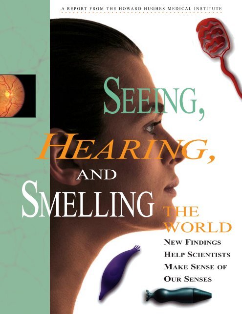
Seeing, Hearing, and Smelling the World - Howard Hughes Medical ...
Seeing, Hearing, and Smelling the World - Howard Hughes Medical ...
Seeing, Hearing, and Smelling the World - Howard Hughes Medical ...

Create successful ePaper yourself
Turn your PDF publications into a flip-book with our unique Google optimized e-Paper software.
A REPORT FROM THE HOWARD HUGHES MEDICAL INSTITUTE<br />
SEEING,<br />
HEARING,<br />
AND<br />
SMELLING<br />
THE<br />
WORLD<br />
NEW FINDINGS<br />
HELP SCIENTISTS<br />
MAKE SENSE OF<br />
OUR SENSES
The <strong>Howard</strong> <strong>Hughes</strong> <strong>Medical</strong> Institute<br />
was founded in 1953 by <strong>the</strong> aviatorindustrialist<br />
<strong>Howard</strong> R. <strong>Hughes</strong>. Its charter<br />
reads, in part:<br />
“The primary purpose <strong>and</strong> objective of <strong>the</strong><br />
<strong>Howard</strong> <strong>Hughes</strong> <strong>Medical</strong> Institute shall be<br />
<strong>the</strong> promotion of human knowledge within<br />
<strong>the</strong> field of <strong>the</strong> basic sciences (principally<br />
<strong>the</strong> field of medical research <strong>and</strong> medical<br />
education) <strong>and</strong> <strong>the</strong> effective application<br />
<strong>the</strong>reof for <strong>the</strong> benefit of mankind.”<br />
This is <strong>the</strong> fifth in a series of reports about biomedical<br />
science. For fur<strong>the</strong>r information, please contact <strong>the</strong><br />
<strong>Howard</strong> <strong>Hughes</strong> <strong>Medical</strong> Institute,<br />
4000 Jones Bridge Road,<br />
Chevy Chase, Maryl<strong>and</strong> 20815-6789<br />
© 1995 <strong>Howard</strong> <strong>Hughes</strong> <strong>Medical</strong> Institute
F O R E W O R D<br />
It is a pleasure to introduce <strong>the</strong> latest of <strong>the</strong> biomedical research<br />
reports that <strong>the</strong> <strong>Howard</strong> <strong>Hughes</strong> <strong>Medical</strong> Institute publishes for<br />
general readers. <strong>Seeing</strong>, <strong>Hearing</strong>, <strong>and</strong> <strong>Smelling</strong> <strong>the</strong> <strong>World</strong>, like<br />
<strong>the</strong> four previous publications in <strong>the</strong> series, takes us to <strong>the</strong> frontiers<br />
of science. It guides us on a journey into <strong>the</strong> fascinating world of <strong>the</strong><br />
senses <strong>and</strong> <strong>the</strong> nervous system, where researchers are working to<br />
underst<strong>and</strong> problems of great potential benefit.<br />
The most routine, everyday occurrences, such as recognizing a<br />
friend on <strong>the</strong> street <strong>and</strong> exchanging greetings, demonstrate <strong>the</strong> biological<br />
complexity of <strong>the</strong> puzzles that scientists are attempting to<br />
solve. Although such encounters seem simple, <strong>the</strong>y require hundreds<br />
of millions of cells to act in precise ways to receive <strong>the</strong> sights<br />
<strong>and</strong> sounds <strong>and</strong> translate <strong>the</strong>m into electrical impulses. These<br />
impulses flow through <strong>the</strong> nervous system to carry <strong>the</strong> messages to<br />
<strong>the</strong> brain, where <strong>the</strong>y can be understood <strong>and</strong> acted upon at astonishing<br />
speed.<br />
Centuries of effort by thous<strong>and</strong>s of scientists in laboratories<br />
throughout <strong>the</strong> world have been required to bring us to our current,<br />
deepening underst<strong>and</strong>ing about how we hear, see, <strong>and</strong> smell.<br />
Thanks to <strong>the</strong> new analytical tools provided by molecular biology,<br />
progress toward underst<strong>and</strong>ing <strong>the</strong> senses <strong>and</strong> <strong>the</strong> nervous system<br />
has been rapid during <strong>the</strong> past decade. Indeed, many neuroscientists<br />
believe that biomedical science is poised to make substantial<br />
progress toward underst<strong>and</strong>ing how <strong>the</strong> brain works, not only in<br />
terms of <strong>the</strong> senses, but also complex functions like learning <strong>and</strong><br />
memory. It is an exciting prospect.<br />
This series is published by <strong>the</strong> Institute as a public service in<br />
order to make <strong>the</strong> results of current biomedical research available<br />
to readers who are not scientists. It is clear that a basic grasp of<br />
biology is increasingly essential for citizens who have to make difficult<br />
decisions about health care, drug abuse, <strong>the</strong> environment, <strong>and</strong><br />
o<strong>the</strong>r critical issues.<br />
Teachers are particularly enthusiastic about <strong>the</strong>se reports, <strong>and</strong><br />
surveys tell us that <strong>the</strong>y preserve <strong>the</strong>ir copies <strong>and</strong> use <strong>the</strong>m year<br />
after year. Nearly 4,000 class sets have been requested by high<br />
school, college, <strong>and</strong> even medical school teachers in <strong>the</strong> United<br />
States <strong>and</strong> abroad; altoge<strong>the</strong>r, more than 400,000 copies of <strong>the</strong><br />
publications have been printed.<br />
The Institute’s interest in science education continues to deepen<br />
<strong>and</strong> its commitment to education reform to grow. Its grants program,<br />
which was established in 1987, has now become <strong>the</strong> largest<br />
private science education effort in U.S. history. Through its financial<br />
support <strong>and</strong> o<strong>the</strong>r activities, <strong>the</strong> Institute is seeking to make<br />
science come alive for today’s students, which is exactly what we<br />
hope <strong>Seeing</strong>, <strong>Hearing</strong>, <strong>and</strong> <strong>Smelling</strong> <strong>the</strong> <strong>World</strong> will do.<br />
Purnell W. Choppin, M.D.<br />
President<br />
<strong>Howard</strong> <strong>Hughes</strong> <strong>Medical</strong> Institute<br />
SEEING, HEARING, AND SMELLING THE WORLD • 3
Laboratory of Richard Masl<strong>and</strong>, Massachusetts General Hospital<br />
A nerve cell that can detect in what<br />
direction an object is moving branches<br />
out to make contact with many o<strong>the</strong>r<br />
cells in a rabbit’s visual system. The cell<br />
glows yellow because it was injected<br />
with fluorescent dye.<br />
4 • SEEING, HEARING, AND SMELLING THE WORLD
A REPORT FROM THE HOWARD HUGHES MEDICAL INSTITUTE<br />
SEEING, HEARING, AND<br />
SMELLING THE WORLD<br />
New Findings Help Scientists Make Sense of Our Senses<br />
Foreword by Purnell W. Choppin, M.D. . . . . . . . . . . . . . . . . . . . . . . . . . . . . . . . . . . . . . . . . .1<br />
Our Common Senses by Maya Pines . . . . . . . . . . . . . . . . . . . . . . . . . . . . . . . . . . . . . . . . . . .4<br />
A Language <strong>the</strong> Brain Can Underst<strong>and</strong> . . . . . . . . . . . . . . . . . . . . . . . . . . . . . . . . . . . . .11<br />
Breaking <strong>the</strong> Code of Color by Geoffrey Montgomery . . . . . . . . . . . . . . . . . . . . . . . . . . .12<br />
A Narrow Tunnel of Light . . . . . . . . . . . . . . . . . . . . . . . . . . . . . . . . . . . . . . . . . . . . . . . . .18<br />
How We See Things That Move by Geoffrey Montgomery . . . . . . . . . . . . . . . . . . . . . . . .22<br />
The Urgent Need to Use Both Eyes . . . . . . . . . . . . . . . . . . . . . . . . . . . . . . . . . . . . . . . . .26<br />
Brain Scans That Spy on <strong>the</strong> Senses . . . . . . . . . . . . . . . . . . . . . . . . . . . . . . . . . . . . . . . .29<br />
The Quivering Bundles That Let Us Hear by Jeff Goldberg . . . . . . . . . . . . . . . . . . . . .32<br />
On <strong>the</strong> Trail of a “Deafness” Gene . . . . . . . . . . . . . . . . . . . . . . . . . . . . . . . . . . . . . . . . . .36<br />
Locating a Mouse by Its Sound by Jeff Goldberg . . . . . . . . . . . . . . . . . . . . . . . . . . . . . . .40<br />
Help from a Bat . . . . . . . . . . . . . . . . . . . . . . . . . . . . . . . . . . . . . . . . . . . . . . . . . . . . . . . . . .45<br />
The Mystery of Smell by Maya Pines . . . . . . . . . . . . . . . . . . . . . . . . . . . . . . . . . . . . . . . . .46<br />
A Secret Sense in <strong>the</strong> Human Nose? by Maya Pines . . . . . . . . . . . . . . . . . . . . . . . . . . . .54<br />
The Next Generation . . . . . . . . . . . . . . . . . . . . . . . . . . . . . . . . . . . . . . . . . . . . . . . . . . . . . . .57<br />
SEEING, HEARING, AND SMELLING THE WORLD • 5
A LIFE-SIZE<br />
HUMAN BRAIN<br />
Vision<br />
Each of <strong>the</strong> five senses activates a<br />
separate area of <strong>the</strong> cerebral cortex, <strong>the</strong><br />
sheet of neurons that makes up <strong>the</strong> outer<br />
layer of <strong>the</strong> brain’s hemispheres. This<br />
brain, shown in actual size, is a computer<br />
reconstruction based on data from magnetic<br />
resonance imaging (MRI).<br />
Approximate locations of <strong>the</strong> primary<br />
sensory areas are shown in color.<br />
Most of <strong>the</strong> activity takes place within<br />
convolutions that cannot be seen<br />
from <strong>the</strong> surface of <strong>the</strong> brain.<br />
Touch<br />
<strong>Hearing</strong><br />
Taste<br />
Smell
We can recognize a friend instantly—full-<br />
face, in profile, or even by <strong>the</strong> back of his<br />
head. We can distinguish hundreds of<br />
colors <strong>and</strong> possibly as many as 10,000 smells. We<br />
can feel a fea<strong>the</strong>r as it brushes our skin, hear <strong>the</strong><br />
faint rustle of a leaf. It all seems so effortless: we<br />
open our eyes or ears <strong>and</strong> let <strong>the</strong> world stream in.<br />
Yet anything we see, hear, feel, smell, or taste<br />
requires billions of nerve cells to flash urgent mes-<br />
sages along linked pathways <strong>and</strong> feedback loops in<br />
our brains, performing intricate calculations that<br />
scientists have only begun to decipher.<br />
“You can think of sensory systems as little scien-<br />
tists that generate hypo<strong>the</strong>ses about <strong>the</strong> world,”<br />
says Anthony Movshon, an HHMI investigator at<br />
New York University. Where did that sound come<br />
from? What color is this, really? The brain makes<br />
an educated guess, based on <strong>the</strong> information at<br />
h<strong>and</strong> <strong>and</strong> on some simple assumptions.<br />
SEEING, HEARING, AND SMELLING THE WORLD • 7
8 • SEEING, HEARING, AND SMELLING THE WORLD<br />
When you look at <strong>the</strong> illustration below,<br />
for instance, you see an X made of spheres<br />
surrounded by cavities. But if you turn <strong>the</strong><br />
page upside down, all <strong>the</strong> cavities become<br />
spheres, <strong>and</strong> vice versa. In each case, <strong>the</strong><br />
shapes seem real because “your brain<br />
assumes <strong>the</strong>re is a single light source—<strong>and</strong><br />
that this light comes from above,” says<br />
Vilayanur Ramach<strong>and</strong>ran, a professor of<br />
neuroscience at <strong>the</strong> University of California,<br />
San Diego. As he points out, this is a good<br />
rule of thumb in our sunlit world.<br />
To resolve ambiguities <strong>and</strong> make sense<br />
of <strong>the</strong> world, <strong>the</strong> brain also creates shapes<br />
from incomplete data, Ramach<strong>and</strong>ran says.<br />
He likes to show an apparent triangle that<br />
was developed by <strong>the</strong> Italian psychologist<br />
Gaetano Kanizsa. If you hide part of this<br />
picture, depriving <strong>the</strong> brain of certain clues<br />
it uses to form conclusions, <strong>the</strong> large white<br />
triangle disappears.<br />
We construct such images unconsciously<br />
<strong>and</strong> very rapidly. Our brains are just as fertile<br />
when we use our o<strong>the</strong>r senses. In<br />
moments of anxiety, for instance, we some-<br />
times “hear things” that are not really<br />
<strong>the</strong>re. But suppose a leopard approached,<br />
half-hidden in <strong>the</strong> jungle—<strong>the</strong>n our ability<br />
to make patterns out of incomplete sights,<br />
sounds, or smells could save our lives.<br />
Everything we know about <strong>the</strong> world<br />
comes to us through our senses. Traditionally,<br />
we were thought to have just five of<br />
<strong>the</strong>m—vision, hearing, touch, smell, <strong>and</strong><br />
taste. Scientists now recognize that we have<br />
several additional kinds of sensations, such<br />
as pain, pressure, temperature, joint position,<br />
muscle sense, <strong>and</strong> movement, but<br />
<strong>the</strong>se are generally included under “touch.”<br />
(The brain areas involved are called <strong>the</strong><br />
“somatosensory” areas.)<br />
Although we pay little attention to <strong>the</strong>m,<br />
ILLUSIONS REVEAL SOME OF THE BRAIN’S ASSUMPTIONS<br />
The shaded circles seem to form an X made of<br />
spheres. But if you turn <strong>the</strong> page upside<br />
down, <strong>the</strong> same circles form an X made of<br />
cavities, since <strong>the</strong> brain assumes that light<br />
comes from above.<br />
Are <strong>the</strong>se triangles real? They appear to be,<br />
because <strong>the</strong> brain automatically fills in lines<br />
that are missing. But if you block out parts of<br />
<strong>the</strong> picture, <strong>the</strong> triangles vanish.<br />
each of <strong>the</strong>se senses is precious <strong>and</strong> almost<br />
irreplaceable—as we discover, to our sorrow,<br />
if we lose one. People usually fear<br />
blindness above all o<strong>the</strong>r disabilities. Yet<br />
deafness can be an even more severe h<strong>and</strong>icap,<br />
especially in early life, when children<br />
learn language. This is why Helen Keller’s<br />
achievements were so extraordinary. As a
esult of an acute illness at <strong>the</strong> age of 19<br />
months, she lost both vision <strong>and</strong> hearing<br />
<strong>and</strong> sank into a totally dark, silent universe.<br />
She was rescued from this terrible isolation<br />
by her teacher, Anne Sullivan, who managed<br />
to explain, by tapping signs into <strong>the</strong><br />
little girl’s palm, that things have names,<br />
that letters make up words, <strong>and</strong> that <strong>the</strong>se<br />
can be used to express wants or ideas.<br />
Helen Keller later grew into a writer (her<br />
autobiography, The Story of My Life, was<br />
published while she was still an undergraduate<br />
at Radcliffe College) <strong>and</strong> a well-known<br />
advocate for <strong>the</strong> h<strong>and</strong>icapped. Her remarkable<br />
development owed a great deal to her<br />
determination, her teacher, <strong>and</strong> her family.<br />
But it also showed that when a sense (or<br />
The black line in <strong>the</strong> back seems much longer<br />
than <strong>the</strong> one in <strong>the</strong> front because your brain<br />
assumes it is seeing <strong>the</strong> effects of perspective.<br />
Take a ruler to find out for yourself.<br />
two, in Helen Keller’s case) is missing,<br />
ano<strong>the</strong>r sense (in her case, touch) may be<br />
trained to make up for <strong>the</strong> loss, at least in<br />
part.<br />
What we perceive through our senses is<br />
quite different from <strong>the</strong> physical characteristics<br />
of <strong>the</strong> stimuli around us. We cannot<br />
see light in <strong>the</strong> ultraviolet range, though<br />
bees can, <strong>and</strong> we cannot detect light in <strong>the</strong><br />
infrared range, though rattlesnakes can.<br />
Our nervous system reacts only to a selected<br />
range of wavelengths, vibrations, or<br />
o<strong>the</strong>r properties. It is limited by our genes,<br />
as well as our previous experience <strong>and</strong> our<br />
current state of attention.<br />
What draws our attention, in many<br />
cases, is change. Our senses are finely<br />
attuned to change. Stationary or unchanging<br />
objects become part of <strong>the</strong> scenery <strong>and</strong><br />
are mostly unseen. Customary sounds<br />
become background noise, mostly unheard.<br />
The feel of a sweater against our skin is<br />
soon ignored. Our touch receptors, “so alert<br />
at first, so hungry for novelty, after a while<br />
say <strong>the</strong> electrical equivalent of ‘Oh, that<br />
again,’ <strong>and</strong> begin to doze, so we can get on<br />
with life,” writes Diane Ackerman in A Natural<br />
History of <strong>the</strong> Senses.<br />
If something in <strong>the</strong> environment<br />
changes, we need to take notice because it<br />
might mean danger—or opportunity. Suppose<br />
an insect l<strong>and</strong>s on your leg. Instantly<br />
<strong>the</strong> touch receptors on <strong>the</strong> affected leg fire a<br />
message that travels through your spinal<br />
column <strong>and</strong> up to your brain. There it crosses<br />
into <strong>the</strong> opposite hemisphere (<strong>the</strong> right<br />
hemisphere of <strong>the</strong> brain receives signals<br />
from <strong>the</strong> left side of <strong>the</strong> body, <strong>and</strong> vice<br />
versa) to alert brain cells at a particular<br />
spot on a sensory map of <strong>the</strong> body.<br />
This map extends vertically along a strip<br />
of cerebral cortex near <strong>the</strong> center of <strong>the</strong><br />
skull. The cortex—a deeply wrinkled sheet<br />
of neurons, or nerve cells, that covers <strong>the</strong><br />
two hemispheres of <strong>the</strong> brain—governs all<br />
our sensations, movements, <strong>and</strong> thoughts.<br />
The sensory map in humans was originally<br />
charted by <strong>the</strong> Canadian neurosurgeon<br />
Wilder Penfield in <strong>the</strong> 1930s. Before<br />
operating on patients who suffered from<br />
epilepsy, Penfield stimulated different parts<br />
of <strong>the</strong>ir brains with electrodes to locate <strong>the</strong><br />
cells that set off <strong>the</strong>ir attacks. He could do<br />
this while <strong>the</strong> patients were awake, since<br />
<strong>the</strong> brain does not feel what is happening to<br />
it. In this way, Penfield soon learned exactly<br />
where each part of <strong>the</strong> body that was<br />
touched or moved was represented in <strong>the</strong><br />
brain, as he showed in his famous<br />
“homunculus” cartoons of <strong>the</strong> somatosensory<br />
…<strong>the</strong> patients<br />
were awake,<br />
since <strong>the</strong> brain<br />
does not feel<br />
what is happening<br />
to it.<br />
SEEING, HEARING, AND SMELLING THE WORLD • 9
10 • SEEING, HEARING, AND SMELLING THE WORLD<br />
Somatosensory<br />
cortex<br />
These famous maps by Wilder Penfield show that each part of <strong>the</strong> body is represented on two strips of <strong>the</strong><br />
brain’s cerebral cortex, <strong>the</strong> somatosensory cortex (left), which receives sensations of touch, <strong>and</strong> <strong>the</strong> motor<br />
cortex (right), which controls movements. Fingers, mouth, <strong>and</strong> o<strong>the</strong>r sensitive areas take up most space<br />
on both maps. Penfield called <strong>the</strong>se cross sections <strong>the</strong> “sensory homunculus” <strong>and</strong> <strong>the</strong> “motor homunculus.”<br />
<strong>and</strong> motor areas.<br />
Surprisingly, <strong>the</strong>se maps do not accurately<br />
reflect <strong>the</strong> size of body parts but<br />
ra<strong>the</strong>r, <strong>the</strong>ir sensitivity. Arms <strong>and</strong> legs take<br />
up very little space, despite <strong>the</strong>ir length.<br />
The face <strong>and</strong> h<strong>and</strong>s, which have greater<br />
sensitivity, are given more space—especially<br />
<strong>the</strong> tips of <strong>the</strong> fingers. Never<strong>the</strong>less, <strong>the</strong><br />
signal that a mosquito has l<strong>and</strong>ed on <strong>the</strong><br />
back of your left leg comes through loud <strong>and</strong><br />
clear. In a fraction of a second, through a<br />
decision process that is not yet understood,<br />
this signal leads you to swat <strong>the</strong> insect at<br />
just <strong>the</strong> right place.<br />
Ever since humans have wondered<br />
about where <strong>the</strong>ir thoughts came from, <strong>the</strong>y<br />
have tried to underst<strong>and</strong> <strong>the</strong> senses. Much<br />
was learned from observing <strong>the</strong> results of<br />
head injuries <strong>and</strong> tumors, as well as by dissecting<br />
postmortem human brains <strong>and</strong> <strong>the</strong><br />
Motor<br />
cortex<br />
brains of animals. In <strong>the</strong> 1930s <strong>and</strong> 1940s,<br />
scientists applied electrodes to <strong>the</strong> surface<br />
of <strong>the</strong> brain or placed <strong>the</strong>m on <strong>the</strong> skull of<br />
humans to study “evoked responses,” <strong>the</strong><br />
changing rhythms of electrical signals in<br />
<strong>the</strong> brain in response to specific stimuli<br />
such as light or sound. Unfortunately, <strong>the</strong>se<br />
signals from billions of brain cells proved<br />
almost impossible to unscramble.<br />
When extremely thin microelectrodes<br />
became available in <strong>the</strong> late 1950s,<br />
researchers implanted <strong>the</strong>m into <strong>the</strong> brains<br />
of living animals to spy on <strong>the</strong> activity of<br />
individual cells. Sharp popping sounds<br />
could be heard as specific neurons fired, <strong>and</strong><br />
<strong>the</strong> scientists tried to find out what provoked<br />
<strong>the</strong>se electrical discharges.<br />
This is how David Hubel <strong>and</strong> Torsten<br />
Wiesel, who were <strong>the</strong>n at Johns Hopkins<br />
University, began <strong>the</strong> groundbreaking
experiments on <strong>the</strong> visual cortex of cats <strong>and</strong><br />
monkeys, for which <strong>the</strong>y later won a Nobel<br />
prize. They discovered that one neuron in <strong>the</strong><br />
primary visual cortex at <strong>the</strong> back of a cat’s<br />
brain might fire only when <strong>the</strong> animal’s eye<br />
was exposed to a line at a particular location<br />
<strong>and</strong> angle, while ano<strong>the</strong>r next to it would fire<br />
only in response to a line at a slightly different<br />
angle. No one had suspected that <strong>the</strong>se neurons<br />
would dissect a scene—<strong>and</strong> respond to<br />
particular elements of it—with such amazing<br />
specificity. Hubel <strong>and</strong> Wiesel’s success led to a<br />
general focus on <strong>the</strong> abilities of single neurons,<br />
especially in <strong>the</strong> visual system.<br />
The past decade has seen an explosion of<br />
research on all <strong>the</strong> senses, partly because of<br />
<strong>the</strong> new tools supplied by molecular biology.<br />
Scientists can now analyze sensory neurons<br />
far more precisely, down to <strong>the</strong> level of specific<br />
genes <strong>and</strong> proteins within <strong>the</strong>se neurons. This<br />
publication will describe some recent research<br />
on three of our senses—vision, hearing, <strong>and</strong><br />
smell—in which <strong>the</strong>re have been particularly<br />
interesting developments.<br />
The visual system, which involves roughly<br />
a quarter of <strong>the</strong> human cerebral cortex, has<br />
attracted more research than all <strong>the</strong> o<strong>the</strong>r<br />
sensory systems combined. It is also <strong>the</strong> most<br />
accessible of our senses. The retina, a sheet of<br />
neurons at <strong>the</strong> back of <strong>the</strong> eye that any physician<br />
can see through an ophthalmoscope, is<br />
<strong>the</strong> only part of <strong>the</strong> brain that is visible from<br />
outside <strong>the</strong> skull. Research on <strong>the</strong> visual system<br />
has taught scientists much of what <strong>the</strong>y<br />
know about <strong>the</strong> brain, <strong>and</strong> it remains at <strong>the</strong><br />
forefront of progress in <strong>the</strong> neurosciences.<br />
Research on hearing is also ga<strong>the</strong>ring<br />
momentum. One group of scientists recently<br />
discovered how receptor neurons in <strong>the</strong> ear—<br />
<strong>the</strong> so-called “hair cells”—respond to sounds.<br />
Ano<strong>the</strong>r group explored how animals use<br />
sounds to compute an object’s location in<br />
space. This may be a model of similar operations<br />
in <strong>the</strong> auditory system of humans.<br />
The olfactory system, which was almost a<br />
total mystery until a few years ago, has<br />
become <strong>the</strong> source of much excitement. The<br />
receptor proteins that make <strong>the</strong> first contact<br />
with odorant molecules appear to have been<br />
identified with <strong>the</strong> help of molecular genetics,<br />
<strong>and</strong> researchers are beginning to examine how<br />
information about smells is coded in <strong>the</strong> brain.<br />
The use of molecular biology has enabled<br />
scientists to discover just how receptor neurons<br />
respond to light, to vibrations in <strong>the</strong> air,<br />
to odorant molecules, or to o<strong>the</strong>r stimuli. The<br />
receptor neurons in each sensory system deal<br />
with different kinds of energy—electromagnetic,<br />
mechanical, or chemical. The receptor<br />
cells look different from one ano<strong>the</strong>r, <strong>and</strong> <strong>the</strong>y<br />
exhibit different receptor proteins. But <strong>the</strong>y<br />
all do <strong>the</strong> same job: converting a stimulus<br />
from <strong>the</strong> environment into an electrochemical<br />
nerve impulse, which is <strong>the</strong> common language<br />
of <strong>the</strong> brain (see p. 11). Recently, researchers<br />
have uncovered many of <strong>the</strong> genes <strong>and</strong> proteins<br />
involved in this process of sensory transduction.<br />
From <strong>the</strong>ir underst<strong>and</strong>ing of this first step<br />
on <strong>the</strong> sensory pathway, researchers have<br />
edged up to analyzing how messages about a<br />
sensory stimulus travel through <strong>the</strong> brain to<br />
<strong>the</strong> cerebral cortex <strong>and</strong> how <strong>the</strong>se messages<br />
are coded.<br />
They know that nearly all sensory signals<br />
go first to a relay station in <strong>the</strong> thalamus, a<br />
central structure in <strong>the</strong> brain. The messages<br />
<strong>the</strong>n travel to primary sensory areas in <strong>the</strong><br />
cortex (a different area for each sense), where<br />
<strong>the</strong>y are modified <strong>and</strong> sent on to “higher”<br />
regions of <strong>the</strong> brain. Somewhere along <strong>the</strong><br />
way, <strong>the</strong> brain figures out what <strong>the</strong> messages<br />
mean.<br />
Many factors enter into this interpretation,<br />
including what signals are coming in from<br />
o<strong>the</strong>r parts of <strong>the</strong> brain, prior learning, overall<br />
goals, <strong>and</strong> general state of arousal. Going in<br />
<strong>the</strong> opposite direction, signals from a sensory<br />
area may help o<strong>the</strong>r parts of <strong>the</strong> brain maintain<br />
arousal, form an image of where <strong>the</strong> body<br />
is in space, or regulate movement.<br />
These interactions are so complex that<br />
focusing on <strong>the</strong> activity of single neurons—or<br />
even single pathways—is clearly not enough.<br />
Researchers are now asking what <strong>the</strong> central<br />
nervous system does with all <strong>the</strong> information<br />
it gets from its various pathways.<br />
In more authoritarian times, scientists<br />
believed that <strong>the</strong> brain had a strictly hierarchical<br />
organization. Each relay station was<br />
supposed to send increasingly complex information<br />
to a higher level until it reached <strong>the</strong><br />
very top, where everything would somehow be<br />
put toge<strong>the</strong>r. But now “we are witnessing a<br />
Sharp<br />
popping<br />
sounds could<br />
be heard as<br />
specific neurons<br />
fired…<br />
SEEING, HEARING, AND SMELLING THE WORLD • 11
senses evolved<br />
“to help<br />
animals<br />
solve vital<br />
problems...”<br />
12 • SEEING, HEARING, AND SMELLING THE WORLD<br />
paradigm shift,” says Terrence Sejnowski,<br />
an HHMI investigator who directs <strong>the</strong> Computational<br />
Neurobiology Laboratory at <strong>the</strong><br />
Salk Institute in La Jolla, California.<br />
Instead of viewing <strong>the</strong> cortex as “a rigid<br />
machine,” scientists see it as “a dynamic<br />
pattern-processor <strong>and</strong> categorizer” that recognizes<br />
which categories go toge<strong>the</strong>r with a<br />
particular stimulus, as best it can, every<br />
step of <strong>the</strong> way. “There is no ‘gr<strong>and</strong>mo<strong>the</strong>r<br />
cell’ at <strong>the</strong> top that responds specifically to<br />
an image of Gr<strong>and</strong>ma,” Sejnowski emphasizes.<br />
“We recognize a face by how its features<br />
are put toge<strong>the</strong>r in relation to one<br />
ano<strong>the</strong>r.”<br />
Sejnowski, a leader in <strong>the</strong> new field of<br />
computational neuroscience, studies neural<br />
networks in which <strong>the</strong> interaction of many<br />
neurons produces surprisingly complex<br />
behavior. He recently designed a computer<br />
Rod<br />
model of how such a network might learn to<br />
“see” <strong>the</strong> three-dimensional shape of objects<br />
just from <strong>the</strong>ir shading, without any o<strong>the</strong>r<br />
information about where <strong>the</strong> light came from.<br />
After being “trained” by being shown many<br />
examples of shaded shapes, <strong>the</strong> network<br />
made its own generalizations <strong>and</strong> found a<br />
way to determine <strong>the</strong> objects’ curvature.<br />
Vision <strong>and</strong> <strong>the</strong> o<strong>the</strong>r senses evolved “to<br />
help animals solve vital problems—for<br />
example, knowing where to flee,” says<br />
Sejnowski. Large populations of sensory<br />
neurons shift <strong>and</strong> work toge<strong>the</strong>r in <strong>the</strong><br />
brain to make this possible. They enable us<br />
to see <strong>the</strong> world in a unified way. They link<br />
up with <strong>the</strong> motor systems that control our<br />
actions. These neurons produce an output<br />
“that is more than <strong>the</strong> sum of its parts,”<br />
Sejnowski says. Just how <strong>the</strong>y do it is a<br />
question for <strong>the</strong> next century.<br />
●<br />
Maya Pines, Editor<br />
SPECIAL RECEPTOR CELLS FOR EACH OF THE SENSES<br />
VISION<br />
Cone<br />
HEARING<br />
SMELL<br />
Rod <strong>and</strong> cone cells in <strong>the</strong> eye respond to electromagnetic<br />
radiation—light.<br />
The ear’s receptor neurons are topped by<br />
hair bundles that move in response to vibrations—sound.<br />
Olfactory neurons at <strong>the</strong> back of <strong>the</strong> nose<br />
respond—<strong>and</strong> bind—to odorant chemicals .<br />
TASTE<br />
TOUCH<br />
Free Nerve<br />
Ending<br />
Meissner<br />
Corpuscle<br />
Taste receptor cells on <strong>the</strong> tongue <strong>and</strong> back<br />
of <strong>the</strong> mouth respond—<strong>and</strong> bind—to chemical<br />
substances.<br />
Meissner corpuscles are specialized for<br />
rapid response to touch, while free nerve endings<br />
bring sensations of pain.
A LANGUAGE THE BRAIN CAN UNDERSTAND<br />
Rod Cell<br />
Light<br />
Rod Membrane<br />
Rhodopsin<br />
Disc Membrane<br />
Enzyme<br />
Cyclic GMP<br />
Transducin<br />
(a G protein)<br />
Ion Channel<br />
Almost at <strong>the</strong> very instant that light hits a cell in<br />
<strong>the</strong> retina, or a sound wave nudges <strong>the</strong> tip of a<br />
receptor cell in <strong>the</strong> ear, <strong>the</strong> receptor cell converts<br />
this stimulus into an electrical signal—<strong>the</strong> language<br />
of <strong>the</strong> brain.<br />
This conversion, or transduction, is swift <strong>and</strong><br />
precise. But it is also surprisingly intricate—so<br />
intricate that <strong>the</strong> process is not yet fully understood<br />
for most of <strong>the</strong> senses. In <strong>the</strong> past decade,<br />
however, it has been worked out quite thoroughly<br />
for vision.<br />
It begins when a photon of light meets one of<br />
<strong>the</strong> photoreceptor cells of <strong>the</strong> retina (ei<strong>the</strong>r a rod<br />
or a cone cell). A photon that strikes a rod cell is<br />
immediately absorbed by one of <strong>the</strong> 100 million<br />
molecules of a receptor protein—rhodopsin—that<br />
are embedded in <strong>the</strong> membranes of a stack of<br />
disks in <strong>the</strong> top part, or “outer segment,” of each<br />
cell. These rhodopsin molecules have a snakelike<br />
shape, crisscrossing <strong>the</strong> membrane seven times,<br />
<strong>and</strong> contain retinal (a form of vitamin A), which<br />
actually absorbs <strong>the</strong> light. In <strong>the</strong> dark, <strong>the</strong> retinal<br />
fits snugly into a binding pocket in rhodopsin.<br />
But on exposure to light, it straightens out. This<br />
alters <strong>the</strong> three-dimensional structure of <strong>the</strong><br />
entire rhodopsin molecule, activating it <strong>and</strong> triggering<br />
a biochemical cascade.<br />
The activated rhodopsin <strong>the</strong>n stimulates<br />
transducin, a protein that belongs to <strong>the</strong> large<br />
family of so-called G proteins. This in turn activates<br />
an enzyme that breaks down cyclic GMP, a<br />
“second messenger,” dramatically lowering its<br />
level. Cyclic GMP carries signals from <strong>the</strong> disks,<br />
Open Channel Closed Channel<br />
where light is absorbed, to <strong>the</strong> cell’s surface membrane,<br />
which contains a large number of channels<br />
that control <strong>the</strong> flow of ions (charged atoms) into<br />
<strong>the</strong> cell. As ions move into <strong>the</strong> cell, <strong>the</strong>y alter its<br />
electrical potential.<br />
“In <strong>the</strong> dark, <strong>the</strong> channels are constantly open<br />
because of a high level of cyclic GMP. This allows<br />
sodium <strong>and</strong> calcium ions, which carry positive<br />
charges, to flow into <strong>the</strong> cell,” explains King-Wai<br />
Yau, an HHMI investigator at <strong>the</strong> Johns Hopkins<br />
University School of Medicine who played an<br />
important role in deciphering <strong>the</strong> transduction<br />
process. “But in <strong>the</strong> light, <strong>the</strong> channels close. Then<br />
<strong>the</strong> electrical potential inside <strong>the</strong> cell becomes<br />
more negative. This reduces <strong>the</strong> amount of neurotransmitter<br />
that is released from <strong>the</strong> base of <strong>the</strong><br />
cell to act on o<strong>the</strong>r cells”—<strong>and</strong> thus alerts neurons<br />
in <strong>the</strong> next layer of retinal cells that a photon of<br />
light has arrived.<br />
This complex cascade of transduction events is<br />
repeated in a remarkably similar way in olfactory<br />
receptor cells, which respond to odors, says Yau.<br />
But <strong>the</strong> receptor cells that respond to sound use a<br />
very different system: <strong>the</strong>ir channels open <strong>and</strong><br />
close as a direct response to a mechanical force—<br />
ei<strong>the</strong>r tension or relaxation.<br />
Whatever <strong>the</strong> means, <strong>the</strong> end result of transduction<br />
is <strong>the</strong> same: <strong>the</strong> cell generates an electrical<br />
signal that flashes through a dense thicket of<br />
nerve cell connections in <strong>the</strong> brain, bringing news<br />
from <strong>the</strong> outside world in a Morse-code-like language<br />
<strong>the</strong> brain can underst<strong>and</strong>.<br />
SEEING, HEARING, AND SMELLING THE WORLD • 13
Breaking <strong>the</strong> C
ode of COLOR<br />
Abright red beach ball comes whirling<br />
toward you. You see its color, shape, <strong>and</strong> motion all<br />
at once—but your brain deals with each of <strong>the</strong>se<br />
characteristics separately.<br />
“We need parallel processing because neurons<br />
are relatively slow computing machines,” says Jere-<br />
my Nathans, an HHMI investigator at <strong>the</strong> Johns<br />
Hopkins University School of Medicine. “They take<br />
several milliseconds to go from input to output. Yet<br />
you see things in a fraction of a second—time for no<br />
more than 100 serial steps. So <strong>the</strong> system has to<br />
have a massively parallel architecture.”<br />
Nathans adjusts a slide projector to show <strong>the</strong> colors that are<br />
detected by receptor proteins in red <strong>and</strong> green cone cells.<br />
The proteins were made from human DNA in his lab. The<br />
peaks in <strong>the</strong> graph indicate <strong>the</strong> wavelengths (in nanometers)<br />
of light best absorbed by each protein.<br />
by Geoffrey Montgomery<br />
SEEING, HEARING, AND SMELLING THE WORLD • 15
The First Glimmer of Color<br />
Nathans became interested in<br />
how we see in color <strong>the</strong> day he<br />
heard of new discoveries about<br />
how we see in black <strong>and</strong> white. It<br />
was 1980, <strong>and</strong> he was a student at<br />
Stanford <strong>Medical</strong> School, he<br />
recalls, when Lubert Stryer <strong>and</strong><br />
Denis Baylor, both of Stanford,<br />
described <strong>the</strong>ir remarkable findings<br />
about <strong>the</strong> workings of rod<br />
cells. These cells—one of two<br />
kinds of photoreceptor cells in <strong>the</strong><br />
retina—enable us to see in dim<br />
light, even by <strong>the</strong> muted starlight<br />
of a hazy night.<br />
“Baylor showed that rod cells<br />
achieve <strong>the</strong> ultimate in light sensitivity—that<br />
<strong>the</strong>y can respond to a<br />
single photon, or particle of light,”<br />
says Nathans. “It was a beautiful<br />
experiment.” (Baylor’s work was<br />
done in collaboration with Trevor<br />
Lamb <strong>and</strong> King-Wai Yau.)<br />
Then Stryer explained how<br />
rhodopsin, <strong>the</strong> light-sensitive receptor protein<br />
in <strong>the</strong> disk membranes of rod cells,<br />
announces <strong>the</strong> arrival of this tiny pulse of<br />
light to <strong>the</strong> signaling machinery inside <strong>the</strong><br />
cell. Stryer had found that rhodopsin could<br />
do this only with <strong>the</strong> help of an intermediary,<br />
called a G protein, which belonged to a<br />
family of proteins that was already known<br />
to biochemists from <strong>the</strong>ir study of how cells<br />
respond to hormones <strong>and</strong> growth factors.<br />
Nathans immediately realized this meant<br />
that <strong>the</strong> structure of rhodopsin itself might<br />
be similar to that of receptors for hormones.<br />
His mind began racing with possibilities.<br />
“And I ran—literally ran—to <strong>the</strong> library <strong>and</strong><br />
started reading about vision,” he says.<br />
Until <strong>the</strong>n, Nathans had been studying<br />
<strong>the</strong> genetics of fruit flies. But as he read a<br />
paper by Harvard University biologist<br />
George Wald—a transcript of Wald’s 1967<br />
Nobel prize lecture on “The Molecular Basis<br />
of Visual Excitation”—Nathans set off on a<br />
different course. He determined to do what<br />
Geoffrey Montgomery, a New York-based<br />
science writer, is working on a book about<br />
vision <strong>and</strong> <strong>the</strong> brain.<br />
16 • SEEING, HEARING, AND SMELLING THE WORLD<br />
The intricate layers<br />
<strong>and</strong> connections<br />
of nerve cells in<br />
<strong>the</strong> retina were<br />
drawn by <strong>the</strong> famed<br />
Spanish anatomist<br />
Santiago Ramón y<br />
Cajal around 1900.<br />
Rod <strong>and</strong> cone cells<br />
are at <strong>the</strong> top.<br />
Optic nerve fibers<br />
leading to <strong>the</strong> brain<br />
may be seen at<br />
bottom right.<br />
Wald himself had wished to do 40<br />
years earlier: find <strong>the</strong> receptor<br />
proteins in <strong>the</strong> retina that<br />
respond to color.<br />
Rod cells function only in dim<br />
light <strong>and</strong> are blind to color. “Get<br />
up on a dark moonlit night <strong>and</strong><br />
look around,” suggests David<br />
Hubel of Harvard <strong>Medical</strong><br />
School, a winner of <strong>the</strong> Nobel<br />
prize for his research on vision.<br />
“Although you can see shapes<br />
fairly well, colors are completely<br />
absent. It is remarkable how few<br />
people realize that <strong>the</strong>y do without<br />
color vision in dim light.”<br />
But <strong>the</strong> human retina also<br />
contains ano<strong>the</strong>r kind of photoreceptor<br />
cell: <strong>the</strong> cones, which operate<br />
in bright light <strong>and</strong> are<br />
responsible for high-acuity vision,<br />
as well as color.<br />
Rods <strong>and</strong> cones form an<br />
uneven mosaic within <strong>the</strong> retina,<br />
with rods generally outnumbering<br />
cones more than 10 to 1—except in <strong>the</strong> retina’s<br />
center, or fovea. The cones are highly<br />
concentrated in <strong>the</strong> fovea, an area that<br />
Nathans calls “<strong>the</strong> most valuable square millimeter<br />
of tissue in <strong>the</strong> body.”<br />
Even though <strong>the</strong> fovea is essential for<br />
fine vision, it is less sensitive to light than<br />
<strong>the</strong> surrounding retina. Thus, if we wish to<br />
detect a faint star at night, we must gaze<br />
slightly to <strong>the</strong> side of <strong>the</strong> star in order to<br />
project its image onto <strong>the</strong> more sensitive<br />
rods, as <strong>the</strong> star casts insufficient light to<br />
trigger a cone into action.<br />
In bright light, <strong>the</strong>n, when <strong>the</strong> cones are<br />
active, how do we perceive colors? This<br />
question has attracted some of <strong>the</strong> finest<br />
minds in science. As early as 1672, by<br />
experimenting with prisms, Isaac Newton<br />
made <strong>the</strong> fundamental discovery that ordinary<br />
“white” light is really a mixture of<br />
lights of many different wavelengths, as<br />
seen in a rainbow. Objects appear to be a<br />
particular color because <strong>the</strong>y reflect some<br />
wavelengths more than o<strong>the</strong>rs. A red apple<br />
is red because it reflects rays from <strong>the</strong> red<br />
end of <strong>the</strong> visible spectrum <strong>and</strong> absorbs<br />
rays from <strong>the</strong> blue end. A blueberry, on <strong>the</strong>
Lateral Geniculate<br />
Nucleus (LGN)<br />
Fovea<br />
Light<br />
Light<br />
Retina<br />
Axons<br />
Retina<br />
Ganglion<br />
Cell<br />
Bipolar<br />
Cell<br />
Optic<br />
Nerve<br />
Rod<br />
Cell Cone<br />
Cell<br />
o<strong>the</strong>r h<strong>and</strong>, reflects <strong>the</strong> blue end of <strong>the</strong> spectrum<br />
<strong>and</strong> absorbs <strong>the</strong> red.<br />
Thinking about Newton’s discovery in<br />
1802, <strong>the</strong> physician Thomas Young, who<br />
later helped decipher <strong>the</strong> hieroglyphics of<br />
<strong>the</strong> Rosetta Stone, concluded that <strong>the</strong> retina<br />
could not possibly have a different receptor<br />
for each of <strong>the</strong>se wavelengths, which span<br />
<strong>the</strong> entire continuum of colors from violet to<br />
red. Instead, he proposed that colors were<br />
perceived by a three-color code. As artists<br />
knew well, any color of <strong>the</strong> spectrum (except<br />
white) could be matched by judicious mixing<br />
of just three colors of paint. Young suggest-<br />
FROM FRONT<br />
OF THE EYE TO<br />
BACK OF<br />
THE BRAIN<br />
Primary<br />
Visual<br />
Cortex (V1)<br />
The visual pathway:<br />
Light rays reflected by an<br />
object—for example, a pencil—<br />
enter <strong>the</strong> eye <strong>and</strong> pass through its<br />
lens. The lens projects an inverted<br />
image of <strong>the</strong> pencil onto <strong>the</strong> retina at <strong>the</strong><br />
back of <strong>the</strong> eye. Signals produced by rod <strong>and</strong> cone<br />
cells in <strong>the</strong> retina <strong>the</strong>n start on <strong>the</strong>ir way into <strong>the</strong><br />
brain through <strong>the</strong> optic nerve <strong>and</strong> reach a major relay<br />
station, <strong>the</strong> LGN (lateral geniculate nucleus).<br />
Signals about particular elements of <strong>the</strong> pencil <strong>the</strong>n travel to selected<br />
areas of <strong>the</strong> primary visual cortex, or V1, which curves around a deep fissure<br />
at <strong>the</strong> back of <strong>the</strong> brain. From <strong>the</strong>re, signals fan out to “higher” areas<br />
of cortex that process more global aspects of <strong>the</strong> pencil such as its shape,<br />
color, or motion.<br />
Surprisingly, light rays must penetrate two transparent layers of neurons<br />
in <strong>the</strong> retina before reaching <strong>the</strong> precious rods <strong>and</strong> cones at <strong>the</strong> back: a<br />
middle layer of bipolar cells, <strong>and</strong> a front layer of ganglion cells whose long<br />
axons (fibers that transmit electrical impulses to o<strong>the</strong>r neurons) form <strong>the</strong><br />
optic nerve leading into <strong>the</strong> brain.<br />
ed that this was not an intrinsic property of<br />
light, but arose from <strong>the</strong> combined activity<br />
of three different “particles” in <strong>the</strong> retina,<br />
each sensitive to different wavelengths.<br />
We now know that color vision actually<br />
depends on <strong>the</strong> interaction of three types of<br />
cones—one especially sensitive to red light,<br />
ano<strong>the</strong>r to green light, <strong>and</strong> a third to blue<br />
light. In 1964, George Wald <strong>and</strong> Paul Brown<br />
at Harvard <strong>and</strong> Edward MacNichol <strong>and</strong><br />
William Marks at Johns Hopkins showed that<br />
each human cone cell absorbs light in only one<br />
of <strong>the</strong>se three sectors of <strong>the</strong> spectrum.<br />
Wald went on to propose that <strong>the</strong> recep-<br />
SEEING, HEARING, AND SMELLING THE WORLD • 17
Laboratory of Richard Masl<strong>and</strong>, Massachusetts General Hospital<br />
tor proteins in all <strong>the</strong>se cones were built on<br />
<strong>the</strong> same plan as rhodopsin. Each protein<br />
uses retinal, a derivative of vitamin A, to<br />
absorb light; <strong>and</strong> each tunes <strong>the</strong> retinal to<br />
absorb a different range of wavelengths.<br />
Wald believed that <strong>the</strong> three receptor proteins<br />
in cones probably evolved from <strong>the</strong><br />
same primordial gene—<strong>and</strong> so did<br />
rhodopsin. They were all “variations on a<br />
central <strong>the</strong>me,” Wald wrote in his Nobel lecture.<br />
This evolutionary message was music to<br />
Nathans’ ears. It meant that if <strong>the</strong> gene<br />
encoding only one receptor protein could be<br />
located, <strong>the</strong> genes encoding <strong>the</strong> o<strong>the</strong>r receptor<br />
proteins could be found by <strong>the</strong> similarity<br />
of <strong>the</strong> sequence of bases in <strong>the</strong>ir DNA.<br />
“I realized while reading Wald’s lecture,”<br />
says Nathans, “that Wald had laid out <strong>the</strong><br />
whole problem of <strong>the</strong> genetic basis of color<br />
vision, <strong>and</strong> that this problem was now solvable,<br />
completely solvable, by molecular<br />
genetic methods.” Wald had taken <strong>the</strong> problem<br />
as far as he could, Nathans pointed out.<br />
“But lacking <strong>the</strong>se molecular methods, he<br />
18 • SEEING, HEARING, AND SMELLING THE WORLD<br />
The many rods<br />
(small circles) <strong>and</strong><br />
fewer cones<br />
(dark outlines) in<br />
most of <strong>the</strong> retina<br />
form a mottled<br />
pattern, as shown in<br />
this photomicrograph.<br />
The central retina,<br />
or fovea, has<br />
only cones.<br />
couldn’t go any fur<strong>the</strong>r.” Nathans’ ambitious<br />
plan to isolate <strong>the</strong> genes that coded for<br />
<strong>the</strong> three color receptor proteins depended<br />
on Wald’s view that <strong>the</strong> genes all evolved<br />
from <strong>the</strong> same primordial ancestor. The<br />
only visual receptor protein that had been<br />
studied with any intensity at that time was<br />
bovine rhodopsin—from <strong>the</strong> rod cells of<br />
cows’ eyes. Scientists had purified bovine<br />
rhodopsin <strong>and</strong> deduced <strong>the</strong> sequence of a<br />
fragment of <strong>the</strong> DNA that coded for it.<br />
Nathans used this information to construct<br />
a lure—a single str<strong>and</strong> of DNA—with which<br />
he fished out <strong>the</strong> complete gene for bovine<br />
rhodopsin from a sea of bovine DNA.<br />
Next he used part of this bovine gene as<br />
a lure to catch <strong>the</strong> gene for human<br />
rhodopsin from <strong>the</strong> jumble of DNA in a<br />
human cell. This took less than a year<br />
“because <strong>the</strong> genes for human <strong>and</strong> bovine<br />
rhodopsin are virtually identical, despite an<br />
evolutionary distance of 200 million years<br />
between cattle <strong>and</strong> humans,” Nathans says.<br />
Finding <strong>the</strong> human genes for <strong>the</strong> color<br />
receptors proved more challenging, however,<br />
since <strong>the</strong>se genes are less closely related<br />
to <strong>the</strong> gene for rhodopsin. Nathans began to<br />
sift through DNA from his own cells. “I figured<br />
I’d be an unlimited source of DNA as<br />
long as I kept eating,” he says. Eventually<br />
he fished out some pieces of DNA that<br />
belonged to three different genes, each of<br />
<strong>the</strong>m clearly related to <strong>the</strong> rhodopsin gene.<br />
“This coincidence—three genes, three types<br />
of cones—didn’t escape our notice,” he said.<br />
Fur<strong>the</strong>rmore, two of <strong>the</strong>se genes were on<br />
<strong>the</strong> X chromosome—”exactly what one<br />
would expect,” says Nathans, “since it has<br />
long been known that defects in red <strong>and</strong><br />
green color vision are X-linked.”<br />
Some 10 million American men—fully 7<br />
percent of <strong>the</strong> male population—ei<strong>the</strong>r cannot<br />
distinguish red from green, or see red<br />
<strong>and</strong> green differently from most people.<br />
This is <strong>the</strong> commonest form of color blindness,<br />
but it affects only 0.4 percent of<br />
women. The fact that color blindness is so<br />
much more prevalent among men implies<br />
that, like hemophilia, it is carried on <strong>the</strong> X<br />
chromosome, of which men have only one<br />
copy. (As in hemophilia, women are protected<br />
because <strong>the</strong>y have two X chromosomes; a
Membrane<br />
normal gene on one chromosome can often<br />
make up for a defective gene on <strong>the</strong> o<strong>the</strong>r.)<br />
Wald <strong>and</strong> o<strong>the</strong>rs had found that in colorblind<br />
men, <strong>the</strong> green or red cones worked<br />
improperly or not at all. Wald suggested that<br />
<strong>the</strong> genes for <strong>the</strong> red <strong>and</strong> green receptors<br />
were altered in <strong>the</strong>se men. He also thought<br />
that <strong>the</strong>se genes must lie near each o<strong>the</strong>r on<br />
<strong>the</strong> X chromosome. This t<strong>and</strong>em arrangement—which<br />
Nathans confirmed—probably<br />
results from <strong>the</strong> duplication of a DNA fragment<br />
in primates that occurred some 40 million<br />
years ago. The New-<strong>World</strong> monkeys of<br />
South America, which broke from <strong>the</strong> continent<br />
of Africa at about that time, possess<br />
only a single functional copy of a red or green<br />
gene, much like color-blind men. But in Old<br />
<strong>World</strong> primates—<strong>the</strong> monkeys <strong>and</strong> apes of<br />
Africa <strong>and</strong> <strong>the</strong> ancestors of humans—a primordial<br />
red-green gene must have duplicated<br />
<strong>and</strong> <strong>the</strong>n diverged slightly in sequence,<br />
leading to separate receptors of <strong>the</strong> red <strong>and</strong><br />
green type. In keeping with this picture,<br />
Rhodopsin, <strong>the</strong><br />
receptor protein in rod<br />
cells, crosses <strong>the</strong> disk<br />
membrane seven<br />
times; its odd shape is<br />
shared by <strong>the</strong> three<br />
receptor proteins in<br />
cone cells. Retinal<br />
(which absorbs light)<br />
is shown in purple.<br />
The o<strong>the</strong>r colored<br />
balls represent amino<br />
acids that make<br />
up <strong>the</strong> rhodopsin<br />
structure.<br />
Retinal<br />
Nathans found that <strong>the</strong> DNA sequences of<br />
<strong>the</strong> genes for red <strong>and</strong> green receptors differ<br />
by only 2 percent—evidence of <strong>the</strong>ir common<br />
origin <strong>and</strong> recent divergence.<br />
Nathans himself is not color-blind. Before<br />
using his own DNA, he thoroughly tested his<br />
color vision to ensure that it was normal.<br />
Never<strong>the</strong>less, one of his initial findings presented<br />
a puzzle: Lying head to tail along his<br />
X chromosome were not just <strong>the</strong> two genes<br />
for <strong>the</strong> red <strong>and</strong> green receptors, but also an<br />
extra copy of <strong>the</strong> green receptor gene.<br />
Here was <strong>the</strong> explanation for <strong>the</strong> prevalence<br />
of color blindness, he realized.<br />
Because <strong>the</strong> DNA sequences of <strong>the</strong> red <strong>and</strong><br />
green receptor genes are so similar, <strong>and</strong><br />
because <strong>the</strong>y lie head to tail, it is easy for<br />
mistakes to occur during <strong>the</strong> development of<br />
egg <strong>and</strong> sperm, as genetic material is replicated<br />
<strong>and</strong> exchanged between chromosomes.<br />
One X chromosome—like Nathans’—may<br />
receive an extra green receptor gene, for<br />
instance, or maybe even two. This does no<br />
SEEING, HEARING, AND SMELLING THE WORLD • 19
Laboratory of Samuel Jacobson, University of Miami (2)<br />
A NARROW TUNNEL OF LIGHT<br />
Inability to see well in <strong>the</strong> dark may be an ominous<br />
sign in a child. Though it could just signal a need to<br />
eat more carrots or take vitamin A supplements, for<br />
more than a million people around <strong>the</strong> world (one<br />
out of every 4,000), it is <strong>the</strong> first symptom of retinitis<br />
pigmentosa (RP), a genetic disorder that may<br />
leave <strong>the</strong>m totally blind by <strong>the</strong> age of 40.<br />
“Sometime between <strong>the</strong>ir teens <strong>and</strong> <strong>the</strong>ir thirties,<br />
depending on <strong>the</strong> family, <strong>the</strong>ir retinas begin to<br />
degenerate,” says Jeremy Nathans, who has been<br />
studying <strong>the</strong> genetic errors that cause <strong>the</strong> disease.<br />
First, <strong>the</strong> rod cells die at <strong>the</strong> retina’s periphery.<br />
Then <strong>the</strong>se zones of cell death slowly exp<strong>and</strong>, leaving<br />
only a small patch of functioning retinal cells<br />
near <strong>the</strong> center of vision. The patients’ visible world<br />
contracts to a narrow tunnel of light. Finally, <strong>the</strong><br />
dying tissue may take everything with it, including<br />
<strong>the</strong> precious cones in <strong>the</strong> central retina, which are<br />
responsible for high-acuity vision.<br />
“The retina doesn’t regenerate,” explains Nathans.<br />
“If any part of it goes, you won’t get it back. And so<br />
A healthy retina (below), as seen through an ophthalmoscope,<br />
has a firm, regular structure. In <strong>the</strong> retina of a<br />
person with retinitis pigmentosa (right), cells die, starting<br />
20 • SEEING, HEARING, AND SMELLING THE WORLD<br />
far, <strong>the</strong>re is no effective <strong>the</strong>rapy for RP.”<br />
Until five years ago, no one even knew <strong>the</strong> cause<br />
of RP. Thinking <strong>the</strong> disease might be related to a<br />
defect in rhodopsin, <strong>the</strong> receptor protein of rod cells<br />
(which are responsible for night vision), Nathans<br />
began to collect blood samples from patients so he<br />
could study <strong>the</strong>ir DNA. In 1989, Peter Humphries<br />
at Trinity College in Dublin, Irel<strong>and</strong>, found <strong>the</strong><br />
location of a gene defect in a very large Irish family<br />
that had a dominant form of <strong>the</strong> disease (in which<br />
<strong>the</strong> inheritance of a mutated gene from just one<br />
parent causes <strong>the</strong> disease). Remarkably,<br />
Humphries mapped <strong>the</strong> defect to <strong>the</strong> same region of<br />
chromosome 3 in which Nathans had located <strong>the</strong><br />
gene for rhodopsin.<br />
Since <strong>the</strong>n, two teams of scientists—Thaddeus<br />
Dryja at <strong>the</strong> Massachusetts Eye <strong>and</strong> Ear Infirmary,<br />
<strong>and</strong> Nathans <strong>and</strong> HHMI associate Ching-Hwa<br />
Sung at Johns Hopkins—have shown that about<br />
one-fourth of patients with <strong>the</strong> dominant form of<br />
<strong>the</strong> disease have mutations in <strong>the</strong>ir gene for<br />
rhodopsin. O<strong>the</strong>r forms of RP result from mutations<br />
in different genes. Most of <strong>the</strong> errors in <strong>the</strong><br />
rhodopsin gene cause <strong>the</strong> protein to be unstable,<br />
at <strong>the</strong> periphery. Cells laden with <strong>the</strong> black pigment<br />
melatonin invade <strong>the</strong> dead retinal tissue, producing<br />
black deposits that are characteristic of <strong>the</strong> disease.
Laboratory of Ann Milam, University of Washington (2)<br />
Nathans says. “Ei<strong>the</strong>r it doesn’t fold correctly<br />
to start with, or once it folds, it<br />
falls apart.” There seems to be a correlation<br />
between <strong>the</strong> kind of mutation <strong>and</strong><br />
<strong>the</strong> severity of <strong>the</strong> disease.<br />
To find out how <strong>the</strong>se mutations<br />
damage <strong>the</strong> retina <strong>and</strong> what drugs might<br />
be designed to prevent this process, several<br />
groups of researchers recently<br />
inserted defective genes for rhodopsin<br />
into mice, where <strong>the</strong>se mutant genes<br />
cause an RP-like disease. They hope to<br />
use this mouse model to develop new<br />
treatments.<br />
Nathans points out that <strong>the</strong> retina<br />
normally consumes more energy per<br />
gram than any o<strong>the</strong>r tissue in <strong>the</strong> body.<br />
A rod cell that needs to dispose of<br />
mutant rhodopsin must expend fur<strong>the</strong>r<br />
energy still, which may “push <strong>the</strong> cell<br />
over <strong>the</strong> edge, so that it runs out of energy<br />
<strong>and</strong> dies,” taking along <strong>the</strong> adjoining<br />
cones. Nathans speculates that perhaps<br />
a drug that reduced energy consumption<br />
in <strong>the</strong> rod cells might minimize or delay<br />
<strong>the</strong> retina’s degeneration—<strong>and</strong> <strong>the</strong>reby<br />
save <strong>the</strong> patient’s cones. “RP is a slow<br />
disease,” says Nathans. “It may take 30<br />
years to develop, so if we can delay its<br />
progression by ano<strong>the</strong>r 30 years, that’s<br />
virtually a cure.”<br />
Cones are tightly packed in<br />
<strong>the</strong> fovea, which is specialized<br />
for high-acuity vision.<br />
In a healthy retina (top),<br />
cones appear tall <strong>and</strong><br />
straight. In <strong>the</strong> retinas of<br />
people with advanced<br />
retinitis pigmentosa, cones<br />
lose <strong>the</strong>ir light-sensitive<br />
outer segment (shown with<br />
an *) <strong>and</strong> <strong>the</strong>n die.<br />
So many cones have<br />
died in <strong>the</strong> retina seen in<br />
<strong>the</strong> bottom picture that only<br />
one layer of cones remains<br />
(n indicates <strong>the</strong> cones’<br />
nuclei) <strong>and</strong> <strong>the</strong> whole area<br />
has shrunk. The two pictures<br />
were taken under a<br />
microscope at <strong>the</strong> same<br />
magnification.<br />
SEEING, HEARING, AND SMELLING THE WORLD • 21
harm. But <strong>the</strong>n <strong>the</strong> o<strong>the</strong>r chromosome with<br />
which it is exchanging bits of genetic information<br />
is left with only a red receptor gene.<br />
The man who inherits this slightly truncated<br />
chromosome will be color-blind, bereft of<br />
<strong>the</strong> genetic information needed to make a<br />
green receptor.<br />
More than 95 percent of all variations in<br />
human color vision involve <strong>the</strong> red <strong>and</strong><br />
green receptors in men’s eyes. It is very rare<br />
for anyone—male or female—to be “blind”<br />
to <strong>the</strong> blue end of <strong>the</strong> spectrum. Nathans<br />
provided a genetic explanation for this phenomenon.<br />
He showed that <strong>the</strong> gene coding<br />
for <strong>the</strong> blue receptor lies on chromosome 7,<br />
which is shared equally by men <strong>and</strong> women,<br />
<strong>and</strong> that this gene does not have any neighbor<br />
whose DNA sequence is similar. Blue<br />
color blindness is caused by a simple mutation<br />
in this gene.<br />
What Color Is It?<br />
<strong>Seeing</strong> a color involves making comparisons.<br />
“All that a single cone can do is capture<br />
light <strong>and</strong> tell you something about its<br />
intensity,” Nathans points out; “it tells you<br />
nothing about color.” To see any color, <strong>the</strong><br />
brain must compare <strong>the</strong> input from different<br />
kinds of cone cells—<strong>and</strong> <strong>the</strong>n make<br />
many o<strong>the</strong>r comparisons as well.<br />
The lightning-fast work of judging a<br />
color begins in <strong>the</strong> retina, which has three<br />
layers of cells. Signals from <strong>the</strong> red <strong>and</strong><br />
green cones in <strong>the</strong> first layer, for instance,<br />
are compared by specialized red-green<br />
“opponent” cells in <strong>the</strong> second layer. These<br />
opponent cells compute <strong>the</strong> balance between<br />
red <strong>and</strong> green light coming from a particular<br />
part of <strong>the</strong> visual field. O<strong>the</strong>r opponent<br />
cells <strong>the</strong>n compare signals from blue cones<br />
with <strong>the</strong> combined signals from red <strong>and</strong><br />
green cones.<br />
On a broader scale, comparisons of<br />
neighboring portions of an image lead to our<br />
amazing ability to see colors as constants in<br />
an ever-changing world. Nathans vividly<br />
remembers demonstrations of this “color<br />
constancy” by <strong>the</strong> late Edwin L<strong>and</strong>, <strong>the</strong><br />
inventor of instant photography <strong>and</strong><br />
founder of <strong>the</strong> Polaroid Corporation. L<strong>and</strong><br />
<strong>and</strong> his colleagues had made a large collage<br />
of multi-colored geometric shapes, called a<br />
22 • SEEING, HEARING, AND SMELLING THE WORLD<br />
But <strong>the</strong> eye<br />
is not a<br />
camera.<br />
“Mondrian” after its resemblance to <strong>the</strong><br />
works of <strong>the</strong> Dutch painter Piet Mondrian.<br />
They used three projectors that beamed<br />
light matching <strong>the</strong> wavelength-sensitivity of<br />
<strong>the</strong> three human cone types. With <strong>the</strong>se<br />
projectors, <strong>the</strong> wavelength composition<br />
reflected from any given patch on <strong>the</strong> Mondrian<br />
could be exactly controlled.<br />
“L<strong>and</strong> pointed out a patch on <strong>the</strong> Mondrian<br />
that looked orange in <strong>the</strong> context of<br />
<strong>the</strong> surrounding colors,” Nathans recalls.<br />
“Then he gave me a tube, like <strong>the</strong> tube<br />
inside a paper towel roll, <strong>and</strong> had me look<br />
at this patch in isolation. And it wasn’t<br />
orange anymore. It was a perfect red.”<br />
The patch was in fact painted orange,<br />
but L<strong>and</strong> had beamed a high-intensity longwave<br />
light from <strong>the</strong> red end of <strong>the</strong> spectrum<br />
on it so that it reflected a high proportion of<br />
red light. Under normal viewing conditions,<br />
however—when <strong>the</strong> patch was surrounded<br />
by o<strong>the</strong>r Mondrian colors—Nathans still<br />
saw <strong>the</strong> orange figure by its true color.<br />
Somehow, by comparing a patch of color<br />
with <strong>the</strong> surrounding colored region, <strong>the</strong><br />
brain is able to discount <strong>the</strong> wavelength of<br />
<strong>the</strong> illuminating light <strong>and</strong> reconstruct <strong>the</strong><br />
patch’s color as it would be seen in daylight.<br />
“Color constancy is <strong>the</strong> most important<br />
property of <strong>the</strong> color system,” declares neurobiologist<br />
Semir Zeki of University College,<br />
London. Color would be a poor way of labeling<br />
objects if <strong>the</strong> perceived colors kept shifting<br />
under different conditions, he points<br />
out. But <strong>the</strong> eye is not a camera. Instead,<br />
<strong>the</strong> eye-brain pathway constitutes a kind of<br />
computer—vastly more complex <strong>and</strong> powerful<br />
than any that human engineers have<br />
built—designed to construct a stable visual<br />
representation of reality.<br />
The key to color constancy is that we do<br />
not determine <strong>the</strong> color of an object in isolation;<br />
ra<strong>the</strong>r, <strong>the</strong> object’s color derives from a<br />
comparison of <strong>the</strong> wavelengths reflected<br />
from <strong>the</strong> object <strong>and</strong> its surroundings. In <strong>the</strong><br />
rosy light of dawn, for instance, a yellow<br />
lemon will reflect more long-wave light <strong>and</strong><br />
<strong>the</strong>refore may appear orange; but its surrounding<br />
leaves also reflect more long-wave<br />
light. The brain compares <strong>the</strong> two <strong>and</strong> cancels<br />
out <strong>the</strong> increases.<br />
L<strong>and</strong>’s “Retinex” <strong>the</strong>ory of color vision—a
ma<strong>the</strong>matical model of this comparison process—left<br />
open <strong>the</strong> question of where in <strong>the</strong><br />
pathway between retina <strong>and</strong> cortex color constancy<br />
was achieved. This issue could only be<br />
addressed by studying <strong>the</strong> brain itself.<br />
Working with anes<strong>the</strong>tized monkeys in<br />
<strong>the</strong> 1960s, David Hubel <strong>and</strong> Torsten Wiesel<br />
of Harvard <strong>Medical</strong> School had shown that<br />
<strong>the</strong> primary visual cortex or area V1, a credit-card-sized<br />
region at <strong>the</strong> back of <strong>the</strong> brain,<br />
possesses a highly organized system of neurons<br />
for analyzing <strong>the</strong> orientation of an<br />
object’s outlines. But in <strong>the</strong>ir early studies<br />
<strong>the</strong>y found relatively few color-sensitive<br />
cells. Then in 1973, Semir Zeki identified a<br />
separate area that he called V4, which was<br />
full of cells that crackled with activity when<br />
<strong>the</strong> monkeys’ visual field was exposed to different<br />
colors.<br />
A few years later, Edwin L<strong>and</strong> paid Zeki<br />
a visit in London. “He showed me his<br />
demonstration, <strong>and</strong> I was much taken by<br />
that,” Zeki says. “I was converted, in fact.<br />
So I used his Mondrian display to study <strong>the</strong><br />
single cells in area V4.”<br />
In this way, Zeki discovered that some of<br />
<strong>the</strong> cells in area V4 consistently respond to<br />
<strong>the</strong> actual surface color of a Mondrian<br />
patch, regardless of <strong>the</strong> lighting conditions.<br />
He believes <strong>the</strong>se are <strong>the</strong> cells that perform<br />
color constancy. More recently, with <strong>the</strong> aid<br />
of PET scans, he found an area similar in<br />
location to <strong>the</strong> monkeys’ V4 that is specifically<br />
activated in humans when <strong>the</strong>y look at<br />
Mondrian color displays. The color displays<br />
also stimulate <strong>the</strong> primary visual area <strong>and</strong><br />
an area that is adjacent to it, V2.<br />
Much controversy exists about all<br />
aspects of <strong>the</strong> color pathway beyond <strong>the</strong><br />
retina, however. Researchers disagree about<br />
<strong>the</strong> exact role of cells in human V1 <strong>and</strong> V2,<br />
about <strong>the</strong> importance of V4, about <strong>the</strong> similarities<br />
between monkey <strong>and</strong> human brains.<br />
To resolve such issues, scientists await<br />
<strong>the</strong> results of fur<strong>the</strong>r experiments on<br />
humans. The new, noninvasive imaging<br />
techniques that can show <strong>the</strong> brain at work<br />
(see p. 30) may supply key answers. Within<br />
a few years, researchers hope, <strong>the</strong>se techniques<br />
will reveal <strong>the</strong> precise paths of <strong>the</strong><br />
neural messages that make it possible for<br />
us to see <strong>the</strong> wealth of colors around us. ●<br />
A microelectrode records<br />
<strong>the</strong> firing of individual cells<br />
in a monkey’s visual cortex.<br />
SEEING, HEARING, AND SMELLING THE WORLD • 23
HOW<br />
WE SEE<br />
THINGS<br />
THAT<br />
M O V E<br />
The patient had great difficulty<br />
pouring coffee into a cup. She could<br />
clearly see <strong>the</strong> cup’s shape, color,<br />
<strong>and</strong> position on <strong>the</strong> table, she told<br />
her doctor. She was able to pour <strong>the</strong><br />
coffee from <strong>the</strong> pot. But <strong>the</strong> column<br />
of fluid flowing from <strong>the</strong> spout<br />
appeared frozen, like a waterfall<br />
turned to ice. She could not see its<br />
motion. So <strong>the</strong> coffee would rise in<br />
<strong>the</strong> cup <strong>and</strong> spill over <strong>the</strong> sides.<br />
More dangerous problems arose<br />
when she went outdoors. She could<br />
not cross a street, for instance,<br />
because <strong>the</strong> motion of cars was<br />
invisible to her: a car was up <strong>the</strong><br />
BY<br />
GEOFFREY<br />
MONTGOMERY<br />
24 • SEEING, HEARING, AND SMELLING THE WORLD<br />
street <strong>and</strong> <strong>the</strong>n upon her, without<br />
ever seeming to occupy <strong>the</strong> intervening<br />
space.<br />
Even people milling through a<br />
room made her feel very uneasy, she<br />
complained to Josef Zihl, a neuropsychologist<br />
who saw her at <strong>the</strong> Max<br />
Planck Institute for Psychiatry in<br />
Munich, Germany, in 1980, because<br />
“<strong>the</strong> people were suddenly here or<br />
<strong>the</strong>re but I did not see <strong>the</strong>m moving.”<br />
The woman’s rare motion blindness<br />
resulted from a stroke that<br />
damaged selected areas of her brain.<br />
What she lost—<strong>the</strong> ability to see<br />
objects move through space—is a key<br />
aspect of vision. In animals, this ability<br />
is crucial to survival: Both preda-<br />
tors <strong>and</strong> <strong>the</strong>ir prey depend upon<br />
being able to detect motion rapidly.<br />
In fact, frogs <strong>and</strong> some o<strong>the</strong>r<br />
simple vertebrates may not even<br />
see an object unless it is moving. If<br />
a dead fly on a string is dangled<br />
motionlessly in front of a starving<br />
frog, <strong>the</strong> frog cannot sense this<br />
winged meal. The “bug-detecting”<br />
cells in its retina are wired to<br />
respond only to movement. The frog<br />
might starve to death, tongue firmly<br />
folded in its mouth, unaware that<br />
salvation lies suspended on a string<br />
in front of its eyes.
Unable to see motion,<br />
Gisela Leibold feels anxious<br />
as she rides down an escalator<br />
in Munich.<br />
SEEING, HEARING, AND SMELLING THE WORLD • 25
While <strong>the</strong> retina of frogs can detect<br />
movement, <strong>the</strong> retina of humans <strong>and</strong> o<strong>the</strong>r<br />
primates cannot. “The dumber <strong>the</strong> animal,<br />
<strong>the</strong> smarter its retina,” observes Denis Baylor<br />
of Stanford <strong>Medical</strong> School. The large,<br />
versatile brain of humans takes over <strong>the</strong><br />
job, analyzing motion through a highly specialized<br />
pathway of neural connections.<br />
This is <strong>the</strong> pathway that was damaged<br />
in <strong>the</strong> motion-blind patient from Munich.<br />
Compared with <strong>the</strong> complex ensemble of<br />
regions in <strong>the</strong> visual cortex that are devoted<br />
to perceiving color <strong>and</strong> form, this motionperception<br />
pathway seems relatively<br />
streamlined <strong>and</strong> simple. More than any<br />
o<strong>the</strong>r part of <strong>the</strong> cortex, it has yielded to<br />
efforts to unveil “<strong>the</strong> precise relationship<br />
between perception <strong>and</strong> <strong>the</strong> activity of a<br />
sensory neuron somewhere in <strong>the</strong> brain,”<br />
says Anthony Movshon, an HHMI investigator<br />
at New York University. By studying<br />
<strong>the</strong> reactions of humans <strong>and</strong> monkeys to<br />
different moving stimuli <strong>and</strong> probing <strong>the</strong><br />
parts of <strong>the</strong> visual cortex that are aroused<br />
at such times, researchers have begun to<br />
build a bridge between <strong>the</strong> objective world<br />
of electrically signaling neurons that can be<br />
observed in a laboratory <strong>and</strong> <strong>the</strong> subjective<br />
world of perception accessible only to an<br />
individual’s own consciousness.<br />
One way to visualize <strong>the</strong> key challenges<br />
for <strong>the</strong> motion-perception system, suggests<br />
Thomas Albright of <strong>the</strong> Salk Institute, is to<br />
consider what happens when we watch a<br />
movie. Each of <strong>the</strong> 24 frames projected per<br />
second on <strong>the</strong> <strong>the</strong>ater screen is a still photograph;<br />
nothing in a movie truly moves<br />
except <strong>the</strong> film. The illusion of movement is<br />
created by <strong>the</strong> motion-processing system in<br />
our brains, which automatically fuses, for<br />
instance, <strong>the</strong> images of legs that shift position<br />
slightly from frame to frame into <strong>the</strong><br />
appearance of a walking actor. The Munich<br />
patient is unable to perform this fusion. In<br />
life or in <strong>the</strong> movie <strong>the</strong>ater, she sees <strong>the</strong><br />
world as a series of stills.<br />
“The motion system must match up image<br />
elements from frame to frame, over space <strong>and</strong><br />
time,” says Albright. “It has to detect which<br />
direction a h<strong>and</strong> is moving in, for instance,<br />
<strong>and</strong> not confuse that h<strong>and</strong> with a head when<br />
it waves in front of someone’s face.”<br />
26 • SEEING, HEARING, AND SMELLING THE WORLD<br />
Researchers have now traced <strong>the</strong> path of<br />
neural connections that make up <strong>the</strong> motion<br />
pathway <strong>and</strong> tested <strong>the</strong> responses of cells at<br />
different steps along this path. This has<br />
revealed <strong>the</strong> basic stages by which <strong>the</strong> motion<br />
system senses which way a h<strong>and</strong> is waving.<br />
Starting in <strong>the</strong> retina, large ganglion<br />
cells called magnocellular neurons, or M<br />
cells, are triggered into action when part of<br />
<strong>the</strong> image of a moving h<strong>and</strong> sweeps across<br />
<strong>the</strong>ir receptive field—<strong>the</strong> small area of <strong>the</strong><br />
visual field to which each cell is sensitive.<br />
The M cells’ impulses travel along <strong>the</strong> optic<br />
nerve to a relay station in <strong>the</strong> thalamus,<br />
near <strong>the</strong> middle of <strong>the</strong> brain, called <strong>the</strong> lateral<br />
geniculate nucleus. Then <strong>the</strong>y flash to<br />
<strong>the</strong> middle layer of neurons in <strong>the</strong> primary<br />
visual cortex. There, by pooling toge<strong>the</strong>r <strong>the</strong><br />
inputs from many M cells, certain neurons<br />
gain a new property: <strong>the</strong>y become sensitive<br />
to <strong>the</strong> direction in which <strong>the</strong> h<strong>and</strong> is moving<br />
across <strong>the</strong>ir window of vision.<br />
Such direction-sensitive cells were first<br />
discovered in <strong>the</strong> mammalian visual cortex<br />
by David Hubel <strong>and</strong> Torsten Wiesel, who<br />
projected moving bars of light across <strong>the</strong><br />
receptive fields of cells in <strong>the</strong> primary visual<br />
cortex of anes<strong>the</strong>tized cats <strong>and</strong> monkeys.<br />
Electrodes very close to <strong>the</strong>se cells picked<br />
up <strong>the</strong>ir response to different moving lines,<br />
<strong>and</strong> <strong>the</strong> pattern of activity could be heard as<br />
a crackling “pop-pop-pop” when <strong>the</strong> signals<br />
were amplified <strong>and</strong> fed into a loudspeaker.<br />
“Listening to a strongly direction-selective<br />
cell responding,” Hubel has written,<br />
“<strong>the</strong> feeling you get is that <strong>the</strong> line moving<br />
in one direction grabs <strong>the</strong> cell <strong>and</strong> pulls it<br />
along <strong>and</strong> that <strong>the</strong> line moving in <strong>the</strong> o<strong>the</strong>r<br />
direction fails utterly to engage it, something<br />
like <strong>the</strong> feeling you get with a ratchet,<br />
in winding a watch.”<br />
The keystone of <strong>the</strong> motion pathway is<br />
an area of <strong>the</strong> cortex that lies just beyond<br />
<strong>the</strong> primary <strong>and</strong> secondary visual areas (V1<br />
<strong>and</strong> V2)—a largely unexplored wilderness<br />
that used to be known as <strong>the</strong> “sensory association<br />
cortex.” “It was thought that somewhere<br />
in this mishmash of association cortex<br />
visual forms were recognized <strong>and</strong> associated<br />
with information from o<strong>the</strong>r senses,”<br />
says John Allman of Caltech. But studies in<br />
<strong>the</strong> owl monkey by Allman <strong>and</strong> Jon Kaas<br />
Nothing<br />
in a<br />
movie<br />
truly<br />
moves...
(who is now at V<strong>and</strong>erbilt) <strong>and</strong> in <strong>the</strong> rhesus<br />
monkey by Semir Zeki revealed that <strong>the</strong><br />
area was not a mishmash at all. Instead,<br />
much of it was made up of separate visual<br />
maps, each containing a distinct representation<br />
of <strong>the</strong> visual field. In 1971, Zeki showed<br />
that one of <strong>the</strong>se visual maps was remarkably<br />
specialized. Though its cells did not<br />
respond to color or form, over 90 percent of<br />
<strong>the</strong>m responded to movement in a particular<br />
direction. American scientists usually<br />
call this map MT (middle temporal area),<br />
but Zeki called it V5. He also nicknamed it<br />
“<strong>the</strong> motion area.”<br />
“This very striking finding of this little<br />
hot spot, this little pocket, in which almost<br />
all <strong>the</strong> cells are sensitive for <strong>the</strong> direction of<br />
movement,” says Anthony Movshon, was<br />
<strong>the</strong> impetus for many vision researchers to<br />
turn <strong>the</strong>ir attention to motion. Nowhere<br />
else in <strong>the</strong> visual cortex was <strong>the</strong>re an area<br />
that seemed so functionally specialized.<br />
The cells of this motion area, MT, are<br />
directly connected to <strong>the</strong> layer of directionsensitive<br />
cells in <strong>the</strong> primary visual area,<br />
V1. And <strong>the</strong> two areas have a remarkably<br />
similar architecture. Hubel <strong>and</strong> Wiesel had<br />
discovered that V1 is organized into a series<br />
of columns. The cells in one column may fire<br />
only when shown lines oriented like an hour<br />
h<strong>and</strong> pointing to one o’clock, for instance,<br />
while <strong>the</strong> cells in <strong>the</strong> next column fire most<br />
readily to lines oriented at two o’clock, <strong>and</strong><br />
so on around <strong>the</strong> dial. Amazingly, MT has<br />
<strong>the</strong> same kind of orientation system as V1,<br />
but in addition <strong>the</strong> cells in its columns<br />
respond preferentially to <strong>the</strong> direction of<br />
movement.<br />
“When you see that an area, like V1 or<br />
MT, has this highly organized columnar<br />
structure,” says Wiesel, “you get a sense of<br />
uncovering something fundamental about<br />
<strong>the</strong> way <strong>the</strong> cells in <strong>the</strong> visual area work.”<br />
In perceiving motion, as in determining<br />
color, <strong>the</strong> brain constructs a view of <strong>the</strong><br />
world from pieces of information that can<br />
<strong>the</strong>mselves be mistaken or ambiguous.<br />
Suppose you paint an X on a piece of paper<br />
<strong>and</strong> <strong>the</strong>n move that paper up <strong>and</strong> down in<br />
front of someone’s eyes. Direction-selective<br />
cells in <strong>the</strong> motion-pathway layer of V1—<br />
each of which sees only a small part of <strong>the</strong><br />
scene—will respond to <strong>the</strong> diagonal orientation<br />
of each of <strong>the</strong> lines making up <strong>the</strong> X<br />
but will not register <strong>the</strong> movement of <strong>the</strong> X<br />
as a whole. How, <strong>the</strong>n, is this overall movement<br />
sensed?<br />
There must be two stages of motion<br />
analysis in <strong>the</strong> cortex, suggested Movshon<br />
<strong>and</strong> Edward Adelson, who was <strong>the</strong>n a postdoctoral<br />
fellow at New York University (he<br />
is now a professor at <strong>the</strong> Massachusetts<br />
Institute of Technology). At <strong>the</strong> second<br />
stage, certain cells must integrate <strong>the</strong> signals<br />
regarding <strong>the</strong> orientation of moving<br />
lines <strong>and</strong> produce an overall signal about<br />
<strong>the</strong> motion of <strong>the</strong> whole object.<br />
When Movshon presented this idea at<br />
an annual meeting of vision researchers in<br />
1981, William Newsome, <strong>the</strong>n a postdoctoral<br />
fellow at <strong>the</strong> National Institutes of<br />
Health (he is now a professor of neurobiology<br />
at Stanford University School of<br />
Medicine), approached him. A lively threehour<br />
dinner ensued <strong>and</strong> <strong>the</strong> two men<br />
resolved to collaborate. Toge<strong>the</strong>r with Adelson,<br />
<strong>the</strong>y would search for such cells in <strong>the</strong><br />
motion area.<br />
The researchers soon found that onethird<br />
of MT’s cells could, in fact, signal <strong>the</strong><br />
direction in which a h<strong>and</strong> waves through<br />
space. Later on, Albright’s research group<br />
showed that MT cells can detect “transparent”<br />
motion, such as a shadow sweeping<br />
across <strong>the</strong> ground.<br />
Then Allman <strong>and</strong> his colleagues discovered<br />
that many MT cells are able to integrate<br />
motion information from a large<br />
swath of <strong>the</strong> scene. “Even though an MT<br />
cell may respond directly to just one spot in<br />
<strong>the</strong> visual field,” says Allman, “<strong>the</strong> cells<br />
have knowledge of what’s going on in <strong>the</strong><br />
region surrounding <strong>the</strong>m.” Using a computer<br />
display with a background texture that<br />
looks vaguely like a leafy forest, Allman<br />
showed that some MT cells will fire particularly<br />
furiously if <strong>the</strong> leafy background<br />
moves in a direction opposite to a moving<br />
object—<strong>the</strong> sort of visual pattern a cheetah<br />
would see when chasing an antelope along<br />
a st<strong>and</strong> of trees. If, however, <strong>the</strong> background<br />
moved in <strong>the</strong> same direction as <strong>the</strong><br />
moving object, <strong>the</strong> cell’s firing was suppressed.<br />
The cell acted as a large-scale<br />
SEEING, HEARING, AND SMELLING THE WORLD • 27
THE URGENT NEED TO USE BOTH EYES<br />
When you look at yourself in <strong>the</strong> mirror, “you are<br />
looking into a predator’s eyes,” writes Diana Ackerman<br />
in A Natural History of <strong>the</strong> Senses. Predators<br />
generally have eyes set right on <strong>the</strong> front of <strong>the</strong>ir<br />
heads so <strong>the</strong>y can use precise, binocular vision to<br />
track <strong>the</strong>ir prey, she explains, whereas prey have<br />
eyes at <strong>the</strong> sides of <strong>the</strong>ir heads so <strong>the</strong>y can be<br />
aware of predators sneaking up on <strong>the</strong>m.<br />
Binocular vision lets us see much more<br />
sharply—but only if our two eyes work smoothly<br />
toge<strong>the</strong>r, starting early in life. Most of <strong>the</strong> time<br />
<strong>the</strong>re is no problem. Each year, however, at least<br />
30,000 babies in <strong>the</strong> United States develop strabismus,<br />
which means that <strong>the</strong>ir left <strong>and</strong> right eyes fail<br />
to align properly in <strong>the</strong> first few months after birth.<br />
Until <strong>the</strong> 1970s, doctors did not realize <strong>the</strong><br />
urgency of doing something about this condition.<br />
Treatment was generally delayed until <strong>the</strong> children<br />
were 4 or older—too late to do much good.<br />
The need for earlier intervention became clear as<br />
a result of David Hubel <strong>and</strong> Torsten Wiesel’s experiments<br />
with kittens. They showed that <strong>the</strong>re is a critical<br />
period, shortly after birth, during which <strong>the</strong><br />
visual cortex requires normal signals from both eyes<br />
in order to develop properly. In kittens, <strong>the</strong> critical<br />
period lasts for about a month or six weeks. In<br />
humans, it continues until <strong>the</strong> age of 5 or 6.<br />
A curious feature of cells in <strong>the</strong> visual cortex is<br />
that those responding to information from <strong>the</strong> left<br />
<strong>and</strong> right eyes form separate “ocular dominance<br />
columns,” one for each eye. Normally <strong>the</strong>se columns<br />
are arranged in a series of alternating b<strong>and</strong>s that can<br />
be labelled by injecting a marker into one eye. This<br />
produces a pattern that resembles <strong>the</strong> black-<strong>and</strong>white<br />
stripes of a zebra. But <strong>the</strong> columns are not fully<br />
wired at birth; <strong>the</strong>y take shape during <strong>the</strong> first<br />
months of life, in response to visual experience.<br />
If vision through one eye is blocked during <strong>the</strong><br />
critical period, <strong>the</strong> ocular dominance columns<br />
responding to <strong>the</strong> open eye exp<strong>and</strong> in <strong>the</strong> cortex,<br />
while <strong>the</strong> columns that would normally respond to<br />
<strong>the</strong> blocked eye progressively shrink. An adult who<br />
loses his vision because of a cataract (a clouding of<br />
<strong>the</strong> lens of <strong>the</strong> eye) will generally see normally<br />
again if <strong>the</strong> opaque lens is removed <strong>and</strong> replaced<br />
with a clear artificial lens. But a child whose<br />
cataract is not removed until <strong>the</strong> age of 7 will be<br />
blind in <strong>the</strong> eye that was blocked by <strong>the</strong> cataract,<br />
even though <strong>the</strong> cataract is gone <strong>and</strong> <strong>the</strong> retina of<br />
28 • SEEING, HEARING, AND SMELLING THE WORLD<br />
LGN<br />
Right<br />
Visual<br />
Field<br />
Optic<br />
Chiasm<br />
Primary<br />
Visual Cortex<br />
Eye<br />
Optic<br />
Nerve<br />
Left<br />
Visual<br />
Field<br />
Primary<br />
Visual Cortex<br />
The two eyes provide slightly different views of <strong>the</strong> same scene.<br />
Information from <strong>the</strong> left visual field goes to <strong>the</strong> right side of <strong>the</strong><br />
retina in both eyes. At <strong>the</strong> optic chiasm, half <strong>the</strong> nerve fibers from<br />
<strong>the</strong> left eye cross over to <strong>the</strong> right hemisphere <strong>and</strong> <strong>the</strong> rest stay<br />
uncrossed, so that all <strong>the</strong> information from <strong>the</strong> left visual field<br />
ends up in <strong>the</strong> right hemisphere. In this way, a given hemisphere<br />
gets information from <strong>the</strong> opposite half of <strong>the</strong> visual world—but<br />
each hemisphere gets input from both eyes.
<strong>the</strong> eye is able to function normally.<br />
The same kind of amblyopia—a loss of vision<br />
without any apparent defect of <strong>the</strong> eye—occurs in<br />
children whose eyes are misaligned, as well as in<br />
those whose eyes focus at different distances. To<br />
avoid double vision, such children generally favor<br />
one eye <strong>and</strong> stop using <strong>the</strong> o<strong>the</strong>r. The brain <strong>the</strong>n<br />
suppresses <strong>the</strong> signals coming from <strong>the</strong> unfavored<br />
or “lazy” eye. The neurons in <strong>the</strong> ocular dominance<br />
columns that should receive signals from this eye<br />
become wired incorrectly, <strong>and</strong> <strong>the</strong> child loses his<br />
ability to see with <strong>the</strong> neglected eye. After a few<br />
years, nei<strong>the</strong>r surgery nor exercises nor a patch<br />
over <strong>the</strong> favored eye can restore <strong>the</strong> lost vision.<br />
Armed with this information, ophthalmologists<br />
now treat infants who have visual defects as early<br />
as possible, with ei<strong>the</strong>r spectacles or surgery, since<br />
normal vision can be restored if treatment begins<br />
before <strong>the</strong> age of 3 or 4.<br />
Anthony Movshon <strong>and</strong> o<strong>the</strong>r researchers have<br />
studied monkeys with artificially produced amblyopia.<br />
They found that <strong>the</strong> cells in <strong>the</strong>se monkeys’<br />
retinas <strong>and</strong> lateral geniculate nuclei (LGN), <strong>the</strong><br />
visual system’s relay stations in <strong>the</strong> center of <strong>the</strong><br />
brain, are all normal. But in <strong>the</strong> case of <strong>the</strong> neglected<br />
eye, “<strong>the</strong> signals that go from <strong>the</strong> LGN to <strong>the</strong> primary<br />
visual cortex don’t make it,” Movshon says.<br />
The loss occurs because <strong>the</strong> connections between<br />
cells in <strong>the</strong> LGN <strong>and</strong> cells in <strong>the</strong> corresponding eye<br />
dominance columns fail to develop or be maintained.<br />
Ocular dominance columns form a<br />
zebra-like pattern of stripes in<br />
part of <strong>the</strong> primary<br />
visual cortex.<br />
detector of motion contrast, performing<br />
exactly <strong>the</strong> sort of operation an animal<br />
would need to sense a figure moving<br />
through <strong>the</strong> camouflage of <strong>the</strong> forest.<br />
While MT cells do not respond to static<br />
forms <strong>and</strong> colors, Albright has found that<br />
<strong>the</strong>y will detect a moving object much more<br />
easily if its form or color strongly contrasts<br />
with its background.<br />
“Imagine you’re looking down <strong>the</strong> concourse<br />
in Gr<strong>and</strong> Central Station <strong>and</strong> you’re<br />
supposed to find <strong>the</strong> woman in <strong>the</strong> red<br />
dress,” says Albright. “There are hundreds<br />
of surrounding people moving in different<br />
directions. Yet <strong>the</strong>re’s no problem at all in<br />
detecting <strong>the</strong> woman in <strong>the</strong> red dress walking<br />
along. Your visual system uses <strong>the</strong><br />
dress’s color to filter out all <strong>the</strong> irrelevant<br />
noise around it <strong>and</strong> homes in on <strong>the</strong> moving<br />
object of interest.”<br />
Suppose scientists could record from <strong>the</strong><br />
MT cells in a laboratory monkey that looked<br />
at <strong>the</strong> woman in <strong>the</strong> red dress crossing<br />
Gr<strong>and</strong> Central Station. They could determine<br />
that a particular cell fired when <strong>the</strong><br />
woman in <strong>the</strong> red dress passed through its<br />
receptive field. But how would <strong>the</strong>y know<br />
that <strong>the</strong> firing of this specific MT cell—<strong>and</strong><br />
not a network of thous<strong>and</strong>s of o<strong>the</strong>r cells in<br />
<strong>the</strong> brain, of which this cell is only one<br />
node—actually causes <strong>the</strong> monkey to perceive<br />
<strong>the</strong> direction of <strong>the</strong> woman’s movement?<br />
How could <strong>the</strong>y ever get inside <strong>the</strong><br />
monkey’s mind <strong>and</strong> determine what it perceives?<br />
Since Hubel <strong>and</strong> Wiesel’s pioneering<br />
studies in <strong>the</strong> visual cortex, most visual scientists<br />
have assumed that <strong>the</strong> perception of<br />
form, color, depth, <strong>and</strong> motion corresponds<br />
to <strong>the</strong> firing of cells specialized to detect<br />
<strong>the</strong>se visual qualities. In a spectacular<br />
series of experiments conducted since <strong>the</strong><br />
mid-1980s, Newsome <strong>and</strong> his colleagues at<br />
Stanford have been directly testing this link<br />
between perception <strong>and</strong> <strong>the</strong> activity of specific<br />
neurons.<br />
They use a device that was developed in<br />
Movshon’s laboratory at NYU: a blizzard of<br />
white dots moving on a computer monitor.<br />
When all <strong>the</strong> white dots are moving r<strong>and</strong>omly,<br />
<strong>the</strong> display looks like a TV tuned to<br />
a nonbroadcasting channel. However, <strong>the</strong><br />
SEEING, HEARING, AND SMELLING THE WORLD • 29
experimenters can gradually increase <strong>the</strong><br />
percentage of dots moving in <strong>the</strong> same<br />
direction. When 10 percent of <strong>the</strong> dots move<br />
coherently toge<strong>the</strong>r, <strong>the</strong>ir motion becomes<br />
apparent. By 25 percent, it is unmistakable.<br />
Movshon had found that whenever a<br />
human subject could detect <strong>the</strong> dots’ motion<br />
at all, he or she could also tell <strong>the</strong> direction<br />
in which <strong>the</strong> dots were moving. “This means<br />
that <strong>the</strong> part of <strong>the</strong> visual pathway carrying<br />
<strong>the</strong> information used for motion detection is<br />
also carrying a label that says what direction<br />
is being detected,” says Movshon. This<br />
is precisely how one would expect MT, with<br />
its columns of direction-selective cells, to<br />
encode a moving target.<br />
Next, Newsome began to teach rhesus<br />
monkeys to “tell” him what <strong>the</strong>y saw on <strong>the</strong><br />
computer screen. When <strong>the</strong>y saw dots moving<br />
downward, for instance, <strong>the</strong> monkeys<br />
were supposed to move <strong>the</strong>ir eyes to a<br />
downward point on <strong>the</strong> screen. Correct<br />
responses were rewarded with fruit juice.<br />
Soon <strong>the</strong> monkeys could signal with eye<br />
movements that <strong>the</strong>y saw <strong>the</strong> dots move in<br />
any of six directions around <strong>the</strong> clock. And<br />
after much training on low-percentage moving<br />
dot displays, <strong>the</strong> monkeys were able to<br />
perform nearly as well as Movshon’s human<br />
subjects.<br />
Everything was in place. Newsome,<br />
Movshon, <strong>and</strong> <strong>the</strong>ir colleagues were ready<br />
to study <strong>the</strong> relationship between <strong>the</strong> monkeys’<br />
perception of motion <strong>and</strong> <strong>the</strong> activity<br />
of cells in particular columns of MT.<br />
“We found, very much to our surprise,”<br />
says Newsome, “that <strong>the</strong> average MT cell<br />
was as sensitive to <strong>the</strong> direction of motion<br />
as <strong>the</strong> monkey was.” As more dots moved<br />
toge<strong>the</strong>r <strong>and</strong> <strong>the</strong> monkey’s ability to recognize<br />
<strong>the</strong>ir direction increased, so did <strong>the</strong> firing<br />
of <strong>the</strong> MT neuron surveying <strong>the</strong> dots.<br />
If <strong>the</strong> monkeys were actually “listening”<br />
to <strong>the</strong> cells in a single MT column as<br />
<strong>the</strong>y made <strong>the</strong>ir decision about <strong>the</strong> direction<br />
of movement of <strong>the</strong> dots on <strong>the</strong> screen,<br />
could <strong>the</strong> decision be altered by stimulating<br />
a different MT column, <strong>the</strong><br />
researchers wondered. So <strong>the</strong>y electrically<br />
stimulated an MT “up” column while <strong>the</strong><br />
monkeys looked at a downward-moving<br />
display. This radically changed <strong>the</strong> mon-<br />
30 • SEEING, HEARING, AND SMELLING THE WORLD<br />
keys’ reports of what <strong>the</strong>y saw.<br />
“The tenth experiment was an unforgettable<br />
experience,” remembers Newsome.<br />
“We got <strong>the</strong> first of what became known in<br />
<strong>the</strong> lab as ‘whoppers’—when <strong>the</strong> effects of<br />
microstimulation were just massive. Fifty<br />
percent of <strong>the</strong> dots would move down, <strong>and</strong><br />
yet if we’d stimulate an ‘up’ column, <strong>the</strong><br />
monkey would signal ‘up’ with its eyes. That<br />
was really a remarkable day.”<br />
The monkeys’ perceptual responses no<br />
longer seemed to be driven by <strong>the</strong> direction<br />
of dots on <strong>the</strong> screen. Instead, <strong>the</strong> animals’<br />
perceptual responses were being controlled<br />
by an electric stimulus applied to specific<br />
cells in <strong>the</strong> brain by an experimenter.<br />
“Intellectually,” says Newsome, “it’s like<br />
putting a novel gene into a bacterium <strong>and</strong><br />
seeing a novel protein come out. We’re<br />
putting a signal into this motion circuitry,<br />
<strong>and</strong> we’re seeing a predictable behavior<br />
come out that corresponds to <strong>the</strong> signal we<br />
put in.”<br />
These experiments, says Movshon,<br />
“close a loop between what <strong>the</strong> cells are<br />
doing <strong>and</strong> what <strong>the</strong> monkey’s doing.” Allman<br />
calls <strong>the</strong> finding “<strong>the</strong> most direct link<br />
that’s yet been established between visual<br />
perception <strong>and</strong> <strong>the</strong> behavior of neurons in<br />
<strong>the</strong> visual cortex.”<br />
It is still possible, however, that when <strong>the</strong><br />
dots are moving down <strong>and</strong> <strong>the</strong> experimenters<br />
stimulate an MT “up” column, <strong>the</strong> stimulation<br />
changes what <strong>the</strong> monkey decides without<br />
actually changing what it sees.<br />
“This is a key question,” says Newsome.<br />
“We now know a lot about <strong>the</strong> first <strong>and</strong> last<br />
stages of this process. But we are almost<br />
totally ignorant about <strong>the</strong> decision process<br />
out <strong>the</strong>re in <strong>the</strong> middle—<strong>the</strong> mechanism<br />
that links sensory input to <strong>the</strong> appropriate<br />
motor output. How does <strong>the</strong> decision get<br />
made?”<br />
It is a burning question not only for<br />
research on <strong>the</strong> visual system, but for all of<br />
cognitive neuroscience, Newsome believes.<br />
The answer would provide a bridge from <strong>the</strong><br />
study of <strong>the</strong> senses, where so much progress<br />
has been made, to <strong>the</strong> much more difficult<br />
study of human thought. At long last, Newsome<br />
says, “we’re now poised to approach<br />
this question.”<br />
●
BRAIN SCANS THAT<br />
For centuries, scientists<br />
dreamed of being<br />
able to peer into a<br />
human brain as it performs<br />
various activities—for<br />
example,<br />
while a person is seeing,<br />
hearing, smelling, tasting, or touching something.<br />
Now several imaging techniques such as<br />
PET (positron emission tomography) <strong>and</strong> <strong>the</strong><br />
newer fMRI (functional magnetic resonance<br />
imaging) make it possible to observe human<br />
brains at work.<br />
The PET scan on <strong>the</strong> left shows two areas of<br />
<strong>the</strong> brain (red <strong>and</strong> yellow) that become particularly<br />
active when volunteers read words on a<br />
video screen: <strong>the</strong> primary visual cortex <strong>and</strong> an<br />
additional part of <strong>the</strong> visual system, both in <strong>the</strong><br />
left hemisphere.<br />
O<strong>the</strong>r brain regions become especially active<br />
SEEING<br />
WORDS<br />
ON THE SENSES<br />
when subjects hear<br />
words through earphones,<br />
as seen in<br />
<strong>the</strong> PET scan on <strong>the</strong><br />
right.<br />
To create <strong>the</strong>se<br />
images, researchers<br />
gave volunteers injections of radioactive water<br />
<strong>and</strong> <strong>the</strong>n placed <strong>the</strong>m, head first, into a doughnut-shaped<br />
PET scanner. Since brain activity<br />
involves an increase in blood flow, more blood—<br />
<strong>and</strong> radioactive water—streamed into <strong>the</strong> areas<br />
of <strong>the</strong> volunteers’ brains that were most active<br />
while <strong>the</strong>y saw or heard words. The radiation<br />
counts on <strong>the</strong> PET scanner went up accordingly.<br />
This enabled <strong>the</strong> scientists to build electronic<br />
images of brain activity along any desired “slice”<br />
of <strong>the</strong> subjects’ brains. The images below were<br />
produced by averaging <strong>the</strong> results of tests on<br />
nine different volunteers.<br />
HEARING<br />
WORDS<br />
SEEING, HEARING AND SMELLING THE WORLD • 31<br />
Laboratory of Marcus Raichle, Washington University
Section on Functional Brain Imaging, NIMH<br />
The volunteer’s brain is particularly active in an area<br />
of her right hemisphere called <strong>the</strong> fusiform gyrus<br />
(arrow) as she matches one of <strong>the</strong> two faces at <strong>the</strong> bottom<br />
of <strong>the</strong> display with <strong>the</strong> face at <strong>the</strong> top. This “slice”<br />
of her brain is seen as though looking through her face.<br />
A GIANT MAGNET<br />
REVEALS THE<br />
BRAIN’S ACTIVITY<br />
Much excitement surrounds a newer technique,<br />
fMRI, that needs no radioactive materials<br />
<strong>and</strong> produces images at a higher resolution<br />
than PET. In this system, a giant magnet surrounds<br />
<strong>the</strong> subject’s head. Changes in <strong>the</strong><br />
direction of <strong>the</strong> magnetic field induce hydrogen<br />
atoms in <strong>the</strong> brain to emit radio signals. These<br />
signals increase when <strong>the</strong> level of blood oxygen<br />
goes up, indicating which parts of <strong>the</strong> brain are<br />
most active.<br />
Since <strong>the</strong> method is non-invasive,<br />
researchers can do hundreds of scans on <strong>the</strong><br />
same person <strong>and</strong> obtain very detailed information<br />
about a particular brain’s activity, as well<br />
as its structure. They no longer need to average<br />
<strong>the</strong> resuts from tests on different subjects,<br />
whose brains are as individual as fingerprints.<br />
Here a normal volunteer prepares for a fMRI study<br />
of face recognition. She will have to match one of<br />
<strong>the</strong> two faces at <strong>the</strong> bottom of <strong>the</strong> display with <strong>the</strong><br />
face at <strong>the</strong> top. James Haxby, chief of <strong>the</strong> section on<br />
functional brain imaging at <strong>the</strong> National Institute<br />
of Mental Health in Be<strong>the</strong>sda, Maryl<strong>and</strong>, adjusts <strong>the</strong><br />
mirror that will allow her to see <strong>the</strong> display from<br />
inside <strong>the</strong> magnet.<br />
32 • SEEING, HEARING AND SMELLING THE WORLD
SEEING, HEARING AND SMELLING THE WORLD • 33
David Corey <strong>and</strong> James Hudspeth, who played<br />
major roles in discovering how <strong>the</strong> ear’s hair cells<br />
respond to sound, discuss <strong>the</strong> cells’ conversion of<br />
vibrations into nerve signals. In <strong>the</strong> background, a<br />
slide shows hair cells in <strong>the</strong> inner ear.
THE<br />
UVRN<br />
Qieig<br />
UVRN<br />
BUNDLES<br />
THAT LET US<br />
HEAR<br />
An unusual dance recital was videotaped in David<br />
Corey’s lab at Massachusetts General Hospital recently.<br />
The star of <strong>the</strong> performance, magnified many times<br />
under a high-powered microscope, was a sound-receptor<br />
cell from <strong>the</strong> ear of a bullfrog, called a hair cell because of<br />
<strong>the</strong> distinctive tuft of fine bristles sprouting from its top.<br />
The music ranged from <strong>the</strong> opening bars of Beethoven’s<br />
Fifth Symphony <strong>and</strong> Richard Strauss’s “Thus Spake<br />
Zarathustra” to David Byrne <strong>and</strong> <strong>the</strong> Beatles.<br />
As <strong>the</strong> music rose <strong>and</strong> fell, an electronic amplifier<br />
translated it into vibrations of a tiny glass probe that<br />
stimulated <strong>the</strong> hair cell, mimicking its normal stimula-<br />
tion in <strong>the</strong> ear. The bristly bundle of “stereocilia” at <strong>the</strong><br />
top of <strong>the</strong> cell quivered to <strong>the</strong> high-pitched tones of vio-<br />
lins, swayed to <strong>the</strong> rumblings of kettle drums, <strong>and</strong> bowed<br />
<strong>and</strong> recoiled, like tiny trees in a hurricane, to <strong>the</strong> blasts<br />
of rock-<strong>and</strong>-roll.<br />
BY<br />
JEFF GOLDBERG<br />
SEEING, HEARING, AND SMELLING THE WORLD • 35
The dance of <strong>the</strong> hair cell’s cilia plays a<br />
vital role in hearing, Corey explains. Now an<br />
HHMI investigator at MGH <strong>and</strong> Harvard<br />
<strong>Medical</strong> School, Corey was a graduate student<br />
at <strong>the</strong> California Institute of Technology when<br />
he began working with James Hudspeth, a<br />
leading authority on hair cells. Toge<strong>the</strong>r, <strong>the</strong><br />
two researchers have helped discover how<br />
movements of <strong>the</strong> cilia, which quiver with <strong>the</strong><br />
mechanical vibrations of sound waves, cause<br />
<strong>the</strong> cell to produce a series of brief electrical<br />
signals that are conveyed to <strong>the</strong> brain as a<br />
burst of acoustic information.<br />
In humans <strong>and</strong> o<strong>the</strong>r mammals, hair<br />
cell bundles are arranged in four long, parallel<br />
columns on a gauzy strip of tissue<br />
called <strong>the</strong> basilar membrane. This membrane,<br />
just over an inch long, coils within<br />
<strong>the</strong> cochlea, a bony, snail-shaped structure<br />
about <strong>the</strong> size of a pea that is located deep<br />
inside <strong>the</strong> inner ear.<br />
Sound waves generated by mechanical<br />
forces, such as a bow being drawn across a<br />
string, water splashing on a hard surface, or<br />
air being expelled across <strong>the</strong> larynx, cause<br />
<strong>the</strong> eardrum—<strong>and</strong>, in turn, <strong>the</strong> three tiny<br />
bones of <strong>the</strong> middle ear—to vibrate. The<br />
last of <strong>the</strong>se three bones (<strong>the</strong> stapes, or<br />
“stirrup”) jiggles a flexible layer of tissue at<br />
<strong>the</strong> base of <strong>the</strong> cochlea. This pressure sends<br />
waves rippling along <strong>the</strong> basilar membrane,<br />
stimulating some of its hair cells. These<br />
cells <strong>the</strong>n send out a rapid-fire code of electrical<br />
signals about <strong>the</strong> frequency, intensity,<br />
<strong>and</strong> duration of a sound. The messages<br />
travel through auditory nerve fibers that<br />
run from <strong>the</strong> base of <strong>the</strong> hair cells to <strong>the</strong><br />
center of <strong>the</strong> cochlea, <strong>and</strong> from <strong>the</strong>re to <strong>the</strong><br />
brain. After several relays within <strong>the</strong> brain,<br />
<strong>the</strong> messages finally reach <strong>the</strong> auditory<br />
areas of <strong>the</strong> cerebral cortex, which processes<br />
<strong>and</strong> interprets <strong>the</strong>se signals as a musical<br />
phrase, a dripping faucet, a human voice, or<br />
any of <strong>the</strong> myriad sounds in <strong>the</strong> world<br />
around us at any particular moment.<br />
“The mechanics of <strong>the</strong> hair cell are fascinating—<strong>the</strong><br />
fact that simply pushing a little<br />
bundle of cilia magically allows us to hear.<br />
Jeff Goldberg, <strong>the</strong> author of Anatomy of a<br />
Scientific Discovery, is writing a book about<br />
fetal tissue transplantation.<br />
36 • SEEING, HEARING, AND SMELLING THE WORLD<br />
When this bundle of<br />
50 to 60 cilia at <strong>the</strong><br />
top of a hair cell<br />
vibrates in response<br />
to sound, <strong>the</strong> hair<br />
cell (from a bullfrog’s<br />
inner ear) produces<br />
an electrical signal.<br />
Tiny tip links<br />
can be seen joining<br />
<strong>the</strong> tops of shorter<br />
cilia to <strong>the</strong> sides of<br />
taller ones (arrow).<br />
Laboratory of David Corey, Massachusetts General Hospital<br />
And <strong>the</strong> cells are beautiful. I never get tired<br />
of looking at <strong>the</strong>m,” says Corey.<br />
Corey <strong>and</strong> Hudspeth have explored <strong>the</strong><br />
microscopic inner workings of hair cells in<br />
finer <strong>and</strong> finer detail over <strong>the</strong> past 20 years,<br />
gaining a solid underst<strong>and</strong>ing of how <strong>the</strong> cells<br />
work. Some pieces of <strong>the</strong> puzzle have fallen<br />
into place recently with <strong>the</strong> discovery of a<br />
unique mechanism that endows hair cells<br />
with <strong>the</strong>ir two most distinctive properties—
extreme sensitivity <strong>and</strong> extreme speed.<br />
This success has attracted a large number<br />
of scientists to <strong>the</strong> study of <strong>the</strong> auditory system.<br />
But until <strong>the</strong> early 1970s, when Hudspeth<br />
set out to determine precisely what hair<br />
cells did <strong>and</strong> how <strong>the</strong>y did it, research into<br />
<strong>the</strong> basic biology of <strong>the</strong> auditory system<br />
lagged so far behind <strong>the</strong> exciting advances<br />
being made in vision that it was dubbed <strong>the</strong><br />
“Cinderella sense” by some researchers.<br />
Because of <strong>the</strong> hair cell bundles’ uncanny<br />
resemblance to little antennae <strong>and</strong> <strong>the</strong>ir<br />
location in <strong>the</strong> inner ear, <strong>the</strong> cells had long<br />
been suspected of playing an important role<br />
in hearing. This view was bolstered by clinical<br />
evidence that <strong>the</strong> majority of hearing<br />
impairments—which affect some 30 million<br />
Americans—involve damage to hair cells.<br />
There are only 16,000 hair cells in a<br />
human cochlea, compared to 100 million<br />
photoreceptors in <strong>the</strong> retina of <strong>the</strong> eye, <strong>and</strong><br />
<strong>the</strong>y are extremely vulnerable. Life in a<br />
high-decibel society of pounding jackhammers,<br />
screeching subway cars, <strong>and</strong> heavy<br />
metal rock music can take a devastating toll<br />
on <strong>the</strong>m. But whatever <strong>the</strong> cause—overexposure<br />
to loud noises, disease, heredity, or<br />
aging—people tend to lose 40 percent of<br />
<strong>the</strong>ir hair cells by <strong>the</strong> age of 65. And once<br />
destroyed, <strong>the</strong>se cells do not regenerate.<br />
Hudspeth’s investigation of <strong>the</strong>se cells<br />
was initially a solitary, frustrating effort. “I<br />
was struck by <strong>the</strong> fact that so little was<br />
known about <strong>the</strong>m,” recalls Hudspeth, who<br />
is now an HHMI investigator at <strong>the</strong> University<br />
of Texas Southwestern <strong>Medical</strong> Center.<br />
“So I decided to apply myself to solving this<br />
one basic problem.” He wanted to see<br />
whe<strong>the</strong>r movements of <strong>the</strong> ciliabundle on<br />
top of <strong>the</strong> cell could convert mechanical<br />
vibrations into electrical signals to <strong>the</strong><br />
brain, a process known as transduction.<br />
Toge<strong>the</strong>r with Corey, who joined him in<br />
1975, Hudspeth began a series of experiments<br />
that focused on transduction in hair<br />
cells. Such experiments, now routine in<br />
<strong>the</strong>ir labs, are tricky. Protected deep inside<br />
<strong>the</strong> skull, hair cells cannot easily be studied<br />
in living creatures—<strong>and</strong> once removed from<br />
laboratory animals, <strong>the</strong>se cells quickly die.<br />
Even now, Corey acknowledges, “a good<br />
experiment would be to study three or four<br />
cells for maybe 15 minutes each.”<br />
The measurements are so delicate that<br />
<strong>the</strong>y are usually carried out on a table<br />
mounted on air-cushioned legs, to reduce<br />
any external movements or vibrations; o<strong>the</strong>rwise,<br />
<strong>the</strong> building’s own vibration would<br />
deafen a hair cell in seconds. Hudspeth<br />
found that an unused swimming pool built<br />
on bedrock in a basement at <strong>the</strong> University<br />
of California, San Francisco, where he<br />
SEEING, HEARING, AND SMELLING THE WORLD • 37
worked previously, made <strong>the</strong> perfect laboratory<br />
for hair cell experiments—especially<br />
after he had it filled with 30 truckloads of<br />
concrete for more stability.<br />
Hair cells from bullfrogs were exposed by<br />
removing <strong>the</strong> sacculus, a part of <strong>the</strong> inner<br />
ear, <strong>and</strong> pinning <strong>the</strong> pinhead-sized tissue to<br />
a microscope slide. Working under a microscope,<br />
Hudspeth <strong>and</strong> Corey were <strong>the</strong>n able to<br />
manipulate an individual hair cell’s bundle of<br />
cilia with a thin glass tube. They slipped <strong>the</strong><br />
tube over <strong>the</strong> bundle’s 50 to 60 stereocilia,<br />
which are arranged like a tepee on <strong>the</strong> top of<br />
each hair cell, <strong>and</strong> moved <strong>the</strong> tube back <strong>and</strong><br />
forth, deflecting <strong>the</strong> bundle less than a tenthous<strong>and</strong>th<br />
of an inch. The hair cell’s<br />
response was detected by a microelectrode<br />
inserted through <strong>the</strong> cell membrane.<br />
Corey <strong>and</strong> Hudspeth found that <strong>the</strong> bundle<br />
of stereocilia operated like a light<br />
switch. When <strong>the</strong> bundle was prodded in<br />
one direction—from <strong>the</strong> shortest cilia to <strong>the</strong><br />
tallest—it turned <strong>the</strong> cell on; when <strong>the</strong> bundle<br />
moved in <strong>the</strong> opposite direction, it<br />
turned <strong>the</strong> cell off.<br />
Based on data from thous<strong>and</strong>s of experiments<br />
in which <strong>the</strong>y wiggled <strong>the</strong> bundle<br />
back <strong>and</strong> forth, <strong>the</strong> researchers calculated<br />
that hair cells are so sensitive that deflecting<br />
<strong>the</strong> tip of a bundle by <strong>the</strong> width of an<br />
atom is enough to make <strong>the</strong> cell respond.<br />
This infinitesimal movement, which might<br />
be caused by a very low, quiet sound at <strong>the</strong><br />
threshold of hearing, is equivalent to displacing<br />
<strong>the</strong> top of <strong>the</strong> Eiffel Tower by only<br />
half an inch.<br />
At <strong>the</strong> same time, <strong>the</strong> investigators reasoned<br />
that <strong>the</strong> hair cells’ response had to be<br />
amazingly rapid. “In order to be able to process<br />
sounds at <strong>the</strong> highest frequency range<br />
of human hearing, hair cells must be able to<br />
turn current on <strong>and</strong> off 20,000 times per second.<br />
They are capable of even more astonishing<br />
speeds in bats <strong>and</strong> whales, which can<br />
distinguish sounds at frequencies as high as<br />
200,000 cycles per second,” says Hudspeth.<br />
Photoreceptors in <strong>the</strong> eye are much<br />
slower, he points out. “The visual system is<br />
so slow that when you look at a movie at 24<br />
frames per second, it seems continuous,<br />
38 • SEEING, HEARING, AND SMELLING THE WORLD<br />
ON THE TRAIL OF A<br />
“DEAFNESS” GENE<br />
Being able to hear speech is taken for granted—“Is<br />
it possible for a hearing person to comprehend <strong>the</strong><br />
enormity of its absence in someone else?” asks Hannah<br />
Merker in her poignant book Listening. “The<br />
silence around me is invisible....”<br />
Most of <strong>the</strong> 28 million deaf or hearing-impaired<br />
people in <strong>the</strong> United States were born with normal<br />
hearing, as was Merker, who became deaf after a<br />
skiing accident in her twenties. Deafness generally<br />
results from overexposure to loud noise, disease, or<br />
old age. But genetic factors are also an important<br />
cause of hearing loss, especially in children.<br />
It has been estimated that 1 in every 1,000 newborns<br />
is profoundly deaf, while nearly 1 in 20 has<br />
significant hearing impairment. In more than half of<br />
<strong>the</strong>se cases, <strong>the</strong> cause is genetic.<br />
Large families in which a single type of deafness<br />
is clearly inherited are rare, however. Geoffrey<br />
Duyk, until recently an HHMI investigator at Harvard<br />
<strong>Medical</strong> School, often spends hours on <strong>the</strong> telephone<br />
with health care workers <strong>and</strong> geneticists in<br />
<strong>the</strong> United States <strong>and</strong> abroad, trying to track down<br />
leads on families with similar hearing disorders so<br />
he can search for <strong>the</strong> genetic error leading to <strong>the</strong>ir<br />
deafness. He recently found an unusually large family<br />
in Worcester, Massachusetts, whose DNA he can<br />
analyze, as well as an entire tribe of Bedouin Arabs<br />
in Nor<strong>the</strong>rn Israel.<br />
<strong>Hearing</strong> loss has taken an extremely high toll<br />
among <strong>the</strong> members of both families. Thirteen of<br />
fifty members of <strong>the</strong> Worcester family, recently<br />
examined by a team of specialists coordinated by<br />
Duyk, were going through <strong>the</strong> same disastrous<br />
sequence of events: although <strong>the</strong>y could hear well at<br />
birth, <strong>the</strong>y would start losing <strong>the</strong>ir hearing in <strong>the</strong>ir<br />
teens; by <strong>the</strong>ir early forties <strong>the</strong>y were profoundly<br />
deaf. The Bedouin tribe suffered from an even more<br />
damaging kind of hereditary disorder: about a quarter<br />
of <strong>the</strong>ir children were born deaf.<br />
Duyk took samples of blood from both families<br />
<strong>and</strong> set out to find mutations in <strong>the</strong>ir DNA that<br />
could account for <strong>the</strong>ir hearing loss. The task was<br />
formidable. “Deafness is associated with over 100<br />
different genetic disorders, <strong>and</strong> <strong>the</strong>re are upwards of<br />
30 forms of hereditary hearing loss alone, each<br />
caused by a different mutation,” Duyk says. Yet he
Laboratory of Charles Liberman, Massachusetts Eye <strong>and</strong> Ear Infirmary<br />
is hot on <strong>the</strong> trail of a genetic error that appears to<br />
be responsible for <strong>the</strong> Bedouin tribe’s deafness.<br />
Meanwhile, David Corey <strong>and</strong> his colleague X<strong>and</strong>ra<br />
Breakefield at Massachusetts General Hospital<br />
are examining <strong>the</strong> gene that is defective in Norrie<br />
disease, a different disorder that causes not only a<br />
progressive loss of hearing similar to that of <strong>the</strong><br />
Worcester family but, in addition, blindness at birth.<br />
The scientists are now analyzing <strong>the</strong> protein made<br />
by normal copies of this gene <strong>and</strong> trying to underst<strong>and</strong><br />
its function, which might lead to ways of preventing<br />
<strong>the</strong> disorder.<br />
For a deeper underst<strong>and</strong>ing of such disorders,<br />
A two-hour exposure<br />
to loud noise—such as<br />
that of loud rock<br />
b<strong>and</strong>s—is enough to<br />
seriously damage cilia<br />
bundles on <strong>the</strong> hair<br />
cells of a cat’s inner<br />
ear. Normal mammalian<br />
hair cell bundles<br />
have two or three<br />
parallel rows of cilia,<br />
one taller than <strong>the</strong><br />
next. The tall cilia are<br />
most vulnerable to<br />
noise. After exposure<br />
to loud noise, all <strong>the</strong><br />
tall cilia on <strong>the</strong> right<br />
of this picture have<br />
disappeared or fused<br />
toge<strong>the</strong>r <strong>and</strong> fallen<br />
over.<br />
scientists need to work with animal models. Duyk’s<br />
research group is presently studying two strains of<br />
mice, called “jerkers” <strong>and</strong> “shakers,” that have been<br />
found to suffer from inherited forms of progressive<br />
hearing loss (as well as <strong>the</strong> peculiar movement disorders<br />
that give <strong>the</strong>m <strong>the</strong>ir names.) The researchers are<br />
looking for fragments of DNA from <strong>the</strong>se mice that<br />
might be similar to pieces of DNA from families with<br />
genetic deafness.<br />
“We would like to develop new kinds of treatment<br />
for hearing loss,” Duyk explains. “But first we need to<br />
identify <strong>the</strong> proteins <strong>and</strong> genes that are essential to<br />
hearing.”<br />
SEEING, HEARING, AND SMELLING THE WORLD • 39
A TIP LINK<br />
PULLS UP<br />
THE GATE OF<br />
A CHANNEL.<br />
In this sketch, James<br />
Hudspeth suggests how<br />
<strong>the</strong> movement of a hair<br />
cell’s cilia bundle (top)<br />
opens ion channels at<br />
<strong>the</strong> tips of <strong>the</strong> cilia. When<br />
<strong>the</strong> bundle tilts to <strong>the</strong> right, tip links from<br />
higher cilia pull up <strong>the</strong> gates of ion channels<br />
on adjoining, shorter cilia.<br />
A close-up shows how a tip link between<br />
two cilia opens an ion channel on <strong>the</strong> shorter cilium.<br />
Even more highly magnified (right), <strong>the</strong> open channel allows ions<br />
into <strong>the</strong> cell. A cluster of myosin molecules in <strong>the</strong> taller cilium is<br />
shown in green <strong>and</strong> some actin filaments are shown in blue.<br />
40 • SEEING, HEARING, AND SMELLING THE WORLD<br />
without any flicker. Contrast 24 frames per<br />
second with 20,000 cycles per second. The<br />
auditory system is a thous<strong>and</strong> times faster.”<br />
How do hair cells do this?<br />
Unlike o<strong>the</strong>r types of sensory receptor cells,<br />
hair cells do not rely on a cascade of chemical<br />
reactions to generate a signal. Photoreceptor<br />
cells in <strong>the</strong> eye, for instance, require a series<br />
of intricate interactions with a G protein <strong>and</strong><br />
a second messenger before <strong>the</strong>ir ion channels<br />
close, sending a signal to <strong>the</strong> brain. This process<br />
would be much too slow to deal with<br />
sounds. Hair cells have to possess a mechanism<br />
that allows <strong>the</strong>ir ion channels to open<br />
<strong>and</strong> close more rapidly than those of any<br />
o<strong>the</strong>r sensory receptor cells.<br />
The answer is that hair cells use something<br />
very much like a spring to open <strong>the</strong>ir<br />
channels when <strong>the</strong> cilia bend, without <strong>the</strong><br />
need for a time-consuming chemical exchange.<br />
Corey <strong>and</strong> Hudspeth first <strong>the</strong>orized that<br />
such a “gating spring” mechanism existed in<br />
<strong>the</strong> early 1980s. They proposed that hair<br />
cells had a previously unknown type of ion<br />
channel—a channel directly activated by<br />
mechanical force. They also developed a bio-<br />
,<br />
physical <strong>the</strong>ory to account for <strong>the</strong> hair cells’<br />
rapid response. But <strong>the</strong>ir <strong>the</strong>ory didn’t tell<br />
<strong>the</strong>m where <strong>the</strong> channels were or what <strong>the</strong><br />
spring was.<br />
By painstakingly measuring <strong>the</strong> electrical<br />
field around <strong>the</strong> cilia with an electrode,<br />
Hudspeth detected a tiny drop in voltage at<br />
<strong>the</strong> cilia’s tips, as if <strong>the</strong> current were being<br />
sucked into a minute whirlpool. This led<br />
him to conclude that <strong>the</strong> channels through<br />
which charged particles move into <strong>the</strong> cell,<br />
changing its electrical potential, were located<br />
at <strong>the</strong> cilia’s tips. He <strong>the</strong>n reasoned that<br />
<strong>the</strong> gating springs that opened <strong>the</strong>se channels<br />
should be <strong>the</strong>re as well.<br />
The springs <strong>the</strong>mselves were first<br />
observed in 1984, in electron microscope<br />
images taken by James Pickles <strong>and</strong> his colleagues<br />
in Engl<strong>and</strong>. Called tip links, <strong>the</strong>se<br />
minute filaments join each stereocilium to<br />
its tallest neighbor. Pickles pointed out that<br />
<strong>the</strong> geometry of <strong>the</strong> cilia bundle would cause<br />
<strong>the</strong> bundle to stretch <strong>the</strong> links when it was<br />
deflected in one direction <strong>and</strong> relax <strong>the</strong>m<br />
when it was moved in <strong>the</strong> o<strong>the</strong>r. If <strong>the</strong> tip<br />
links were <strong>the</strong> hypo<strong>the</strong>tical gating springs,<br />
it would explain everything.
“This was a completely new kind of<br />
mechanism, unlike anything ever observed<br />
before,” says Corey, who provided compelling<br />
evidence two years ago that <strong>the</strong> tip<br />
links pull on <strong>the</strong> channels. By “cutting” <strong>the</strong><br />
tip links with a chemical, Corey could stop<br />
<strong>the</strong> cell’s response cold. “Within less than a<br />
second, as <strong>the</strong> tip links became unstable,<br />
<strong>the</strong> whole mechanical sensitivity of <strong>the</strong> cell<br />
was destroyed,” Corey observed.<br />
Recently, both he <strong>and</strong> Hudspeth have<br />
been independently investigating ano<strong>the</strong>r<br />
property of hair cells: <strong>the</strong>ir ability to adapt<br />
to being deflected. At first, when a hair cell<br />
bundle is deflected, <strong>the</strong> ion channels open.<br />
But if <strong>the</strong> bundle remains deflected for a<br />
tenth of a second, <strong>the</strong> channels close spontaneously.<br />
It appears from electron microscope<br />
images <strong>and</strong> physiological evidence<br />
that <strong>the</strong> channels close when <strong>the</strong> tip links<br />
relax. This is related to <strong>the</strong> activity of <strong>the</strong><br />
tip links’ attachment points, which can<br />
move up <strong>and</strong> down along <strong>the</strong> cilia to finetune<br />
<strong>the</strong> tension on <strong>the</strong> channels. When <strong>the</strong><br />
attachment points move down, <strong>the</strong> tip links<br />
are relaxed <strong>and</strong> <strong>the</strong> ion channels close.<br />
While <strong>the</strong> researchers are still trying to<br />
figure out what enables <strong>the</strong> attachment<br />
points to move, <strong>the</strong>y strongly suspect that<br />
myosin plays a role. Myosin is <strong>the</strong> protein<br />
that gives muscle cells <strong>the</strong>ir ability to contract<br />
<strong>and</strong> relax, <strong>and</strong> Hudspeth’s group has<br />
found evidence of myosin in cilia bundles.<br />
Both labs have now cloned <strong>and</strong> sequenced<br />
<strong>the</strong> gene for a myosin molecule in hair cells.<br />
A cluster of such molecules in each stereocilium<br />
could provide <strong>the</strong> force to move <strong>the</strong><br />
attachment point up or down.<br />
Slight movements of <strong>the</strong> attachment<br />
points allow <strong>the</strong> hair cell to set just <strong>the</strong><br />
right amount of tension on each channel so<br />
it is maximally sensitive. They also permit<br />
<strong>the</strong> cell to avoid being overloaded when it is<br />
barraged by sound.<br />
A second type of hair cell in <strong>the</strong> highly<br />
specialized cochlea of mammals may enable<br />
us to distinguish <strong>the</strong> quietest sounds. These<br />
outer hair cells, which are shaped like tiny<br />
hot dogs, look distinctly different from inner<br />
hair cells. The outer hair cells also have a<br />
peculiar ability to become shorter or longer<br />
❑<br />
...our ears<br />
not only<br />
receive<br />
sounds,<br />
but emit<br />
<strong>the</strong>m as<br />
well.<br />
❑<br />
doing so with a flamboyant, bouncy, up-<strong>and</strong>down<br />
motion not found in any o<strong>the</strong>r cell<br />
type. They outnumber inner hair cells 3 to<br />
1. However, <strong>the</strong> 4,000 inner hair cells are<br />
connected to most of <strong>the</strong> auditory nerve<br />
fibers leading to <strong>the</strong> brain <strong>and</strong> are clearly<br />
<strong>the</strong> main transmitters of sound.<br />
The precise function of <strong>the</strong> outer hair<br />
cells is still unclear. Auditory researchers<br />
speculate that <strong>the</strong>se cells may serve as an<br />
amplification mechanism for tuning up lowfrequency<br />
sound waves, possibly by accelerating<br />
<strong>the</strong> motion of <strong>the</strong> basilar membrane.<br />
Hudspeth is also intrigued by <strong>the</strong> possibility<br />
that outer hair cells may be responsible<br />
for something that has puzzled<br />
researchers for years: <strong>the</strong> fact that our ears<br />
not only receive sounds, but emit <strong>the</strong>m as<br />
well. When sensitive microphones are placed<br />
in <strong>the</strong> ear <strong>and</strong> a tone is played, a faint echo<br />
can be detected resonating back out. Such<br />
otoacoustic emissions are considered normal;<br />
in fact, <strong>the</strong>ir presence in screening<br />
exams of newborn babies is thought to be<br />
indicative of healthy hearing. However, in<br />
certain cases, otoacoustic emissions can be<br />
spontaneous <strong>and</strong> so intense that <strong>the</strong>y are<br />
audible without <strong>the</strong> aid of special equipment.<br />
“In some people, you can actually hear<br />
<strong>the</strong>m. The loudest ones ever recorded were<br />
in a dog in Minnesota, whose owner noticed<br />
<strong>the</strong> sound coming out of <strong>the</strong> animal’s ear<br />
<strong>and</strong> took <strong>the</strong> dog to a specialist, who did<br />
recordings <strong>and</strong> analysis,” says Hudspeth.<br />
“What may be happening is that <strong>the</strong><br />
amplification system driven by <strong>the</strong> movements<br />
of outer hair cells is generating feedback,<br />
like a public address system that’s<br />
tuned up too high,” he speculates, adding<br />
that such otoacoustic emissions gone awry<br />
may account for certain unusual forms of<br />
tinnitus, or ringing in <strong>the</strong> ear.<br />
Hudspeth <strong>and</strong> Corey’s research is providing<br />
such a detailed picture of <strong>the</strong> hair<br />
cell that it is now possible to begin to identify<br />
<strong>the</strong> individual proteins making up <strong>the</strong> tip<br />
links, ion channels, <strong>and</strong> motor mechanisms<br />
involved, as well as <strong>the</strong> genes that produce<br />
<strong>the</strong>m. Malfunctions in those genes, resulting<br />
in defects in <strong>the</strong>se important structures,<br />
may be <strong>the</strong> cause of inherited forms of deafness<br />
(see p. 36)<br />
within microseconds when stimulated, ●<br />
SEEING, HEARING, AND SMELLING THE WORLD • 41
L O C A TING<br />
A MOUSE<br />
SEEING, HEARING AND SMELLING THE WORLD • 42<br />
BY ITS<br />
SOUND<br />
BY JEFF GOLDBERG
While some scientists investigate <strong>the</strong> mystery<br />
of how we hear from <strong>the</strong> bottom up, beginning<br />
with <strong>the</strong> ear’s sound receptors, o<strong>the</strong>rs search for<br />
answers from <strong>the</strong> top down, mapping networks<br />
of auditory neurons in <strong>the</strong> brain in an effort to<br />
underst<strong>and</strong> how <strong>the</strong> brain processes sounds.<br />
At Caltech in <strong>the</strong> mid-1970s, Masakazu<br />
(“Mark”) Konishi began studying <strong>the</strong> auditory<br />
system of barn owls in an effort to resolve a<br />
seemingly simple question: Why do we have two<br />
ears?<br />
While most sounds can be distinguished quite<br />
well with one ear alone, <strong>the</strong> task of pinpointing<br />
where sounds are coming from in space requires<br />
a complex process called binaural fusion, in<br />
which <strong>the</strong> brain must compare information<br />
received from each ear, <strong>the</strong>n translate subtle differences<br />
into a unified perception of a single<br />
sound—say a dog’s bark—coming from a particular<br />
location.<br />
Konishi, a zoologist <strong>and</strong> expert on <strong>the</strong> nervous<br />
system of birds, chose to study this<br />
process in owls. The ability to identify<br />
where sounds are coming from based on<br />
auditory cues alone is common to all<br />
hearing creatures, but owls—especially<br />
barn owls—excel at <strong>the</strong> task.<br />
These birds exhibit such extraordinary<br />
sound localization abilities<br />
that <strong>the</strong>y are able to hunt<br />
in total darkness.<br />
In total darkness,<br />
a barn owl swoops<br />
down on a mouse.
Dan Feldman, an<br />
HHMI predoctoral<br />
fellow in Eric<br />
Knudsen’s lab, wears<br />
protective gloves as<br />
he prepares an owl<br />
for an experiment<br />
that will record <strong>the</strong><br />
owl’s head-turning<br />
movements in<br />
response to sounds.<br />
44 • SEEING, HEARING, AND SMELLING THE WORLD<br />
Working with Eric Knudsen, who is now<br />
conducting his own research on owls at<br />
Stanford University, Konishi undertook a<br />
series of experiments on owls in 1977 to<br />
identify networks of neurons that could distinguish<br />
sounds coming from different locations.<br />
He used a technique pioneered by<br />
vision researchers, probing <strong>the</strong> brains of<br />
anes<strong>the</strong>tized owls with fine electrodes. With<br />
<strong>the</strong> electrodes in place, a remote-controlled<br />
sound speaker was moved to different locations<br />
around <strong>the</strong> owl’s head along an imaginary<br />
sphere. As <strong>the</strong> speaker moved, imitating<br />
sounds <strong>the</strong> owl would hear in <strong>the</strong> wild,<br />
<strong>the</strong> investigators recorded <strong>the</strong> firing of neurons<br />
in <strong>the</strong> vicinity of <strong>the</strong> electrodes.<br />
Over <strong>the</strong> course of several months, Konishi<br />
<strong>and</strong> Knudsen were able to identify an<br />
area in <strong>the</strong> midbrain of <strong>the</strong> birds containing<br />
cells called space-specific neurons—about<br />
10,000 in all—which would fire only when<br />
sounds were presented in a particular location.<br />
Astonishingly, <strong>the</strong> cells were organized<br />
in a precise topographic array, similar to<br />
maps of cells in <strong>the</strong> visual cortex of <strong>the</strong><br />
brain. Aggregates of space-specific neurons,<br />
corresponding to <strong>the</strong> precise vertical <strong>and</strong><br />
horizontal coordinates of <strong>the</strong> speaker, fired<br />
when a tone was played at that location.<br />
“Regardless of <strong>the</strong> level of <strong>the</strong> sound or<br />
<strong>the</strong> content of <strong>the</strong> sound, <strong>the</strong>se cells always<br />
responded to <strong>the</strong> sources at <strong>the</strong> same place<br />
in space. Each group of cells across <strong>the</strong> circuit<br />
was sensitive to sound coming from a<br />
different place in space, so when <strong>the</strong> sound<br />
moved, <strong>the</strong> pattern of firing shifted across<br />
<strong>the</strong> map of cells,” Knudsen recalls.<br />
The discovery of auditory brain cells that<br />
could identify <strong>the</strong> location of sounds in<br />
space quickly produced a new mystery. “The
lens of <strong>the</strong> eye projects visual space onto<br />
receptors on a 2-dimensional sheet, <strong>the</strong> retina,<br />
<strong>and</strong> <strong>the</strong> optic nerve fibers project <strong>the</strong><br />
same spatial relationships to <strong>the</strong> brain,”<br />
says Konishi. “But in <strong>the</strong> auditory system,<br />
only <strong>the</strong> frequency of sound waves is<br />
mapped on <strong>the</strong> receptor layer, <strong>and</strong> <strong>the</strong> auditory<br />
nerve fibers project this map of frequency<br />
to <strong>the</strong> brain. How can <strong>the</strong> brain create<br />
a map of auditory space, based only on<br />
frequency cues?”<br />
The answer, Konishi believes, may shed<br />
light on how <strong>the</strong> brain <strong>and</strong> <strong>the</strong> auditory system<br />
process all sounds.<br />
To enable <strong>the</strong> brain to process efficiently<br />
<strong>the</strong> rapid stream of impulses emanating<br />
from <strong>the</strong> hair cells in <strong>the</strong> ear, <strong>the</strong> auditory<br />
system must first filter out simple, discrete<br />
aspects of complex sounds. Information<br />
about how high- or low-pitched a sound is,<br />
how loud it is, <strong>and</strong> how often it is heard is<br />
<strong>the</strong>n channeled along separate nerve pathways<br />
to higher-order processing centers in<br />
<strong>the</strong> brain, where millions of auditory neurons<br />
can compute <strong>the</strong> raw data into a recognizable<br />
sound pattern.<br />
This filtering process begins with <strong>the</strong><br />
hair cells, which respond to different frequencies<br />
at different locations along <strong>the</strong><br />
basilar membrane. Hair cells at <strong>the</strong> bottom<br />
of <strong>the</strong> basilar membrane respond more<br />
readily when <strong>the</strong>y detect high-frequency<br />
sound waves, while those at <strong>the</strong> top are<br />
more sensitive to low-frequency sounds.<br />
David Corey compares <strong>the</strong> arrangement to<br />
<strong>the</strong> strings of a gr<strong>and</strong><br />
piano, with <strong>the</strong> high<br />
notes at <strong>the</strong> base of<br />
<strong>the</strong> cochlea, where<br />
<strong>the</strong> basilar membrane<br />
is narrow <strong>and</strong><br />
stiff, <strong>and</strong> <strong>the</strong> bass<br />
notes at <strong>the</strong> apex,<br />
where <strong>the</strong> membrane<br />
is wider <strong>and</strong> more<br />
flexible.<br />
Hair cells also<br />
convey basic information<br />
about <strong>the</strong><br />
intensity <strong>and</strong> dura-<br />
tion of sounds. The<br />
louder a sound is at<br />
Laboratory of Eric Knudsen, Stanford University<br />
After wearing prism spectacles for a few months,<br />
this owl began to miss auditory targets<br />
because <strong>the</strong> sound localization system in its brain<br />
tried to harmonize with <strong>the</strong> visual system,<br />
which received erroneous cues.<br />
any particular frequency, <strong>the</strong> more vigorously<br />
hair cells tuned to that frequency<br />
respond, while <strong>the</strong>ir signaling pattern provides<br />
information about <strong>the</strong> timing <strong>and</strong><br />
rhythm of a sound.<br />
Konishi hypo<strong>the</strong>sized that such timing<br />
<strong>and</strong> intensity information was vital for<br />
sound localization. So he placed microphones<br />
in <strong>the</strong> ears of owls to measure precisely<br />
what <strong>the</strong>y were hearing as <strong>the</strong><br />
portable loudspeaker rotated around <strong>the</strong>ir<br />
head. He <strong>the</strong>n recorded <strong>the</strong> differences in<br />
time <strong>and</strong> intensity as sounds reached each<br />
of <strong>the</strong> owl’s ears. The differences are very<br />
slight. A sound that originates at <strong>the</strong><br />
extreme left of <strong>the</strong> animal will arrive at <strong>the</strong><br />
left ear about 200 microseconds (millionths<br />
of a second) before it reaches <strong>the</strong> right ear.<br />
(In humans, whose sound localization abilities<br />
are keen but not on a par with those of<br />
owls, <strong>the</strong> difference between a similar<br />
sound’s time of arrival in each ear would be<br />
about three times greater.)<br />
As <strong>the</strong> sound source was moved toward<br />
<strong>the</strong> center of <strong>the</strong> owl’s head, <strong>the</strong>se interaural<br />
time differences diminished, Konishi<br />
observed. Differences in <strong>the</strong> intensity of<br />
sounds entering <strong>the</strong> two ears occurred as<br />
<strong>the</strong> speaker was moved up <strong>and</strong> down, mostly<br />
because <strong>the</strong> owl’s ears are asymmetrical—<strong>the</strong><br />
left ear is higher than eye level <strong>and</strong><br />
points downward, while <strong>the</strong> right ear is<br />
lower <strong>and</strong> points upward.<br />
Based on his findings, Konishi delivered<br />
signals separated by various time intervals<br />
<strong>and</strong> volume differences<br />
through tiny<br />
earphones inserted<br />
into <strong>the</strong> owls’ ear<br />
canals. Then he<br />
observed how <strong>the</strong><br />
animals responded.<br />
Because owls’ eyes<br />
are fixed in <strong>the</strong>ir<br />
sockets <strong>and</strong> cannot<br />
rotate, <strong>the</strong> animals<br />
turn quickly in <strong>the</strong><br />
direction of a sound,<br />
a characteristic<br />
movement. By electronically<br />
monitoring<br />
<strong>the</strong>se head-turning<br />
SEEING, HEARING, AND SMELLING THE WORLD • 45
NMR Center, Massachusetts General Hospital<br />
CAN FUNCTIONAL MRI TELL WHETHER<br />
A PERSON IS HEARING MUSIC OR JUST MEANINGLESS CLICKS?<br />
–<br />
Parts of a volunteer’s brain were activated (white box on left of first picture) when he heard a series of sharp but<br />
meaningless clicks while inside a fMRI magnet at Massachusetts General Hospital. Some of <strong>the</strong> same<br />
areas became much more active <strong>and</strong> several new areas were activated as well (square box on right of second picture)<br />
when he listened to instrumental music, reflecting <strong>the</strong> richer meaning of <strong>the</strong> sounds.<br />
46 • SEEING, HEARING, AND SMELLING THE WORLD<br />
movements, Konishi <strong>and</strong> his assistants<br />
showed that <strong>the</strong> owls would turn toward a<br />
precise location in space corresponding to<br />
<strong>the</strong> interaural time <strong>and</strong> intensity differences<br />
in <strong>the</strong> signals. This suggested that owls fuse<br />
<strong>the</strong> two sounds that are delivered to <strong>the</strong>ir<br />
two ears into an image of a single source—in<br />
this case, a phantom source.<br />
“When <strong>the</strong> sound in one ear preceded that<br />
in <strong>the</strong> o<strong>the</strong>r ear, <strong>the</strong> head turned in <strong>the</strong> direction<br />
of <strong>the</strong> leading ear. The longer we delayed<br />
delivering <strong>the</strong> sound to <strong>the</strong> second ear, <strong>the</strong><br />
fur<strong>the</strong>r <strong>the</strong> head turned,” Konishi recalls.<br />
Next, Konishi tried <strong>the</strong> same experiment<br />
on anes<strong>the</strong>tized owls to learn how <strong>the</strong>ir<br />
brains carry out binaural fusion. Years earlier,<br />
he <strong>and</strong> Knudsen had identified spacespecific<br />
neurons in <strong>the</strong> auditory area of <strong>the</strong><br />
owl’s midbrain that fire only in response to<br />
sounds coming from specific areas in space.<br />
Now Konishi <strong>and</strong> his associates found that<br />
<strong>the</strong>se space-specific neurons react to specific<br />
combinations of signals, corresponding to<br />
<strong>the</strong> exact direction in which <strong>the</strong> animal<br />
+<br />
turned its head when phantom sounds were<br />
played. “Each neuron was set to a particular<br />
combination of interaural time <strong>and</strong> intensity<br />
difference,” Konishi recalls.<br />
Konishi <strong>the</strong>n decided to trace <strong>the</strong> pathways<br />
of neurons that carry successively<br />
more refined information about <strong>the</strong> timing<br />
<strong>and</strong> intensity of sounds to <strong>the</strong> owl’s midbrain.<br />
Such information is first processed in<br />
<strong>the</strong> cochlear nuclei, two bundles of neurons<br />
projecting from <strong>the</strong> inner ear. Working with<br />
Terry Takahashi, who is now at <strong>the</strong> University<br />
of Oregon, Konishi showed that one of<br />
<strong>the</strong> nuclei in this first way station signals<br />
only <strong>the</strong> timing of each frequency b<strong>and</strong>,<br />
while <strong>the</strong> o<strong>the</strong>r records intensity. The signals<br />
are <strong>the</strong>n transmitted to two higherorder<br />
processing stations before reaching <strong>the</strong><br />
space-specific neurons in <strong>the</strong> owl’s midbrain.<br />
One more experiment proved conclusively<br />
that <strong>the</strong> timing <strong>and</strong> intensity of sounds<br />
are processed along separate pathways.<br />
When <strong>the</strong> researchers injected a minute<br />
amount of local anes<strong>the</strong>tic into one of <strong>the</strong>
cochlear nuclei (<strong>the</strong> magnocellular nucleus),<br />
<strong>the</strong> space-specific neurons higher in <strong>the</strong><br />
brain stopped responding to differences in<br />
interaural time, though <strong>the</strong>ir response to<br />
differences in intensity was unchanged. The<br />
converse occurred when neurons carrying<br />
intensity information were blocked.<br />
“I think we are dealing with basic principles<br />
of how an auditory stimulus is processed<br />
<strong>and</strong> analyzed in <strong>the</strong> brain. Different<br />
features are processed along parallel,<br />
almost independent pathways to higher stations,<br />
which create more <strong>and</strong> more refined<br />
neural codes for <strong>the</strong> stimulus,” says Konishi.<br />
“Our knowledge is not complete, but<br />
we know a great deal. We are very lucky.<br />
The problem with taking a top-down<br />
approach is that often you find nothing.”<br />
Konishi has been able to express <strong>the</strong><br />
mechanical principles of <strong>the</strong> owl’s sound<br />
localization process as a step-by-step<br />
sequence. He has collaborated with computer<br />
scientists at Caltech in developing an<br />
“owl chip” that harnesses <strong>the</strong> speed <strong>and</strong><br />
accuracy of <strong>the</strong> owl’s neural networks for<br />
possible use in computers.<br />
At Stanford University, Eric Knudsen<br />
has recently been conducting experiments<br />
on owls fitted with prism spectacles to<br />
determine whe<strong>the</strong>r distortions in <strong>the</strong>ir<br />
vision affect <strong>the</strong>ir sound localization abilities.<br />
Despite <strong>the</strong>ir exceptionally acute hearing,<br />
he has found, <strong>the</strong> owls trust <strong>the</strong>ir vision<br />
even more. When <strong>the</strong>y wear distorting<br />
prisms, <strong>the</strong>ir hunting skills deteriorate over<br />
a period of weeks as <strong>the</strong>ir auditory systems<br />
try to adapt to <strong>the</strong> optical displacement of<br />
<strong>the</strong> prisms. “The visual system has ultimate<br />
control <strong>and</strong> basically dictates how <strong>the</strong> brain<br />
will interpret auditory localization cues,”<br />
Knudsen says.<br />
He is also examining a particular network<br />
of neurons in <strong>the</strong> animals’ brains<br />
where he believes auditory <strong>and</strong> visual system<br />
signals converge. “This network makes<br />
it possible for <strong>the</strong> owls to direct <strong>the</strong>ir eyes<br />
<strong>and</strong> attention to a sound once it’s heard,”<br />
Knudsen explains. His research is part of a<br />
new wave of studies that focus not just on<br />
single sensory pathways, but on how <strong>the</strong><br />
brain combines information it receives from<br />
many different sources.<br />
●<br />
HELP FROM A BAT<br />
Perhaps <strong>the</strong> finest achievement in sound<br />
processing is <strong>the</strong> ability to underst<strong>and</strong><br />
speech. Since this is a uniquely human<br />
trait, it would seem difficult to study in<br />
animals. Yet a researcher at Washington<br />
University in St. Louis believes it can be<br />
examined—by working with bats.<br />
Bats navigate <strong>and</strong> locate prey by<br />
echolocation, a form of sonar in which <strong>the</strong>y<br />
emit sound signals of <strong>the</strong>ir own <strong>and</strong> <strong>the</strong>n<br />
analyze <strong>the</strong> reflected sounds. Nobuo Suga,<br />
who has spent nearly 20 years investigating<br />
<strong>the</strong> neural mechanisms used by bats to<br />
process <strong>the</strong> reflected signals, is convinced<br />
that such research can shed light on <strong>the</strong><br />
underst<strong>and</strong>ing of human speech.<br />
When Suga slowed down recordings of<br />
<strong>the</strong> high-frequency, short-duration sounds<br />
that bats hear, he found that <strong>the</strong> sounds’<br />
acoustic components were surprisingly<br />
similar to those of mammalian communication,<br />
including human speech. There<br />
were some constant frequencies <strong>and</strong> noise<br />
bursts, not unlike vowel <strong>and</strong> consonant<br />
sounds, as well as frequency-modulated<br />
components that were similar to those in<br />
combinations of phonemes such as “papa.”<br />
Each of <strong>the</strong>se acoustic elements is<br />
processed along a distinct pathway to<br />
higher-order neurons, which combine <strong>and</strong><br />
refine different aspects of <strong>the</strong> sonar pattern<br />
in much <strong>the</strong> same way that spacespecific<br />
neurons combine <strong>the</strong> timing <strong>and</strong><br />
intensity cues of sound signals.<br />
Suga also identified maps of neurons<br />
in <strong>the</strong> bats’ auditory cortex which register<br />
slight variations in <strong>the</strong>se components of<br />
sound. Humans may use similar maps to<br />
process <strong>the</strong> basic acoustic patterns of<br />
speech, though speech requires additional,<br />
higher-level mechanisms, he points out.<br />
“The ability to recognize variations in<br />
sound is what enables us to underst<strong>and</strong><br />
each o<strong>the</strong>r. No two people pronounce<br />
vowels <strong>and</strong> consonants in exactly <strong>the</strong><br />
same way, but we are able to recognize<br />
<strong>the</strong> similarities,” says Suga. He believes<br />
that neuronal maps may also play a role<br />
in human voice recognition—<strong>the</strong> ability<br />
to recognize who is speaking as well as<br />
what is being said.<br />
SEEING, HEARING, AND SMELLING THE WORLD • 47
A<br />
fter taking a mixture of mind-altering<br />
drugs one night, Stephen D., a 22year-old<br />
medical student, dreamed<br />
that he had become a dog <strong>and</strong> was<br />
surrounded by extraordinarily rich, meaningful smells.<br />
The dream seemed to continue after he woke up—his<br />
world was suddenly filled with pungent odors.<br />
Walking into <strong>the</strong> hospital clinic that morning, “I<br />
sniffed like a dog. And in that sniff I recognized, before<br />
seeing <strong>the</strong>m, <strong>the</strong> twenty patients who were <strong>the</strong>re,” he<br />
later told neurologist Oliver Sacks. “Each had his own<br />
smell-face, far more vivid <strong>and</strong> evocative than any<br />
sight-face.” He also recognized local streets <strong>and</strong> shops<br />
by <strong>the</strong>ir smell. Some smells gave him pleasure <strong>and</strong><br />
48 • SEEING, HEARING AND SMELLING THE WORLD<br />
M<br />
The<br />
YSTERY<br />
o<strong>the</strong>rs disgusted him, but all were so compelling that<br />
he could hardly think about anything else.<br />
The strange symptoms disappeared after a few<br />
weeks. Stephen D. was greatly relieved to be normal<br />
again, but he felt “a tremendous loss, too,” Sacks<br />
reported in his book The Man Who Mistook His Wife<br />
for a Hat <strong>and</strong> O<strong>the</strong>r Clinical Tales. Years later, as a<br />
successful physician, Stephen D. still remembered<br />
“that smell-world—so vivid, so real! It was like a visit<br />
to ano<strong>the</strong>r world, a world of pure perception, rich,<br />
alive, self-sufficient, <strong>and</strong> full...I see now what we give
of S M E L L BY<br />
up in being civilized <strong>and</strong> human.”<br />
Being civilized <strong>and</strong> human means, for one thing,<br />
that our lives are not ruled by smells. The social<br />
behavior of most animals is controlled by smells <strong>and</strong><br />
o<strong>the</strong>r chemical signals. Dogs <strong>and</strong> mice rely on odors<br />
to locate food, recognize trails <strong>and</strong> territory, identify<br />
kin, find a receptive mate. Social insects such as ants<br />
send <strong>and</strong> receive intricate chemical signals that tell<br />
<strong>the</strong>m precisely where to go <strong>and</strong> how to behave at all<br />
times of day. But humans “see” <strong>the</strong> world largely<br />
through eyes <strong>and</strong> ears. We neglect <strong>the</strong> sense of<br />
MAYA PINES<br />
Linda Buck (right)<br />
sniffs an odorant used<br />
to study <strong>the</strong> sense of<br />
smell. She <strong>and</strong> Richard<br />
Axel (left) discovered<br />
what appear to be <strong>the</strong><br />
long-sought odorant<br />
receptor proteins.<br />
smell—<strong>and</strong> often suppress our awareness of what our<br />
nose tells us. Many of us have been taught that <strong>the</strong>re<br />
is something shameful about odors.<br />
Yet mo<strong>the</strong>rs can recognize <strong>the</strong>ir babies by smell,<br />
<strong>and</strong> newborns recognize <strong>the</strong>ir mo<strong>the</strong>rs in <strong>the</strong> same<br />
way. The smells that surround us affect our wellbeing<br />
throughout our lives. Smells also retain an<br />
uncanny power to move us. A whiff of pipe tobacco, a<br />
particular perfume, or a long-forgotten scent can<br />
instantly conjure up scenes <strong>and</strong> emotions from <strong>the</strong><br />
past. Many writers <strong>and</strong> artists have marveled at <strong>the</strong><br />
haunting quality of such memories.<br />
In Remembrance of Things Past, French novelist<br />
Marcel Proust described what happened to him after<br />
SEEING, HEARING, AND SMELLING THE WORLD • 49
“ Suddenly<br />
<strong>the</strong> memory<br />
revealed<br />
itself…”<br />
50 • SEEING, HEARING, AND SMELLING THE WORLD<br />
drinking a spoonful of tea in which he had<br />
soaked a piece of a madeleine, a type of<br />
cake: “No sooner had <strong>the</strong> warm liquid mixed<br />
with <strong>the</strong> crumbs touched my palate than a<br />
shudder ran through my whole body, <strong>and</strong> I<br />
stopped, intent upon <strong>the</strong> extraordinary<br />
thing that was happening to me,” he wrote.<br />
“An exquisite pleasure had invaded my<br />
senses...with no suggestion of its origin....<br />
“Suddenly <strong>the</strong> memory revealed itself.<br />
The taste was of a little piece of madeleine<br />
which on Sunday mornings...my Aunt<br />
Leonie used to give me, dipping it first in her<br />
own cup of tea....Immediately <strong>the</strong> old gray<br />
house on <strong>the</strong> street, where her room was,<br />
rose up like a stage set...<strong>and</strong> <strong>the</strong> entire town,<br />
with its people <strong>and</strong> houses, gardens, church,<br />
<strong>and</strong> surroundings, taking shape <strong>and</strong> solidity,<br />
sprang into being from my cup of tea.”<br />
Just seeing <strong>the</strong> madeleine had not<br />
brought back <strong>the</strong>se memories, Proust noted.<br />
He needed to taste <strong>and</strong> smell it. “When<br />
nothing else subsists from <strong>the</strong> past,” he<br />
wrote, “after <strong>the</strong> people are dead, after <strong>the</strong><br />
things are broken <strong>and</strong> scattered...<strong>the</strong> smell<br />
<strong>and</strong> taste of things remain poised a long<br />
time, like souls...bearing resiliently, on<br />
impalpable droplets, <strong>the</strong> immense edifice of<br />
memory.”<br />
Proust referred to both taste <strong>and</strong> smell—<br />
<strong>and</strong> rightly so, because most of <strong>the</strong> flavor of<br />
food comes from its aroma, which wafts up<br />
<strong>the</strong> nostrils to sensory cells in <strong>the</strong> nose <strong>and</strong><br />
also reaches <strong>the</strong>se cells through a passageway<br />
in <strong>the</strong> back of <strong>the</strong> mouth. Our taste<br />
buds provide only four distinct sensations:<br />
sweet, salty, sour, <strong>and</strong> bitter. O<strong>the</strong>r flavors<br />
come from smell, <strong>and</strong> when <strong>the</strong> nose is<br />
blocked, as by a cold, most foods seem bl<strong>and</strong><br />
or tasteless.<br />
Both smell <strong>and</strong> taste require us to incorporate—to<br />
brea<strong>the</strong> in or swallow—chemical<br />
substances that attach <strong>the</strong>mselves to receptors<br />
on our sensory cells. Early in evolution,<br />
<strong>the</strong> two senses had <strong>the</strong> same precursor, a<br />
common chemical sense that enabled bacteria<br />
<strong>and</strong> o<strong>the</strong>r single-celled organisms to locate<br />
food or be aware of harmful substances.<br />
How we perceive such chemical substances<br />
as odors is a mystery that, until<br />
recently, defeated most attempts to solve it.<br />
Anatomical studies showed that signals<br />
from <strong>the</strong> olfactory cells in <strong>the</strong> nose reach <strong>the</strong><br />
olfactory area of <strong>the</strong> cortex after only a single<br />
relay in <strong>the</strong> olfactory bulb. The olfactory<br />
cortex, in turn, connects directly with a key<br />
structure called <strong>the</strong> hypothalamus, which<br />
controls sexual <strong>and</strong> maternal behavior.<br />
When scientists tried to explore <strong>the</strong> details<br />
of this system, however, <strong>the</strong>y hit a blank<br />
wall. None of <strong>the</strong> methods that had proved<br />
fruitful in <strong>the</strong> study of vision seemed to<br />
work.<br />
To make matters worse, very little was<br />
known about <strong>the</strong> substances to which <strong>the</strong><br />
olfactory system responds. The average<br />
human being, it is said, can recognize some<br />
10,000 separate odors. We are surrounded<br />
by odorant molecules that emanate from<br />
trees, flowers, earth, animals, food, industrial<br />
activity, bacterial decomposition, o<strong>the</strong>r<br />
humans. Yet when we want to describe<br />
<strong>the</strong>se myriad odors, we often resort to crude<br />
analogies: something smells like a rose, like<br />
sweat, or like ammonia. Our culture places<br />
such low value on olfaction that we have<br />
never developed a proper vocabulary for it.<br />
In A Natural History of <strong>the</strong> Senses, poet <strong>and</strong><br />
essayist Diane Ackerman notes that it is<br />
almost impossible to explain how something<br />
smells to someone who hasn’t smelled it.<br />
There are names for all <strong>the</strong> pastels in a hue,<br />
she writes—but none for <strong>the</strong> tones <strong>and</strong> tints<br />
of a smell.<br />
Nor can odors be measured on <strong>the</strong> kind<br />
of linear scale that scientists use to measure<br />
<strong>the</strong> wavelength of light or <strong>the</strong> frequency of<br />
sounds. “It would be nice if one smell corresponded<br />
to a short wavelength <strong>and</strong> ano<strong>the</strong>r<br />
to a long wavelength, such as rose versus<br />
skunk, <strong>and</strong> you could place every smell on<br />
this linear scale,” says R<strong>and</strong>all Reed, an<br />
HHMI investigator at <strong>the</strong> Johns Hopkins<br />
University School of Medicine who has long<br />
been interested in olfaction. “But <strong>the</strong>re is no<br />
smell scale,” since odorous molecules vary<br />
widely in chemical composition <strong>and</strong> threedimensional<br />
shape.<br />
To find out how <strong>the</strong>se diverse odorants<br />
trigger our perception of smell, researchers<br />
needed to examine <strong>the</strong> olfactory cells <strong>and</strong><br />
identify <strong>the</strong> receptor proteins that actually<br />
bind with <strong>the</strong> odorants. This task was made<br />
more difficult by <strong>the</strong> awkward location of
HOW ODOR AND<br />
TASTE SIGNALS<br />
REACH THE BRAIN<br />
Smell Sensory<br />
Cortex<br />
Taste Sensory<br />
Cortex<br />
Olfactory<br />
Tract<br />
Olfactory<br />
Bulb<br />
SEEING, HEARING, AND SMELLING THE WORLD • 51<br />
Olfactory<br />
Neurons<br />
Taste<br />
Receptor<br />
cells
52 • SEEING, HEARING, AND SMELLING THE WORLD<br />
<strong>the</strong> olfactory cells.<br />
“We think that we smell with our noses,<br />
[but] this is a little like saying that we hear<br />
with our ear lobes,” writes Gordon Shepherd,<br />
professor of neuroscience at Yale University.<br />
“In fact, <strong>the</strong> part of <strong>the</strong> nose we can<br />
see from <strong>the</strong> outside serves only to take in<br />
<strong>and</strong> channel <strong>the</strong> air containing odorous<br />
molecules.” The neurons that sense <strong>the</strong>se<br />
molecules lie high up in <strong>the</strong> nose, in a patch<br />
of cells called <strong>the</strong> olfactory epi<strong>the</strong>lium.<br />
Perched behind a sort of hairpin turn at<br />
<strong>the</strong> very top of <strong>the</strong> nasal cavity, <strong>the</strong><br />
olfactory epi<strong>the</strong>lium is only a few centimeters<br />
square. It contains some five million<br />
olfactory neurons, plus <strong>the</strong>ir supporting<br />
cells <strong>and</strong> stem cells. Actually, <strong>the</strong>re are two<br />
such patches—one on each side of <strong>the</strong><br />
nose—lying in a horizontal line just below<br />
<strong>the</strong> level of <strong>the</strong> eye. Each olfactory neuron<br />
in <strong>the</strong> epi<strong>the</strong>lium is topped by at least ten<br />
hairlike cilia that protrude into a thin bath<br />
of mucus at <strong>the</strong> cell surface. Somewhere on<br />
<strong>the</strong>se cilia, scientists were convinced, <strong>the</strong>re<br />
must be receptor proteins that recognize<br />
<strong>and</strong> bind odorant molecules, <strong>the</strong>reby stimulating<br />
<strong>the</strong> cell to send signals to <strong>the</strong> brain.<br />
The receptor proteins would be <strong>the</strong> key<br />
to answering two basic questions about<br />
olfaction, explains Richard Axel, an HHMI<br />
investigator at Columbia University. First,<br />
how does <strong>the</strong> system respond to <strong>the</strong> thous<strong>and</strong>s<br />
of molecules of different shapes <strong>and</strong><br />
sizes that we call odorants? “Does it use a<br />
restricted number of promiscuous receptors,<br />
or a large number of relatively specific<br />
receptors?” And second, how does <strong>the</strong> brain<br />
make use of <strong>the</strong>se responses to discriminate<br />
between odors?<br />
In <strong>the</strong> mid-1980s, Solomon Snyder of <strong>the</strong><br />
Johns Hopkins University School of Medicine,<br />
whose research team had identified many<br />
o<strong>the</strong>r receptor proteins, took up <strong>the</strong> challenge.<br />
He decided to use <strong>the</strong> same approach that had<br />
worked so well for him before. He would<br />
attach radioactive tags to a set of molecules (in<br />
this case, odorant molecules that smelled of<br />
bell pepper), mix <strong>the</strong> tagged molecules with<br />
cells (in this case, olfactory neurons) in a test<br />
tube, <strong>and</strong> examine where <strong>the</strong> molecules bound<br />
to <strong>the</strong> cells. The cell’s binding site would be<br />
<strong>the</strong> odorant receptor.<br />
Snyder <strong>and</strong> his colleagues, including<br />
Jonathan Pevsner, R<strong>and</strong>all Reed, <strong>and</strong> Paul<br />
Feinstein, did find a receptor protein in this<br />
way, but it was not <strong>the</strong> one <strong>the</strong>y were looking<br />
for. “It turned out we had discovered a<br />
protein that seems to function as a kind of<br />
amplifying device for odorants,” Snyder<br />
says. This protein, an abundant constituent<br />
of mucus, is “made in a gl<strong>and</strong> in <strong>the</strong> nose<br />
<strong>and</strong> sprayed, as by an atomizer, into <strong>the</strong><br />
inhaled air, where it can bind to <strong>the</strong> maximum<br />
number of odorant molecules,” he<br />
explains. Snyder believes this odorant-binding<br />
protein ga<strong>the</strong>rs <strong>the</strong> molecules into large<br />
enough concentrations to activate <strong>the</strong> sense<br />
of smell <strong>and</strong> <strong>the</strong>n deposits <strong>the</strong>m at <strong>the</strong> back<br />
of <strong>the</strong> nose, where <strong>the</strong> olfactory neurons are.<br />
If so, it might explain why we are able to<br />
perceive a few molecules of odorants in a<br />
trillion molecules of air, even though olfactory<br />
neurons are not activated until <strong>the</strong> concentration<br />
of odorant is 1,000 times greater.<br />
This did not lead to <strong>the</strong> long-sought<br />
odorant receptor protein, however. The big<br />
break came in 1991. “In <strong>the</strong> past five years,<br />
we have gone from an era in which we knew<br />
nothing about <strong>the</strong> biochemical process<br />
involved in perceiving odors to knowing<br />
nearly all <strong>the</strong> biochemical steps involved—<br />
as well as how <strong>the</strong>y generate electrical signals<br />
to <strong>the</strong> brain,” says Reed. During this<br />
period, Reed himself discovered a protein,<br />
Golf (<strong>the</strong> olfactory G protein), that is activated<br />
early in <strong>the</strong> cascade of biochemical<br />
events leading to electrical signals. Then he<br />
cloned one of <strong>the</strong> genes for <strong>the</strong> ion channel<br />
that opens in <strong>the</strong> cell membrane in response<br />
to this cascade, generating an electrical signal.<br />
He is now trying to reconstruct <strong>the</strong><br />
entire signaling pathway in lines of cultured<br />
cells.<br />
The string of discoveries that totally<br />
changed <strong>the</strong> study of olfaction resulted from<br />
a new emphasis on genetics. Instead of<br />
hunting for <strong>the</strong> receptor proteins directly,<br />
Richard Axel <strong>and</strong> Linda Buck, who was<br />
<strong>the</strong>n a postdoctoral fellow in Axel’s group<br />
<strong>and</strong> is now an HHMI investigator at Harvard<br />
<strong>Medical</strong> School, looked for genes that<br />
contained instructions for proteins found<br />
only in <strong>the</strong> olfactory epi<strong>the</strong>lium. Their<br />
efforts produced nothing at first. “Now we
know why our initial schemes failed,” says<br />
Axel. “It’s because <strong>the</strong>re are a large number<br />
of odorant receptors, <strong>and</strong> each was<br />
expressed only at a very low level.”<br />
Finally, Buck came up with what Axel<br />
calls “an extremely clever twist.” She made<br />
three assumptions that drastically narrowed<br />
<strong>the</strong> field, allowing her to zero in on a<br />
group of genes that appear to code for <strong>the</strong><br />
odorant receptor proteins, though <strong>the</strong> final<br />
proof is not yet in.<br />
Her first assumption—based on bits of<br />
evidence from various labs—was that <strong>the</strong><br />
odorant receptors look a lot like rhodopsin,<br />
<strong>the</strong> receptor protein in rod cells of <strong>the</strong> eye.<br />
Rhodopsin <strong>and</strong> at least 40 o<strong>the</strong>r receptor<br />
proteins criss-cross <strong>the</strong> cell surface seven<br />
times, which gives <strong>the</strong>m a characteristic,<br />
snake-like shape. They also function in similar<br />
ways, by interacting with G proteins<br />
(see p.11) to transmit signals to <strong>the</strong> cell’s<br />
interior. Since many receptors of this type<br />
share certain DNA sequences, Buck<br />
designed probes that would recognize <strong>the</strong>se<br />
sequences.<br />
Next, she assumed that <strong>the</strong> odorant<br />
receptors are members of a large family of<br />
related proteins. So she looked for groups of<br />
genes that had certain similarities. Third,<br />
<strong>the</strong> genes had to be expressed only in a rat’s<br />
olfactory epi<strong>the</strong>lium.<br />
“Had we employed only one of <strong>the</strong>se criteria,<br />
we would have had to sort through<br />
thous<strong>and</strong>s more genes,” says Axel. “This<br />
saved several years of drudgery.”<br />
Buck recalls that “I had tried so many<br />
things <strong>and</strong> had been working so hard for<br />
three <strong>and</strong> a half years, with nothing to show<br />
for it. So when I finally found <strong>the</strong> genes, I<br />
couldn’t believe it! None of <strong>the</strong>m had ever<br />
been seen before. They were all different<br />
but all related to each o<strong>the</strong>r. That was very<br />
satisfying.”<br />
The discovery made it possible to study<br />
<strong>the</strong> sense of smell with <strong>the</strong> techniques of<br />
modern molecular <strong>and</strong> cell biology <strong>and</strong> to<br />
explore how <strong>the</strong> brain discriminates among<br />
odors. It also allowed researchers to “pull<br />
out” <strong>the</strong> genes for similar receptor proteins<br />
in o<strong>the</strong>r species by searching through<br />
libraries of DNA from <strong>the</strong>se species. Odorant<br />
receptors of humans, mice, catfish, dogs,<br />
<strong>and</strong> salam<strong>and</strong>ers have been identified in<br />
this way.<br />
The team’s most surprising finding was<br />
that <strong>the</strong>re are so many olfactory receptors.<br />
The 100 different genes <strong>the</strong> researchers<br />
identified first are just <strong>the</strong> tip of <strong>the</strong> iceberg,<br />
according to Axel. He thinks <strong>the</strong>re must be<br />
a total of “about 1,000 separate receptor<br />
proteins” on rat—<strong>and</strong> probably human—<br />
olfactory neurons.<br />
“That’s really a lot of genes,” Axel says.<br />
“It’s 1 percent of <strong>the</strong> genome! This means<br />
that, at least in <strong>the</strong> rat, 1 out of every 100<br />
genes is likely to be engaged in <strong>the</strong> detection<br />
of odors.” This staggering number of<br />
genes reflects <strong>the</strong> crucial importance of<br />
smell to animals.<br />
Large as <strong>the</strong> number of receptors may be,<br />
however, it is probably smaller than <strong>the</strong> number<br />
of odors we can recognize. “Most likely,<br />
<strong>the</strong> number of odorants far exceeds <strong>the</strong> number<br />
of receptor proteins—by a ratio of at least<br />
10 to 1,” Axel says. “In that case, how does<br />
<strong>the</strong> brain know what <strong>the</strong> nose is smelling?”<br />
The visual system needs only three kinds<br />
of receptors to distinguish among all <strong>the</strong> colors<br />
that we can perceive, he points out. These<br />
receptors all respond to <strong>the</strong> same thing—<br />
light. Light of different wavelengths makes<br />
<strong>the</strong> three kinds of receptors react with different<br />
intensity, <strong>and</strong> <strong>the</strong>n <strong>the</strong> brain compares<br />
<strong>the</strong>ir signals to determine color. But <strong>the</strong><br />
olfactory system must use a different strategy<br />
in dealing with <strong>the</strong> wide variety of<br />
molecules that produce odors.<br />
To figure out this strategy, Axel began<br />
by asking how many kinds of receptor proteins<br />
are made by a single olfactory neuron.<br />
“If a single neuron expresses only one or a<br />
small number of receptors, <strong>the</strong>n <strong>the</strong> problem<br />
of determining which receptors have<br />
been activated reduces to determining<br />
which neurons have been activated,” he<br />
says.<br />
He thought he would make more rapid<br />
progress by working with simpler organisms<br />
than rats. So he turned to fish, which<br />
respond to fewer odorants <strong>and</strong> were likely to<br />
have fewer receptors. From studies with<br />
catfish, whose odorant receptors proved<br />
very similar to those of rats, Axel <strong>and</strong> his<br />
associates soon concluded that a given<br />
“When<br />
I finally<br />
found <strong>the</strong><br />
genes,<br />
I couldn’t<br />
believe<br />
it!”<br />
SEEING, HEARING, AND SMELLING THE WORLD • 53
Separate zones of <strong>the</strong><br />
olfactory epi<strong>the</strong>lium<br />
of mice are shown in<br />
red, blue, <strong>and</strong> yellow.<br />
A different set<br />
of odorant receptor<br />
genes is expressed<br />
in each zone<br />
Laboratory of Linda Buck, Harvard <strong>Medical</strong> School<br />
54 • SEEING, HEARING, AND SMELLING THE WORLD<br />
olfactory neuron can make only one or, at<br />
most, a few odorant receptors. (Buck <strong>and</strong><br />
her colleagues have come to <strong>the</strong> same conclusion<br />
from <strong>the</strong>ir work with mice.)<br />
The next step was to find out how <strong>the</strong>se<br />
odorant receptors—<strong>and</strong> <strong>the</strong> neurons that<br />
make <strong>the</strong>m—are distributed in <strong>the</strong> nose.<br />
Also, what parts of <strong>the</strong> brain do <strong>the</strong>se neurons<br />
connect with? “We want to learn <strong>the</strong><br />
nature of <strong>the</strong> olfactory code,” Axel says.<br />
“Will neurons that respond to jasmine relay<br />
to a different station in <strong>the</strong> brain than those<br />
responding to basil?” If so, he suggests, <strong>the</strong><br />
brain might rely on <strong>the</strong> position of activated<br />
neurons to define <strong>the</strong> quality of odors.<br />
Each olfactory neuron in <strong>the</strong> nose has a<br />
long fiber, or axon, that pokes through a<br />
tiny opening in <strong>the</strong> bone above it, <strong>the</strong> cribriform<br />
plate, to make a connection, or<br />
synapse, with o<strong>the</strong>r neurons in <strong>the</strong> olfactory<br />
bulb, which is a part of <strong>the</strong> brain. A round,<br />
knob-like structure, <strong>the</strong> olfactory bulb is<br />
quite large in animals that have an acute<br />
sense of smell. It decreases in relative size<br />
as this ability wanes. Thus, bloodhounds,<br />
which can follow <strong>the</strong> scent of a person’s<br />
tracks for long distances over varied terrain,<br />
have larger olfactory bulbs than<br />
humans do—even though humans are more<br />
than twice <strong>the</strong> total size of <strong>the</strong>se dogs <strong>and</strong><br />
have brains that are several times as large.<br />
In <strong>the</strong> olfactory epi<strong>the</strong>lium of <strong>the</strong> nose,<br />
Axel’s group found, neurons that have a<br />
given odorant receptor do not cluster toge<strong>the</strong>r.<br />
Instead, <strong>the</strong>se neurons are distributed<br />
r<strong>and</strong>omly within certain broad regions of<br />
<strong>the</strong> nasal epi<strong>the</strong>lium. Then <strong>the</strong>ir axons converge<br />
on <strong>the</strong> same place in <strong>the</strong> olfactory<br />
bulb, Axel believes.<br />
“The brain is essentially saying something<br />
like, ‘I’m seeing activity in positions 1,<br />
15, <strong>and</strong> 54 of <strong>the</strong> olfactory bulb, which correspond<br />
to odorant receptors 1, 15, <strong>and</strong> 54,<br />
so that must be jasmine,” Axel suggests.<br />
Most odors consist of mixtures of odorant<br />
molecules, so o<strong>the</strong>r odors would be identified<br />
by different combinations.<br />
Buck, who has been trying to solve <strong>the</strong><br />
same problem at Harvard, recently found<br />
that <strong>the</strong> olfactory epi<strong>the</strong>lium of mice is<br />
divided into regions that she calls expression<br />
zones, each of which contains a differ
ent set of odorant receptors. These zones are<br />
symmetrical on <strong>the</strong> two sides of <strong>the</strong> animals’<br />
nasal cavities (see p.52). “This suggests<br />
that <strong>the</strong>re may be an initial broad<br />
organization of sensory information that<br />
occurs in <strong>the</strong> nose, even before <strong>the</strong> information<br />
is sent on to <strong>the</strong> brain,” she declares.<br />
Earlier researchers had traced <strong>the</strong><br />
anatomical connections between neurons in<br />
<strong>the</strong> olfactory epi<strong>the</strong>lium <strong>and</strong> <strong>the</strong> olfactory<br />
bulb, using radioactive labels. When Buck<br />
<strong>and</strong> her associates examined this older<br />
work recently, “we were amazed,” she says,<br />
“because <strong>the</strong>ir patterns [of connections]<br />
looked just like our zones.” Putting <strong>the</strong> two<br />
sets of findings toge<strong>the</strong>r, she has produced a<br />
tentative map of <strong>the</strong> connections between<br />
expression zones in <strong>the</strong> olfactory epi<strong>the</strong>lium<br />
of mice <strong>and</strong> certain parts of <strong>the</strong> olfactory<br />
bulb. She believes <strong>the</strong> initial organization of<br />
information about smells is maintained as it<br />
reaches <strong>the</strong> bulb. She has preliminary evidence<br />
that once <strong>the</strong> axons get to <strong>the</strong> bulb,<br />
<strong>the</strong>y reassort <strong>the</strong>mselves so that all those<br />
that express <strong>the</strong> same receptor converge at<br />
a specific site.<br />
And so <strong>the</strong> first stages of olfaction are<br />
beginning to yield to researchers. But many<br />
mysteries remain.<br />
One riddle is how we manage to remember<br />
smells despite <strong>the</strong> fact that each olfactory<br />
neuron in <strong>the</strong> epi<strong>the</strong>lium only survives for<br />
about 60 days, <strong>and</strong> is <strong>the</strong>n replaced by a new<br />
cell. “The olfactory neurons are <strong>the</strong> only neurons<br />
in <strong>the</strong> body that are continually<br />
replaced in adults,” points out R<strong>and</strong>all Reed.<br />
O<strong>the</strong>r neurons die without any successors,<br />
<strong>and</strong> it is thought that we lose increasing<br />
numbers of brain cells as we age. The olfactory<br />
neurons are far more exposed <strong>and</strong> vulnerable<br />
than o<strong>the</strong>r neurons, since <strong>the</strong>y come into<br />
direct contact with <strong>the</strong> outside environment.<br />
But as <strong>the</strong>y die, a layer of stem cells beneath<br />
<strong>the</strong>m constantly generates new olfactory<br />
neurons to maintain a steady supply.<br />
“Then how can we remember smells?”<br />
asks Buck. “How do we maintain perceptual<br />
fidelity when <strong>the</strong>se neurons are constantly<br />
dying <strong>and</strong> being replaced, <strong>and</strong> new synapses<br />
are being formed? You’d have to recreate<br />
<strong>the</strong> same kind of connections between olfactory<br />
epi<strong>the</strong>lium <strong>and</strong> bulb over <strong>and</strong> over<br />
again, throughout life, or you wouldn’t be<br />
able to remember smells in <strong>the</strong> same way.”<br />
An even deeper mystery is what happens<br />
to information about smells after it<br />
has made its way from <strong>the</strong> olfactory epi<strong>the</strong>lium<br />
to <strong>the</strong> olfactory bulb. How is it processed<br />
<strong>the</strong>re, as well as in <strong>the</strong> olfactory cortex?<br />
How does it go into long-term memory?<br />
How does it reach <strong>the</strong> higher brain centers,<br />
in which information about smells is linked<br />
to behavior?<br />
Some researchers believe that such<br />
questions can best be answered by studying<br />
<strong>the</strong> salam<strong>and</strong>er, in which <strong>the</strong> nasal cavity is<br />
a flattened sac. “You can open it up more or<br />
less like a book” to examine how its olfactory<br />
neurons respond to odors, says John<br />
Kauer, a neuroscientist at Tufts <strong>Medical</strong><br />
School <strong>and</strong> New Engl<strong>and</strong> <strong>Medical</strong> Center in<br />
Boston, Massachusetts, who has been working<br />
on olfaction since <strong>the</strong> mid-1970s.<br />
Salam<strong>and</strong>ers will make it possible to<br />
analyze <strong>the</strong> entire olfactory system—from<br />
odorant receptors to cells in <strong>the</strong> olfactory<br />
bulb, to higher levels of <strong>the</strong> brain, <strong>and</strong> even<br />
to behavior, Kauer thinks. His research<br />
group has already trained salam<strong>and</strong>ers to<br />
change <strong>the</strong>ir skin potential—<strong>the</strong> type of<br />
behavioral response that is measured in lie<br />
detector tests—whenever <strong>the</strong>y perceive a<br />
particular odor. To study <strong>the</strong> entire system<br />
non-invasively, Kauer uses arrays of photodetectors<br />
that record from many sites at<br />
once. He applies special dyes that reveal<br />
voltage changes in <strong>the</strong> membranes of cells.<br />
Then he turns on a videocamera that provides<br />
an image of activity in many parts of<br />
<strong>the</strong> system.<br />
“We think this optical recording will give<br />
us a global view of what all <strong>the</strong> components<br />
do when <strong>the</strong>y operate toge<strong>the</strong>r,” says Kauer.<br />
He hopes that “maybe 10 years from now, or<br />
20 years from now, we’ll be able to make a<br />
very careful description of each step in <strong>the</strong><br />
process.”<br />
This would be amazing progress for a<br />
sensory system that was relatively neglected<br />
until five years ago. Axel <strong>and</strong> Buck’s discoveries<br />
have galvanized <strong>the</strong> study of olfaction,<br />
<strong>and</strong> scientists now flock to this field,<br />
aroused by <strong>the</strong> possibility of success, at last,<br />
in solving its mysteries.<br />
●<br />
“how<br />
can we<br />
remember<br />
smells…<br />
when <strong>the</strong>se<br />
neurons are<br />
constantly<br />
dying<br />
<strong>and</strong> being<br />
replaced?”<br />
SEEING, HEARING, AND SMELLING THE WORLD • 55
In addition to our sense of smell, do we have<br />
<strong>the</strong> ability to sense certain chemical signals<br />
emitted by people around us—without being<br />
aware of it? Many o<strong>the</strong>r mammals use a separate<br />
set of sensory receptor cells in <strong>the</strong>ir<br />
nose to receive social <strong>and</strong> sexual information<br />
from members of <strong>the</strong>ir species, <strong>and</strong> <strong>the</strong>re is<br />
growing suspicion that we do, too.<br />
A whiff of airborne chemicals from a<br />
female mouse, for instance, may spur a male<br />
mouse to mate immediately. Certain chemical<br />
messages from o<strong>the</strong>r males may make<br />
him aggressive. O<strong>the</strong>r messages may pro-<br />
A<br />
duce changes in his physiology—as well as in<br />
that of <strong>the</strong> responding female.<br />
The effects of such messages would be<br />
far less obvious in humans. If we do receive<br />
chemical signals from people in our vicinity,<br />
<strong>the</strong>se signals must compete with many<br />
o<strong>the</strong>r factors that influence our behavior.<br />
Yet our physiology may be just as responsive<br />
to chemical messages as that of o<strong>the</strong>r<br />
mammals. It is known that certain chemical<br />
messages from o<strong>the</strong>r mice lead to <strong>the</strong> onset<br />
of puberty in young males, while a different<br />
set of signals brings young female mice into<br />
estrus. Similarly, <strong>the</strong>re are some suggestions<br />
that women may alter <strong>the</strong>ir hormonal<br />
cycles when exposed to chemical signals<br />
from o<strong>the</strong>r people.<br />
In <strong>the</strong> past five years, scientists have<br />
become extremely interested in <strong>the</strong>se signals,<br />
as well as in <strong>the</strong> “accessory olfactory<br />
system” that responds to <strong>the</strong>m in many ani-<br />
Smals. This system starts with nerve cells in a<br />
pair of tiny, cigar-shaped sacs called <strong>the</strong><br />
vomeronasal organs (VNOs), where <strong>the</strong> signals<br />
are first picked up.<br />
“The VNO appears to be a much more<br />
primitive structure that uses a different set<br />
of molecular machinery than <strong>the</strong> main olfactory<br />
system,” says Richard Axel, who recently<br />
became intrigued with this system. “It<br />
seems to work in a different way—<strong>and</strong> we<br />
don’t know how.”<br />
The VNOs are located just behind <strong>the</strong><br />
nostrils, in <strong>the</strong> nose’s dividing wall (<strong>the</strong>y<br />
take <strong>the</strong>ir name from <strong>the</strong> vomer bone, where<br />
<strong>the</strong> nasal septum meets <strong>the</strong> hard palate). In<br />
rodents, at least, signals travel from <strong>the</strong><br />
VNO to <strong>the</strong> accessory olfactory bulb (ra<strong>the</strong>r<br />
than <strong>the</strong> main olfactory bulb) <strong>and</strong> <strong>the</strong>n, as<br />
Sally Winans of <strong>the</strong> University of Michigan<br />
showed in 1970, to parts of <strong>the</strong> brain that<br />
control reproduction <strong>and</strong> maternal behavior.<br />
“It’s an alternate route to <strong>the</strong> brain,”<br />
explains Rochelle Small, who runs <strong>the</strong> chemical<br />
senses program at <strong>the</strong> National Institute<br />
ECRE
on Deafness <strong>and</strong> O<strong>the</strong>r Communicative Dis-<br />
pits—tiny openings to <strong>the</strong> VNO in <strong>the</strong> nasal<br />
orders in Be<strong>the</strong>sda, Maryl<strong>and</strong>. If <strong>the</strong> acces-<br />
septum—have been found in nearly all<br />
sory olfactory system functions in humans<br />
patients examined by Bruce Jafek, an oto-<br />
as it does in rodents, bypassing <strong>the</strong> cerebral<br />
laryngologist at <strong>the</strong> University of Colorado<br />
cortex, <strong>the</strong>re is likely to be no conscious<br />
at Denver, <strong>and</strong> David Moran, who is now at<br />
awareness of it at all.<br />
<strong>the</strong> University of Pennsylvania’s Smell <strong>and</strong><br />
This system is particularly important to<br />
Taste Center in Philadelphia. Last year<br />
animals that are inexperienced sexually.<br />
Thomas <strong>and</strong> Marilyn Getchell of <strong>the</strong> Uni-<br />
Experiments by Michael Meredith, a neuroversity<br />
of Kentucky College of Medicine in<br />
scientist at Florida State University in Tal-<br />
Lexington <strong>and</strong> <strong>the</strong>ir colleagues found that<br />
lahassee, Charles Wysocki, of <strong>the</strong> Monell<br />
<strong>the</strong> cells lining <strong>the</strong>se organs have several<br />
Chemical Senses Center in Philadelphia,<br />
molecular markers in common with <strong>the</strong><br />
<strong>and</strong> o<strong>the</strong>rs have shown that <strong>the</strong> VNOs play<br />
olfactory neurons that respond to odors.<br />
a key role in triggering sexual behavior in<br />
“This has opened up <strong>the</strong> possibility of a<br />
naive hamsters, mice, <strong>and</strong> rats.<br />
new sensory system in humans,” says<br />
A virgin male hamster or mouse whose<br />
Rochelle Small. “We were often told that <strong>the</strong><br />
vomeronasal organs are removed generally<br />
VNO does not exist in adults, so we have<br />
will not mate with a receptive female, even<br />
taken a big step just to show that <strong>the</strong> struc-<br />
if <strong>the</strong> male’s main olfactory nerves are<br />
ture is <strong>the</strong>re.” She cautions that we still<br />
undamaged. Apparently, <strong>the</strong> VNOs are<br />
don’t know whe<strong>the</strong>r this organ actually has<br />
needed to start certain chains of behavior<br />
connections to <strong>the</strong> brain, however. “The<br />
that are already programmed in <strong>the</strong> brain.<br />
question now,” she says, “is what its func-<br />
Losing <strong>the</strong> VNOs has a much less drastion<br />
might be.”<br />
tic effect on experienced animals, says<br />
Just what do <strong>the</strong> VNOs of rodents—or,<br />
perhaps, humans—<br />
T S ENSE<br />
BY MAYA PINES<br />
respond to? Probably<br />
pheromones, a<br />
kind of chemical signal<br />
originally studied<br />
in insects. The<br />
first pheromone ever<br />
identified (in 1956)<br />
was a powerful sex<br />
attractant for silkworm<br />
moths. A<br />
team of German<br />
researchers worked<br />
20 years to isolate it.<br />
After removing cer-<br />
Wysocki, who has been studying <strong>the</strong> VNOs<br />
tain gl<strong>and</strong>s at <strong>the</strong> tip of <strong>the</strong> abdomen of<br />
for nearly 20 years. When male mice have<br />
begun to associate sexual activity with<br />
o<strong>the</strong>r cues from females, including smells, IN<br />
500,000 female moths, <strong>the</strong>y extracted a<br />
curious compound. The minutest amount of<br />
it made male moths beat <strong>the</strong>ir wings madly<br />
<strong>the</strong>y become less dependent on <strong>the</strong> VNOs.<br />
in a “flutter dance.” This clear sign that <strong>the</strong><br />
Sexually experienced males whose VNOs<br />
are removed mate almost as frequently as<br />
intact males.<br />
THE<br />
males had sensed <strong>the</strong> attractant enabled<br />
<strong>the</strong> scientists to purify <strong>the</strong> pheromone. Step<br />
by step, <strong>the</strong>y removed extraneous matter<br />
Do human beings have VNOs? In <strong>the</strong><br />
early 1800s, L. Jacobson, a Danish physician,<br />
detected likely structures in a patient’s HUMAN<br />
<strong>and</strong> sharply reduced <strong>the</strong> amount of attractant<br />
needed to provoke <strong>the</strong> flutter dance.<br />
When at last <strong>the</strong>y obtained a chemically<br />
nose, but he assumed <strong>the</strong>y were non-senso-<br />
pure pheromone, <strong>the</strong>y named it “bombykol”<br />
ry organs. O<strong>the</strong>rs thought that although<br />
VNOs exist in human embryos, <strong>the</strong>y disappear<br />
during development or remain “vesti- NOSE?<br />
for <strong>the</strong> silkworm moth, Bombyx mori, from<br />
which it was extracted. It signaled, “come to<br />
me!” from great distances. “It has been<br />
gial”—imperfectly developed.<br />
soberly calculated that if a single female<br />
Recently, both VNOs <strong>and</strong> vomeronasal<br />
moth were to release all <strong>the</strong> bombykol in<br />
SEEING, HEARING AND SMELLING THE WORLD • 57
her sac in a single spray, all at once, she<br />
could <strong>the</strong>oretically attract a trillion males in<br />
<strong>the</strong> instant,” wrote Lewis Thomas in The<br />
Lives of a Cell.<br />
In dealing with mammals, however, scientists<br />
faced an entirely different problem.<br />
Compared to insects, whose behavior is<br />
stereotyped <strong>and</strong> highly predictable, mammals<br />
are independent, ornery, complex creatures.<br />
Their behavior varies greatly, <strong>and</strong> its<br />
meaning is not always clear.<br />
What scientists need is “a behavioral<br />
assay that is really specific, that leaves no<br />
doubt,” explains Alan Singer of <strong>the</strong> Monell<br />
Chemical Senses Center. A few years ago,<br />
Singer <strong>and</strong> Foteos Macrides of <strong>the</strong> Worcester<br />
Foundation for Experimental Biology in<br />
Massachusetts did find an assay that<br />
worked with hamsters—but <strong>the</strong> experiment<br />
would be hard to repeat with larger mammals.<br />
It went as follows: First <strong>the</strong><br />
researchers anes<strong>the</strong>tized a male golden<br />
hamster <strong>and</strong> placed it in a cage. Then <strong>the</strong>y<br />
let a normal male hamster into <strong>the</strong> same<br />
cage. The normal hamster ei<strong>the</strong>r ignored<br />
<strong>the</strong> anes<strong>the</strong>tized stranger or bit its ears <strong>and</strong><br />
dragged it around <strong>the</strong> cage. Next <strong>the</strong><br />
researchers repeated <strong>the</strong> procedure with an<br />
anes<strong>the</strong>tized male hamster on which <strong>the</strong>y<br />
had rubbed some vaginal secretions from a<br />
female hamster. This time <strong>the</strong> normal male<br />
hamster’s reaction was quite different:<br />
instead of rejecting <strong>the</strong> anes<strong>the</strong>tized male,<br />
<strong>the</strong> hamster tried to mate with it.<br />
Eventually Singer isolated <strong>the</strong> protein<br />
that triggered this clear-cut response.<br />
“Aphrodisin,” as <strong>the</strong> researchers called it,<br />
appears to be a carrier protein for a smaller<br />
molecule that is tightly bound to it <strong>and</strong> may<br />
be <strong>the</strong> real pheromone. The substance<br />
seems to work through <strong>the</strong> VNO, since male<br />
hamsters do not respond to it when <strong>the</strong>ir<br />
VNOs have been removed.<br />
Many o<strong>the</strong>r substances have powerful<br />
effects on lower mammals, but <strong>the</strong> pheromones<br />
involved have not been precisely identified <strong>and</strong><br />
it is not clear whe<strong>the</strong>r <strong>the</strong>y activate <strong>the</strong> VNO or<br />
<strong>the</strong> main olfactory system, or both.<br />
Humans are “<strong>the</strong> hardest of all” mammals<br />
to work with, Singer says. Yet some studies<br />
suggest that humans may also respond to<br />
some chemical signals from o<strong>the</strong>r people. In<br />
1971, Martha McClintock, a researcher who is<br />
now at <strong>the</strong> University of Chicago (she was<br />
<strong>the</strong>n at Harvard University), noted that college<br />
women who lived in <strong>the</strong> same dormitory<br />
58 • SEEING, HEARING, AND SMELLING THE WORLD<br />
…some<br />
evidence<br />
of real,<br />
measurable<br />
sexual<br />
chemistry.<br />
The opening of an<br />
adult woman’s VNO<br />
is seen as a small pit<br />
(arrow) in <strong>the</strong> picture<br />
below, which was<br />
taken with an angled<br />
telescope. The VNOs<br />
are narrow sacs, only<br />
a few millimeters<br />
long. They lie on<br />
ei<strong>the</strong>r side of <strong>the</strong><br />
nasal septum, quite<br />
far from <strong>the</strong> olfactory<br />
epi<strong>the</strong>lium.<br />
Pit<br />
<strong>and</strong> spent a lot of time toge<strong>the</strong>r gradually<br />
developed closer menstrual cycles. Though <strong>the</strong><br />
women’s cycles were r<strong>and</strong>omly scattered<br />
when <strong>the</strong>y arrived, after a while <strong>the</strong>ir timing<br />
became more synchronized.<br />
McClintock is now doing a new study of<br />
women’s menstrual cycles, based on her<br />
findings from an experiment with rats.<br />
When she exposed a group of female rats—<br />
let’s call <strong>the</strong>m <strong>the</strong> “A” rats—to airborne<br />
“chemosignals” taken from various phases<br />
of o<strong>the</strong>r rats’ estrous cycles, she discovered<br />
that one set of signals significantly shortened<br />
<strong>the</strong> A rats’ cycles, while ano<strong>the</strong>r set<br />
leng<strong>the</strong>ned <strong>the</strong>m. Now she wants to know<br />
whe<strong>the</strong>r <strong>the</strong> same is true for humans—<br />
whe<strong>the</strong>r <strong>the</strong>re are two opposing<br />
pheromones that can ei<strong>the</strong>r delay or<br />
advance women’s cycles. In this study, she<br />
is focusing on <strong>the</strong> exact time of ovulation<br />
ra<strong>the</strong>r than on synchrony.<br />
The most direct scientific route to underst<strong>and</strong>ing<br />
pheromones <strong>and</strong> <strong>the</strong> VNO may,<br />
once again, be through genetics. Many<br />
researchers, including Axel, Buck, <strong>and</strong><br />
Reed, are now racing to find <strong>the</strong> genes for<br />
<strong>the</strong> receptor proteins that actually bind to<br />
pheromones in <strong>the</strong> VNOs of rodents. These<br />
genes would lead <strong>the</strong>m to <strong>the</strong> first receptors<br />
for pheromones ever identified in mammals—a<br />
prize tool for studying <strong>the</strong> mechanism<br />
<strong>and</strong> function of <strong>the</strong> VNO.<br />
Once <strong>the</strong> genes for such receptors are<br />
identified, it should be easy to find out<br />
whe<strong>the</strong>r equivalent genes exist in humans.<br />
Scientists could <strong>the</strong>n determine, once <strong>and</strong><br />
for all, whe<strong>the</strong>r such genes are expressed in<br />
<strong>the</strong> human nose. If <strong>the</strong>y are, <strong>the</strong> receptors<br />
may provide a new scientific clue to <strong>the</strong><br />
mystery of attraction between men <strong>and</strong><br />
women—some evidence of real, measurable<br />
sexual chemistry.<br />
●<br />
VNO<br />
Pit<br />
Olfactory<br />
Epi<strong>the</strong>lium
Messages from <strong>the</strong> senses travel so swiftly through <strong>the</strong> brain that imaging machines<br />
such as PET <strong>and</strong> fMRI cannot keep up with <strong>the</strong>m. To track <strong>the</strong>se messages in real<br />
time, scientists now use faster methods—electrical recording techniques such as MEG<br />
(magnetoencephalography) or EEG (electroencephalography). These techniques rely<br />
on large arrays of sensors or electrodes that are placed harmlessly on <strong>the</strong> scalp to<br />
record <strong>the</strong> firing of brain cells almost instantaneously. Their data are <strong>the</strong>n combined with anatomical information<br />
obtained by structural MRI scans.<br />
One of <strong>the</strong> first experiments in which structural MRI was used<br />
jointly with MEG produced a three-dimensional map of <strong>the</strong> areas of <strong>the</strong><br />
brain that are activated by touching <strong>the</strong> five fingers of one h<strong>and</strong><br />
(below). A New York University research team headed by Rodolfo<br />
Llinás found this map to be distorted in <strong>the</strong> brain of a patient who had<br />
two webbed fingers since birth. A few weeks after <strong>the</strong> man’s fingers<br />
were separated by surgery, however, parts of his brain reorganized <strong>and</strong><br />
<strong>the</strong> map became almost normal. THE<br />
NEXT<br />
Laboratory of Rodolfo Llinas, New York University<br />
Each of <strong>the</strong> color-coded areas in this combined MRI/MEG image of<br />
<strong>the</strong> brain responds to <strong>the</strong> touch of a different finger of <strong>the</strong> right h<strong>and</strong>.<br />
GENERATION<br />
SEEING, HEARING, AND SMELLING THE WORLD • 59
EEG Systems Laboratory, San Francisco<br />
Comparing<br />
(High load)<br />
EEG Systems Laboratory, San Francisco<br />
60 • SEEING, HEARING, AND SMELLING THE WORLD<br />
In this high-tech<br />
version of EEG, <strong>the</strong><br />
positions of 124<br />
recording electrodes<br />
(attached to a soft<br />
helmet) are carefully<br />
plotted on an<br />
MRI model of <strong>the</strong><br />
head.<br />
Comparing<br />
(Low load)<br />
The rapidly-shifting patterns of activity in<br />
<strong>the</strong> six images below reflect what goes on in<br />
<strong>the</strong> brain of a woman who is looking at a<br />
letter on a screen during a test at <strong>the</strong> EEG<br />
Systems Laboratory, a private research center<br />
directed by Alan Gevins in San Francisco.<br />
The woman’s task is to decide whe<strong>the</strong>r<br />
<strong>the</strong> letter is located in <strong>the</strong> same place as a<br />
letter she has seen before.<br />
In <strong>the</strong> “low load” test she compares <strong>the</strong><br />
new letter’s location to a previous one. In<br />
<strong>the</strong> “high load” test she compares <strong>the</strong> new<br />
location to three previous ones, <strong>and</strong> <strong>the</strong><br />
brighter colors reflect a higher degree of<br />
brain activation.<br />
The images are based on data from 124<br />
recording electrodes positioned in a soft<br />
helmet that covered <strong>the</strong> woman’s head.<br />
The scientists used an MRI-derived model<br />
MATCHING A LOCATION<br />
These computer-generated images recreate <strong>the</strong> electrical signals that flash across <strong>the</strong> brain of<br />
a volunteer during <strong>the</strong> matching test. A strong electrical signal (first image) sweeps across<br />
<strong>the</strong> frontal cortex of her right hemisphere 320 milliseconds after a new letter has appeared on<br />
<strong>the</strong> screen, as she compares <strong>the</strong> letter’s location to three locations that she has seen before.<br />
The same areas of her brain are activated—but less intensively—in <strong>the</strong> second image, as she<br />
compares a new letter’s location to only one location that she has seen before.<br />
Updating<br />
(High load)<br />
Only 140 milliseconds later, a different<br />
set of electrical signals is<br />
recorded from <strong>the</strong> volunteer’s brain<br />
<strong>and</strong> recreated in <strong>the</strong>se images. This<br />
time <strong>the</strong> frontal cortex of her left
of her head to correct for any distortions in<br />
electrical transmission that might be caused<br />
by variations in <strong>the</strong> thickness of her skull.<br />
The resulting images clearly show that<br />
various areas of <strong>the</strong> woman’s brain are activated<br />
in turn. However, <strong>the</strong>se images are<br />
limited to <strong>the</strong> brain’s surface.<br />
The next generation of imaging technology<br />
will use functional MRI in various combinations<br />
with MEG <strong>and</strong> EEG, predicts John<br />
Belliveau, director of cognitive neuroimaging<br />
at <strong>the</strong> Massachusetts General Hospital<br />
in Cambridge. Functional MRI shows activity<br />
deep in <strong>the</strong> brain with high spatial resolution,<br />
but is relatively slow since it is based<br />
on <strong>the</strong> blood-flow response, which takes<br />
about 450 milliseconds. “If you do a visual<br />
stimulation experiment, four to five differ-<br />
Updating<br />
(Low load)<br />
hemisphere is activated as she<br />
enters <strong>the</strong> location of <strong>the</strong> new letter<br />
into her working memory. The signals<br />
are more intense in <strong>the</strong> high<br />
load than in <strong>the</strong> low load condition.<br />
ent areas may have turned on within that<br />
time,” Belliveau says. “We know where<br />
those areas are, but we don’t know which<br />
one turned on first.” By contrast, EEG’s<br />
spatial resolution is relatively poor, but<br />
because of its speed it may reveal <strong>the</strong><br />
sequence of events. His group has already<br />
done some EEG recordings right inside <strong>the</strong><br />
magnet of an fMRI machine, to get simultaneous<br />
measurements.<br />
Toge<strong>the</strong>r, such techniques will offer scientists<br />
a glimpse of how information from <strong>the</strong><br />
senses is processed in different parts of <strong>the</strong><br />
brain. Building on <strong>the</strong> studies shown here, <strong>the</strong><br />
new hybrids may <strong>the</strong>n begin to tackle neural<br />
networks. They may help researchers examine<br />
how various parts of <strong>the</strong> brain exchange<br />
information <strong>and</strong>—most intriguing—how sensory<br />
information leads to thought.<br />
●<br />
Rehearsing<br />
(High load)<br />
Rehearsing<br />
(Low load)<br />
After <strong>the</strong> screen goes blank, <strong>the</strong> volunteer rehearses <strong>the</strong> new memory. As <strong>the</strong><br />
next two images show, this activity produces yet ano<strong>the</strong>r electrical signal over<br />
her right hemisphere. The signal is stronger in <strong>the</strong> high load than in <strong>the</strong> low<br />
load condition, but in both cases it is maintained until a new letter appears on<br />
<strong>the</strong> screen.<br />
S EEING, HEARING, AND SMELLING THE WORLD • 61
TRUSTEES<br />
Alex<strong>and</strong>er G. Bearn, M.D.<br />
Adjunct Professor<br />
The Rockefeller University<br />
Professor Emeritus of Medicine<br />
Cornell University <strong>Medical</strong> College<br />
Former Senior Vice President<br />
Merck Sharp & Dohme, International<br />
Helen K. Copley<br />
Chairman of <strong>the</strong> Corporation <strong>and</strong><br />
Chief Executive Officer<br />
The Copley Press, Inc.<br />
Frank William Gay<br />
Former President <strong>and</strong><br />
Chief Executive Officer<br />
SUMMA Corporation<br />
James H. Gilliam, Jr., Esq.<br />
Executive Vice President<br />
Beneficial Corporation<br />
Hanna H. Gray, Ph.D.<br />
President Emeritus <strong>and</strong><br />
Professor of <strong>the</strong> Department<br />
of History <strong>and</strong> <strong>the</strong> College<br />
The University of Chicago<br />
William R. Lummis, Esq.<br />
Chairman of <strong>the</strong> Board of Directors<br />
The <strong>Howard</strong> <strong>Hughes</strong> Corporation<br />
Irving S. Shapiro, Esq.<br />
Chairman<br />
Of Counsel<br />
Skadden, Arps, Slate, Meagher & Flom<br />
Former Chairman <strong>and</strong> Chief<br />
Executive Officer<br />
E.I. du Pont de Nemours <strong>and</strong> Company<br />
George W. Thorn, M.D.<br />
Chairman Emeritus<br />
Professor Emeritus<br />
Harvard <strong>Medical</strong> School<br />
Editor ................................................................... Maya Pines<br />
Copy Editors ........................................... Jennifer Ackerman<br />
...........................................................William T. Carrigan<br />
James D. Wolfensohn<br />
President<br />
James D. Wolfensohn Incorporated<br />
Art Direction <strong>and</strong> Design: RCW Communication Design Inc.<br />
Editorial Director: Robert A. Potter<br />
OFFICERS<br />
Purnell W. Choppin, M.D.<br />
President<br />
W. Maxwell Cowan, M.D., Ph.D.<br />
Vice President <strong>and</strong> Chief Scientific<br />
Officer<br />
Joan S. Leonard, Esq.<br />
Vice President <strong>and</strong> General Counsel<br />
Joseph G. Perpich, M.D., J.D.<br />
Vice President for Grants <strong>and</strong><br />
Special Programs<br />
Jon C. Strauss<br />
Vice President <strong>and</strong> Chief Financial Officer<br />
C. F. Wolfe<br />
Vice President <strong>and</strong> Chief Investment<br />
Officer<br />
Production Editor ............................................. Gail Markley<br />
Permissions Editor .......................... Kimberly A. Blanchard<br />
Illustration <strong>and</strong> Photography Credits:<br />
Cover: Head-A. Gevins <strong>and</strong> M.E. Smith, EEG Systems Laboratory, San Francisco; retinal cells-Richard H. Masl<strong>and</strong>, Massachusetts General Hospital; nerve cell-Guang Yang <strong>and</strong> Richard<br />
H. Masl<strong>and</strong>, Massachusetts General Hospital; hair cell-John Assad, Gordon Shepherd <strong>and</strong> David Corey, Massachusetts General Hospital; retina-Photographic Laboratory of Bascom<br />
Palmer Eye Institute, University of Miami; profile-Michael Freeman; sensory receptor cells-Eade Creative Services, Inc./George Eade illustrator (cone cell adapted from<br />
Scientific American Vol. 256, No. 2, page 42, 1987)<br />
Page Source<br />
2-3 Guang Yang <strong>and</strong> Richard H. Masl<strong>and</strong>, Massachusetts General Hospital<br />
4-5 Photo of head-Michael Freeman; MRI scan of brain-John Belliveau, NMR Center,<br />
Massachusetts General Hospital<br />
6-7 Mark R. Holmes, ©National Geographic Society<br />
8 Brain-John Belliveau, NMR Center, Massachusetts General Hospital<br />
Homunculus-Reprinted with permission of Simon & Schuster, Inc. from The<br />
Cerebral Cortex of Man by Wilder Penfield <strong>and</strong> Theodore Rasmussen ©1950<br />
Macmillan Publishing Co.; renewed ©1978 Theodore Rasmussen<br />
10 Eade Creative Services, Inc./George Eade illustrator (rod <strong>and</strong> cone cells adapted<br />
from Scientific American Vol. 256, No. 2, page 42, 1987)<br />
11 Eade Creative Services, Inc./George Eade illustrator (rod cell adapted from<br />
Scientific American Vol. 256, No. 2, page 42, 1987; transduction adapted from<br />
figure 28-3, page 404, Principles of Neural Science by Eric R. K<strong>and</strong>el, James H.<br />
Schwartz <strong>and</strong> Thomas M. Jessell ©1991 Elsevier Science Publishing Company, Inc.)<br />
12-13 Kay Chernush<br />
14 Instituto Cajal. CSIC. Madrid.<br />
15 Eade Creative Services, Inc./George Eade illustrator (visual system adapted from a<br />
drawing by Laszlo Kubinyi <strong>and</strong> Precision Graphics on page 9 of Images of Mind by<br />
Michael I. Posner <strong>and</strong> Marcus E. Raichle, Scientific American Library ©1994; retinal<br />
cells adapted from a drawing by Carol Donner/Tom Cardamone Associates on page<br />
37 of Eye, Brain <strong>and</strong> Vision by David H. Hubel, Scientific American Library, ©1988)<br />
16 Richard H. Masl<strong>and</strong>, Massachusetts General Hospital<br />
17 Eade Creative Services, Inc./George Eade illustrator (adapted from figure 28-4,<br />
page 405, Principles of Neural Science by Eric R. K<strong>and</strong>el, James H. Schwartz <strong>and</strong><br />
Thomas M. Jessell ©1991 Elsevier Science Publishing Company, Inc.)<br />
18 Photographic Laboratory of Bascom Palmer Eye Institute, University of Miami<br />
19 Ann H. Milam, University of Washington, Seattle<br />
21 Fritz Goro, Life Magazine ©Time Inc.<br />
23 Joe McNally/Sygma<br />
26 Eade Creative Services, Inc./George Eade illustrator (adapted from a drawing by<br />
Carol Donner on page 60 of Eye, Brain <strong>and</strong> Vision, by David H. Hubel,<br />
Scientific American Library, ©1988)<br />
62 • SEEING, HEARING, AND SMELLING THE WORLD<br />
27 Simon LeVay, Journal of Neuroscience Vol. 5, No. 2, February 1985.<br />
30 Vincent Clark <strong>and</strong> James Haxby, Section on Functional Brain Imaging,<br />
Laboratory of Psychology <strong>and</strong> Psychopathology, NIMH, NIH<br />
30-31 Kay Chernush<br />
32-33 Kay Chernush<br />
34-35 John Assad, Gordon Shepherd <strong>and</strong> David Corey, Massachusetts General Hospital<br />
37 Michael J. Mulroy <strong>and</strong> M. Charles Liberman, Massachusetts Eye <strong>and</strong> Ear Infirmary<br />
38 Jennifer Jordan/RCW Communication Design Inc. (adapted from a sketch by<br />
James Hudspeth, HHMI, University of Texas Southwestern <strong>Medical</strong> Center)<br />
40-41 Masakazu Konishi, California Institute of Technology<br />
42-43 Kay Chernush<br />
43 Eric Knudsen, Stanford University<br />
44 R<strong>and</strong>all R. Benson <strong>and</strong> Thomas Talavage, NMR Center, Massachusetts<br />
General Hospital<br />
46-47 Kay Chernush<br />
49 Scott T. Barrows, ©National Geographic Society<br />
52 Kerry J. Ressler, Susan L. Sullivan, <strong>and</strong> Linda B. Buck from Cell Vol. 73, page<br />
601, figure 4(5), May 7, 1993 ©Cell Press<br />
54 The Lovers by Pablo Picasso, National Gallery of Art/Chester Dale Collection<br />
56 Photo-The Journal of NIH Research Vol. 6, January 1994. Reprinted with<br />
permission. Illustration-Eade Creative Services, Inc./George Eade illustrator<br />
(adapted from The Journal of NIH Research Vol. 6, January 1994)<br />
57 Urs Ribary, Rodolfo Llinás et al from <strong>the</strong> Proceedings of <strong>the</strong> National Academy<br />
of Sciences Vol. 90, page 3594, April 1993, ©National Academy of Sciences<br />
58 A. Gevins <strong>and</strong> M.E. Smith, EEG Systems Laboratory, San Francisco<br />
58-59 A. Gevins <strong>and</strong> M.E. Smith, EEG Systems Laboratory, San Francisco<br />
Printing: S&S Graphics Inc.<br />
02-95-002-110K
The views <strong>and</strong> opinions expressed in this publication are not necessarily those of <strong>the</strong> Trustees or management of <strong>the</strong> <strong>Howard</strong> <strong>Hughes</strong> <strong>Medical</strong> Institute.<br />
The <strong>Howard</strong> <strong>Hughes</strong> <strong>Medical</strong> Institute is a nonprofit scientific <strong>and</strong> philanthropic organization incorporated in Delaware. It is dedicated to <strong>the</strong> promotion of human knowledge within <strong>the</strong><br />
basic sciences, principally medical research <strong>and</strong> education, <strong>and</strong> <strong>the</strong> use of such knowledge for <strong>the</strong> benefit of humanity. The Institute is directly engaged in medical research <strong>and</strong> is qualified<br />
as a medical research organization under <strong>the</strong> federal tax law. It is an equal opportunity employer.<br />
This publication is a project of <strong>the</strong> Institute’s Office of Communications, Robert A. Potter, director, (301) 215-8855. Published February 1995. Printed in <strong>the</strong> USA.