C5aR-Antagonist Significantly Reduces the ... - John D. Lambris
C5aR-Antagonist Significantly Reduces the ... - John D. Lambris
C5aR-Antagonist Significantly Reduces the ... - John D. Lambris

You also want an ePaper? Increase the reach of your titles
YUMPU automatically turns print PDFs into web optimized ePapers that Google loves.
2 RECKNAGEL ET AL.<br />
blunt chest trauma on bone healing in a rat model.<br />
The <strong>C5aR</strong>-antagonist was applied after <strong>the</strong> thoracic<br />
trauma to prevent <strong>the</strong> immediate C5a-dependent systemic<br />
inflammation. The fracture healing outcome was<br />
investigated after 35 days.<br />
METHODS<br />
Animal Experiment<br />
The animal experiment was performed according to international<br />
regulations for <strong>the</strong> care and use of laboratory animals,<br />
and approved by <strong>the</strong> local ethical committee (Regierungspräsidium<br />
Tübingen, Germany). Sixteen male Wistar rats<br />
(weight 400–450 g; age 10–12 weeks) received a blunt chest<br />
trauma combined with a femur osteotomy that was stabilized<br />
with an external fixator. Then <strong>the</strong> animals received ei<strong>the</strong>r a<br />
<strong>C5aR</strong>-antagonist (n ¼ 8) or a control peptide (control group,<br />
n ¼ 8).<br />
Surgery and Blunt Chest Trauma<br />
Surgery was performed as described previously. 6,18 Briefly, a<br />
standardized osteotomy gap of 1 mm was created at <strong>the</strong><br />
mid-shaft of <strong>the</strong> right femur and fixated with a custom-made<br />
external fixator. The offset of <strong>the</strong> fixator block was 6 mm,<br />
resulting in an axial stiffness of 119 N/mm. 6 Immediately<br />
after surgery <strong>the</strong> rats received an additional blunt chest<br />
trauma under general anes<strong>the</strong>sia using a blast wave generator<br />
as previously described in detail. 1,19 This model allows a<br />
bilateral, isolated lung contusion by <strong>the</strong> application of a standardized<br />
single blast wave centered on <strong>the</strong> middle of <strong>the</strong> thorax<br />
and induces a reproducible transient systemic<br />
inflammation. 1,6 An analgesic (20 mg/kg, Tramal 1 , Gruenenthal<br />
GmbH, Aachen, Germany) was administered subcutaneously<br />
during <strong>the</strong> operation and was diluted in <strong>the</strong> drinking<br />
water (25 mg/L) for <strong>the</strong> first 3 days following surgery. Each<br />
animal was individually housed, given unrestricted access to<br />
food and monitored daily for infection and mobility.<br />
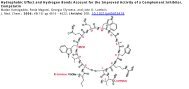
<strong>C5aR</strong>-<strong>Antagonist</strong><br />
Immediately after <strong>the</strong> blunt chest trauma, one group<br />
received a <strong>C5aR</strong>-antagonist (Ac-F[OPdChaWR]; PMX-53) at<br />
a dosage of 1 mg/kg intravenously into <strong>the</strong> penis vein. 20,21<br />
The injection was repeated 12 h after <strong>the</strong> trauma to prevent<br />
<strong>the</strong> C5a-dependent systemic inflammation, which was<br />
detectable during <strong>the</strong> first 12–24 h after <strong>the</strong> blunt chest<br />
trauma in rats. 1,6 Control animals received a peptide<br />
(Ac-F[OPdChaAdR]) with two changed amino acids, which<br />
does not have antagonistic activity and thus does not develop<br />
any biological effect at <strong>the</strong> same concentration and at <strong>the</strong><br />
same time points. 22<br />
Biomechanical Testing<br />
After 35 days <strong>the</strong> rats were sacrificed and <strong>the</strong> operated as<br />
well as <strong>the</strong> contralateral intact femora were explanted. Biomechanical<br />
testing was performed using a non-destructive,<br />
three-point bending test, as described previously. 6 Briefly,<br />
after removing <strong>the</strong> fixators, <strong>the</strong> distal end of each bone<br />
was potted in a cylinder using polymethylmethacrylate<br />
(Technovit 1 3040, Heraeus Kulzer GmbH, Wer<strong>the</strong>im,<br />
Germany) and fixed in a hinge joint whereas <strong>the</strong> proximal<br />
end of <strong>the</strong> femur rested on <strong>the</strong> bending support. A quasistatic<br />
load was applied in a three-point bending mode with a materials<br />
testing machine (1454, Zwick GmbH, Ulm, Germany)<br />
using a 500 N load cell (A.S.T. Angewandte System-Technik<br />
GmbH, Dresden, Germany) and <strong>the</strong> flexural rigidity (EI) was<br />
JOURNAL OF ORTHOPAEDIC RESEARCH 2011<br />
calculated from <strong>the</strong> slope of <strong>the</strong> force deflection curve. The<br />
absolute values of <strong>the</strong> operated femora were related to<br />
<strong>the</strong> contralateral values of <strong>the</strong> un-operated femora to<br />
eliminate individual differences.<br />
Micro-Computed Tomography<br />
The femora were scanned using a mCT scanning device<br />
(Skyscan 1172), operating at a peak voltage of 50 kV and<br />
200 mA at a resolution of 15 mm. The mineralized callus<br />
within <strong>the</strong> former osteotomy gap was segmented and <strong>the</strong><br />
total tissue volume and <strong>the</strong> bone volume fraction (BV/TV)<br />
were calculated by global thresholding to distinguish<br />
between mineralized and non-mineralized tissue. 23 The maximum<br />
moment of inertia was calculated based on <strong>the</strong> tissue<br />
area on <strong>the</strong> transversal slices in <strong>the</strong> fracture gap. The apparent<br />
modulus of elasticity was calculated as <strong>the</strong> flexural rigidity<br />
divided by <strong>the</strong> maximum moment of inertia. 24 According<br />
to <strong>the</strong> standard clinical evaluation of X-rays <strong>the</strong> number of<br />
bridged cortices per callus were evaluated in two planes at<br />
right angles to one ano<strong>the</strong>r by using an CT analyzing software<br />
(Data viewer, Skyscan, Kontich, Belgium). 25 The distal<br />
pin hole served as orientation for <strong>the</strong> exact positioning of <strong>the</strong><br />
specimens. At least three bridged cortices per callus were<br />
considered as a ‘‘healed fracture.’’ Two observers evaluated<br />
<strong>the</strong> cortical bridging independently in a blinded fashion.<br />
Histomorphometry<br />
After fixating <strong>the</strong> femora in buffered 4% formaldehyde <strong>the</strong>y<br />
were dehydrated with ethanol (40–100%) and embedded in<br />
methyl methacrylate (Merck KGaA, Darmstadt, Germany).<br />
Seventy micrometers longitudinal sections were prepared,<br />
which were cut in anterior–posterior direction of <strong>the</strong> right<br />
femur. The pin holes guaranteed <strong>the</strong> standardized orientation<br />
of <strong>the</strong> sections. Then <strong>the</strong> sections were stained with<br />
Paragon (Toluidin blue and Fuchsin; both Waldeck GmbH &<br />
Co KG, Münster, Germany), which stains fibrous tissue in<br />
blue, cartilage tissue in purple and mineralized matrix in<br />
white-yellow. In <strong>the</strong> former osteotomy gap <strong>the</strong> newly formed<br />
tissue was evaluated by using a light microscope (Leica<br />
DMI6000B) at a fivefold magnification. The amount of bone,<br />
cartilage and fibrous tissue was assessed by circumscribing<br />
<strong>the</strong> corresponding areas with image analysis software (Leica<br />
MMAF 1.4.0 Imaging System, Leica, Heerbrugg, Switzerland<br />
powered by MetaMorph 1 ).<br />
Statistical Analysis<br />
Results are presented as medians and interquartile ranges<br />
(IR). For statistical analysis, <strong>the</strong> software PASW Statistics<br />
18.0 (SPSS, Inc., Chicago, IL) was used. Differences between<br />
groups regarding flexural rigidity, mCT-parameters, and histomorphometrical<br />
data were calculated using a Mann–Whitney<br />
U-test, whereas differences between groups regarding<br />
<strong>the</strong> number of bridged cortices were calculated using <strong>the</strong><br />
Fisher exact test. The level of significance was p < 0.05.<br />
RESULTS<br />
Biomechanical Testing<br />
The treatment of <strong>the</strong> animals with <strong>the</strong> <strong>C5aR</strong>-antagonist<br />
after blunt chest trauma significantly increased<br />
<strong>the</strong> flexural rigidity (Ctrl: EI ¼ 46.54% (IR: 27.44);<br />
<strong>C5aR</strong>-Ag: EI ¼ 72.18% (IR: 66.50)) of <strong>the</strong> callus by<br />
about 55% compared to <strong>the</strong> control group, which<br />
received <strong>the</strong> control peptide (Fig. 1).