A Partial Skeleton of Pseudaelurus (Carnivora: Felidae) - American ...
A Partial Skeleton of Pseudaelurus (Carnivora: Felidae) - American ...
A Partial Skeleton of Pseudaelurus (Carnivora: Felidae) - American ...
Create successful ePaper yourself
Turn your PDF publications into a flip-book with our unique Google optimized e-Paper software.
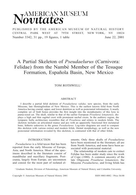
PUBLISHED BY THE AMERICAN MUSEUM OF NATURAL HISTORY<br />
CENTRAL PARK WEST AT 79TH STREET, NEW YORK, NY 10024<br />
Number 3342, 31 pp., 19 figures, 1 table June 22, 2001<br />
A <strong>Partial</strong> <strong>Skeleton</strong> <strong>of</strong> <strong>Pseudaelurus</strong> (<strong>Carnivora</strong>:<br />
<strong>Felidae</strong>) from the Nambé Member <strong>of</strong> the Tesuque<br />
Formation, Española Basin, New Mexico<br />
TOM ROTHWELL 1<br />
ABSTRACT<br />
I describe a partial felid skeleton <strong>of</strong> <strong>Pseudaelurus</strong> validus, new species, from the early<br />
Miocene, late Hemingfordian <strong>of</strong> New Mexico. This is the earliest known felid from North<br />
America having cranial, upper, and lower dentition as well as postcranial information. A nearly<br />
complete set <strong>of</strong> limb bones provides the first insight into the morphometry <strong>of</strong> this lynx to<br />
puma-sized cat. The skull, similar to that <strong>of</strong> the earlier European Proailurus lemanensis, displays<br />
a high and thin sagittal crest with prominent nuchal crests. In the auditory region, the<br />
tympanic bulla architecture resembles that <strong>of</strong> Proailurus and relates to modern felids. The<br />
skeleton includes an articulated manus and pes with an apparently functional first metatarsal<br />
bone, hitherto unknown in the genus <strong>Pseudaelurus</strong>. Log-ratio diagrams are used to compare<br />
this skeleton with various extinct and modern felids. Dental morphology, as well as the new<br />
postcranial information revealed by this skeleton, is contrasted with that <strong>of</strong> other felids.<br />
INTRODUCTION<br />
<strong>Pseudaelurus</strong> is a felid taxon that has been<br />
reported from the early Miocene <strong>of</strong> Europe,<br />
Asia, and North America. Most <strong>of</strong> the specimens<br />
described in the literature consist <strong>of</strong><br />
mandibular and maxillary fragments. Postcrania,<br />
largely from Europe, are uncommon<br />
and consist for the most part <strong>of</strong> isolated el-<br />
ements. Only three skulls <strong>of</strong> <strong>Pseudaelurus</strong><br />
have been mentioned in the literature; all are<br />
from North America, and none have been associated<br />
with postcranial material.<br />
The relationship <strong>of</strong> modern cats to extinct<br />
<strong>Felidae</strong> has been under study since the work<br />
<strong>of</strong> Cope (1880). A common ancestry <strong>of</strong> the<br />
late Oligocene Proailurus lemanensis, the<br />
Miocene <strong>Pseudaelurus</strong>, and living felids has<br />
1 Graduate Student, Division <strong>of</strong> Paleontology, <strong>American</strong> Museum <strong>of</strong> Natural History and Columbia University.<br />
Copyright � <strong>American</strong> Museum <strong>of</strong> Natural History 2001 ISSN 0003-0082 / Price $4.00
2 AMERICAN MUSEUM NOVITATES<br />
NO. 3342<br />
been suspected since before the turn <strong>of</strong> the<br />
last century (Adams, 1897). Although a<br />
small amount <strong>of</strong> cranial and postcranial material<br />
has been described <strong>of</strong> the earliest genus<br />
Proailurus (Filhol, 1888; Helbing, 1928), the<br />
postcrania <strong>of</strong> <strong>Pseudaelurus</strong> have been largely<br />
unknown. Considerable cranial and postcranial<br />
material for Felis is found throughout<br />
the fossil record from its earliest appearance<br />
in the late Miocene <strong>of</strong> Europe (de Beaumont,<br />
1961b) and Asia (Heintz, 1981; Qi, 1985)<br />
(fig. 1). Information on the postcrania <strong>of</strong><br />
<strong>Pseudaelurus</strong>, linked to a skull, is therefore<br />
needed to further the understanding <strong>of</strong> the<br />
relationships among these three felid genera.<br />
ABBREVIATIONS<br />
Anatomical<br />
a-p anterior to posterior<br />
c lower canine tooth<br />
C upper canine tooth<br />
d-v dorsal to ventral<br />
i1 lower first incisor<br />
I3 upper third incisor<br />
m1 lower first molar<br />
mm millimeter<br />
Mc1 first metacarpal bone<br />
Mt1 first metatarsal bone<br />
M1 upper first molar<br />
p1 lower first premolar<br />
ph1 first phalanx<br />
P3<br />
Institutional<br />
upper third premolar<br />
AMNH <strong>American</strong> Museum <strong>of</strong> Natural History,<br />
New York<br />
F:AM Frick Collection, <strong>American</strong> Museum<br />
<strong>of</strong> Natural History<br />
MHNL Muséum Guimet d’Histoire Naturelle,<br />
Lyon<br />
USNM United States Natural History Museum,<br />
Washington, DC<br />
HISTORY OF THE GENUS PSEUDAELURUS<br />
In 1843, H. M. Ducrotay de Blainville described<br />
a maxillary fragment and a lower jaw<br />
fragment <strong>of</strong> a ‘‘highly specialized machairodont’’<br />
from the medial Miocene <strong>of</strong> Sansan,<br />
France. The ramus contained three lower premolars<br />
(p2, p3, p4), whereas modern cats<br />
normally have only two (p3, p4). He gave<br />
this specimen the name Felis quadridentata.<br />
In 1850, Paul Gervais established the genus<br />
<strong>Pseudaelurus</strong> for the Blainville specimen. He<br />
distinguished it from the genus Felis by a<br />
single character: ‘‘possessing an additional<br />
inferior premolar (p2) in advance <strong>of</strong> the others’’.<br />
This was the first recognition <strong>of</strong> an ancestral<br />
state, or plesiomorphic anatomical<br />
condition <strong>of</strong> a fossil specimen, with respect<br />
to extant members <strong>of</strong> <strong>Felidae</strong>.<br />
In North America, Joseph Leidy (1858)<br />
described another new species <strong>of</strong> Miocene<br />
cat, Felis intrepidus, from ‘‘the loose sands<br />
<strong>of</strong> the Niobrara River’’ in western Nebraska.<br />
This fossil also possessed evidence <strong>of</strong> the<br />
lower second premolar, but Leidy apparently<br />
did not immediately recognize the generic<br />
value <strong>of</strong> this character. By the time he had<br />
published a drawing <strong>of</strong> the left ramus (Leidy,<br />
1869) however, he had designated it as <strong>Pseudaelurus</strong><br />
intrepidus. He explained the significance<br />
<strong>of</strong> the additional tooth: ‘‘In both rami<br />
the premolar (p2), considered as the chief<br />
character <strong>of</strong> the genus, is absent, but its alveolus<br />
remains midway in the hiatus back <strong>of</strong><br />
the canine tooth’’ (Leidy, 1869).<br />
Filhol (1879) proposed a new genus,<br />
Proailurus, to accommodate specimens from<br />
the early Miocene (Aquitanian) <strong>of</strong> France<br />
with characters more plesiomorphic and fossils<br />
stratigraphically older than those already<br />
assigned to <strong>Pseudaelurus</strong>. Proailurus lemanensis<br />
mandibles, dating back to the late Oligocene<br />
Quercy fissures <strong>of</strong> France, possessed<br />
four lower premolars where <strong>Pseudaelurus</strong><br />
had three. When E.D. Cope (1880) published<br />
his treatise: ‘‘On the Extinct Cats <strong>of</strong> North<br />
America’’, he clearly suggested that Proailurus<br />
and <strong>Pseudaelurus</strong> were ancestral felids.<br />
<strong>Pseudaelurus</strong> specimens first appear in the<br />
early Miocene (MN3) <strong>of</strong> Europe (Dehm,<br />
1950). The specimens are numerous in comparison<br />
to Proailurus. However, the overwhelming<br />
majority <strong>of</strong> the material reported<br />
in the literature consists <strong>of</strong> isolated lower<br />
jaws. Four species have been named from the<br />
Miocene <strong>of</strong> Europe: (1) <strong>Pseudaelurus</strong> quadridentatus<br />
Blainville, 1843; (2) <strong>Pseudaelurus</strong><br />
turnauensis Hoernes, 1882; (3) <strong>Pseudaelurus</strong><br />
lorteti Gaillard, 1899; and (4) <strong>Pseudaelurus</strong><br />
romieviensis Roman-Viret, 1934.<br />
Charles Depéret (1892) described a<br />
small specimen, <strong>Pseudaelurus</strong> transitorius<br />
from the La Grive-Saint-Alban (Isere) locale<br />
<strong>of</strong> France. This species was placed in
2001 ROTHWELL: PSEUDAELURUS FROM NEW MEXICO<br />
3<br />
Fig. 1. Stratigraphic ranges for Proailurus spp. (Pro), <strong>Pseudaelurus</strong> spp. in Europe (Eur), <strong>Pseudaelurus</strong><br />
spp. in North America (NA) and Felis (Felis). There appears to be no overlap in the fossil record<br />
among these three genera <strong>of</strong> <strong>Felidae</strong>. Proailurus is not found after MN2b. European <strong>Pseudaelurus</strong> range<br />
is that <strong>of</strong> the smallest species, P. turnauensis: MN3 to MN9. Correlation references: McKenna and Bell<br />
(1997), MacFadden and Hunt (1998).<br />
synonymy with P. turnauensis Hoernes by<br />
G. de Beaumont (1961). The synonymization<br />
was confirmed by Heizmann (1973)<br />
and Wang (1998), but ignored by Ginsburg<br />
(1983). All four species was originally<br />
described based on mandibular and maxillary<br />
fragments. There have been no European<br />
skulls yet described. The first postcranial<br />
bone was described in 1899: a right<br />
humerus <strong>of</strong> P. transitorius (� P. turnauen-<br />
sis) from the medial Miocene La Grive-<br />
Saint-Alban locality in France (Gaillard,<br />
1899). G. de Beaumont (1961) described<br />
19 separate postcranial bones from La Grive-Saint-Alban<br />
and the early Miocene Wintersh<strong>of</strong>-West<br />
localities, but none was alleged<br />
to be from the same individual. L.<br />
Ginsburg (1961b) reported an even larger<br />
assortment <strong>of</strong> isolated mandibular, maxillary<br />
and postcranial material from the me-
4 AMERICAN MUSEUM NOVITATES<br />
NO. 3342<br />
dial Miocene <strong>of</strong> Sansan in France, again<br />
without a skull.<br />
Richard Dehm (1950), in his seminal report<br />
on the carnivores from Wintersh<strong>of</strong>-West<br />
in Germany, described 18 lower jaws and 1<br />
maxillary specimen. These are early Miocene<br />
specimens <strong>of</strong> Burdigalian (MN3) age. Dehm<br />
assigned them to P. transitorius Depéret (�<br />
P. turnauensis Hoernes). Dehm described<br />
these specimens as elements from a transitional<br />
species between Proailurus and the<br />
more derived specimens <strong>of</strong> <strong>Pseudaelurus</strong> reported<br />
from La Grive-Saint-Alban in France.<br />
What Dehm considered most interesting in<br />
this collection was the variable key lower<br />
jaw characters: (1) presence or absence <strong>of</strong><br />
p1; (2) presence or absence <strong>of</strong> p2; (3) one or<br />
two roots <strong>of</strong> p2; (4) presence or absence <strong>of</strong><br />
m2; (5) presence or absence <strong>of</strong> m1 metaconid;<br />
(6) length <strong>of</strong> m1.<br />
Elmar Heizmann (1973) published a review<br />
<strong>of</strong> the European <strong>Pseudaelurus</strong> radiation<br />
and recognized four species in order <strong>of</strong> increasing<br />
size: (1) <strong>Pseudaelurus</strong> turnauensis<br />
Hoernes; (2) <strong>Pseudaelurus</strong> lorteti Gaillard;<br />
(3) <strong>Pseudaelurus</strong> romieviensis Roman and<br />
Viret; (4) <strong>Pseudaelurus</strong> quadridentatus<br />
Blainville. He suggested that <strong>Pseudaelurus</strong><br />
evolved from Proailurus with the following<br />
character changes: (1) loss <strong>of</strong> p1; (2) loss <strong>of</strong><br />
m2; (3) reduction <strong>of</strong> p2 to one root; (4)<br />
lengthening <strong>of</strong> canines; (5) reductions in<br />
height <strong>of</strong> lower premolars such that p3 and<br />
p4 became similar in height; (6) reduction <strong>of</strong><br />
m1 protoconid height; (7) movement <strong>of</strong> m1<br />
metaconid from a position on the posterior<br />
surface <strong>of</strong> the protoconid, lingual <strong>of</strong> median,<br />
to a median position; (8) reduction <strong>of</strong> m1 talonid.<br />
In North America, three species have been<br />
described from the medial Miocene. Again,<br />
all species determinations were based on<br />
mandibular and maxillary fragments. <strong>Pseudaelurus</strong><br />
intrepidus Leidy, 1858, and <strong>Pseudaelurus</strong><br />
marshi Thorpe, 1922, are both late<br />
Barstovian specimens from the Valentine<br />
Formation in western Nebraska. <strong>Pseudaelurus</strong><br />
aeluroides Macdonald, 1954, is from the<br />
early Barstovian Olcott Formation in Sioux<br />
County, Nebraska. J. R. Macdonald (1948a,<br />
1948b) described two large Clarendonian<br />
<strong>Pseudaelurus</strong> species that have enlarged<br />
mental ridges on the ventral margins <strong>of</strong> the<br />
mandibles: <strong>Pseudaelurus</strong> pedionomus from<br />
the Minnechaduza Fauna <strong>of</strong> Nebraska and<br />
<strong>Pseudaelurus</strong> thinobates from the Black<br />
Hawk Ranch Local Fauna <strong>of</strong> California. David<br />
Kitts (1958) erected a new genus, Nimravides,<br />
for this material and made <strong>Pseudaelurus</strong><br />
thinobates its type species.<br />
Some North <strong>American</strong> cranial material has<br />
been described. Chester Stock (1934) described<br />
the first <strong>Pseudaelurus</strong> skull in a report<br />
on five individuals from the early Barstovian<br />
Tonopah, Nevada locality. He assigned<br />
all to P. intrepidus Leidy. MacDonald<br />
(1948a) described seven assorted postcranial<br />
bones referenced to P. pedionomus, but material<br />
referred to this species has subsequently<br />
been shifted into the genus Nimravides (de<br />
Beaumont, 1990). Shotwell and Russel<br />
(1963) also described some assorted postcranial<br />
fragments <strong>of</strong> <strong>Pseudaelurus</strong>, which they<br />
thought ‘‘apparently represent a single individual’’,<br />
but no species determination was<br />
made.<br />
In Asia, three species <strong>of</strong> <strong>Pseudaelurus</strong><br />
have been reported. All are described from<br />
mandibular, maxillary, or dental material:<br />
<strong>Pseudaelurus</strong> guangheensis Cao, Du, Zhao,<br />
and Cheng, 1990, is a new species from the<br />
medial Miocene Guanghe District <strong>of</strong> Gansu,<br />
China. <strong>Pseudaelurus</strong> cuspidatus Wang, 1998,<br />
is a new species from the early medial Miocene<br />
Halamagai Formation in northern Junggar<br />
Basin, in the Xinjiang Autonomous Region,<br />
China. P. lorteti left ramus and dental<br />
fragments were reported from the medial<br />
Miocene <strong>of</strong> Xiacaowan, Sihong County in<br />
Jiangsu Province, China (Qiu and Gu, 1986).<br />
From Africa, an m1 and tibia fragment<br />
from the Al-Sarrar locality in what is now<br />
Saudi Arabia were assigned to P. turnauensis<br />
(Thomas et al., 1982). This formation is early<br />
Miocene (MN4a) in age. There are no other<br />
reports <strong>of</strong> any nonmachairodont members <strong>of</strong><br />
<strong>Felidae</strong> from the African Miocene.<br />
The Nambé skeleton (F:AM 62128) described<br />
in this paper was collected in 1939<br />
by J. C. Blick <strong>of</strong> the Frick Laboratory. The<br />
specimen was recovered from the late Hemingfordian<br />
Nambé Member locality (Galusha<br />
and Blick, 1971) in the Tesuque Formation,<br />
Española Basin, near East Cuyamunque,<br />
New Mexico.
2001 ROTHWELL: PSEUDAELURUS FROM NEW MEXICO<br />
5<br />
SYSTEMATIC PALEONTOLOGY<br />
ORDER CARNIVORA BOWDICH, 1821<br />
SUBORDER FELIFORMIA KRETZOI, 1945<br />
FAMILY FELIDAE FISCHER DE WALDHEIM, 1817<br />
Genus <strong>Pseudaelurus</strong> Gervais, 1850<br />
DISTRIBUTION: Early to medial (MN3 to<br />
MN9) Miocene <strong>of</strong> Europe; early to medial<br />
(late Hemingfordian to late Barstovian) Miocene<br />
<strong>of</strong> North America; early medial Miocene<br />
<strong>of</strong> Asia; early (MN4a) Miocene <strong>of</strong> Africa.<br />
GENERIC DIAGNOSIS: Members <strong>of</strong> <strong>Felidae</strong><br />
with the unique combination <strong>of</strong> the following<br />
derived and primitive characters: presence <strong>of</strong><br />
p2 with the usual absence <strong>of</strong> p1 and m2, m1<br />
with reduced metaconid and talonid, P2 with<br />
one root, alisphenoid canal present, paroccipital<br />
process cupped about the posterior surface<br />
<strong>of</strong> an enlarged caudal entotympanic.<br />
Differing from extant felid genera by c cross<br />
section showing flattened inner surface and<br />
posterior trenchant edge, presence <strong>of</strong> p2, m1<br />
with variable metaconid and reduced talonid,<br />
presence <strong>of</strong> alisphenoid canal. Differing from<br />
Metailurus by presence <strong>of</strong> alisphenoid canal<br />
and p2, absence <strong>of</strong> enlargement <strong>of</strong> mental<br />
ridge. Differing from Nimravides by smaller<br />
size and absence <strong>of</strong> any ventral mandibular<br />
mental ridge enlargement. Differing from<br />
Proailurus by absence <strong>of</strong> p1 and m2.<br />
TYPE SPECIES: <strong>Pseudaelurus</strong> quadridentatus,<br />
(Blainville, 1843) (�Felis quadridenta<br />
Blainville, 1843).<br />
INCLUDED SPECIES: <strong>Pseudaelurus</strong> intrepidus<br />
Leidy, 1858; <strong>Pseudaelurus</strong> turnauensis<br />
Hoernes, 1882; <strong>Pseudaelurus</strong> lorteti Gaillard,<br />
1899; <strong>Pseudaelurus</strong> marshi Thorpe,<br />
1922; <strong>Pseudaelurus</strong> romieviensis Roman-Viret,<br />
1934; <strong>Pseudaelurus</strong> aeluroides Macdonald,<br />
1954; <strong>Pseudaelurus</strong> guangheensis Cao,<br />
Du, Zhao, and Cheng, 1990; <strong>Pseudaelurus</strong><br />
cuspidatus Wang, 1998.<br />
<strong>Pseudaelurus</strong> validus, new species<br />
HOLOTYPE: F:AM 62128, skull with left<br />
and right I3, left C, broken right C, singlerooted<br />
alveoli for P1 and P2, left and right<br />
P3-M1. Articulated lower jaws with left i3,<br />
left and right c, left and right single-rooted<br />
alveoli for p2, left and right p3-m1. Postcrania:<br />
right and left humeri, right and left radii,<br />
right and left ulnae, left manus, articulated<br />
right manus, distal fragment <strong>of</strong> left femur<br />
with patella, left tibia, right tibial fragment,<br />
left and right fibular fragments, right pes, articulated<br />
left pes, partially prepared elements<br />
in plaster jacket, including exposed scapulae,<br />
ribs, vertebrae.<br />
TYPE LOCALITY: Nambé Member, Tesuque<br />
Formation, Española Basin near East Cuyamunque,<br />
New Mexico.<br />
AGE: Late Hemingfordian (Galusha and<br />
Blick, 1971).<br />
ETYMOLOGY: validus, Latin for strong, robust,<br />
able.<br />
REFERRED SPECIMENS: Sheep Creek Fauna,<br />
Sheep Creek Formation (late Hemingfordian),<br />
Sioux County, Nebraska: F:AM 61827,<br />
right ramus with c, alveolus p2, p3, p4, broken<br />
m1, Greenside Quarry; F:AM 61837, left<br />
maxillary fragment with I3, C-M1, right tibia<br />
and fibula, partial right radius, partial right<br />
ulna, astragalus, vertebrae and right fourth<br />
metatarsal, Head <strong>of</strong> Pliohippus Draw; Lower<br />
Snake Creek Fauna, Olcott Formation (early<br />
Barstovian), Sioux County, Nebraska: F:AM<br />
61834, complete skull with upper dentition,<br />
zygomatic arches, intact basicranium and associated<br />
lower jaws (F:AM 61829), Humbug<br />
Quarry; F:AM 61803, left maxilla with I3<br />
alveolus, C, alveolus P1 and P2; P3-M1,<br />
Humbug Quarry; F:AM 61833, partial skull<br />
with upper dentition and partial right zygomatic<br />
arch, Quarry 3 (Far Surface Quarry),<br />
F:AM 61832, left ramus with i3, c, p2, broken<br />
p3, p4 and broken m1, East Wall Quarry;<br />
F:AM 61830, partial left ramus with p4, m1,<br />
Quarry 3 (Far Surface Quarry); F:AM<br />
61828, left and right rami with left and right<br />
c, p2 alveoli, p3-m1, right humerus, left radius,<br />
left and right tibiae, Echo Quarry; F:<br />
AM 61835, a nearly complete skull with upper<br />
dentition and intact basicranium, Echo<br />
Quarry; F:AM 61836, skull fragment with<br />
left P4, left and right partial rami with partial<br />
dentitions, maxillary fragment with canine<br />
tooth, left radius, Echo Quarry.<br />
DISTRIBUTION: Late Hemingfordian <strong>of</strong> New<br />
Mexico, late Hemingfordian and early Barstovian<br />
<strong>of</strong> Nebraska.<br />
DIAGNOSIS: Differing from other species<br />
by the combination <strong>of</strong> large size, long distance<br />
between c and p3, extremely reduced<br />
or absent metaconid on m1, and robust den-
6 AMERICAN MUSEUM NOVITATES<br />
NO. 3342<br />
tary with large and erect rectangular-shaped<br />
coronoid process. P. validus overlaps in size<br />
with P. intrepidus and P. marshi in North<br />
America and P. quadridentatus in Europe,<br />
and can be differentiated from these species<br />
by the longer c-p3 distance in P. validus,<br />
which exceeds the length <strong>of</strong> its m1, and by<br />
its large, erect, and rectangular coronoid process.<br />
DESCRIPTION AND COMPARISON<br />
SKULL<br />
The skull (figs. 2a, 2b) has been crushed,<br />
giving it a low and wide appearance. This<br />
compromises the integrity <strong>of</strong> some skull<br />
measurements, especially those <strong>of</strong> height and<br />
width. However, the upper dentition survived,<br />
and can be associated with the lower<br />
teeth for only the second time in this genus.<br />
The premaxillary bones possess a pair <strong>of</strong><br />
fragmented I3, the I1–2 having been lost. A<br />
short and smooth diastema separates I3 from<br />
C. The left C is separated from the skull,<br />
revealing a large, cavernous alveolus. The<br />
right C root is in situ; however its crown is<br />
fractured and missing. These two C are more<br />
elliptical than round in cross-section and lack<br />
any longitudinal grooves. Posterior to the alveolus<br />
for the left C are a preserved P1 and<br />
an alveolus for P2, both single-rooted. This<br />
agrees with the presence <strong>of</strong> these teeth in the<br />
skull described by Stock (1934) from Tonopah,<br />
although the presence <strong>of</strong> P1 was interpreted<br />
by Stock as being ‘‘a rather unusual<br />
feature’’.<br />
The lengths <strong>of</strong> P3 and P4 in P. validus<br />
agree with the Tonopah skull but are smaller<br />
than the P. quadridentatus type. These two<br />
teeth are aligned parallel to the maxillary<br />
axis and do not exhibit any overlap, obliqueness,<br />
or crowding as seen in some extant species<br />
(Salles, 1992). The left P3 is wider and<br />
more robust when compared to P3 in the P.<br />
lemanensis type skull (MNHN1903–20). On<br />
the right P4 (fig. 2b) can be seen a prominent<br />
protocone that projects in an anterolingual<br />
direction. The morphology <strong>of</strong> P4 in F:AM<br />
62128 resembles the P4 in the P. quadridentatus<br />
maxillary fragment from la Grive-<br />
Saint-Alban (Isère) (cast � AMNH 103396)<br />
described by Gaillard (1899). In the P. lemanensis<br />
type specimen, the protocone <strong>of</strong> P4<br />
is at a slightly more obtuse angle to the lingual<br />
surface <strong>of</strong> P4. In modern felids, the protocone<br />
is closer to the lingual surface <strong>of</strong> the<br />
tooth, resulting in a narrower, compressed<br />
P4. The P4 <strong>of</strong> F:AM 62128 has a parastyle<br />
on the anterior surface which does not differ<br />
from the condition in P. lemanensis, other<br />
species <strong>of</strong> <strong>Pseudaelurus</strong>, or modern felids.<br />
Both M1 are present with two roots. The<br />
M1 are compressed in the a-p direction, but<br />
are elongate transversely. Individual cusps<br />
can be seen on the left M1. The buccal end<br />
<strong>of</strong> this tooth is formed by the parastyle wing.<br />
The nearly conjoined paracone and metacone<br />
are midway along the occlusal surface. The<br />
lingual margin <strong>of</strong> M1 is formed by a distinct<br />
protocone. The parastyle <strong>of</strong> F:AM 626128<br />
M1 is as tall as the caudal surface <strong>of</strong> the<br />
metastyle blade <strong>of</strong> P4 and appears to extend<br />
the carnassial blade. The left M1 <strong>of</strong> F:AM<br />
62128 compares favorably with the M1 in<br />
the P. lemanensis skull (MNHN1903–20),<br />
but is in sharp contrast to the rudimentary<br />
M1 <strong>of</strong> modern cats.<br />
The skull has a high, thin sagittal crest<br />
which is joined by equally large nuchal crests<br />
that unite at the occiput to form a protuberance<br />
that projects to a point caudal to the<br />
level <strong>of</strong> the occipital condyles. This area <strong>of</strong><br />
the skull is very similar to the type skull <strong>of</strong><br />
P. lemanensis (MNHN1903–20) from the<br />
early Miocene <strong>of</strong> France. The basicranial<br />
anatomy <strong>of</strong> F:AM 62128 is somewhat distorted,<br />
but much information endures (figs.<br />
2b, 3). On the right side <strong>of</strong> the skull, much<br />
<strong>of</strong> the ossified bulla is lost, but the close approximation<br />
<strong>of</strong> the mastoid and paroccipital<br />
process to the bulla can be appreciated. The<br />
paroccipital process displays a concavity on<br />
its anterior surface where it cradled the posterior<br />
surface <strong>of</strong> the tympanic bulla in typical<br />
aeluroid fashion (Cope, 1880; Hunt, 1987,<br />
1989, 1998; Werdelin, 1996; Wyss and<br />
Flynn, 1993). Medially, the ridge formed by<br />
the caudal entotympanic indenting the basioccipital<br />
bone can be seen (fig. 3). On the<br />
lateral margins <strong>of</strong> the right bulla, just proximal<br />
to the mastoid process, is the external<br />
acoustic meatus surrounded by most <strong>of</strong> the<br />
ectotympanic. This evidence <strong>of</strong> the anterolateral<br />
compartment <strong>of</strong> the bulla gives a<br />
strong signal as to the inflated size <strong>of</strong> the<br />
caudal entotympanic. This is further evi-
2001 ROTHWELL: PSEUDAELURUS FROM NEW MEXICO<br />
7<br />
Fig. 2a. Dorsal view <strong>of</strong> F:AM 62128 skull. The morphology <strong>of</strong> the cranium resembles that <strong>of</strong> the<br />
P. lemanensis skull (MNHN S.G. 3509a) from Europe and the early North <strong>American</strong> Ginn Quarry skull<br />
(F:AM 61847) from Nebraska. sc � sagittal crest, nc � nuchal crest.<br />
dence that this Nambé <strong>Pseudaelurus</strong> specimen<br />
possessed felid characters in the basicranial<br />
area (Hunt and Tedford, 1993).<br />
The petrosal bone and its promontorial<br />
process are preserved in the tympanic bulla<br />
region on the left side (figs. 2b, 3). Although<br />
the petrosal has been nudged in a caudal direction<br />
by the crushing, facets for its articulation<br />
with the ectotympanic can be seen on<br />
its ventral surface. The grooving <strong>of</strong> the an-
8 AMERICAN MUSEUM NOVITATES<br />
NO. 3342<br />
Fig. 2b. Ventral view <strong>of</strong> F:AM 62128 skull containing four upper premolars with P2 evidenced by<br />
a single-rooted alveolus. PP � paroccipital process, P � petrosal part <strong>of</strong> the temporal bone, M � mastoid<br />
bone.<br />
terodorsal ro<strong>of</strong> <strong>of</strong> the tympanic bulla, formed<br />
by the anterior invasion <strong>of</strong> the caudal entotympanic<br />
to the level <strong>of</strong> the squamosal bone,<br />
can also be seen. The hypoglossal foramina<br />
are visible bilaterally, within the depression<br />
where the posterior lacerate foramina would<br />
be.<br />
LOWER JAW<br />
The two rami <strong>of</strong> the lower jaw were cemented<br />
together during early, cursory preparation<br />
(fig. 4). The size <strong>of</strong> this jaw is smaller<br />
than Leidy’s P. intrepidus type, and is more<br />
similar in size to P. marshi (table 1). How-
2001 ROTHWELL: PSEUDAELURUS FROM NEW MEXICO<br />
9<br />
Fig. 3. Close view <strong>of</strong> basicranial area <strong>of</strong> skull. The x markers delineate the medial emargination <strong>of</strong><br />
the basioccipital caused by an enlarged caudal entotympanic. Also seen: the concavity <strong>of</strong> the paroccipital<br />
process (PP) where it contacts the caudal surface <strong>of</strong> the expanded caudal entotympanic. T � ectotympanic,<br />
P � petrosal bone, PP � paroccipital process.<br />
ever, it is obvious that this jaw was reconstructed<br />
from multiple fragments, thus compromising<br />
its measurements. The mandibles<br />
are not slender, as described in Thorpe’s P.<br />
marshi specimen, but rather display considerable<br />
depth below the tooth row. The ventral<br />
surface <strong>of</strong> the lower jaw is slightly convex,<br />
but the area immediately below the tooth row<br />
is essentially straight. There is no ventral<br />
bulge in this location.<br />
Both <strong>of</strong> the large lower canines are preserved.<br />
The inner surface <strong>of</strong> each is more<br />
flattened than the outer surfaces. Their<br />
crowns do not possess any grooves. In the<br />
space between c and p3 on both rami is an<br />
alveolus for p2, located not at the midpoint<br />
in the diastema as in Leidy’s description<br />
(Leidy, 1869), but considerably closer to p3<br />
(fig. 4). This single alveolus is also located<br />
medial <strong>of</strong> the line <strong>of</strong> the tooth row, a character<br />
first described by Thorpe (1922) in the<br />
P. marshi type specimen. This medial location<br />
<strong>of</strong> p2 is a consistent feature in the North<br />
<strong>American</strong> specimens, and can be seen in the<br />
P. intrepidus and P. intrepidus sinclairi types<br />
(Matthew, 1918; Thorpe, 1922). Although<br />
the number <strong>of</strong> roots <strong>of</strong> p2 varied in early European<br />
specimens (Dehm, 1950), I am unaware<br />
<strong>of</strong> any references, in North America,<br />
to a double-rooted p2.<br />
The anterior mental foramina are located<br />
beneath the posterior portion <strong>of</strong> the c-p3 diastema,<br />
and are best seen on the left ramus<br />
<strong>of</strong> this jaw (fig. 4). The left posterior mental<br />
foramen is underneath the posterior root <strong>of</strong><br />
the third lower premolar. This location agrees<br />
well with Stock’s (1934) description <strong>of</strong> the<br />
Tonopah specimens that he assigned to P. intrepidus<br />
and is in slight contrast to the more<br />
anterior location in the P. marshi specimen<br />
(Thorpe, 1922). The coronoid process is wider<br />
and more erect than in the Leidy or Marsh<br />
types and is much taller than in P. lemanensis.<br />
There is no terminal ‘‘hook’’ in the coronoid<br />
process as is seen in modern felids.<br />
The deep masseteric fossa <strong>of</strong> F:AM 62128<br />
extends anteriorly to a point just below the<br />
talonid <strong>of</strong> m1. The F:AM 62128 condyle has<br />
a mediolateral articular surface that is 20%<br />
longer than the Leidy type, and sits higher
10 AMERICAN MUSEUM NOVITATES<br />
NO. 3342<br />
Fig. 4. Lateral (A) and oblique (B) views <strong>of</strong> lower jaw indicating dental characters <strong>of</strong> generic<br />
significance: presence <strong>of</strong> p2, lack <strong>of</strong> p1 and m2, and reduced metaconid and talonid on m1.<br />
by an equal amount above the angular process.<br />
The p3 and p4 are damaged by crushing.<br />
However, their size is recoverable (table 1).<br />
The posterior accessory cusps <strong>of</strong> the left p3<br />
and both p4 can be seen on the posterior surface<br />
<strong>of</strong> the primary cusps. These posterior<br />
accessory cusps are separate from a well-defined<br />
posterior cingulum. This condition <strong>of</strong> a<br />
conspicuous posterior accessory cusp on p3<br />
and p4 is present in specimens <strong>of</strong> Proailurus<br />
lemanensis and persists, with little change, in<br />
modern felids. The left m1 is well preserved<br />
and demonstrates a much-reduced metaconid<br />
that blends smoothly into an abridged talonid.<br />
The protoconid is taller than the paraconid<br />
and the carnassial notch is open and<br />
deep. This description <strong>of</strong> the carnassial<br />
would fit the diagnosis <strong>of</strong> any <strong>of</strong> the previously<br />
described large <strong>Pseudaelurus</strong> species
2001 ROTHWELL: PSEUDAELURUS FROM NEW MEXICO<br />
11<br />
TABLE 1<br />
Comparison <strong>of</strong> Lower Jaws <strong>of</strong> Five North <strong>American</strong> Type Specimens <strong>of</strong> <strong>Pseudaelurus</strong><br />
<strong>of</strong> North America: P. intrepidus, P. marshi,<br />
or Matthew’s sinclairi variety <strong>of</strong> P. intrepidus.<br />
Because <strong>of</strong> the predominance <strong>of</strong> lower jaw<br />
material in the fossil felid literature, this portion<br />
<strong>of</strong> the skeleton must be given special<br />
consideration with respect to diagnosis and<br />
species referrals. Most species within the genus<br />
<strong>Pseudaelurus</strong> are represented only by<br />
lower jaws. A recent study utilizing bivariate<br />
analysis (Glass and Martin, 1978) concluded<br />
that mandibular dentition is useful in differentiating<br />
extant felid species. However, to<br />
rely on lower jaw material for purposes <strong>of</strong><br />
specific diagnosis is at present problematic.<br />
In table 1 and in the log-ratio diagram (Simpson,<br />
1941) <strong>of</strong> figure 5, the lower jaw <strong>of</strong> the<br />
Nambé skeleton is compared with four North<br />
<strong>American</strong> type specimens.<br />
HUMERUS<br />
In the first description <strong>of</strong> a postcranial<br />
<strong>Pseudaelurus</strong> specimen, Claude Gaillard<br />
(1899) compared a P. turnauensis right humerus<br />
from La Grive-Saint-Alban with that<br />
<strong>of</strong> a domestic cat (Felis catus) and <strong>of</strong> a lynx<br />
(Lynx lynx). The <strong>Pseudaelurus</strong> humeri from<br />
the Nambé Member are 50% larger than the<br />
specimen from France. The shaft <strong>of</strong> the F:<br />
AM 6128 right humerus (fig. 6) is thinner<br />
when viewed from the anterior or posterior<br />
perspective. Its lateral and medial surfaces<br />
appear broad and flat. The proximal end <strong>of</strong><br />
each humerus consists <strong>of</strong> a convex arthral<br />
surface that is visible only from the posterior<br />
view. This articular surface blends into a<br />
deep bicipital groove between greater and<br />
lesser tubercles <strong>of</strong> equal height. The greater<br />
tubercle continues down the anterior surface<br />
<strong>of</strong> the shaft <strong>of</strong> the humerus as a sharp, but<br />
low, deltoid ridge, very typical <strong>of</strong> a cursorial<br />
carnivore (Ginsburg, 1961a; Wang, 1993). In<br />
the dog, this ridge is more exaggerated, and<br />
is better termed the deltoid tuberosity. In the<br />
domestic cat, this deltoid ridge blends into<br />
the humeral shaft by midpoint. In this specimen,<br />
however, the deltoid ridge extends well<br />
into the distal half <strong>of</strong> the humerus. This<br />
agrees with Ginsburg’s description <strong>of</strong> this<br />
character in the P. quadridentatus humerus<br />
from Sansan. On the lateral surface <strong>of</strong> the<br />
proximal end <strong>of</strong> the right humerus, the pectoral<br />
ridge extends down from the posterior<br />
surface <strong>of</strong> the greater tubercle (fig. 6).<br />
Modern felid humeri are most cylindrical<br />
just distal to the midpoint <strong>of</strong> the diaphysis,<br />
and this is true <strong>of</strong> F:AM 62128. From this<br />
midpoint in the humeral diaphysis, the supracondyloid<br />
ridge extends down the lateral<br />
margin <strong>of</strong> the shaft to the point <strong>of</strong> the lateral<br />
epicondyle. This feature is large in this specimen,<br />
forming a shelf that curls in the ante-
12 AMERICAN MUSEUM NOVITATES<br />
NO. 3342<br />
Fig. 5. Log-ratio diagram <strong>of</strong> lower jaw characters and measurements featured in table 1. The standard<br />
specimen on the y axis is P. lemanensis, an early Miocene felid from Europe. The five <strong>Pseudaelurus</strong><br />
specimens fall into two categories <strong>of</strong> jaw types based on size <strong>of</strong> jaw and the c-p3 diastema character.<br />
The two P. intrepidus species and F:AM 62128 have larger jaws than Proailurus, but still have a<br />
relatively shortened c-p3 diastema when compared to their European relative. The other two <strong>Pseudaelurus</strong><br />
species (P. marshi, P. aeluroides) have a greater reduced c-p3 diastema in comparison to Proailurus.<br />
rior direction. This shelf forms a ridge with<br />
a cupped appearance when viewed from the<br />
anterior perspective. Forming the medial<br />
edge <strong>of</strong> this distal end <strong>of</strong> the bone is the medial<br />
condyloid ridge. The supracondlyoid foramen<br />
is located here. This oval shaped foramen,<br />
which carries the brachial artery and<br />
the median nerve in <strong>Felidae</strong>, is somewhat<br />
smaller in this specimen when compared to<br />
similar sized modern specimens. This is due<br />
to the crushing. In other, better preserved<br />
<strong>Pseudaelurus</strong> sp. humeri specimens in the<br />
Frick-AMNH collection, these foramina<br />
agree in size and shape with modern specimens<br />
such as Panthera pardus or Felis concolor.<br />
The distal end <strong>of</strong> the P. validus humeri,<br />
while extremely similar to modern felids,<br />
possesses certain characters that resemble the<br />
humerus <strong>of</strong> Proailurus. Differing in the proportions<br />
<strong>of</strong> the medial epicondyle, depth <strong>of</strong><br />
olecranon fossa, and the distally projecting<br />
medial margin <strong>of</strong> the trochlea, the <strong>Pseudaelurus</strong><br />
humerus maintains a position interme-<br />
diate between Proailurus and modern felids.<br />
The two P. validus humeri possess large, bulbous,<br />
and convoluted medial epicondyles,<br />
deep olecranon fossae, and robust medial<br />
margins <strong>of</strong> the trochleae. This morphology<br />
agrees well with Ginsburg’s (1961b) description<br />
<strong>of</strong> <strong>Pseudaelurus</strong> quadridentatus, which<br />
he stated was closer morphologically to<br />
Proailurus than to modern felids. The large<br />
and rough-surfaced medial epicondyles, typical<br />
<strong>of</strong> animals that climb (or dig) (Taylor,<br />
1976; Heinrich and Rose, 1997) are in contrast<br />
to the smaller and smoother epicondyles<br />
in modern felids. This combination <strong>of</strong> enlarged<br />
medial epicondyle and deep olecranon<br />
fossa suggests that this species was a reasonably<br />
cursorial cat (Wang, 1993). The capitulum<br />
on the humeral trochlea is convex and<br />
faces primarily in an anterior direction. Medially,<br />
the arthral surface becomes concave<br />
and terminates in a distally projecting, robust<br />
trochlea. Just proximal to these structures, on<br />
the anterior surface is the radial fossa adja-
2001 ROTHWELL: PSEUDAELURUS FROM NEW MEXICO<br />
13<br />
Fig. 6. Right humerus: dorsal view (left) and<br />
ventral view (right). The deltoid ridge (dr) can be<br />
seen extending beyond midpoint into the distal<br />
half <strong>of</strong> the diaphysis. gt � greater tubercle, sf �<br />
supracondyloid foramen, sr � supracondyloid<br />
ridge, <strong>of</strong> � olecranon fossa, me � medial epicondyle.<br />
cent to the more lateral and slightly smaller<br />
ulnar fossa.<br />
RADIUS<br />
The right radius (fig. 7) has a proximal<br />
surface that is oval in shape. The bone immediately<br />
narrows into a brief neck area just<br />
prior to the bicipital tuberosity that lies on<br />
the posteromedial surface. The shaft is<br />
roughly cylindrical and is convex in the lateral<br />
and cranial direction. All <strong>of</strong> these characters<br />
agree with modern <strong>Felidae</strong>. The radius<br />
increases in diameter distally. Unless this is<br />
due to crushing, it agrees with the description<br />
<strong>of</strong> Proailurus (Ginsburg, 1961b), and may<br />
establish a plesiomorphic character state for<br />
felid radii.<br />
Fig. 7. A Medial view <strong>of</strong> right ulna, B: dorsal<br />
view <strong>of</strong> right radius.<br />
ULNA<br />
The right and left ulnae <strong>of</strong> F:AM 62128<br />
are complete. The right (fig. 7) was selected<br />
for description. There is considerable width<br />
or surface area <strong>of</strong> the trochlear notch, more<br />
so than in extant felids. This joint surface is<br />
interrupted only by the laterally excavated<br />
radial notch that accepts the head <strong>of</strong> the radius.<br />
This laterally positioned radial notch<br />
has been shown to be a consistent character<br />
within modern felids (Gonyea, 1978), contrasting<br />
with the more cranially located notch<br />
in canids and hyaenids. This ulna has a maximum<br />
width just below the olecranon fossa<br />
and tapers towards its distal termination as<br />
the styloid process. Below the olecranon, in<br />
the next one-third <strong>of</strong> the diaphysis, the shaft<br />
changes from the two-sided olecranon to a
14 AMERICAN MUSEUM NOVITATES<br />
NO. 3342<br />
three-surfaced bone. In the final one-third <strong>of</strong><br />
this bone, the shaft tapers and loses a sense<br />
<strong>of</strong> surfaces.<br />
CARPUS<br />
The F:AM 62128 scapholunar proximal<br />
surface consists <strong>of</strong> a dome-shaped convexity<br />
for articulation with the distal radius. Its distal<br />
surface is extremely complicated, containing<br />
facets for articulation with all components<br />
<strong>of</strong> the next row <strong>of</strong> carpal bones. Most<br />
anterior and medial is the facet for articulation<br />
with the trapezoid. Moving posteriorly<br />
and laterally, there are facets for the trapezium<br />
and the magnum, and, on the posteriormost<br />
margin <strong>of</strong> this carpal bone there is the<br />
surface for articulation with the unciform.<br />
This complicated articulation can be seen in<br />
a photograph <strong>of</strong> a European specimen assigned<br />
to P. lorteti (de Beaumont, 1961b,<br />
plate 2, #7), and agrees well with modern<br />
felids.<br />
The lateral member <strong>of</strong> the proximal row<br />
<strong>of</strong> carpal bones is the cuneiform. Its lateral<br />
surface consists <strong>of</strong> two equal-sized concave<br />
facets for articulation with the ulna and the<br />
pisiform. This ulnar articulation is <strong>of</strong>ten lacking<br />
in modern felids (Merriam and Stock,<br />
1932), but can be clearly seen in this specimen.<br />
The pisiform <strong>of</strong> F:AM 62128 contains<br />
a deeper groove on its proximal end than is<br />
seen in extant felids, but agrees in this respect<br />
with illustrations <strong>of</strong> Felis atrox and<br />
Smilodon californicus (Merriam and Stock,<br />
1932). Otherwise, the cuneiform and the pisiform<br />
do not differ from these same elements<br />
in modern felids.<br />
In the distal row <strong>of</strong> carpal bones, most medial,<br />
is the trapezium articulating with Mc1.<br />
Lateral to the trapezium and articulating with<br />
Mc2 is the trapezoid. It is clear that both articulate<br />
with the scapholunar proximally, and<br />
therefore distribute load to and from the radius.<br />
Completing the distal row <strong>of</strong> carpal<br />
bones, the magnum and unciform articulate<br />
with the proximal ends <strong>of</strong> metacarpals 3, 4<br />
and 5.<br />
METACARPUS<br />
The compressed and compact appearance<br />
<strong>of</strong> the articulated metacarpals (fig. 8) coordinates<br />
with the hind foot’s digitigrade ap-<br />
Fig. 8. Articulated left manus: third phalanges<br />
(ph3), lateral concavity in the second phalanges<br />
(ph2) for retractile claws.<br />
pearance. All five metacarpal bones are present.<br />
In this early Miocene specimen, we see<br />
metacarpal proportions like those <strong>of</strong> the living<br />
felids: The length decreases from the<br />
third to the fifth and the second is shorter<br />
than the fourth. The proximal ends <strong>of</strong> these<br />
metacarpals articulate together and with the<br />
distal row <strong>of</strong> carpal bones in a unique and<br />
characteristic manner that can be traced<br />
through the fossil record into today’s modern<br />
<strong>Felidae</strong> (Helbing, 1928; Merriam and Stock,<br />
1932; Ginsburg, 1961a; Wang, 1993).<br />
The first <strong>Pseudaelurus</strong> Mc1 reported in<br />
the literature was from Sansan, France assigned<br />
to P. quadridentatus (Ginsburg,<br />
1961b). The F:AM 62128 Mc1 is nearly<br />
identical in shape to the drawing on page<br />
143 <strong>of</strong> that paper. A second P. quadridentatus<br />
Mc1, from Los Valles in Spain (Ginsburg<br />
et al., 1981), was neither described nor<br />
drawn. Both the Nambé Mc1 and the Sansan<br />
Mc1 have the morphology <strong>of</strong> a vestigial<br />
metacarpal bone. The distal end <strong>of</strong> the F:<br />
AM 62128 Mc1 has an oblique, medially<br />
directed axis with respect to the other four<br />
metacarpals (fig. 9). The lateral condyle extends<br />
considerably further distally than the
2001 ROTHWELL: PSEUDAELURUS FROM NEW MEXICO<br />
15<br />
Fig. 9. Disarticulated and prepared right manus.<br />
The vestigial state <strong>of</strong> Mc1 is almost identical<br />
to that seen in modern members <strong>of</strong> <strong>Felidae</strong>. sc �<br />
scapholunar carpal bone, acc � accessory, cun �<br />
cuneiform, tzm � trapezium. Greatest length measurements<br />
<strong>of</strong> metacarpal bones: first Mc, 20.9<br />
mm; second Mc, 41.0 mm; third Mc, 49.5 mm;<br />
fourth Mc, 47.0 mm; fifth Mc, 38.1 mm.<br />
medial does. This distorts the distal articular<br />
surface, allowing the first phalanx <strong>of</strong> this<br />
digit to articulate in a medial direction. The<br />
bone has a blunt and rectangular shape, having<br />
lost the length <strong>of</strong> the other four metacarpals.<br />
This shared derived character <strong>of</strong> vestigial<br />
Mc1 persists in all extant <strong>Felidae</strong>. There<br />
is only one accurate reference to a Mc1 in<br />
the Proailurus literature (Helbing, 1928). In<br />
this paper, Helbing illustrated a Mc1 labeled<br />
Proailurus sp. that is in the primitive, unreduced<br />
state. The Proailurus Mc1, unlike those<br />
in the <strong>Pseudaelurus</strong> specimens, resembles<br />
the other four metacarpals. It has a slender,<br />
axial diaphysis and a distal articular surface<br />
that is parallel to the ground.<br />
The remaining metacarpals, 2 through 5,<br />
are robust precursors <strong>of</strong> their counterparts in<br />
modern <strong>Felidae</strong> (figs. 8, 9). Articulated metacarpals<br />
2, 3, and 4, assigned to the large European<br />
species P. quadridentatus, were described<br />
and illustrated by Ginsburg (1961b).<br />
Their proximal articular surfaces are identical<br />
to those <strong>of</strong> the Nambé specimen. F:AM<br />
62128 metacarpals 1, 2, 3 and 5 are accompanied<br />
by first phalanges in the prepared<br />
right manus (fig. 9). Only two <strong>of</strong> the second<br />
phalanges and one <strong>of</strong> the third phalanges are<br />
present in this front foot. The articulated left<br />
manus (fig. 8) contains a more complete set<br />
<strong>of</strong> phalanges. Clearly evident in both mani<br />
are lateral concavities in the second phalanges<br />
necessary for a retraction and protraction<br />
claw mechanism (Bryant et al., 1996). The<br />
degree <strong>of</strong> concavity in the F:AM 62128 second<br />
phalanges is not distinguishable from<br />
modern felids.<br />
FEMUR AND PATELLA<br />
Only the distal 39 mm <strong>of</strong> the left femur<br />
and the patella has survived. Although slightly<br />
distorted by crushing, the internal condyle<br />
can be identified by its greater length. The<br />
condyles’ articular surfaces fuse anteriorly to<br />
form the concave patellar groove. This patellar<br />
groove is constrained by heightened<br />
edges with distinct margins. The maximum<br />
width <strong>of</strong> the patella occurs just below the upper<br />
margin <strong>of</strong> this sesamoid bone. It is<br />
shaped like a teardrop, ending in a blunt distal<br />
end. Its dorsal surface is smooth and uniformly<br />
convex. However, the ventral surface,<br />
which articulates with the femur, consists <strong>of</strong><br />
a pair <strong>of</strong> equal-sized concave surfaces that<br />
register with the corresponding femoral condyles.<br />
TIBIA<br />
The intact left tibia (fig. 10) strongly resembles<br />
a modern felid tibia. In the only pri-
16 AMERICAN MUSEUM NOVITATES<br />
NO. 3342<br />
Fig. 10. Left tibia, a-p view.<br />
or North <strong>American</strong> description <strong>of</strong> <strong>Pseudaelurus</strong><br />
postcranial material, Shotwell and Russell<br />
(1963) mentioned ‘‘tibia and vertebrae<br />
fragments’’, but did not describe or illustrate<br />
these bones. G. de Beaumont (1961) described<br />
tibial material assigned to P. turnauensis,<br />
and L. Ginsburg (1961b) described<br />
a distal tibial fragment assigned to the larger<br />
P. quadridentatus. Of the distal fragment assigned<br />
to the P. quadridentatus specimen<br />
from Sansan, Ginsburg stated it ‘‘differs not<br />
from the size or shape <strong>of</strong> Felis.’’<br />
The tibia is the longest bone in the domestic<br />
cat, F. catus (Mivart, 1881), but it is<br />
exceeded in length by the femur in the<br />
Panthera radiation. Both the Proailurus skeleton<br />
(Filhol, 1888) and F:AM 62144, a partial<br />
<strong>Pseudaelurus</strong> skeleton in the Frick-<br />
AMNH collection from the late Barstovian<br />
Rincon Quarry in New Mexico, have femora<br />
that exceed their tibiae in length. The tibia<br />
<strong>of</strong> F:AM 62128 has been compressed laterally<br />
by crushing. The proximal end displays<br />
two large, concave articular condyles that are<br />
separated at their cranial and caudal boundaries<br />
by depressions for insertion <strong>of</strong> menisci<br />
and collateral ligaments. Dividing these two<br />
oval articular surfaces in a sagittal manner is<br />
a large intercondylar eminence.<br />
FIBULA<br />
Approximately the distal two-thirds <strong>of</strong> the<br />
left fibula and a small distal fragment <strong>of</strong> the<br />
right fibula are present in F:AM 62128.<br />
There is no information on the proximal end<br />
<strong>of</strong> this bone. The medial surface <strong>of</strong> this robust<br />
hind limb element is concave, contrasting<br />
with the convex lateral surface. The cross<br />
section <strong>of</strong> the shaft <strong>of</strong> the left fibula is triangular,<br />
and terminates in a lateral malleolus<br />
with a deep groove for the passage <strong>of</strong> the<br />
peroneus brevis muscle. All <strong>of</strong> this is in<br />
strong accord with modern felids.<br />
TARSUS<br />
The entire right pes <strong>of</strong> this specimen is<br />
articulated (fig. 11). The left rear foot (fig.<br />
12) was disarticulated and prepared. The<br />
proximal end <strong>of</strong> the left calcaneus begins<br />
with a shallow groove for the flexor tendons<br />
and quickly narrows into a neck much deeper<br />
anteroposteriorly than it is wide. The calca-
2001 ROTHWELL: PSEUDAELURUS FROM NEW MEXICO<br />
17<br />
Fig. 11. Articulated right pes in dorsal (A) and ventral (B) views. In the dorsal view, the more stout<br />
third metatarsal bone can be appreciated. The ventral view shows sesamoid bones (ses) in situ. nav �<br />
navicular, cu � cuboid, ent � entocuneiform, lat conc � lateral concavity <strong>of</strong> second phalanx.<br />
neal facets are distinct and separate. Halfway<br />
down the calcaneus length, the bone quickly<br />
widens to form the tight s-shaped articulation<br />
with the astragalus. This s-shaped articulation,<br />
a character <strong>of</strong> cursoriality (Ginsburg,<br />
1961a) seen in all modern felids, is well displayed<br />
in this specimen.<br />
The right astragalus possesses a large and<br />
deeply grooved trochlea. There is no evidence<br />
<strong>of</strong> an astragalar foramen. This smooth<br />
joint surface appears to <strong>of</strong>fer little restriction<br />
or limitation to tibial rotation. It describes a<br />
circular arc that extends nearly from the<br />
proximal to the distal margin <strong>of</strong> this tarsal<br />
bone. All <strong>of</strong> these characters <strong>of</strong> the astragalus<br />
demonstrate that P. validus was fully digiti-<br />
grade (Wang, 1993). There is a short and<br />
strong neck, which terminates in a rounded,<br />
convex articular surface.<br />
Immediately below and articulating with<br />
the astragalus is the navicular bone (fig. 12).<br />
Shotwell and Russell (1963) listed a right navicular,<br />
but did not provide illustration or description<br />
<strong>of</strong> this Clarendonian specimen from<br />
Oregon. The Nambé navicular contains a<br />
deeply concave proximal surface for articulation<br />
with the astragalus. This articular area<br />
occupies most <strong>of</strong> the proximal surface <strong>of</strong> this<br />
rectangular bone. On the distal surface <strong>of</strong> the<br />
navicular are separate facets for articulation<br />
with all three members <strong>of</strong> the distal row <strong>of</strong><br />
tarsal bones. This navicular, including its typ-
18 AMERICAN MUSEUM NOVITATES<br />
NO. 3342<br />
Fig. 12. Left pes, dorsal view. With the exception<br />
<strong>of</strong> the hallux, this rear foot is virtually<br />
identical to that <strong>of</strong> today’s felids. nav � navicular,<br />
cu � cuboid, ent � entocuneiform, lateral concavity<br />
� lateral concavity in second phalanx.<br />
ical caudal projection, strongly resembles its<br />
complement in large, extant felids.<br />
Lateral to the navicular is the cuboid,<br />
which articulates with the astragalus proximally.<br />
The remainder <strong>of</strong> the third row consists<br />
<strong>of</strong> the three smallest tarsal bones. Superior<br />
to Mt3, and therefore the most lateral<br />
<strong>of</strong> the three, is the ectocuneiform. Medial to<br />
this is the mesocuneiform, which articulates<br />
with the second metatarsal. The third <strong>of</strong><br />
these, most medial and positioned more caudal<br />
to the navicular, is the entocuneiform. All<br />
<strong>of</strong> this tarsal anatomy can be observed in situ<br />
in the articulated rear foot (fig. 11) and<br />
agrees with modern felids.<br />
METATARSUS AND PHALANGES<br />
The articulated right pes shows four primary<br />
metatarsals tightly compressed side to<br />
side, indicating a digitigrade hindfoot (Ginsburg,<br />
1961a; Wang, 1993). These four main<br />
metatarsals, numbered 2 through 5, are approximately<br />
equal in length. Each bone’s diameter<br />
varies little throughout its length.<br />
However, as in modern felids, the third metatarsal<br />
is considerably stouter than the others<br />
(fig. 12). The distal ends <strong>of</strong> the metatarsals<br />
widen to form large, prominently displayed<br />
arthral surfaces. When viewed from the ventral<br />
surface, their articulations with the proximal<br />
phalanges are linked by paired sesamoids<br />
that are separated by a midline trochlea<br />
(fig. 11).<br />
The first metatarsal did not survive in the<br />
articulated specimen, but is intact and in<br />
place in the prepared left pes (fig. 12). F:AM<br />
62128 Mt1 is plesiomorphic, is not vestigial,<br />
and articulates with a proximal phalanx. The<br />
Nambé Mt1 has proximal and distal articular<br />
surfaces, a slender diaphysis, and an accompanying<br />
first phalanx (fig. 13). The proximal<br />
end <strong>of</strong> F:AM 62128 Mt1 is very broad, with<br />
an asymmetric arthral surface that articulates<br />
with the accompanying entocuneiform. The<br />
lateral condyle <strong>of</strong> the proximal end <strong>of</strong> this<br />
bone is enlarged and extends further proximally.<br />
The result <strong>of</strong> this asymmetry is to direct<br />
the digit medially, at an angle from the<br />
foot, not parallel with the other metatarsals<br />
that point toward the ground. On the ventral<br />
surface <strong>of</strong> the proximal end <strong>of</strong> Mt1 is a<br />
groove for passage <strong>of</strong> a flexor tendon. Dis-
2001 ROTHWELL: PSEUDAELURUS FROM NEW MEXICO<br />
19<br />
Fig. 13. Left Mt1 articulating with entocuneiform (ent) proximally and its first phalanx distally.<br />
From left to right: dorsoventral, medial, and ventral views. The shape <strong>of</strong> this metatarsal bone is extremely<br />
similar to that seen in early Tertiary members <strong>of</strong> <strong>Carnivora</strong>. das � distal articular surface with midline<br />
trochlea, lc � lateral condyle <strong>of</strong> first metatarsal bone.<br />
tally the shaft narrows considerably, reaching<br />
its minimal diameter in the middle <strong>of</strong> the diaphysis.<br />
The termination <strong>of</strong> this Mt1 is similar<br />
to the four main metatarsals: It enlarges<br />
to form an articulation with a proximal phalanx.<br />
Ventrally, it displays a central trochlea<br />
similar to the four adjacent metatarsals. This<br />
distal articular surface is not perpendicular to<br />
the bone’s axis. The shaft is longer on its<br />
lateral margin and this results in a medial<br />
deviation <strong>of</strong> the first digit. This first digit on<br />
the rear foot <strong>of</strong> F:AM 62128 is unlike that<br />
in any living member <strong>of</strong> <strong>Felidae</strong>. I am unaware<br />
<strong>of</strong> any Mt1 bones in the <strong>Pseudaelurus</strong><br />
literature. Helbing (1928) published an illustration<br />
<strong>of</strong> a Mt1 (fig. 14) assigned to P. lemanensis<br />
which is extremely similar to the<br />
F:AM 62128 bone just described. However,
20 AMERICAN MUSEUM NOVITATES<br />
NO. 3342<br />
Fig. 14. Mt1, Proailurus sp. Helbing (1928).<br />
This illustration suggests a more midline, sagittal<br />
axis than F:AM 62128, especially on the distal<br />
articular surface.<br />
the Proailurus Mt1 is more plesiomorphic. It<br />
has a more midline sagittal axis than the<br />
<strong>Pseudaelurus</strong> specimen.<br />
The first phalanges <strong>of</strong> F:AM 62128 do not<br />
differ from modern felids. The second phalanges<br />
<strong>of</strong> digits 2 through 4 are roughly half<br />
the length <strong>of</strong> their respective first phalanges.<br />
Two <strong>of</strong> these second phalanges are present in<br />
the prepared left pes (fig. 12) while all four<br />
can be observed in the articulated right rear<br />
foot (fig. 11). The three proximal facets <strong>of</strong><br />
each second phalanx allow for a strong articulation<br />
between the first two phalanges and<br />
would appear to minimize torque between<br />
the two bones. The second phalanges are<br />
asymmetric; their diaphyses are laterally concave.<br />
The degree <strong>of</strong> this concavity is equal<br />
to that in the second phalanges <strong>of</strong> the manus.<br />
Both groups <strong>of</strong> second phalanges <strong>of</strong> F:AM<br />
62128, those <strong>of</strong> the manus and those <strong>of</strong> the<br />
pes, resemble those in large, modern felids.<br />
The concave lateral surfaces <strong>of</strong> these second<br />
Fig. 15. Postcranial log-ratio diagram comparing<br />
front and rear limb measurements <strong>of</strong><br />
Proailurus and <strong>Pseudaelurus</strong>. The standard specimen<br />
on the y axis is P. lemanensis, the earliest<br />
recognized member <strong>of</strong> <strong>Felidae</strong>. Contrasted to this<br />
taxon is F:AM 62128, the early Miocene Nambé<br />
skeleton described in this paper. The length <strong>of</strong> the<br />
fragmented femur <strong>of</strong> F:AM 62128 was estimated.<br />
The data curve <strong>of</strong> the Nambé skeleton is reasonably<br />
vertical (similar in proportion to Proailurus).<br />
For the characters featured, the North <strong>American</strong><br />
<strong>Pseudaelurus</strong> appears to be a larger version <strong>of</strong> the<br />
earlier P. lemanensis. A data point <strong>of</strong> 0.1 to the<br />
right <strong>of</strong> the y axis on this log difference scale<br />
represents an increase in size <strong>of</strong> approximately<br />
25%.<br />
phalanges demonstrate that <strong>Pseudaelurus</strong><br />
possessed a passive claw retraction and protraction<br />
mechanism (Wang, 1993; Bryant et<br />
al., 1996). Both the first and second phalanges<br />
<strong>of</strong> metatarsals 2 through 4 possess a<br />
groove on their ventral surface, as in extant<br />
felids, for passage <strong>of</strong> the flexor tendon <strong>of</strong> the<br />
claw.<br />
The third phalanges <strong>of</strong> the pes are large<br />
and laterally compressed (fig. 12). On their<br />
proximal surfaces is a circular concave area<br />
for articulation with the second phalanx. The
2001 ROTHWELL: PSEUDAELURUS FROM NEW MEXICO<br />
21<br />
Fig. 16. Postcranial log-ratio diagram <strong>of</strong> figure 15 with inclusion <strong>of</strong> four extant species <strong>of</strong> <strong>Felidae</strong>.<br />
All four <strong>of</strong> the extant species display a characteristic lengthening spike in their data curves at Mc3.<br />
Note how all six felids featured on this chart have proportionate (all curves parallel to each other) rear<br />
limb components. The rear limb <strong>of</strong> felids has changed little from the early Miocene P. lemanensis to<br />
today’s modern species.<br />
ventral termination <strong>of</strong> this joint ends at the<br />
plantar process. The beak-like ungual process<br />
or bony claw core projects distally,<br />
forming the foundation for the keratinous<br />
claw. All <strong>of</strong> this morphology <strong>of</strong> the third<br />
phalanx in P. validus resembles that <strong>of</strong> large,<br />
extant felids. In the third digit <strong>of</strong> the articulated<br />
right pes (fig. 11) can be seen an example<br />
<strong>of</strong> a third phalanx with an intact ungual<br />
crest and hood.<br />
DISCUSSION<br />
All students <strong>of</strong> <strong>Felidae</strong> become frustrated,<br />
at some time, with the homogeneity <strong>of</strong> the<br />
anatomy within this family. In both extant<br />
and extinct species <strong>of</strong> felids, morphological<br />
characters are more <strong>of</strong>ten shared by species<br />
than differentiated by them. Early and medial<br />
Miocene felid lower jaws that overlap in size<br />
are distinguished only by the absence or<br />
presence <strong>of</strong> p1, p2, or m2 and the morphology<br />
<strong>of</strong> the lower carnassial. If we look only<br />
at specimens from the early and medial Miocene<br />
<strong>of</strong> North America, dental variations are<br />
restricted to p2 and m1. Partly because <strong>of</strong><br />
this phenomenon <strong>of</strong> homogeneity, <strong>Pseudaelurus</strong><br />
has been referred to as a wastebasket<br />
taxon for Miocene felids. Species within the<br />
genus <strong>Pseudaelurus</strong> are based on different<br />
sizes <strong>of</strong> lower jaws that rarely differ otherwise.<br />
In North America, virtually 100% <strong>of</strong><br />
all early and medial Miocene felid fossils are
22 AMERICAN MUSEUM NOVITATES<br />
NO. 3342<br />
Fig. 17. Bar-graph presentation <strong>of</strong> front limb data seen in figure 16. From left to right, each species’<br />
bar is segmented into the humerus, ulna, and metacarpal 3 contribution to the front limb length. The<br />
percentage <strong>of</strong> the front limb length contributed by the metacarpal bone has increased in the modern<br />
radiation <strong>of</strong> <strong>Felidae</strong>.<br />
assigned to <strong>Pseudaelurus</strong>. The problem to<br />
date has been a lack <strong>of</strong> information (cranial,<br />
basicranial, postcranial) accompanying lower<br />
jaw specimens. Because <strong>of</strong> this lack <strong>of</strong> supporting<br />
data, assignment <strong>of</strong> many <strong>of</strong> these<br />
lower jaws to genera other than <strong>Pseudaelurus</strong><br />
has not been possible, or even necessary.<br />
The specimen described herein from the<br />
Nambé Member is the earliest felid skeleton<br />
from North America having information on<br />
the cranial, postcranial, and upper and lower<br />
dentition. The description <strong>of</strong> this partial skeleton<br />
provides a baseline <strong>of</strong> information for<br />
at least one species within the genus <strong>Pseudaelurus</strong>.<br />
The lower jaw <strong>of</strong> the Nambé skeleton differs<br />
from the other three North <strong>American</strong><br />
species <strong>of</strong> <strong>Pseudaelurus</strong> in size, c-p3 diastema,<br />
and morphology <strong>of</strong> the coronoid process.<br />
In a manner resembling modern felids, the<br />
lower dentition <strong>of</strong> all four species is similar.<br />
This can be seen in the lower jaw and dental<br />
log-ratio diagram <strong>of</strong> figure 5. In this chart I<br />
have compared characters from the lower<br />
jaws <strong>of</strong> Proailurus lemanensis (MNHN S.G.<br />
3509a), the holotypes <strong>of</strong> four other North<br />
<strong>American</strong> species <strong>of</strong> <strong>Pseudaelurus</strong>, as well<br />
as the Nambé form (F:AM 62128). Proailurus<br />
is the standard specimen, represented as<br />
the vertical axis. The graph demonstrates that<br />
the <strong>Pseudaelurus</strong> lower jaws and teeth are<br />
similar in anatomical proportions to each<br />
other and to the Oligocene felid. The graph<br />
also demonstrates the differences in size <strong>of</strong><br />
the lower jaw and length <strong>of</strong> the diastema between<br />
the c and p3. In comparison to the<br />
Proailurus specimen, the lower jaws <strong>of</strong> the<br />
<strong>Pseudaelurus</strong> taxa have been abbreviated by
2001 ROTHWELL: PSEUDAELURUS FROM NEW MEXICO<br />
23<br />
Fig. 18. Hind limb bar chart. For each species, the bar is segmented into the femur, tibia, and Mt3<br />
contribution to rear limb length. From Proailurus in the early Miocene to the living members <strong>of</strong> <strong>Felidae</strong>,<br />
the proportions <strong>of</strong> the hind limb have remained constant.<br />
shortening <strong>of</strong> the diastema space. Two species,<br />
P. aeluroides and P. marshi are distinguished<br />
by an extreme shortening <strong>of</strong> this diastema.<br />
The length <strong>of</strong> c-p3 diastema is an<br />
important tool for fossil felid-specific diagnosis.<br />
The coronoid process <strong>of</strong> P. validus,<br />
while larger than that seen in P. lemanensis,<br />
is in the primitive, erect state.<br />
F:AM 62128 displays many primitive felid<br />
characters in the skull and upper dentition. In<br />
most instances, the Nambé felid resembles<br />
the P. lemanensis skull from St.-Gérand<br />
while maintaining an intermediate position<br />
anatomically between P. lemanensis and Felis.<br />
The P4 <strong>of</strong> F:AM 62128 has a prominent<br />
protocone which projects in an anterolingual<br />
direction from the lingual surface <strong>of</strong> the paracone<br />
(fig. 2b). However, the protocone projects<br />
at a more obtuse angle than is seen in<br />
later specimens <strong>of</strong> this genus such as P. in-<br />
trepidus and P. marshi. This less acute angle<br />
formed by the protocone is seen also in the<br />
earlier P. lemanensis skull and the temporally<br />
equivalent F:AM 61847 Ginn Quarry skull<br />
(see Hunt, 1998, p. 43, fig. 19B). F:AM<br />
62128 has a primitive (for felids) M1 with<br />
evidence <strong>of</strong> parastyle, paracone, metacone,<br />
and protocone. The M1 <strong>of</strong> the Nambé felid<br />
appears to have had carnassial function. The<br />
M1 paracone is contiguous with the metastyle<br />
blade <strong>of</strong> P4. The paracone <strong>of</strong> M1 thus<br />
provides a longer carnassial blade in this plesiomorphic<br />
state. This primitive M1 morphology<br />
can also be seen in the P. lemanensis<br />
skull (MNHN S.G. 3509a), but is in contrast<br />
to the vestigial condition <strong>of</strong> this tooth<br />
in modern felids.<br />
The cranial information provided by the<br />
crushed F:AM 62128 skull is important, but<br />
limited. It is clear that this late Hemingfor-
24 AMERICAN MUSEUM NOVITATES<br />
NO. 3342<br />
Fig. 19. F:AM 62128, the Nambé skeleton from the Tesuque Formation near Cuyamunque, New<br />
Mexico.
2001 ROTHWELL: PSEUDAELURUS FROM NEW MEXICO<br />
25<br />
dian felid was a large and primitive member<br />
<strong>of</strong> the family <strong>Felidae</strong>. The skull anatomy resembles<br />
the earlier European taxon P. lemanensis.<br />
Precise conclusions regarding the<br />
relationships <strong>of</strong> this specimen with other fossil<br />
felids are not possible. Fortunately, two<br />
other skulls referenced to P. validus (F:AM<br />
61834 and F:AM 61835), from the early Barstovian<br />
Lower Snake Creek Fauna, are in<br />
near perfect condition. The basicranial anatomy<br />
<strong>of</strong> these two skulls, along with 10 other<br />
assorted specimens from late Hemingfordian<br />
and early Barstovian localities, will be described<br />
in more detail in a paper in preparation.<br />
The Nambé skeleton (F:AM 62128) provides<br />
the first North <strong>American</strong> evidence for<br />
limb proportions in the genus <strong>Pseudaelurus</strong>.<br />
Since information is available from a Proailurus<br />
skeleton (Filhol, 1888), it is possible to<br />
compare both <strong>of</strong> these fossil taxa with extant<br />
felids (figs. 15, 16, 17, 18). Figure 15 demonstrates<br />
that the limb proportions <strong>of</strong> P. validus<br />
are similar to those <strong>of</strong> P. lemanensis.<br />
The Nambé felid was larger than the Proailurus<br />
specimen, but it still resembled the earlier<br />
European felid. In figure 16, a recognizable<br />
‘‘signature’’ <strong>of</strong> front limb proportions<br />
for the modern cats can be seen. There has<br />
been an increase in the proportion <strong>of</strong> front<br />
limb length contributed by the metacarpals in<br />
the modern felids. These extant felids are<br />
clearly different from the standard specimen,<br />
Proailurus, and the North <strong>American</strong> P. validus<br />
skeleton. Proailurus and <strong>Pseudaelurus</strong><br />
have similar proportions <strong>of</strong> the limb bones.<br />
The modern felids form a separate anatomical<br />
group. Equal-sized specimens <strong>of</strong> P. lemanensis,<br />
P. validus, and L. lynx would all<br />
be <strong>of</strong> the same approximate height at the<br />
shoulder and the hip. However, the lynx<br />
would have a longer set <strong>of</strong> metacarpals and<br />
a somewhat shorter humerus than the two<br />
Miocene felids. In figure 17, front limb data<br />
demonstrating the metacarpal contribution is<br />
compared in a bar graph.<br />
A vestigial Mt1 is seen in all modern <strong>Felidae</strong>.<br />
The presence <strong>of</strong> the primitive, unreduced<br />
Mt1 in Proailurus lemanensis and<br />
<strong>Pseudaelurus</strong> validus, and the advanced<br />
state <strong>of</strong> vestigial Mt1 in Felis supports the<br />
hypothesis that this character developed in<br />
parallel in the family Hyaenidae (Werdelin,<br />
1996). This character should not be considered<br />
a synapomorphy for these two aeluroid<br />
families as previously suggested<br />
(Wyss and Flynn, 1993). The rear limb data<br />
(figs. 16, 18) indicate that there has been<br />
little change in the proportions <strong>of</strong> these elements<br />
within the family <strong>Felidae</strong> since the<br />
early Miocene. The metatarsals did not<br />
change in proportional size, as did the<br />
metacarpal series.<br />
SUMMARY<br />
P. validus is the earliest felid in North<br />
America with cranial, dental and postcranial<br />
information (Galusha and Blick, 1971; Galusha,<br />
1975; Tedford, 1981; Tedford et al.,<br />
1987) (fig. 19). This is a big cat, with a skeleton<br />
approximately 30% larger than the P.<br />
lemanensis skeleton described by Filhol<br />
(1888). By one method, its body mass could<br />
be estimated at 26 kg (Legendre and Roth,<br />
1988). This would place P. validus between<br />
a large lynx (Lynx canadensis) and a small<br />
puma (F. concolor) in size. However, this<br />
<strong>Pseudaelurus</strong> skeleton clearly resembles P.<br />
lemanensis more so than Felis (figs. 15, 16).<br />
Its skull morphology, carnassial apparatus,<br />
and limb bone proportions are very similar<br />
to P. lemanensis. Regardless <strong>of</strong> whether<br />
Proailurus and <strong>Pseudaelurus</strong> are monophyletic<br />
groups, the fossils in these genera are<br />
early, primitive members <strong>of</strong> the family <strong>Felidae</strong>.<br />
Thus, understanding their anatomy is<br />
critical to our understanding <strong>of</strong> the phylogeny<br />
<strong>of</strong> modern cats and in determining polarity<br />
<strong>of</strong> characters within the family. This,<br />
in turn, can only improve phylogenetic resolution<br />
within the Aeluroidea.<br />
ACKNOWLEDGMENTS<br />
For guidance throughout my stay here at<br />
the <strong>American</strong> Museum <strong>of</strong> Natural History, I<br />
express appreciation to Malcolm McKenna.<br />
For assistance with the selection and description<br />
<strong>of</strong> the specimen described in this paper,<br />
I thank Richard Tedford. I owe a large debt<br />
<strong>of</strong> gratitude to my reviewers, Robert Hunt,<br />
Jr. and Harold N. Bryant. Their suggestions<br />
greatly improved the substance <strong>of</strong> the manuscript.<br />
I also thank Xiaoming Wang, who<br />
always helped when asked. Ed Pedersen pre-
26 AMERICAN MUSEUM NOVITATES<br />
NO. 3342<br />
pared some <strong>of</strong> the material. Mick Ellison and<br />
Chester Tarka photographed the skeleton.<br />
REFERENCES<br />
Adams, G. I.<br />
1897. The extinct <strong>Felidae</strong> <strong>of</strong> North America.<br />
Am. J. Sci. 4: 145–149.<br />
Andrews, C. W.<br />
1914. Lower Miocene vertebrates from British<br />
East Africa. Q. J. Geol. Soc. 70:<br />
163–186.<br />
Azanza, B., E. Cerdeño, L. Ginsburg, J. Van Der<br />
Made, J. Morales, and P. Tassy<br />
1993. Les grands mammifères du Miocene inférieur<br />
d’Artesilla, bassin de Calatayud-Teruel<br />
(province de Saragosse, Espagne).<br />
Bull. Mus. Natl. Hist. Nat.<br />
Sect. C Sci. Terre. Paléontol. Géol.<br />
Minéral. [Sér. 4] 15: 105–153.<br />
Baskin, J. A.<br />
1981. Barbour<strong>of</strong>elis (Nimravidae) and Nimravides<br />
(<strong>Felidae</strong>), with a description <strong>of</strong><br />
two new species from the late Miocene<br />
<strong>of</strong> Florida. J. Mammal. 62: 122–139.<br />
Blainville, H. M.<br />
1843. Ostéographie ou description iconographique<br />
comparée du squelette et du système<br />
dentare des mammifères, Paris: J.<br />
B. Bailliere.<br />
Bryant, H. N., E. P. Russell, R. Laroiya, and G.<br />
L. Powell<br />
1996. Claw retraction and protraction in the<br />
<strong>Carnivora</strong>: skeletal microvariation in<br />
the phalanges <strong>of</strong> the <strong>Felidae</strong>. J. Morphol.<br />
229: 289–308.<br />
Cao, Z., H. Du, Q. Zhao, and J. Cheeng<br />
1990. Discovery <strong>of</strong> the Middle Miocene fossil<br />
mammals in Guanghe District, Gansu<br />
and their stratigraphic significance.<br />
Geoscience 4: 16–32.<br />
Carranza-Castaneda, O., and W. E. Miller<br />
1996. Hemphillian and Blancan felids from<br />
Central Mexico. J. Paleontol. 70: 509–<br />
518.<br />
Cope, E. D.<br />
1880. On the extinct cats <strong>of</strong> America. Am.<br />
Nat. 14: 834–858.<br />
Dalquest, W. W.<br />
1969. Pliocene carnivores <strong>of</strong> the C<strong>of</strong>fee<br />
Ranch (type Hemphill) local fauna.<br />
Bull. Texas Mem. Mus. 15: 1–44.<br />
de Beaumont, G.<br />
1961. Recherches sur Felis attica Wagner du<br />
Pontien eurasiatique avec quelques observations<br />
sur les genres <strong>Pseudaelurus</strong><br />
Gervais et Proailurus Filhol. Nouv.<br />
Arch. Mus. Hist. Nat. Lyon 6: 17–45.<br />
1990. Contribution à l’étude du genre Nimravides<br />
Kitts (Mammalia, <strong>Carnivora</strong>,<br />
<strong>Felidae</strong>). L’espèce N. pedionomus<br />
(Macdonald). Arch. Sci. C. R. Séances<br />
Soc. 43: 125–157.<br />
Dehm, R.<br />
1950. Die Raubtiere aus dem Mittel-Miocän<br />
(Burdigalium) von Wintersh<strong>of</strong>-West bei<br />
Eichstätt in Bayern. Abh. Bayer. Akad.<br />
Wiss. Math. -Nat. Abt. 58: 1–141.<br />
Depéret, C.<br />
1892. La faune de mammifères miocènes de<br />
la Grive-Saint-Alban (Isère) et de quelques<br />
autres localités du bassin du<br />
Rhône. Documents nouveau et revision<br />
général. Arch. Mus. Hist. Nat. Lyon V:<br />
1–93.<br />
Filhol, H.<br />
1876. Recherches sur les phosphorites du<br />
Quercy. Étude des fossiles qu’on y rencontre<br />
et spécialement des mammifères.<br />
Ann. Sci. Géol. Paris, 7(7): 1–220.<br />
1879. Étude des mammifères fossiles de Saint<br />
Gerand le Puy (Allier). Bibl. Ecole<br />
Hautes Etud., Sect. Sci. Nat. 19: 1–252.<br />
1888. Observations sur le genre Proailurus.<br />
Ann. Sci. Phys. Nat. Tolouse 248–293.<br />
Flower, W. H.<br />
1871. On the composition <strong>of</strong> the carpus <strong>of</strong> the<br />
dog. J. Anat. 6: 62–664.<br />
Flynn, J. J., and H. Galiano<br />
1982. Phylogeny <strong>of</strong> early Tertiary <strong>Carnivora</strong>,<br />
with a description <strong>of</strong> a new species <strong>of</strong><br />
Protictis from the middle Eocene <strong>of</strong><br />
northwestern Wyoming. Am. Mus.<br />
Novitates 2725: 64 pp.<br />
Gaillard, C.<br />
1899. Mammifères Miocènes de la Grive-<br />
Saint-Alban (Isère). Arch. Mus. Hist.<br />
Nat. Lyon 1–41.<br />
Galusha, T.<br />
1975. Stratigraphy <strong>of</strong> the Box Butte Formation<br />
Nebraska. Bull. Am. Mus. Nat.<br />
Hist. 156: 1–68.<br />
Galusha, T., and J. C. Blick<br />
1971. Stratigraphy <strong>of</strong> the Santa Fe Group,<br />
New Mexico. Bull. Am. Mus. Nat.<br />
Hist. 144: 1–128.<br />
Gervais, P.<br />
1850. Zoologie et paléontologie francais.<br />
Nouvelles recherches sur les animaux<br />
vertébrés dont on trouve les ossements<br />
enfouis dans le sol de la France et sur<br />
leur comparaison avec les espèces propres<br />
aux autres regions du globe. Zool.<br />
Paléontol. Française 8: 1–271.
2001 ROTHWELL: PSEUDAELURUS FROM NEW MEXICO<br />
27<br />
Ginsburg, L.<br />
1961a. Plantigradie et digitigradie chez les carnivores<br />
fissipèdes. Mammalia 25: 1–21.<br />
1961b. La faune des carnivores Miocènes de<br />
Sansan (Gers). Mém. Mus. Nat. Hist.<br />
Nat. Nouv. Sér. 9: 1–187.<br />
1978. Syrtosmilus syrtensis, n. gen., n. sp.,<br />
felide machairodontiforme du Burdigalien<br />
de Libye. C. R. Soc. Geol.<br />
France 2: 73–74.<br />
1983. Sur les modalités d’évolution du genre<br />
néogène <strong>Pseudaelurus</strong> Gervais (<strong>Felidae</strong>,<br />
<strong>Carnivora</strong>, Mammalia). In J. Chaline<br />
(ed.), Modalités, rythmes, mécanismes<br />
de l’évolution biologique. Gradualisme<br />
phylétique ou équilibres ponctués?:<br />
131–136. Paris: Éditions du<br />
Centre National de la Recherche Scientifique.<br />
Ginsburg, L., and C. Bulot<br />
1982. Les carnivores du miocene de Bezian<br />
pres de la Romieu. Proc. K. Ned. Akad.<br />
Wet. Ser. B Palaeontol. Geol. Phys.<br />
Chem. Anthropol. 85: 53–76.<br />
Ginsburg, L., J. M. Romero, and D. S. Mayor<br />
1981. Nuevos datos sobre los carnivoros de<br />
Los Valles de Fuentiduena (Segovia).<br />
Estud. Geol. 383–415.<br />
Glass, G. E., and L. D. Martin<br />
1978. A multivariate comparison <strong>of</strong> some extant<br />
and fossil <strong>Felidae</strong>. Carnivore 1:<br />
80–87.<br />
Gonyea, W. J.<br />
1978. Functional implications <strong>of</strong> felid forelimb<br />
anatomy. Acta. Anat. 102: 111–<br />
121.<br />
Heinrich, R. E., and D. Rose<br />
1997. Postcranial morphology and locomotor<br />
behavior <strong>of</strong> two Early Eocene miacoid<br />
carnivorans, Vulpavus and Didymictis.<br />
Paleontology 40: 279–305.<br />
Heintz, E., M. Brunet, and B. Battail<br />
1981. A Cercopithecid primate from the Late<br />
Miocene <strong>of</strong> Molayan, Afghanistan,<br />
with remarks on Mesopithecus. Int. J.<br />
Primatol. 2: 273–284.<br />
Heizmann, E. P. J.<br />
1973. Die Carnivoren des Steinheimer Beckens,<br />
B. Ursidae, <strong>Felidae</strong>, Viverridae<br />
sowie ergänzungen uns nachträge zu<br />
den Mustelidae. Palaeontographica<br />
Suppl. 8: 1–95.<br />
Heizmann, E. P. J., and H. Kubiak<br />
1992. <strong>Felidae</strong> and Hyaenidae (<strong>Carnivora</strong>,<br />
Mammalia) from the Miocene <strong>of</strong> Przeworno<br />
(Lower Silesia, (Poland), with<br />
general remarks on the fauna complex.<br />
Acta Zool. Cracov 35: 241–263.<br />
Helbing, H.<br />
1928. Carnivoren des oberen Stampien. Abh.<br />
Schweiz. Paleontol. Ges. 47: 1–83.<br />
Hibbard, C. W.<br />
1934. Two new genera <strong>of</strong> <strong>Felidae</strong> from the<br />
middle Pliocene <strong>of</strong> Kansas. Trans. Kansas<br />
Acad. Sci. 37: 239–255.<br />
Hoernes, V. R.<br />
1882. Säugetier-Reste aus der Braunkohle<br />
von Göriach bei Turnau in Steiermark.<br />
Jahrb. D. K. K. Geol. Reischsanstalt<br />
32: 153–164.<br />
Hunt, R. M., Jr.<br />
1987. Evolution <strong>of</strong> the aeluroid <strong>Carnivora</strong>:<br />
significance <strong>of</strong> auditory structure in the<br />
nimravid cat Dinictis. Am. Mus. Novitates<br />
2886: 74 pp.<br />
1989. Evolution <strong>of</strong> the aeluroid <strong>Carnivora</strong>:<br />
significance <strong>of</strong> the ventral promontorial<br />
process <strong>of</strong> the petrosal, and the origin<br />
<strong>of</strong> basicranial patterns in the living<br />
families. Am. Mus. Novitates 2930: 32<br />
pp.<br />
1998. Evolution <strong>of</strong> the Aeluroid <strong>Carnivora</strong>:<br />
diversity <strong>of</strong> the earliest Aeluroids from<br />
Eurasia (Quercy, Hsanda-Gol) and the<br />
origin <strong>of</strong> felids. Am. Mus. Novitates<br />
3252: 65 pp.<br />
Hunt, R. M., Jr., and R. H. Tedford<br />
1993. Phylogenetic relationships within the<br />
aeluroid <strong>Carnivora</strong> and implications <strong>of</strong><br />
their temporal and geographic distribution.<br />
In F. S. Szalay, M. J. Novacek,<br />
and M. C. McKenna (eds.), Mammal<br />
phylogeny (placentals): 53–73. New<br />
York: Springer.<br />
Kitts, D. B.<br />
1958. Nimravides, a new genus <strong>of</strong> <strong>Felidae</strong><br />
from the Pliocene <strong>of</strong> California, Texas,<br />
and Oklahoma. J. Mammal. 39: 368–<br />
375.<br />
Kretzoi, N.<br />
1929. Materialien zur phylogenetischen Klassifikation<br />
der Aeluroïdeen. X Congr.<br />
Int. Zool. Budapest 1: 1293–1355.<br />
1945. Bemerkungen über das Raubtiersystem.<br />
Ann. Hist.—Nat. Mus. Nation. Hungarici<br />
38: 59–83.<br />
Kurtén, B.<br />
1976. Fossil Puma (Mammalia: <strong>Felidae</strong>) in<br />
North America. Netherlands J. Zool.<br />
26: 502–534.<br />
Legendre, S., and C. Roth<br />
1988. Correlation <strong>of</strong> carnassial tooth size and<br />
body weight in recent carnivores<br />
(Mammalia). Hist. Biol. 1: 85–98.
28 AMERICAN MUSEUM NOVITATES<br />
NO. 3342<br />
Leidy, J.<br />
1858. Notice <strong>of</strong> remains <strong>of</strong> extinct vertebrata,<br />
from the valley <strong>of</strong> the Niobrara River,<br />
collected during the exploring expedition<br />
<strong>of</strong> 1857, in Nebraska, under the<br />
command <strong>of</strong> Lieut. G. K. Warren, U.S.<br />
Top. Eng., by Dr. F. V. Hayden, Geologist<br />
to the expedition. Proc. Acad.<br />
Nat. Sci. Philadelphia 10: 20–29.<br />
1869. The extinct mammalian fauna <strong>of</strong> Dakota<br />
and Nebraska, including an account<br />
<strong>of</strong> some allied forms from other<br />
localities, together with a synopsis <strong>of</strong><br />
the mammalian remains <strong>of</strong> North<br />
America. J. Acad. Nat. Sci. Philadelphia<br />
2: 1–472.<br />
Macdonald, J. R.<br />
1948a. A new species <strong>of</strong> <strong>Pseudaelurus</strong> fron the<br />
Lower Pliocene <strong>of</strong> Nebraska. Univ.<br />
California Publ. Geol. Sci. 28: 45–52.<br />
1948b. The Pliocene Carnivores <strong>of</strong> the Black<br />
Hawk Ranch fauna. Univ. California<br />
Publ. Geol. Sci. 28: 53–80.<br />
1954. A new <strong>Pseudaelurus</strong> from the lower<br />
Snake Creek fauna <strong>of</strong> Nebraska. J. Paleontol.<br />
28: 67–69.<br />
MacFadden, B. J., and T. E. Cerling<br />
1994. Fossil horses, carbon isotopes and<br />
global change. Trends Ecol. Evol. 9:<br />
481–485.<br />
MacFadden, B. J., and R. M. Hunt, Jr.<br />
1998. Magnetic polarity stratigraphy and correlation<br />
<strong>of</strong> the Arikaree Group, Arikareean<br />
(late Oligocene-early Miocene)<br />
<strong>of</strong> northwestern Nebraska. In D. O.<br />
Terry, H. E. LaGarry, and R. M. Hunt<br />
(eds.), Depositional environments, lithostratigraphy,<br />
and biostratigraphy <strong>of</strong><br />
the White River and Arikaree Groups<br />
(Late Eocene to Early Miocene, North<br />
America. Geol. Soc. Am. Spec. Pap.<br />
325: 143–165.<br />
Matthew, W. D.<br />
1909. The <strong>Carnivora</strong> and Insectivora <strong>of</strong> the<br />
Bridger Basin, middle Eocene. Mem.<br />
Am. Mus. Nat. Hist. 9: 289–567.<br />
1910. The phylogeny <strong>of</strong> the <strong>Felidae</strong>. Bull.<br />
Am. Mus. Nat. Hist. 28: 289–316.<br />
1918. Contributions to the Snake Creek fauna.<br />
Bull. Am. Mus. Nat. Hist. 38: 183–<br />
229.<br />
1924. Third contribution to the Snake Creek<br />
fauna. Bull. Am. Mus. Nat. Hist. 50:<br />
59–210.<br />
McKenna, M. C., and S. Bell<br />
1997. Classification <strong>of</strong> mammals above the<br />
species level. New York: Columbia<br />
Univ. Press.<br />
Merriam, J. C., and C. Stock<br />
1932. The <strong>Felidae</strong> <strong>of</strong> Rancho La Brea. Washington<br />
DC: Carnegie Institution <strong>of</strong><br />
Washington.<br />
Mivart, G.<br />
1881. The cat, an introduction to the study <strong>of</strong><br />
backboned animals especially mammals.<br />
London: John Murray.<br />
Neff, N. A.<br />
1983. The basicranial anatomy <strong>of</strong> the Nimravidae<br />
(Mammalia: <strong>Carnivora</strong>) character<br />
analyses and phylogenetic inferences.<br />
Ph.D. diss., City Univ. New<br />
York.<br />
Pilgrim, G.<br />
1910. Notices <strong>of</strong> new mammalian genera and<br />
species from the Tertiaries <strong>of</strong> India.<br />
Rec. Geol. Surv. India 60: 65.<br />
1915. Note on the new feline genera Sivaelurus<br />
and Paramachaerodus and on the<br />
possible survival <strong>of</strong> the subphylum in<br />
modern times. Rec. Geol. Surv. India<br />
65: 138–155.<br />
1932. The fossil <strong>Carnivora</strong> <strong>of</strong> India. Pal.<br />
Indica 18: 1–232.<br />
Qi, G.<br />
1985. A preliminary report on <strong>Carnivora</strong><br />
from the Ramapithecus locality, Lufeng,<br />
Yunnan. Acta Anthropol. Sinica<br />
4: 33–43.<br />
Qiu, Z., and Y. Gu<br />
1986. The Middle Miocene vertebrate fauna<br />
from Xiacaowan, Sihong Co., Jiangsu<br />
Province. 3. Two species <strong>of</strong> fossil carnivores:<br />
Semigenetta and <strong>Pseudaelurus</strong>.<br />
Vertebr. PalAsiat. 24: 1–31.<br />
Radinsky, L.<br />
1975. Evolution <strong>of</strong> the felid brain. Brain Behav.<br />
Evol. 11: 214–254.<br />
Roger, O.<br />
1887. Verzeichniss der bisher bekannten fossilen<br />
Säugethiere. Ber. Naturwiss. Ver.<br />
Schwaben Neuburg 29: 1–162.<br />
Roman, F., and J. Viret<br />
1934. La faune de mammifères du Burdigalien<br />
de La Romieu (Gers). Mém. Soc.<br />
Géol. France 21: 1–67.<br />
Salles, L. O.<br />
1992. Felid phylogenetics: extant taxa and<br />
skull morphology (<strong>Felidae</strong>, Aeluroidea).<br />
Am. Mus. Novitates 3047: 66 pp.<br />
Savage, R. J. G.<br />
1965. Fossil mammals <strong>of</strong> Africa: 19. The<br />
Miocene <strong>Carnivora</strong> <strong>of</strong> East Africa.<br />
Bull. Br. Mus. Nat. Hist. 10: 242–316.<br />
Schmidt-Kittler, N.<br />
1987. The <strong>Carnivora</strong> (Fissipedia) from the<br />
lower Miocene <strong>of</strong> east Africa. Paleontographica<br />
197(3) 197: 85–126.
2001 ROTHWELL: PSEUDAELURUS FROM NEW MEXICO<br />
29<br />
Schuchert, C.<br />
1938. Biographical Memoir <strong>of</strong> Othniel<br />
Charles Marsh 1831–1899. Nat. Acad.<br />
Sci. 20: 1–78.<br />
Schultz, C. B., and L. D. Martin<br />
1972. Two lynx-like cats from the Pliocene<br />
and Pleistocene. Bull. Univ. Nebraska<br />
State Mus. 9: 197–203.<br />
Shotwell, J. A., and D. E. Russell<br />
1963. Mammalian fauna <strong>of</strong> the Upper Juntura<br />
Formation, the Black Butte local fauna.<br />
Trans. Am. Philos. Soc. 53: 42–69.<br />
Simpson, G. G.<br />
1941. Large Pleistocene felines <strong>of</strong> North<br />
America. Am. Mus. Novitates 1136: 26<br />
pp.<br />
1945. The principles <strong>of</strong> classification and a<br />
classification <strong>of</strong> mammals. Bull. Am.<br />
Mus. Nat. Hist. 85: 1–350.<br />
Sinclair, W. J.<br />
1915. Additions to the fauna <strong>of</strong> the Lower<br />
Pliocene Snake Creek beds (results <strong>of</strong><br />
the Princeton University 1924 Expedition<br />
to Nebraska). Proc. Am. Philos.<br />
Soc. 54: 73–95.<br />
Skinner, M. F., S. M. Skinner, and R. J. Gooris<br />
1977. Stratigraphy and biostratigraphy <strong>of</strong><br />
Late Cenozoic deposits in central Sioux<br />
Co., western Nebraska. Bull. Am. Mus.<br />
Nat. Hist. 158: 265–371.<br />
Stock, C.<br />
1934. Skull and dentition <strong>of</strong> the <strong>American</strong><br />
Miocene cat, <strong>Pseudaelurus</strong>. Bull. Geol.<br />
Soc. Am. 45: 1051–1058.<br />
Taylor, M. E.<br />
1976. The functional anatomy <strong>of</strong> the forelimb<br />
<strong>of</strong> some African Viverridae (<strong>Carnivora</strong>).<br />
J. Morphol. 143: 307–336.<br />
Tedford, R. H.<br />
1978. History <strong>of</strong> dogs and cats. Nutr. Manag.<br />
dogs, cats 1–10.<br />
1981. Mammalian biochronology <strong>of</strong> the late<br />
Cenozoic basins <strong>of</strong> New Mexico. Bull.<br />
Geol. Soc. Am. 92: 1008–1022.<br />
Tedford, R. H., M. F. Skinner, R. W. Fields, et al.<br />
1987. Faunal succession and biochronology<br />
<strong>of</strong> the Arikareean through Hemphillian<br />
interval (Late Oligocene through earliest<br />
Pliocene Epochs) in North America.<br />
In M. O. Woodburne (ed.), Cenozoic<br />
mammals <strong>of</strong> North America, geochronology<br />
and biostratigraphy: 153–210.<br />
Berkeley: Univ. California Press.<br />
Thomas, H., S. Sen, M. Khan, B. Battail, and G.<br />
Ligabue<br />
1982. The Lower Miocene fauna <strong>of</strong> Al-Sarrar<br />
(Eastern province, Saudi Arabia). J.<br />
Saudi Arabian Archaeol. 5: 109–136.<br />
Thorpe, M. R.<br />
1922. Some Tertiary <strong>Carnivora</strong> in the Marsh<br />
Collection, with descriptions <strong>of</strong> new<br />
forms. Am. J. Sci. 3: 432–455.<br />
Toohey, L.<br />
1951. Frick laboratory notes, Department <strong>of</strong><br />
Vertebrate Paleontology Archives,<br />
<strong>American</strong> Museum <strong>of</strong> Natural History<br />
(unpublished).<br />
Viret, J.<br />
1929. Les faunes de mammifères de<br />
l’Oligocene supérieur de la limagne<br />
Bourbonnaise. Ann. Univ. Lyon 47: 1–<br />
328.<br />
1951. Catalogue critique de la faune des<br />
mammifères miocènes de La Grive<br />
Saint-Alban (Isère). Première partie:<br />
Chiroptéres, carnivores, Édentés, Pholidotes.<br />
Nouv. Arch. Mus. Hist. Nat.,<br />
Lyon 3: 7–104.<br />
Wang, X.<br />
1993. Transformation from plantigrady to<br />
digitigrady: functional morphology <strong>of</strong><br />
locomotion in Hesperocyon (Canidae:<br />
<strong>Carnivora</strong>). Am. Mus. Novitates 3069:<br />
23 pp.<br />
1998. <strong>Carnivora</strong> from Middle Miocene <strong>of</strong><br />
northern Junggar Basin, Xinjiang Autonomous<br />
Region, China. Vertebr.<br />
PalAsiat. 36: 218–243.<br />
Webb, S. D.<br />
1969. The Burge and Minnechaduza Clarendonian<br />
mammalian faunas <strong>of</strong> northcentral<br />
Nebraska. Univ. California<br />
Publ. Geol. Sci. 78: 1–17.<br />
Werdelin, L.<br />
1996. <strong>Carnivora</strong>n ecomorphology: a phylogenetic<br />
perspective. In J. L. Gittleman<br />
(ed.), Carnivore behavior, ecology, and<br />
evolution 2: 582–624. Ithaca: Cornell<br />
Univ. Press.<br />
Wilder, B. G., and S. H. Gage<br />
1882. Anatomical technology as applied to<br />
the domestic cat. 2nd ed. New York: A.<br />
S. Barnes.<br />
Wyss, A. R., and J. J. Flynn<br />
1993. A phylogenetic analysis and definition<br />
<strong>of</strong> the <strong>Carnivora</strong>. In F.S. Szalay, M.J.<br />
Novacek, and M.C. McKenna (eds.),<br />
Mammal phylogeny: placentals: 32–52.<br />
New York: Springer–Verlag.
30 AMERICAN MUSEUM NOVITATES<br />
NO. 3342<br />
APPENDIX 1<br />
<strong>Pseudaelurus</strong> validus, Type Specimen (F:AM 62128), Measurements (in millimeters)
2001 ROTHWELL: PSEUDAELURUS FROM NEW MEXICO<br />
31<br />
APPENDIX 1<br />
Continued
Recent issues <strong>of</strong> the Novitates may be purchased from the Museum. Lists <strong>of</strong> back issues <strong>of</strong> the<br />
Novitates and Bulletin published during the last five years are available at World Wide Web site<br />
http://nimidi.amnh.org. Or address mail orders to: <strong>American</strong> Museum <strong>of</strong> Natural History Library,<br />
Central Park West at 79th St., New York, NY 10024. TEL: (212) 769-5545. FAX: (212) 769-<br />
5009. E-MAIL: scipubs@amnh.org<br />
a This paper meets the requirements <strong>of</strong> ANSI/NISO Z39.48-1992 (Permanence <strong>of</strong> Paper).


