

A Partial Skeleton of Pseudaelurus (Carnivora: Felidae) - American ...
A Partial Skeleton of Pseudaelurus (Carnivora: Felidae) - American ...
A Partial Skeleton of Pseudaelurus (Carnivora: Felidae) - American ...

Create successful ePaper yourself
Turn your PDF publications into a flip-book with our unique Google optimized e-Paper software.
16 AMERICAN MUSEUM NOVITATES<br />
NO. 3342<br />
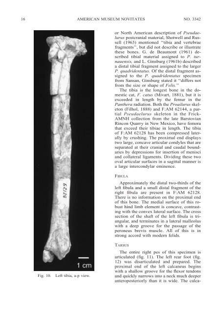
Fig. 10. Left tibia, a-p view.<br />
or North <strong>American</strong> description <strong>of</strong> <strong>Pseudaelurus</strong><br />
postcranial material, Shotwell and Russell<br />
(1963) mentioned ‘‘tibia and vertebrae<br />
fragments’’, but did not describe or illustrate<br />
these bones. G. de Beaumont (1961) described<br />
tibial material assigned to P. turnauensis,<br />
and L. Ginsburg (1961b) described<br />
a distal tibial fragment assigned to the larger<br />
P. quadridentatus. Of the distal fragment assigned<br />
to the P. quadridentatus specimen<br />
from Sansan, Ginsburg stated it ‘‘differs not<br />
from the size or shape <strong>of</strong> Felis.’’<br />
The tibia is the longest bone in the domestic<br />
cat, F. catus (Mivart, 1881), but it is<br />
exceeded in length by the femur in the<br />
Panthera radiation. Both the Proailurus skeleton<br />
(Filhol, 1888) and F:AM 62144, a partial<br />
<strong>Pseudaelurus</strong> skeleton in the Frick-<br />
AMNH collection from the late Barstovian<br />
Rincon Quarry in New Mexico, have femora<br />
that exceed their tibiae in length. The tibia<br />
<strong>of</strong> F:AM 62128 has been compressed laterally<br />
by crushing. The proximal end displays<br />
two large, concave articular condyles that are<br />
separated at their cranial and caudal boundaries<br />
by depressions for insertion <strong>of</strong> menisci<br />
and collateral ligaments. Dividing these two<br />
oval articular surfaces in a sagittal manner is<br />
a large intercondylar eminence.<br />
FIBULA<br />
Approximately the distal two-thirds <strong>of</strong> the<br />
left fibula and a small distal fragment <strong>of</strong> the<br />
right fibula are present in F:AM 62128.<br />
There is no information on the proximal end<br />
<strong>of</strong> this bone. The medial surface <strong>of</strong> this robust<br />
hind limb element is concave, contrasting<br />
with the convex lateral surface. The cross<br />
section <strong>of</strong> the shaft <strong>of</strong> the left fibula is triangular,<br />
and terminates in a lateral malleolus<br />
with a deep groove for the passage <strong>of</strong> the<br />
peroneus brevis muscle. All <strong>of</strong> this is in<br />
strong accord with modern felids.<br />
TARSUS<br />
The entire right pes <strong>of</strong> this specimen is<br />
articulated (fig. 11). The left rear foot (fig.<br />
12) was disarticulated and prepared. The<br />
proximal end <strong>of</strong> the left calcaneus begins<br />
with a shallow groove for the flexor tendons<br />
and quickly narrows into a neck much deeper<br />
anteroposteriorly than it is wide. The calca-


