
Create successful ePaper yourself
Turn your PDF publications into a flip-book with our unique Google optimized e-Paper software.
14 Bostock, H. and Bergmans, J. (1994) Post-tetanic excitability<br />
changes and ectopic d<strong>is</strong>charges in a human motor axon. Brain<br />
117, 913–928<br />
15 David, G. et al. (1993) Activation of internodal potassium<br />
conductance in rat myelinated axons. J. Physiol. 472, 177–202<br />
16 Kapoor, R. et al. (1993) Internodal potassium currents can generate<br />
ectopic impulses in mammalian myelinated axons. Brain Res.<br />
611, 165–169<br />
17 Smith, K.J. and McDonald, W.I. (1980) Spontaneous and<br />
mechanically evoked activity due to a central demyelinating<br />
lesion. Nature 286, 154–156<br />
18 Baker, M. and Bostock, H. (1992) Ectopic activity in demyelinated<br />
spinal root axons of <strong>the</strong> rat. J. Physiol. 451, 539–552<br />
19 Frankenhaueser, B. and Hodgkin, A.L. (1956) The after-effects of<br />
impulses in <strong>the</strong> giant nerve fibres of Loligo. J. Physiol. 131, 341–376<br />
20 Kapoor, R. et al. (1997) Slow sodium-dependent potential<br />
oscillations contribute to ectopic firing in mammalian<br />
demyelinated axons. Brain 120, 647–652<br />
21 Taylor, C.P. (1993) Na � currents that fail to inactivate. Trends<br />
Neurosci. 16, 455–459<br />
22 Crill, W.E. (1996) Pers<strong>is</strong>tent sodium current in mammalian central<br />
neurons. Annu. Rev. Physiol. 58, 349–362<br />
23 Baker, M.D. and Bostock, H. (1998) Inactivation of macroscopic<br />
late Na � currents in sensory neurons. J. Neurophysiol. 80,<br />
2538–2549<br />
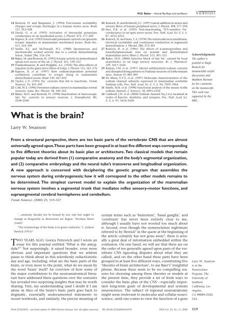
<strong>What</strong> <strong>is</strong> <strong>the</strong> <strong>brain</strong>?<br />
Larry W. Swanson<br />
M.D. Baker – Axonal flip-flops and oscillators V IEWPOINT<br />
24 Bostock, H. and Rothwell, J.C. (1997) Latent addition in motor and<br />
sensory fibres of human peripheral nerve. J. Physiol. 498, 277–294<br />
25 Stys, P.K. et al. (1993) Non-inactivating, TTX-sensitive Na �<br />
conductance in rat optic nerve axons. Proc. Natl. Acad. Sci. U. S. A.<br />
87, 4212–4216<br />
26 Bostock, H. and Sears, T.A. (1978) The internodal axon membrane:<br />
electrical excitability and continuous conduction in segmental<br />
demyelination. J. Physiol. 280, 273–301<br />
27 Bostock, H. et al. (1981) The effects of 4-aminopyridine and<br />
teraethylammonium ions on normal and demyelinated<br />
mammalian nerve fibres J. Physiol. 313, 301–315<br />
28 Baker, M.D. (2000) Selective block of late Na � current by local<br />
anaes<strong>the</strong>tics in rat large sensory neurones. Br. J. Pharmacol.<br />
129, 1617–1626<br />
29 Raman, I.M. et al. (1997) Altered subthreshold sodium currents<br />
and d<strong>is</strong>rupted firing patterns in Purkinje neurons of Scn8a mutant<br />
mice. Neuron 19, 881–891<br />
30 De Miera, E.V.S. et al. (1997) Molecular characterization of <strong>the</strong><br />
sodium channel subunits expressed in mammalian cerebellar<br />
Purkinje cells. Proc. Natl. Acad. Sci. U. S. A. 94, 7059–7064<br />
31 Smith, M.R. et al. (1998) Functional analys<strong>is</strong> of <strong>the</strong> mouse Scn8a<br />
sodium channel. J. Neurosci. 18, 6093–6102<br />
32 Caldwell, J.H. et al. (2000) Sodium channel Na v 1.6 <strong>is</strong> localized at<br />
nodes of Ranvier, dendrites, and synapses. Proc. Natl. Acad. Sci.<br />
U. S. A. 97, 5616–5620<br />
From a structural perspective, <strong>the</strong>re are ten basic parts of <strong>the</strong> vertebrate CNS that are almost<br />
universally agreed upon.These parts have been grouped in at least five different ways corresponding<br />
to five different <strong>the</strong>ories about its basic plan or architecture.Two classical models that remain<br />
popular today are derived from (1) comparative anatomy and <strong>the</strong> body’s segmental organization,<br />
and (2) comparative embryology and <strong>the</strong> neural tube’s transverse and longitudinal organization.<br />
A new approach <strong>is</strong> concerned with deciphering <strong>the</strong> genetic program that assembles <strong>the</strong><br />
nervous system during embryogenes<strong>is</strong>; how it will correspond to <strong>the</strong> o<strong>the</strong>r models remains to<br />
be determined. The simplest current model to explain <strong>the</strong> organization of <strong>the</strong> mammalian<br />
nervous system involves a segmental trunk that mediates reflex sensory–motor functions, and<br />
suprasegmental cerebral hem<strong>is</strong>pheres and cerebellum.<br />
Trends Neurosci. (2000) 23, 519–527<br />
‘…anatomy should not be bound by any rule but ought to<br />
change as frequently as d<strong>is</strong>sections are begun.’ Nicolaus Steno<br />
(1669) 1<br />
‘The terminology of <strong>the</strong> <strong>brain</strong> <strong>is</strong> in great confusion.’ C. Judson<br />
Herrick (1915) 2<br />
TWO YEARS AGO, Gorica Petrovich and I wrote an<br />
essay for th<strong>is</strong> journal entitled ‘<strong>What</strong> <strong>is</strong> <strong>the</strong> amygdala?’<br />
3 Not surpr<strong>is</strong>ingly, it ra<strong>is</strong>ed broader, even more<br />
obvious and important questions that we seldom<br />
pause to think about in th<strong>is</strong> relentlessly reduction<strong>is</strong>tic<br />
day and age, including, what are <strong>the</strong> basic parts of <strong>the</strong><br />
<strong>brain</strong>, or even more to <strong>the</strong> point, what do we mean by<br />
<strong>the</strong> word ‘<strong>brain</strong>’ itself? An overview of how some of<br />
<strong>the</strong> major contributors to <strong>the</strong> neuroanatomical literature<br />
have addressed <strong>the</strong>se questions over <strong>the</strong> centuries<br />
has revealed two surpr<strong>is</strong>ing insights that may be worth<br />
sharing. First, my understanding (and I doubt if I am<br />
alone in th<strong>is</strong>) of <strong>the</strong> <strong>brain</strong>’s basic parts goes back to<br />
dogmatic, essentially undocumented statements in<br />
recent textbooks, and similarly, <strong>the</strong> prec<strong>is</strong>e meaning of<br />
certain terms such as ‘<strong>brain</strong>stem’, ‘basal ganglia’, and<br />
‘cerebrum’ has never been entirely clear to me,<br />
although I usually have not worried too much about<br />
it. Second, even though <strong>the</strong> nomenclature nightmare<br />
referred to by Herrick 2 in <strong>the</strong> quote at <strong>the</strong> beginning of<br />
<strong>the</strong> article certainly has not gone away 4 , <strong>the</strong>re <strong>is</strong> actually<br />
a great deal of information embedded within <strong>the</strong><br />
confusion. On one hand, we will see that <strong>the</strong>re are on<br />
<strong>the</strong> order of ten generally agreed upon parts of <strong>the</strong> vertebrate<br />
CNS (ignoring d<strong>is</strong>putes about what <strong>the</strong>y are<br />
called), and on <strong>the</strong> o<strong>the</strong>r hand <strong>the</strong>se parts have been<br />
grouped in at least five different ways, constituting five<br />
‘<strong>the</strong>ories of <strong>brain</strong> architecture’, to use Baer’s 5 insightful<br />
phrase. Because <strong>the</strong>re seem to be no compelling reasons<br />
for choosing among <strong>the</strong>se <strong>the</strong>ories or models at<br />
<strong>the</strong> present time, <strong>the</strong>y provide a set of fresh ways to<br />
consider <strong>the</strong> basic plan of <strong>the</strong> CNS – especially important<br />
long-term goals of developmental and systems<br />
neuroscience. The subject of regional neuroanatomy<br />
might seem irrelevant to molecular and cellular neuroscience,<br />
until one comes to view <strong>the</strong> function of a gene<br />
0166-2236/00/$ – see front matter © 2000 Elsevier Science Ltd. All rights reserved. PII: S0166-2236(00)01639-8 TINS Vol. 23, No. 11, 2000 519<br />
Acknowledgements<br />
The author <strong>is</strong><br />
grateful to Hugh<br />
Bostock for<br />
innumerable useful<br />
d<strong>is</strong>cussions and<br />
Mat<strong>the</strong>w Kiernan<br />
for h<strong>is</strong> comments<br />
on <strong>the</strong> manuscript.<br />
Th<strong>is</strong> work was<br />
supported by <strong>the</strong><br />
MRC.<br />
Larry W. Swanson<br />
<strong>is</strong> at <strong>the</strong><br />
Neuroscience<br />
Program, The<br />
University of<br />
Sou<strong>the</strong>rn<br />
California, Los<br />
Angeles,<br />
CA 90089-2520,<br />
USA.
V IEWPOINT<br />
‘Brain’<br />
(c. 4200 BC)<br />
L.W. Swanson – Brain architecture<br />
Dual <strong>brain</strong><br />
(c. 340 BC)<br />
Ar<strong>is</strong>totle<br />
or neuron in terms of its role within a particular neural<br />
system or part of <strong>the</strong> <strong>brain</strong>. Th<strong>is</strong> brings us back to <strong>the</strong><br />
two questions posed at <strong>the</strong> beginning of th<strong>is</strong> review.<br />
CNS nomenclature in antiquity<br />
From one perspective, <strong>the</strong> h<strong>is</strong>tory of neuroanatomical<br />
nomenclature can be traced back more than 6000<br />
years (Fig. 1). An equivalent to <strong>the</strong> Engl<strong>is</strong>h word<br />
‘<strong>brain</strong>’ <strong>is</strong> found in an Egyptian manuscript composed<br />
~4200 BC (Ref. 6), although its most famous occurrence<br />
<strong>is</strong> in <strong>the</strong> Edwin Smith Surgical Papyrus (a 1700 BC copy<br />
of an Egyptian manuscript composed some 1500 years<br />
earlier), where 13 cases of skull fracture caused by<br />
war injuries are d<strong>is</strong>cussed 7–10 . In approximately 340 BC<br />
Ar<strong>is</strong>totle publ<strong>is</strong>hed <strong>the</strong> first surviving account of <strong>brain</strong><br />
d<strong>is</strong>sections 10 . He d<strong>is</strong>tingu<strong>is</strong>hed <strong>the</strong> parencephalon or<br />
small <strong>brain</strong> (cerebellum in Latin) from <strong>the</strong> encephalon<br />
or large <strong>brain</strong> (cerebrum in Latin), which extends<br />
down through <strong>the</strong> back as <strong>the</strong> spinal marrow (<strong>the</strong><br />
marrow of <strong>the</strong> back bone or vertebral column; our<br />
spinal cord). Th<strong>is</strong> was <strong>the</strong> first <strong>the</strong>ory or model of CNS<br />
architecture and cons<strong>is</strong>ted of three parts based on <strong>the</strong><br />
simplest gross anatomical criteria, that <strong>is</strong> a small <strong>brain</strong><br />
attached to a large <strong>brain</strong>, which in turn <strong>is</strong> attached to<br />
a spinal marrow (Fig. 2a). Th<strong>is</strong> dual (large and small)<br />
<strong>brain</strong> model was followed unaltered for about 1800<br />
years, until <strong>the</strong> work of Vesalius, described below.<br />
Before progressing to <strong>the</strong> European Rena<strong>is</strong>sance it<br />
<strong>is</strong> important to note that Ar<strong>is</strong>totle started <strong>the</strong> unenviable<br />
tradition of ambiguous terminology in<br />
neuroanatomy referred to by Herrick 2 . Ar<strong>is</strong>totle used<br />
exactly <strong>the</strong> same word, encephalon, in two very different<br />
ways: first, in its oldest sense, as <strong>the</strong> ‘marrow’ of<br />
<strong>the</strong> skull, and second, as a name for h<strong>is</strong> ‘large <strong>brain</strong>’.<br />
As with all such ambiguities, <strong>the</strong> meaning of a specific<br />
example of <strong>the</strong> word can only be inferred from its context,<br />
which <strong>is</strong> a crucial problem today because keywords<br />
with more than one meaning might be used<br />
(without context) for database queries.<br />
Interestingly, th<strong>is</strong> ambiguity was recognized, and<br />
even dealt with, very quickly in classical antiquity. As<br />
Galen noted 10 , in approximately 290 BC <strong>the</strong> founder of<br />
human anatomy, Herophilus, might have proposed<br />
<strong>the</strong> unequivocal term enkranon for <strong>the</strong> neural contents<br />
of <strong>the</strong> skull compared with Ar<strong>is</strong>totle’s specialized<br />
use of ‘encephalon’ for <strong>the</strong> large <strong>brain</strong>. Unfortunately<br />
520 TINS Vol. 23, No. 11, 2000<br />
Will<strong>is</strong><br />
1644<br />
Varolio<br />
1573<br />
Vesalius<br />
1543<br />
Malpighi<br />
1672<br />
Dual <strong>brain</strong><br />
Edinger<br />
1908<br />
1980s<br />
Segmental<br />
Segmental<br />
Segmental<br />
Developmental<br />
Evolutionary<br />
Genomic<br />
trends in Neurosciences<br />
Fig. 1. Evolutionary h<strong>is</strong>tory of major <strong>the</strong>ories on regional organization of <strong>the</strong> CNS. In <strong>the</strong> 16th and 17th centuries <strong>the</strong>re<br />
was an ‘explosion’ of <strong>the</strong>ories regarding <strong>the</strong> basic regional or topographic organization of <strong>the</strong> CNS after <strong>the</strong> ‘dual <strong>brain</strong>’<br />
account of Ar<strong>is</strong>totle was introduced in classical antiquity (~340 BC). Two new classes of <strong>the</strong>ory, evolutionary and genomic,<br />
were added in <strong>the</strong> 20th century; <strong>the</strong> former has had little substantial influence, whereas <strong>the</strong> latter <strong>is</strong>, just now, maturing.<br />
th<strong>is</strong> useful d<strong>is</strong>tinction never caught<br />
on. From an early time, however,<br />
careful anatom<strong>is</strong>ts began to speak of<br />
<strong>the</strong> ‘<strong>brain</strong> proper’ (or <strong>the</strong> cerebrum<br />
proper or <strong>the</strong> encephalon proper)<br />
when referring to Ar<strong>is</strong>totle’s large<br />
<strong>brain</strong>, and <strong>the</strong> ‘<strong>brain</strong>’ or ‘<strong>brain</strong> in<br />
general’ when referring to <strong>the</strong><br />
neural contents of <strong>the</strong> skull.<br />
Incidentally, a curious tradition has<br />
evolved in Engl<strong>is</strong>h (which probably<br />
dates back to 1755 and <strong>the</strong> publication<br />
of Samuel Johnson’s great<br />
dictionary 51 ), where frequently <strong>the</strong><br />
part of <strong>the</strong> CNS in <strong>the</strong> skull <strong>is</strong><br />
referred to as <strong>the</strong> encephalon or<br />
<strong>brain</strong>, whereas <strong>the</strong> large <strong>brain</strong> <strong>is</strong><br />
referred to as <strong>the</strong> cerebrum. Once<br />
one <strong>is</strong> aware of ambiguities associated<br />
with definitions and uses of<br />
<strong>the</strong> words ‘<strong>brain</strong>’, ‘cerebrum’ and<br />
‘encephalon’ it <strong>is</strong> amazing how often <strong>the</strong>ir meaning <strong>is</strong><br />
unclear when reading <strong>the</strong> literature carefully.<br />
The Rena<strong>is</strong>sance explosion of models: segmental<br />
and developmental<br />
With <strong>the</strong> publication in 1543 of h<strong>is</strong> truly revolutionary<br />
masterpiece, De Humani Corpor<strong>is</strong> Fabrica 52 ,<br />
Vesalius codified Rena<strong>is</strong>sance anatomy, which was<br />
based on two principles: direct observation instead of<br />
reliance on classical texts, and use of <strong>the</strong> new, natural<strong>is</strong>tic<br />
art to illustrate results (h<strong>is</strong> principal art<strong>is</strong>t, Kalkar,<br />
was in all probability from Titian’s studio). Similar to<br />
Ar<strong>is</strong>totle, Vesalius proposed a tripartite div<strong>is</strong>ion of our<br />
CNS, but th<strong>is</strong> time <strong>the</strong> parcellation was based on much<br />
better structural analys<strong>is</strong> that now included a consideration<br />
of <strong>the</strong> nerves 17 . At <strong>the</strong> core of Vesalius’s description<br />
was <strong>the</strong> dorsal medulla, which corresponds to our<br />
spinal cord, medulla and pons (Figs 2,3). He extended<br />
Ar<strong>is</strong>totle’s ‘spinal medulla’ up into <strong>the</strong> cranium<br />
because <strong>the</strong> whole structure <strong>is</strong> associated with a series<br />
of major paired nerves beginning rostrally with our<br />
trigeminal nerve, and thus excluding <strong>the</strong> ‘atypical’<br />
nerves associated more rostrally with <strong>the</strong> eye and olfactory<br />
apparatus. Vesalius attached h<strong>is</strong> version of <strong>the</strong><br />
large <strong>brain</strong> (now <strong>the</strong> Latin cerebrum instead of <strong>the</strong><br />
Greek encephalon) to <strong>the</strong> rostral end of <strong>the</strong> dorsal<br />
medulla or ‘trunk’ (derived from Will<strong>is</strong>, see below), and<br />
<strong>the</strong> small <strong>brain</strong> (now <strong>the</strong> Latin cerebellum instead of<br />
<strong>the</strong> Greek parencephalon) to <strong>the</strong> dorsal aspect of <strong>the</strong><br />
trunk, just caudal to <strong>the</strong> cerebrum (Fig. 2).<br />
Th<strong>is</strong> was <strong>the</strong> first example of a general class of<br />
model wherein a central trunk that generates ‘segmental’<br />
nerves <strong>is</strong> associated with two ‘suprasegmental’<br />
structures, <strong>the</strong> cerebrum and cerebellum, as specifically<br />
articulated at <strong>the</strong> close of <strong>the</strong> 19th century by<br />
Meyer 53 . Based on <strong>the</strong> origin of regularly spaced nerve<br />
pairs, Vesalius was able to escape from <strong>the</strong> all too easy<br />
convention (advocated by Galen and still common<br />
today) of dividing what we call <strong>the</strong> CNS into a part<br />
within <strong>the</strong> skull and a part within <strong>the</strong> vertebral column<br />
(<strong>brain</strong> as a whole and spinal cord, respectively).<br />
Never<strong>the</strong>less, <strong>the</strong> double meaning of <strong>the</strong> word<br />
cerebrum referred to in <strong>the</strong> preceding section was evident<br />
even in h<strong>is</strong> text. For example, at <strong>the</strong> beginning<br />
of chapter four in Book VII, Vesalius wrote:<br />
‘D<strong>is</strong>sectionum professores, universum cerebrum in anterius,
quod cerebrum, & in posterius, quod cerebellum vocant:<br />
deinde anterius in dextrum & sin<strong>is</strong>trum dividere resolent…’,<br />
which Singer 17 has translated as ‘By professors<br />
of d<strong>is</strong>section <strong>the</strong> <strong>brain</strong> <strong>is</strong> customarily divided into an<br />
anterior part, which <strong>the</strong>y call ‘cerebrum’, and a posterior,<br />
<strong>the</strong> ‘cerebellum’, <strong>the</strong> anterior (being divided)<br />
into right and left’. Note that Singer has used ‘<strong>brain</strong>’<br />
instead of ‘cerebrum’ at <strong>the</strong> beginning of <strong>the</strong> sentence<br />
(even though Vesalius used cerebrum), and in <strong>the</strong> title<br />
of h<strong>is</strong> book, Vesalius on <strong>the</strong> Human Brain 17 .<br />
Thirty years later, in 1573, Varolio (who d<strong>is</strong>covered<br />
<strong>the</strong> pons, or more accurately, our middle cerebellar<br />
peduncles) came up with a second interpretation of<br />
where <strong>the</strong> trunk ends rostrally 26 . He showed <strong>the</strong> trunk<br />
extending all <strong>the</strong> way to <strong>the</strong> base of <strong>the</strong> cerebral hem<strong>is</strong>pheres<br />
(thus including our inter<strong>brain</strong> or diencephalon),<br />
and he referred to <strong>the</strong> whole trunk as <strong>the</strong><br />
spinal marrow ra<strong>the</strong>r than <strong>the</strong> dorsal marrow of<br />
Vesalius (Figs 2,3), which was probably an unfortunate<br />
choice because <strong>the</strong> rostral end <strong>is</strong> not within <strong>the</strong> spinal<br />
column; that <strong>is</strong>, <strong>the</strong> term <strong>is</strong> obviously inaccurate.<br />
The final variant of <strong>the</strong> ‘segmental’ model from<br />
th<strong>is</strong> era appeared in <strong>the</strong> first book devoted exclusively<br />
to nervous system structure and function,<br />
Will<strong>is</strong>’s classic Cerebri Anatomie, where <strong>the</strong> word neurology<br />
was first used 34 . Will<strong>is</strong> extended <strong>the</strong> trunk<br />
even far<strong>the</strong>r rostrally, to include what he identified<br />
and named <strong>the</strong> striate body (<strong>the</strong> non-cortical part<br />
of <strong>the</strong> cerebral hem<strong>is</strong>phere, Fig. 3 and Table 1).<br />
However, he introduced a major new div<strong>is</strong>ion: <strong>the</strong><br />
part of <strong>the</strong> trunk in <strong>the</strong> skull he referred to by <strong>the</strong><br />
synonyms oblong marrow, medullary stem and<br />
medullary trunk – in d<strong>is</strong>tinction to <strong>the</strong> part of <strong>the</strong><br />
trunk in <strong>the</strong> spinal or vertebral canal (<strong>the</strong> spinal marrow).<br />
So accordingly, <strong>the</strong>re were oblong and spinal<br />
div<strong>is</strong>ions of <strong>the</strong> marrow or trunk. Th<strong>is</strong> advance contains<br />
an initial concept of a ‘<strong>brain</strong>stem’, although<br />
th<strong>is</strong> term does not appear to have been used until <strong>the</strong><br />
19th century (Table 1). Note that <strong>the</strong> first definition<br />
of <strong>the</strong> medulla oblongata (oblong marrow) included<br />
what today we call <strong>the</strong> basal ganglia/cerebral nuclei,<br />
inter<strong>brain</strong>, mid<strong>brain</strong> and hind<strong>brain</strong>. Essentially,<br />
Will<strong>is</strong> pointed out that <strong>the</strong> cranial nerves ar<strong>is</strong>e from<br />
<strong>the</strong> oblong marrow, whereas <strong>the</strong> spinal nerves ar<strong>is</strong>e<br />
from <strong>the</strong> spinal marrow.<br />
Will<strong>is</strong> provides ano<strong>the</strong>r classic example of how<br />
nomenclature can become confused for no apparent<br />
reason. Since <strong>the</strong> time of Galen our superior and inferior<br />
colliculi (corpora quadrigemini, tectum) had been<br />
referred to as <strong>the</strong> testes and nates (buttocks), respectively.<br />
However, Will<strong>is</strong> inexplicably reversed th<strong>is</strong>,<br />
despite what would seem to be an unforgettable explanation<br />
of Galen’s terminology by Vesalius 17 , ‘Th<strong>is</strong> part<br />
of <strong>the</strong> cerebrum graphically represents <strong>the</strong> two nates<br />
connected toge<strong>the</strong>r. But since <strong>the</strong> upper part even<br />
more resembles <strong>the</strong> testes because of that gland (<strong>the</strong><br />
pineal) which <strong>is</strong> like a pen<strong>is</strong>, <strong>the</strong> Ancients, who traditionally<br />
trained youths in <strong>the</strong> home in verbal d<strong>is</strong>tinctions,<br />
called that part DIDYMOS. We, imitating <strong>the</strong>m,<br />
name it testes or gemelli. Since in <strong>the</strong> hind and lower<br />
part of th<strong>is</strong> body <strong>is</strong> placed <strong>the</strong> orifice of <strong>the</strong> passage<br />
from <strong>the</strong> third ventricle to <strong>the</strong> fourth, and since th<strong>is</strong><br />
orifice has some resemblance to <strong>the</strong> anus, and since in<br />
<strong>the</strong> hind part of th<strong>is</strong> area <strong>the</strong>re <strong>is</strong> a transverse linear<br />
impression separating <strong>the</strong> upper parts from <strong>the</strong> lower,<br />
<strong>the</strong> Ancients as I believe, gave to th<strong>is</strong> region <strong>the</strong> name<br />
GLOUTION (GLOUTOS � buttock)’.<br />
L.W. Swanson – Brain architecture V IEWPOINT<br />
Malpighi (who proved Harvey’s model of <strong>the</strong> circulation<br />
of <strong>the</strong> blood by d<strong>is</strong>covering <strong>the</strong> capillary connection<br />
between arteries and veins, and <strong>is</strong> considered<br />
<strong>the</strong> founder of h<strong>is</strong>tology) provided <strong>the</strong> final classical<br />
model of CNS organization in 1672 (Ref. 37), based on<br />
embryology in <strong>the</strong> chick (Figs 2,3). He described and<br />
beautifully illustrated how at very early stages of<br />
development <strong>the</strong> nervous system seems to be associated<br />
with a plate-like structure that <strong>is</strong> broad rostrally<br />
and tapers caudally. Then, one can see three sequential<br />
vesicles or swellings in <strong>the</strong> broad rostral area that<br />
corresponds to <strong>the</strong> future <strong>brain</strong>. Finally, <strong>the</strong>re <strong>is</strong> a<br />
series of five sequential vesicles in <strong>the</strong> <strong>brain</strong> region,<br />
with <strong>the</strong> most rostral vesicle actually being paired, <strong>the</strong><br />
developing eyes lying on ei<strong>the</strong>r side of <strong>the</strong> second<br />
vesicle (which he seems to have associated with <strong>the</strong><br />
optic thalamus of Will<strong>is</strong>, our inter<strong>brain</strong>), and <strong>the</strong> third<br />
vesicle (which he called <strong>the</strong> cr<strong>is</strong>tate vesicle and thus<br />
d<strong>is</strong>covered our mid<strong>brain</strong>) also being paired. How Baer<br />
named <strong>the</strong> same vesicles in a way that <strong>is</strong> still followed<br />
today <strong>is</strong> considered below, but <strong>the</strong> important point<br />
here <strong>is</strong> that Malpighi d<strong>is</strong>covered a fundamental transverse<br />
organization of <strong>the</strong> neural tube, and thus, presumably,<br />
of <strong>the</strong> adult CNS.<br />
In summary, by 1672 <strong>the</strong>re were three general models<br />
of CNS organization: <strong>the</strong> dual <strong>brain</strong> model of<br />
Ar<strong>is</strong>totle; <strong>the</strong> segmental model of Vesalius, Varolio, and<br />
Will<strong>is</strong>; and <strong>the</strong> embryological model of Malpighi. By<br />
th<strong>is</strong> time, six basic parts of <strong>the</strong> CNS had been identified,<br />
although <strong>the</strong> definition of several of <strong>the</strong>m varied with<br />
different authors. These parts included <strong>the</strong> cerebrum,<br />
striate body (basal ganglia/cerebral nuclei), optic<br />
thalamus (inter<strong>brain</strong>), cr<strong>is</strong>tate vesicle (mid<strong>brain</strong>),<br />
cerebellum, and spinal or dorsal marrow (spinal cord).<br />
How <strong>the</strong>se three models evolved will now be examined.<br />
The dual <strong>brain</strong> model<br />
Ar<strong>is</strong>totle’s basic model of a small or posterior <strong>brain</strong><br />
attached to a large or anterior <strong>brain</strong>, which in turn <strong>is</strong><br />
attached to <strong>the</strong> spinal marrow or cord, exerted a considerable<br />
influence well into <strong>the</strong> 19th century, but <strong>is</strong><br />
now largely forgotten. Never<strong>the</strong>less, a crucial part of it<br />
has survived in a convoluted way, caused mainly by<br />
<strong>the</strong> pioneering work of Vieussens 13 , who in 1684 was<br />
<strong>the</strong> first to begin subdividing and <strong>the</strong>n extending<br />
Will<strong>is</strong>’s corpora striata, which he called <strong>the</strong> corpora<br />
striata inferiora (essentially <strong>the</strong> anterior striate bodies).<br />
Because he used Ar<strong>is</strong>totle’s model, he went on to<br />
describe Will<strong>is</strong>’s optic thalamus (inter<strong>brain</strong>) as <strong>the</strong> corpora<br />
striata superna posterior (essentially <strong>the</strong> posterior<br />
striate bodies), and <strong>the</strong> rest of <strong>the</strong> ‘<strong>brain</strong>stem’ (our<br />
mid<strong>brain</strong> and hind<strong>brain</strong>) as <strong>the</strong> corpora striata media.<br />
Many years later <strong>the</strong> influential neuroanatom<strong>is</strong>t,<br />
Reil 10,15 referred to <strong>the</strong> first two div<strong>is</strong>ions as <strong>the</strong> great<br />
cerebral nucleus, with an anterior part (Will<strong>is</strong>’s striate<br />
body) and a posterior part (Will<strong>is</strong>’s optic thalamus, our<br />
inter<strong>brain</strong>). Then, only a year later in 1810, <strong>the</strong> even<br />
more influential neuroanatomical work of Gall and<br />
Spurzheim 16,59 referred to <strong>the</strong> same features as <strong>the</strong> great<br />
cerebral ganglion, with its anterior and posterior parts.<br />
By 1876, Ferrier 36 would refer to <strong>the</strong> great cerebral<br />
nucleus or ganglion as <strong>the</strong> basal ganglia, <strong>the</strong> first reference<br />
to th<strong>is</strong> term that I have found thus far. It remained<br />
for <strong>the</strong> majority of leading 20th-century neuroanatom<strong>is</strong>ts<br />
to restrict <strong>the</strong> term ‘basal ganglia’ (‘basal<br />
nuclei’) to its original meaning, <strong>the</strong> corpus striatum<br />
of Will<strong>is</strong>, that <strong>is</strong>, <strong>the</strong> basal nuclei of <strong>the</strong> cerebral<br />
TINS Vol. 23, No. 11, 2000 521
V IEWPOINT<br />
Dual <strong>brain</strong><br />
Segmental<br />
Developmental<br />
Striate<br />
L.W. Swanson – Brain architecture<br />
Antiquity to end of <strong>the</strong> 17th century<br />
(classical)<br />
ENCEPHALON<br />
Cerebrum (cortex)<br />
body<br />
INF<br />
T N<br />
Vesalius (1543) 17<br />
CEREBELLUM<br />
522 TINS Vol. 23, No. 11, 2000<br />
DORSAL MARROW<br />
SPINAL MARROW<br />
Varolio (1573) 26 , Aranzi (1587) 27<br />
Optic<br />
thalamus<br />
N<br />
T<br />
Encephalon (<strong>brain</strong>) =<br />
encephalon (proper)<br />
PAR-<br />
+ parencephalon<br />
ENCEPH.<br />
Cerebellum<br />
(cortex)<br />
Will<strong>is</strong> (1664) 34<br />
SPINAL MARROW<br />
Ar<strong>is</strong>totle (c. 340 BC) 10 , Mondino (1316) 10 , Berengario (1523) 11 , Dryander (1536) 12<br />
CEREBRUM<br />
CEREBRUM<br />
Terminal vesicle<br />
[Optic Cr<strong>is</strong>tate<br />
thalamus] vesicle<br />
Cerebrum (<strong>brain</strong>) =<br />
cerebrum (proper)+<br />
cerebellum<br />
CEREBELLUM<br />
Cerebellar<br />
vesicle<br />
Malpighi (1673) 37<br />
Oblong marrow,<br />
medullary stem/trunk<br />
Spinal marrow<br />
Brain vesicles and spinal medulla<br />
(transverse div<strong>is</strong>ions)<br />
Spinal medulla<br />
Fig. 2. Neuroanatomical nomenclature. The main ways of subdividing<br />
<strong>the</strong> CNS are shown in <strong>the</strong> column on <strong>the</strong> left. Note that <strong>the</strong> dual <strong>brain</strong><br />
and segmental models involve a large and a small <strong>brain</strong> attached to a<br />
central core of variable length, whereas <strong>the</strong> developmental model features<br />
a rostrocaudally arranged series of vesicles. In <strong>the</strong> early 20th-century,<br />
evolutionary models were introduced and towards <strong>the</strong> end of <strong>the</strong><br />
century a genomic approach began to emerge. By now <strong>the</strong>re are at least<br />
ten basic parts or regions that are widely accepted (d<strong>is</strong>regarding nomenclature<br />
debates that are usually of a <strong>the</strong>oretical nature). These parts are<br />
shown in <strong>the</strong> small diagram to <strong>the</strong> right, without imposing any assumptions<br />
about how <strong>the</strong>y are grouped. By illustrating <strong>the</strong>ir topographic relationships,<br />
<strong>the</strong>y have a very specific meaning and <strong>the</strong>y can be used to<br />
compare and describe any of <strong>the</strong> o<strong>the</strong>r schemes in th<strong>is</strong> chart. The flatmap<br />
shows <strong>the</strong> right side of <strong>the</strong> CNS, with rostral to <strong>the</strong> left and caudal to <strong>the</strong><br />
right; its construction <strong>is</strong> d<strong>is</strong>cussed in Ref. 50. Abbreviations: cereb., cerebral;<br />
cereb. ped., cerebral peduncle; corp. Quad., corpus quadrigemina;<br />
d. thalamus, dorsal thalamus; epi., epithalamus; gang., ganglion; hypothal.,<br />
hypothalamus; INF, infundibulum; m., metathalamus; Met.,<br />
metencephalon; Myel., myelencephalon; N, nates; PARENCEPH., parencephalon;<br />
pars mam. hypo., pars mammillar<strong>is</strong> hypothalamus; p. opt.<br />
hypo., pars opticus hypothalamus; Rhombenceph., rhombencephalon;<br />
SYN, synencephalon; T, testes; teg., tegmentum; tel. imp., telencephalon<br />
impar; thal., thalamus; vent. thal., ventral thalamus.<br />
Cerebral cortex<br />
(hem<strong>is</strong>pheric ganglion)<br />
Anterior<br />
cereb. Posterior<br />
gang.<br />
cerebral<br />
ganglion<br />
Cerebral cortex<br />
Cerebellum<br />
Striate<br />
corp.<br />
quad.<br />
body Optic<br />
thalamus Cereb.<br />
pedun.<br />
Pons Medulla<br />
N<br />
T<br />
Haller (1781) 18 , Bell (1802) 19 , Leuret and Gratiolet (1839–57) 20 , Kölliker (1854) 21 , Luys (1865) 22<br />
Cereb.<br />
pedun.<br />
Cerebellum<br />
Pons<br />
Cerebral cortex<br />
Great cerebral ganglia<br />
(cerebral nuclei, basal ganglia)<br />
Primitive, medial, ascending<br />
(cerebral) ganglion<br />
Spinal cord<br />
Vieussens (1684) 13 , Monro (1783) 14 , Reil (1809) 15 , Gall and Spurzheim (1809) 16<br />
Reichert (1859) 28 , Schwalbe (1881) 29<br />
Cerebrum (cortex)<br />
body<br />
18th to mid-19th century<br />
(late classical)<br />
Cerebral cortex<br />
Striate<br />
body Optic<br />
thalamus<br />
Striate<br />
Optic<br />
thalamus<br />
End<strong>brain</strong><br />
FOREBRAIN<br />
corp.<br />
quad.<br />
corp.<br />
quad.<br />
Mid<strong>brain</strong><br />
MID-<br />
BRAIN<br />
Cerebellum<br />
(cortex)<br />
Ann.<br />
Prot.<br />
Pons Medulla<br />
Longet (1842) 35 , Ferrier (1876) 36<br />
Inter<strong>brain</strong><br />
corp.<br />
quad.<br />
Cerebellum<br />
Baer (1837) 5 , Edinger (1885) 38<br />
CTX<br />
Cerebellum<br />
Pons<br />
Medulla<br />
Cerebrum<br />
Spinal marrow<br />
Cerebrum<br />
Brain stem<br />
Spinal marrow<br />
Brain stem<br />
Spinal marrow<br />
HINDBRAIN<br />
Pons Medulla SPINAL MARROW<br />
L/D<br />
R C<br />
M/V<br />
BG TH<br />
HY<br />
T<br />
TG<br />
CB<br />
P M<br />
Major Regions<br />
SP<br />
BG, basal ganglia (cerebral nuclei)<br />
CB, cerebellum<br />
CTX, cerebral cortex (pallium)<br />
HY, hypothalamus<br />
M, medulla<br />
P, pons<br />
SP, spinal cord<br />
T, tectum<br />
TG, tegmentum<br />
TH, thalamus
Cerebral cortex<br />
Anterior<br />
cereb. Optic thal.<br />
gang.<br />
Tuber cinereum<br />
Mid<strong>brain</strong><br />
Cerebellum<br />
Pons Medulla<br />
Cerebellum<br />
Cerebral cortex<br />
Cerebral (central) ganglion<br />
Central tubular gray matter<br />
Spinal cord<br />
Meynert (1872) 23 , Gaskell (1889) 24 , Dejerine and Dejerine-Klumpke (1895) 25<br />
rhinencephalon<br />
rhinencephalon<br />
Late 19th to early 20th century<br />
(modern–h<strong>is</strong>tology)<br />
Cerebellum<br />
corp. Met.<br />
m. quad. RHOMBENCEPH.<br />
thalamus<br />
pars cereb. Pons<br />
p. opt.<br />
Myel.<br />
mam. ped. H T<br />
hypo. hypo.<br />
I S<br />
M U S<br />
H<strong>is</strong> (BNA, 1895) 39<br />
Cerebrum =<br />
fore<strong>brain</strong> + mid<strong>brain</strong><br />
Sulcus limitans<br />
(adds longitudinal organization<br />
to transverse div<strong>is</strong>ions)<br />
sulcus limitans<br />
SPINAL MEDULLA<br />
bas<strong>is</strong><br />
rhinencephalon<br />
tel.<br />
epi.<br />
tectum<br />
teg.<br />
hypothal. S Y<br />
N<br />
d. thalamus<br />
vent. thal.<br />
imp.I S THMUS<br />
L.W. Swanson – Brain architecture V IEWPOINT<br />
Contemporary<br />
Spinal marrow<br />
Bechterew (1900) 30 Carpenter and Sutin (1983) 31 , Nauta and Feirtag (1986) 32 , Williams (1995) 33<br />
Cerebral cortex<br />
Brain stem<br />
Cerebellum<br />
Striate<br />
Optic thal.<br />
corp.<br />
quad.<br />
body<br />
Tuber cinereum<br />
Mid<strong>brain</strong><br />
Pons Medulla<br />
T R U N K<br />
Ann.<br />
Prot.<br />
Herrick (1915) 2<br />
Cerebral cortex Cerebellum<br />
(cortex)<br />
Striate<br />
corp.<br />
m. quad.<br />
thalamus<br />
Pons<br />
pars cereb.<br />
Medulla<br />
body<br />
p. opt. mam.<br />
hypoth. hypo. ped.<br />
p a l l i u m<br />
corpus striatum<br />
striate body<br />
epi.<br />
epi.<br />
Archencephalon<br />
p a l l i u m<br />
bas<strong>is</strong><br />
rhinencephalon<br />
tel.<br />
imp.<br />
epithal.<br />
d. thalamus<br />
vent. thal.<br />
corp.<br />
quad.<br />
Ahlborn (1883) 42 , Kingsbury (1920) 43<br />
Ann.<br />
Prot.<br />
Brain stem<br />
tectum<br />
Met. Myel.<br />
Spinal cord<br />
teg.<br />
hypothal. S Y Deuterencephalon & spinal cord<br />
N<br />
(prechordal)<br />
Palaeëncephalon (old <strong>brain</strong>)<br />
trends in Neurosciences<br />
Neopallium<br />
Archepallium<br />
C E R E B R U M<br />
C E R E B R U M<br />
Spinal cord Brainstem<br />
Spinal cord<br />
p a l l i u m<br />
Cerebellum<br />
Crosby et al. (1962) 40 , Nieuwenhuys et al. (1998) 41<br />
p a l l i u m<br />
rhinencephalon<br />
Edinger (1908) 46 , Kappers (1909) 47 MacLean (1990) 48<br />
p. opt.<br />
hypoth.<br />
Spinal cord<br />
epi.<br />
Met. Myel.<br />
Cerebellum<br />
corp. Met.<br />
m. quad.<br />
thalamus<br />
pars cereb. Pons<br />
p. opt.<br />
Myel.<br />
mam. ped.<br />
hypoth. hypo.<br />
corpus striatum<br />
CB<br />
CB<br />
Fore<strong>brain</strong><br />
Kupffer (1906) 44 , Kuhlenbeck (1967–1978) 45<br />
Mid<strong>brain</strong><br />
Hind<strong>brain</strong><br />
Neomammalian<br />
SPINAL MEDULLA<br />
Paleomammalian<br />
Paleopallium Reptilian<br />
Hox-2.6 (e12.5 mouse)<br />
Wilkinson et al. (1989) 49<br />
TINS Vol. 23, No. 11, 2000 523<br />
Dual <strong>brain</strong><br />
Segmental<br />
Developmental<br />
Genomic Evolutionary
V IEWPOINT<br />
(a)<br />
(b)<br />
(d)<br />
L.W. Swanson – Brain architecture<br />
(c)<br />
(e)<br />
hem<strong>is</strong>phere (Table 1). At <strong>the</strong> same time <strong>the</strong> meaning of<br />
‘cerebrum proper’ was being greatly restricted to <strong>the</strong><br />
cerebral hem<strong>is</strong>pheres (Table 1), leading to <strong>the</strong> dem<strong>is</strong>e of<br />
<strong>the</strong> dual <strong>brain</strong> model. As an interesting h<strong>is</strong>torical footnote,<br />
in 1836 Solly60 introduced <strong>the</strong> term ‘hem<strong>is</strong>pheric<br />
ganglion’ to refer to <strong>the</strong> cerebral cortex, in an effort to<br />
provide a complement to Gall and Spurzheim’s ‘great<br />
cerebral ganglion’ (later, <strong>the</strong> basal ganglion).<br />
Segmental models<br />
The evolution of <strong>the</strong>se models – essentially cons<strong>is</strong>ting<br />
of a trunk generating regularly spaced paired<br />
nerves, and suprasegmental cerebrum and cerebellum<br />
– <strong>is</strong> complex and only a broad overview will be given<br />
524 TINS Vol. 23, No. 11, 2000<br />
trends in Neurosciences<br />
Fig. 3. Pioneering illustrations of CNS topography. (a) In th<strong>is</strong> drawing, Vesalius (1543) 17<br />
illustrates h<strong>is</strong> concept of <strong>the</strong> human dorsal medulla – essentially a trunk that generates a<br />
regular series of paired nerves, beginning rostrally with <strong>the</strong> trigeminal nerves (labeled 3 and 4<br />
at <strong>the</strong> far left) and ending caudally with a 30th pair of spinal nerves at <strong>the</strong> far right. The region<br />
between A and B <strong>is</strong> what we know today as <strong>the</strong> pons and medulla (5 <strong>is</strong> <strong>the</strong> facial nerve; 6 <strong>is</strong><br />
cranial nerves IX–XI; and 7 <strong>is</strong> <strong>the</strong> hypoglossal nerve); that between B and H <strong>is</strong> what we know<br />
as <strong>the</strong> spinal cord. (b) Thirty years later, Varolio 26 illustrated <strong>the</strong> base of <strong>the</strong> human <strong>brain</strong> and<br />
showed <strong>the</strong> spinal medulla (labeled 1–3 on <strong>the</strong> left side or top; and labeled a,b on <strong>the</strong> right<br />
side or bottom, rostral <strong>is</strong> to <strong>the</strong> left) as extending all <strong>the</strong> way to <strong>the</strong> base of <strong>the</strong> cerebral hem<strong>is</strong>pheres,<br />
just caudal to what <strong>is</strong> obviously <strong>the</strong> optic chiasm. (c) Shortly <strong>the</strong>reafter, Will<strong>is</strong> 34<br />
extended <strong>the</strong> trunk even far<strong>the</strong>r rostral to include what he identified and named <strong>the</strong> striate<br />
bodies (labeled A in th<strong>is</strong> dorsal view of <strong>the</strong> sheep <strong>brain</strong>, which has been rotated so that rostral<br />
<strong>is</strong> to <strong>the</strong> left). Th<strong>is</strong> figure beautifully illustrates <strong>the</strong> entirety of what Will<strong>is</strong> referred to (leaving<br />
aside <strong>the</strong> cerebellum, T and s, which was b<strong>is</strong>ected and pulled to ei<strong>the</strong>r side) as <strong>the</strong> oblong marrow,<br />
medullary stem, or medullary trunk. (d) A lateral view of <strong>the</strong> right half of a chick embryo<br />
after four days of development as drawn by Malpighi 37 . Using H<strong>is</strong>’s later terminology, <strong>the</strong> following<br />
parts of <strong>the</strong> neural tube seem clear: E indicates <strong>the</strong> paired telencephalic vesicles, D <strong>the</strong><br />
diencephalic vesicle, F <strong>the</strong> optic cup and choroid f<strong>is</strong>sure, A <strong>the</strong> mesencephalic vesicle, B <strong>the</strong><br />
metencephalic vesicle, and C <strong>the</strong> myelencephalic vesicle. (e) Illustration of how Edinger 46<br />
viewed <strong>the</strong> mammalian neëncephalon as being added on top of <strong>the</strong> lower vertebrate paleëncephalon,<br />
basically at <strong>the</strong> rhinal f<strong>is</strong>sure, which extends caudally from <strong>the</strong> olfactory bulb at <strong>the</strong><br />
far left. Some of <strong>the</strong> figures have been lightly retouched so that <strong>the</strong> labels are clearer.<br />
here (Fig. 2). The Vesalian model was refined in <strong>the</strong> late<br />
18th century by Haller18 who divided <strong>the</strong> cerebrum<br />
sequentially into cerebral cortex, striate body, optic<br />
thalamus, and <strong>the</strong> corpora quadrigemini and cerebral<br />
peduncle, which toge<strong>the</strong>r form our mid<strong>brain</strong>. Caudal<br />
to th<strong>is</strong>, he was <strong>the</strong> first to d<strong>is</strong>tingu<strong>is</strong>h clearly between<br />
<strong>the</strong> pons and medulla as we now refer to <strong>the</strong>m,<br />
although he was somewhat obscure about <strong>the</strong> larger<br />
part of <strong>the</strong> CNS that <strong>the</strong>y should be placed within. One<br />
of <strong>the</strong> last major anatomical analyses based on <strong>the</strong><br />
Vesalian model was publ<strong>is</strong>hed by Meynert23 in 1872. It<br />
took advantage of <strong>the</strong> h<strong>is</strong>tological methods that had<br />
been introduced earlier in <strong>the</strong> century. Essentially, he<br />
introduced an innovative, longitudinal way of thinking<br />
about <strong>the</strong> <strong>brain</strong>stem by dividing <strong>the</strong> traditional<br />
posterior cerebral ganglion into a dorsal zone, which<br />
he called <strong>the</strong> optic thalamus proper (our thalamus),<br />
and a ventral zone, <strong>the</strong> tuber cinereum (our hypothalamus).<br />
He included <strong>the</strong> optic thalamus proper and<br />
<strong>the</strong> corpora quadrigemina in <strong>the</strong> true posterior cerebral<br />
ganglion, and included <strong>the</strong> tuber cinereum, cerebral<br />
peduncle (our ventral mid<strong>brain</strong> or tegmentum), pons,<br />
medulla, and spinal cord in what he called <strong>the</strong> central<br />
tubular gray matter, which was responsible for generating<br />
all of <strong>the</strong> motor nerves.<br />
Here, for <strong>the</strong> first time, we can recognize ten basic<br />
parts of <strong>the</strong> CNS that are common to virtually all<br />
modern gross anatomical descriptions: cerebral cortex,<br />
basal ganglia/cerebral nuclei, thalamus and hypothalamus,<br />
tectum and tegmentum, pons, medulla,<br />
cerebellum, and spinal cord (in modern terms).<br />
Will<strong>is</strong>’s basic model of a <strong>brain</strong>stem stretching from<br />
<strong>the</strong> corpus striatum rostrally to <strong>the</strong> medulla caudally<br />
was refined by <strong>the</strong> addition of Haller’s subdiv<strong>is</strong>ions35,36 ,<br />
and in Herrick’s2 final major reincarnation by <strong>the</strong> addition<br />
of H<strong>is</strong>’s embryonic schema (see below).<br />
It <strong>is</strong> Varolio’s basic model that has remained popular<br />
today, refined by div<strong>is</strong>ion of <strong>the</strong> central trunk into a<br />
<strong>brain</strong>stem part, generating <strong>the</strong> cranial nerves and a<br />
spinal cord part, generating <strong>the</strong> spinal nerves31,32 . Haller’s<br />
subdiv<strong>is</strong>ions of <strong>the</strong> <strong>brain</strong>stem have usually been applied<br />
to th<strong>is</strong> model, see for example Ref. 30, although in a<br />
ra<strong>the</strong>r idiosyncratic way, Reichert28 and <strong>the</strong>n Schwalbe 29<br />
included <strong>the</strong> cerebellum in <strong>the</strong> <strong>brain</strong>stem (Table 1).<br />
Developmental models<br />
The great Baer has <strong>the</strong> d<strong>is</strong>tinction of having provided<br />
simple, descriptive names (Fig. 2) for <strong>the</strong> embryonic<br />
<strong>brain</strong> vesicles first identified by Malpighi (see<br />
above), and for demonstrating 5 that <strong>the</strong>se vesicles are<br />
probably common to all vertebrates. These contributions<br />
have had a profound influence and lasting value.<br />
He recognized three primary vesicles (in Engl<strong>is</strong>h, fore<strong>brain</strong>,<br />
mid<strong>brain</strong> and hind<strong>brain</strong>), and five secondary<br />
vesicles (in Engl<strong>is</strong>h, <strong>the</strong> fore<strong>brain</strong> vesicle divides into<br />
end<strong>brain</strong> and inter<strong>brain</strong> vesicles, and <strong>the</strong> hind<strong>brain</strong><br />
vesicle divides into hind<strong>brain</strong>, cons<strong>is</strong>ting of cerebellum<br />
and pons and after<strong>brain</strong> vesicles). The actual subdiv<strong>is</strong>ions<br />
were quite similar to Haller’s scheme, proposed<br />
a few years earlier, so a number of equivalent<br />
synonyms were generated, for example, optic thalamus<br />
and inter<strong>brain</strong>, and end<strong>brain</strong> and cerebrum<br />
(Fig. 2). By <strong>the</strong> mid-19th century, writers using <strong>the</strong><br />
dual <strong>brain</strong>, segmental, and developmental models of<br />
CNS organization were using each o<strong>the</strong>rs nomenclature<br />
for subdiv<strong>is</strong>ions almost interchangeably. <strong>What</strong><br />
was different was <strong>the</strong> way <strong>the</strong>y grouped <strong>the</strong> various
TABLE 1. H<strong>is</strong>torical overview of <strong>the</strong> meaning of some common gross anatomical terms for parts of <strong>the</strong> CNS<br />
Common gross Modern scheme of basic subdiv<strong>is</strong>ions Selected refs<br />
anatomical terms<br />
Cerebral Basal Inter- Mid- Hind- Cere- Spinal<br />
cortex ganglia <strong>brain</strong> <strong>brain</strong> <strong>brain</strong> bellum cord<br />
subdiv<strong>is</strong>ions. The Malpighi–Baer model was unique<br />
because it viewed <strong>the</strong> basic plan of <strong>the</strong> <strong>brain</strong> in terms<br />
of three rostrocaudally arranged swellings, in contrast<br />
to suprasegmental cerebrum and cerebellum attached<br />
to a trunk generating <strong>the</strong> cranial and spinal nerves.<br />
The final major variation of <strong>the</strong> Malpighi–Baer<br />
model was provided by H<strong>is</strong> towards <strong>the</strong> end of <strong>the</strong><br />
19th century. He described 61 a longitudinally oriented<br />
limiting sulcus that divides <strong>the</strong> right and left halves of<br />
<strong>the</strong> developing neural tube into a dorsal alar plate<br />
(which, at least initially, <strong>is</strong> sensory in function) and a<br />
ventral basal plate (which, at least initially, <strong>is</strong> motor in<br />
function). Subsequently, <strong>the</strong> question of where to<br />
place <strong>the</strong> rostral end of <strong>the</strong> limiting sulcus has generated<br />
endless and unresolved controversy, although<br />
H<strong>is</strong> himself placed it at <strong>the</strong> optic chiasm in <strong>the</strong> 1895<br />
‘Basle Nomina Anatomica’ (BNA), an ‘official’ tabulation<br />
of anatomical nomenclature 39,62 . In some ways<br />
<strong>the</strong> original BNA taxonomy of <strong>brain</strong> parts <strong>is</strong> less sat<strong>is</strong>factory<br />
today than <strong>the</strong> older and simpler scheme of<br />
Baer. For example, H<strong>is</strong> divided <strong>the</strong> end<strong>brain</strong> (telencephalic)<br />
vesicle into pallium, rhinencephalon, and<br />
corpus striatum, <strong>the</strong> latter of which he extended to<br />
include all of <strong>the</strong> hypothalamus except for <strong>the</strong> mammillary<br />
bodies (even though <strong>the</strong> hypothalamus,<br />
including <strong>the</strong> preoptic region, <strong>is</strong> derived from <strong>the</strong><br />
third ventricle neuroepi<strong>the</strong>lium). In addition, H<strong>is</strong><br />
included, between <strong>the</strong> mid<strong>brain</strong> and hind<strong>brain</strong>, a narrow<br />
<strong>is</strong>thmus vesicle whose components in <strong>the</strong> adult<br />
mammal have never been identified sat<strong>is</strong>factorily.<br />
Never<strong>the</strong>less, H<strong>is</strong>’s basic model has had a profound<br />
and lasting influence, see for example Ref. 41, mainly<br />
because it makes a d<strong>is</strong>tinction between dorsal, sensory<br />
regions of <strong>the</strong> CNS and ventral, motor regions.<br />
Analys<strong>is</strong> of th<strong>is</strong> combined structural and functional<br />
approach would require ano<strong>the</strong>r essay, but by way of<br />
introduction it very nicely complemented <strong>the</strong> brilliant<br />
earlier work of Magendie 63 and of Luys 22 and<br />
Meynert 23 . Magendie showed experimentally that <strong>the</strong><br />
L.W. Swanson – Brain architecture V IEWPOINT<br />
Cerebrum (proper) X X X X X Ar<strong>is</strong>totle10 , Vieussens 13 1684, Monro 14 1783, Reil 15 1809<br />
X X X X Vesalius17 1543, H<strong>is</strong> 39 (BNA 1895), Herrick 2 1915<br />
X X X Müller54 1843<br />
X X Varolio26 1573, Reichert 28 1859, Nauta-Feirtig 32 1986<br />
X Will<strong>is</strong>34 1664, Ferrier 36 1876<br />
Cerebral hem<strong>is</strong>phere X X Baer 5 1828, Herrick 2 1915, Nauta-Feirtig 32 1986<br />
X Will<strong>is</strong>34 1664, Ferrier 36 1876<br />
Cerebral or basal X X X X Vieussens 13 1684, Reil 15 1809, Gall-Spurzheim 16 1810<br />
nuclei or ganglia X X X Meynert23 1872<br />
X X Ferrier36 1876<br />
X Herrick2 1915, Crosby et al. 40 1962, Nauta-Feirtig 32 1986<br />
Cerebellum X Ar<strong>is</strong>totle 10 , universal since antiquity<br />
Brainstem X X X X Baer 5 1828, Herrick 2 1915, Ranson-Clark 55 1959<br />
X X X Obersteiner56 1888, Carpenter-Sutin 31 1983, Nauta-Feirtig 32 1986<br />
X X X X Reichert28 1859, Schwalbe 29 1881<br />
X X Jacobsohn57 1909, Olszewski-Baxter 58 1954, Williams 33 1995<br />
Spinal cord X X X X Varolio 26 1573, Aranzi 27 1587<br />
X X Vesalius17 1543, Malpighi 37 1672<br />
X Ar<strong>is</strong>totle 10 , universal since 18th century<br />
dorsal roots carry sensory information into <strong>the</strong> spinal<br />
cord, whereas <strong>the</strong> ventral roots carry motor information<br />
out of <strong>the</strong> spinal cord. Luys and Meynert<br />
suggested that sensory information travels up dorsal<br />
regions of <strong>the</strong> neurax<strong>is</strong> on route to <strong>the</strong> cerebral<br />
hem<strong>is</strong>pheres, whereas motor information leaving <strong>the</strong><br />
hem<strong>is</strong>pheres takes a more ventral route.<br />
A clear alternative to <strong>the</strong> Malpighi–Baer model was<br />
proposed by Ahlborn in 1883 (Ref. 42). Based on an<br />
examination of lamprey development, he proposed<br />
that <strong>the</strong> hind<strong>brain</strong>, similar to <strong>the</strong> spinal cord, develops<br />
adjacent to <strong>the</strong> notochord and <strong>is</strong> thus epichordal,<br />
whereas <strong>the</strong> fore<strong>brain</strong> and mid<strong>brain</strong> (Vesalius’s cerebrum)<br />
develop rostral to <strong>the</strong> notochord and are thus<br />
prechordal. Th<strong>is</strong> idea was supported by Kingsbury43 ,<br />
who showed conclusively that <strong>the</strong> definitive, h<strong>is</strong>tologically<br />
defined embryonic floor plate does not extend rostral<br />
to <strong>the</strong> hind<strong>brain</strong>–mid<strong>brain</strong> junction. Assuming that<br />
<strong>the</strong> notochord and floor plate are essentially co-extensive,<br />
th<strong>is</strong> would support a prechordal derivation of <strong>the</strong><br />
mid<strong>brain</strong> and fore<strong>brain</strong>. By contrast, Kupffer publ<strong>is</strong>hed<br />
an exhaustive review of h<strong>is</strong> own work and that of<br />
o<strong>the</strong>rs in 1906 (Ref. 44), concluding that <strong>the</strong> mid<strong>brain</strong><br />
<strong>is</strong> epichordal. He used <strong>the</strong> terms deuterencephalon for<br />
supposed epichordal parts of <strong>the</strong> <strong>brain</strong>, and archencephalon<br />
for supposed prechordal parts, an interpretation<br />
that has been followed by Kuhlenbeck45 .<br />
Unfortunately, <strong>the</strong> exact rostral limit of <strong>the</strong> notochord<br />
and its precursor remains problematic, partly because of<br />
<strong>the</strong> uncertain nature of <strong>the</strong> so-called prechordal plate.<br />
Evolutionary models<br />
All of <strong>the</strong> models d<strong>is</strong>cussed so far are based on comparative<br />
anatomy and embryology, with an emphas<strong>is</strong><br />
on mammals, and even humans. However, it was<br />
inevitable that phylogenetic models would be proposed<br />
after <strong>the</strong> publication of Darwin’s The Origin of<br />
Species in 1859 (Ref. 64). Interestingly, <strong>the</strong>y did not<br />
appear until approximately <strong>the</strong> turn of <strong>the</strong> century,<br />
TINS Vol. 23, No. 11, 2000 525
V IEWPOINT<br />
L.W. Swanson – Brain architecture<br />
and <strong>the</strong>n had only a brief, d<strong>is</strong>ruptive life, undoubtedly<br />
because relatively small amounts of reliable information<br />
about non-mammalian <strong>brain</strong> circuitry were<br />
(and are) available. In spite of th<strong>is</strong>, <strong>the</strong> main intellectual<br />
stimulus came from Edinger (see Ref. 46), who<br />
proposed a d<strong>is</strong>tinction between palaeëncephalon (old<br />
<strong>brain</strong>) and neëncephalon (new <strong>brain</strong>). According to<br />
th<strong>is</strong> model (Figs 2,3), part of <strong>the</strong> <strong>brain</strong> <strong>is</strong> common to<br />
and essentially unchanged throughout all vertebrates,<br />
and <strong>is</strong> responsible for all of <strong>the</strong> sensory–motor reflexes<br />
and purely instinctive behaviors necessary for survival.<br />
Th<strong>is</strong> <strong>is</strong> <strong>the</strong> palaeëncephalon, which constitutes<br />
<strong>the</strong> entire f<strong>is</strong>h <strong>brain</strong>. During <strong>the</strong> course of subsequent<br />
evolution, a new layer <strong>is</strong> added, <strong>the</strong> neëncephalon,<br />
which corresponds to <strong>the</strong> cerebral pallium. Th<strong>is</strong> <strong>is</strong> tiny<br />
in amphibians, a little bigger in reptiles, considerably<br />
larger in birds and huge in mammals. According to<br />
th<strong>is</strong> model, <strong>brain</strong> evolution took place in a geological<br />
fashion by <strong>the</strong> addition of strata.<br />
Edinger’s young colleague, Kappers, soon elaborated<br />
th<strong>is</strong> basic idea dividing <strong>the</strong> pallium into three successively<br />
evolved regions47 : <strong>the</strong> oldest or paleopallium<br />
(essentially <strong>the</strong> primary olfactory cortex), archipallium<br />
(more or less Ammon’s horn or <strong>the</strong> hippocampus), and<br />
neopallium (all <strong>the</strong> rest, Fig. 2). In addition, he divided<br />
<strong>the</strong> corpus striatum (basal ganglia/cerebral nuclei) into<br />
three parts: <strong>the</strong> paleostri-atum (globus pallidus), arch<strong>is</strong>triatum<br />
(amygdala), and neostriatum (caudate nucleus<br />
and putamen) 65 . Omitting details, th<strong>is</strong> line of thinking<br />
led to designations like paleo-, archi- and neothalamus;<br />
and paleo-, archi- and neocerebellum (see Ref. 45). It<br />
has had very little influence on contemporary neuroscience<br />
and <strong>the</strong> basic connectional principles that it<br />
rested on almost a century ago have not been borne<br />
out (for example, that almost all sensory inputs to <strong>the</strong><br />
pallium of f<strong>is</strong>h and amphibians are inevitably olfactory)<br />
41,45 . Instead, most contemporary evolutionary (i.e.<br />
comparative) thinking seems to be based on <strong>the</strong> embryological<br />
models d<strong>is</strong>cussed above (see Refs 41,45). All<br />
that remain of th<strong>is</strong> model (except for MacLean’s triune<br />
<strong>brain</strong> concept48 ; Fig. 2) are <strong>the</strong> constantly used, basically<br />
inaccurate, terms neocortex and neostriatum. It <strong>is</strong><br />
telling that Elliot Smith, who actually introduced <strong>the</strong><br />
concept of ‘neopallium’ in 1901 (Ref. 66), wrote a strenuous<br />
d<strong>is</strong>claimer less than a decade later67 .<br />
Genomic models<br />
Molecular genetics <strong>is</strong> breathing new life into developmental<br />
and evolutionary models of <strong>brain</strong> organization,<br />
although it <strong>is</strong> premature to start placing <strong>the</strong> results into<br />
a h<strong>is</strong>torical perspective. The ultimate goal <strong>is</strong> to decipher<br />
and understand <strong>the</strong> genetic program that assembles <strong>the</strong><br />
neural tube, in part by analysing spatiotemporal patterns<br />
of gene expression throughout development and<br />
establ<strong>is</strong>hing <strong>the</strong>ir functional significance. Once important<br />
sets of genes are identified, <strong>the</strong>ir expression patterns<br />
could be compared to <strong>the</strong> many structural patterns illustrated<br />
in Fig. 2, and eventually th<strong>is</strong> approach should<br />
provide criteria for helping to decide which, if any, of<br />
<strong>the</strong> many structural arrangements suggested thus far<br />
best represent <strong>the</strong> basic plan of <strong>the</strong> CNS (Ref. 68).<br />
Fur<strong>the</strong>rmore, it might be possible to infer how <strong>the</strong>se<br />
genomic programs, with <strong>the</strong>ir constituent genes,<br />
evolved over <strong>the</strong> ~400 million years of vertebrate ex<strong>is</strong>tence<br />
69 – and perhaps to determine whe<strong>the</strong>r 19th-century<br />
attempts (see Ref. 24) to homologize <strong>the</strong> invertebrate<br />
and vertebrate CNSs have any validity 70 . To date, <strong>the</strong><br />
526 TINS Vol. 23, No. 11, 2000<br />
best example by far of th<strong>is</strong> approach <strong>is</strong> <strong>the</strong> partly overlapping<br />
patterns of Hox gene expression in <strong>the</strong> developing<br />
vertebrate hind<strong>brain</strong>49 , where <strong>the</strong>y correlate to <strong>the</strong><br />
appearance of <strong>the</strong> long-known finer subdiv<strong>is</strong>ions called<br />
rhombomeres (see Ref. 45).<br />
Overview<br />
Th<strong>is</strong> review has focused narrowly on <strong>the</strong> h<strong>is</strong>tory of<br />
attempts to enumerate <strong>the</strong> major parts of <strong>the</strong> adult<br />
vertebrate CNS – an exerc<strong>is</strong>e in classical regional or<br />
topographic structural analys<strong>is</strong>, identical to enumerating<br />
<strong>the</strong> major parts of <strong>the</strong> body: head, neck, trunk, tail,<br />
upper limbs and lower limbs. The conclusion seems to<br />
be that <strong>the</strong>re are at least ten basic parts that, after centuries<br />
of research, almost everyone now agrees on<br />
(ignoring d<strong>is</strong>putes about exactly what <strong>the</strong>y are called,<br />
and where <strong>the</strong>ir borders are placed): (1) a dorsal cerebral<br />
cortex or pallium; (2) ventral or basal cerebral<br />
nuclei or ‘ganglia’ [(1) and (2) forming <strong>the</strong> cerebral<br />
hem<strong>is</strong>pheres or cerebrum]; (3) a dorsal thalamus; (4) a<br />
ventral hypothalamus [(3) and (4) forming <strong>the</strong> inter<strong>brain</strong>],<br />
(5) a dorsal tectum, (6) a ventral tegmentum<br />
[(5) and (6) forming <strong>the</strong> mid<strong>brain</strong>], (7) a cerebellum,<br />
(8) a pons, (9) a medulla, and (10) a spinal cord.<br />
By contrast, <strong>the</strong>re are at least five different <strong>the</strong>oretical<br />
frameworks for grouping or arranging <strong>the</strong>se ten<br />
basic parts (and many more variations on <strong>the</strong> basic<br />
<strong>the</strong>mes; Fig. 2), and it <strong>is</strong> doubtful that <strong>the</strong>re are compelling<br />
reasons at th<strong>is</strong> stage for adopting one particular<br />
scheme. Therefore, <strong>the</strong> time does not seem ripe to<br />
propose here yet ano<strong>the</strong>r unifying hypo<strong>the</strong>s<strong>is</strong> or<br />
model based on essentially <strong>the</strong> same data.<br />
Never<strong>the</strong>less, it <strong>is</strong> worth observing that <strong>the</strong>re have<br />
been no serious attempts to present a comprehensive,<br />
modern systems model of CNS organization.<br />
Reconsidering <strong>the</strong> gross anatomy analogy, <strong>the</strong><br />
regional account <strong>is</strong> traditionally complemented with a<br />
systems account. Thus, in addition to <strong>the</strong> various<br />
topographically arranged parts or regions l<strong>is</strong>ted above,<br />
<strong>the</strong> body <strong>is</strong> described as having skeletomotor, circulatory,<br />
digestive, respiratory, reproductive, immune,<br />
endocrine and nervous systems. One’s initial thought<br />
might be that <strong>the</strong> systems approach <strong>is</strong> more useful<br />
than <strong>the</strong> topographic, but from a broad functional<br />
perspective th<strong>is</strong> <strong>is</strong> not strictly true. The hand <strong>is</strong> used,<br />
for example, to write with. For <strong>the</strong> upper limb to perform<br />
th<strong>is</strong> behavior <strong>the</strong> skeletal system acts as a series<br />
of levers that are moved by <strong>the</strong> muscles, which in turn<br />
are controlled by nerves directed from <strong>the</strong> CNS. The<br />
vascular system supplies oxygen and nutrients to <strong>the</strong><br />
t<strong>is</strong>sues in <strong>the</strong> limb, <strong>the</strong> immune system fights infections<br />
in th<strong>is</strong> part of <strong>the</strong> body, etc. One important reason<br />
why <strong>the</strong> basic organization of <strong>the</strong> nervous system<br />
remains mysterious <strong>is</strong> that a consensus enumeration<br />
of its functional systems has not been achieved.<br />
Ultimately, a profound understanding of what <strong>the</strong><br />
<strong>brain</strong> <strong>is</strong> and how <strong>the</strong> <strong>brain</strong> works will be based on an<br />
integrated view of its topographic and systems organization.<br />
Th<strong>is</strong> has been accompl<strong>is</strong>hed for most of <strong>the</strong><br />
body, partly explaining why <strong>the</strong> reduction<strong>is</strong>tic molecular<br />
biology approach <strong>is</strong> so powerful. However, <strong>the</strong><br />
nervous system remains problematic: <strong>the</strong> nature of its<br />
organizing principles as a system <strong>is</strong> an unsolved problem<br />
that can only hinder <strong>the</strong> rational search for ways<br />
to cure <strong>the</strong> many d<strong>is</strong>eases that strike within it.<br />
Finally, note in Fig. 2 that <strong>the</strong> tripartite model first<br />
proposed by Varolio in 1573 remains <strong>the</strong> simplest for
mammals, refined only by a subdiv<strong>is</strong>ion of <strong>the</strong> central<br />
trunk into <strong>brain</strong>stem and spinal cord. Perhaps a systems<br />
model based on <strong>the</strong> fundamental organization of<br />
connections within and between <strong>the</strong>se three parts<br />
might be a useful way to start. The profound question<br />
really <strong>is</strong> not ‘what <strong>is</strong> <strong>the</strong> <strong>brain</strong>?’ but ra<strong>the</strong>r ‘what are<br />
<strong>the</strong> basic parts of <strong>the</strong> nervous system and how are <strong>the</strong>y<br />
interconnected functionally?’<br />
Selected references<br />
1 Scherz, G. (1965) Nicolaus Steno’s Lecture on <strong>the</strong> Anatomy of <strong>the</strong><br />
Brain, Busck<br />
2 Herrick, C.J. (1915) An Introduction to Neurology, Saunders<br />
3 Swanson, L.W. and Petrovich, G. (1998) <strong>What</strong> <strong>is</strong> <strong>the</strong> amygdala?<br />
Trends Neurosci. 28, 323–331<br />
4 Anthoney, T.R. (1994) Neuroanatomy and <strong>the</strong> Neurologic Exam: A<br />
Thesaurus of Synonyms, Similar-Sounding Non–Synonyms, and Terms<br />
of Variable Meaning, CRC Press<br />
5 Baer, K.E.v. (1828–37) Über Entwickelungsgeschichte der Thiere.<br />
Beobachtung und Reflexion, Bornträger. See <strong>the</strong> facsimile reprint<br />
publ<strong>is</strong>hed in 1967 by Impression Anastaltique Culture et Civil<strong>is</strong>ation<br />
6 Walker, A.E. (1998) The Genes<strong>is</strong> of Neuroscience, American<br />
Association of Neurological Surgeons<br />
7 Breasted, J.H. (1930) The Edwin Smith Surgical Papyrus, Chicago<br />
8 McHenry, L.C. (1969) Garr<strong>is</strong>on’s H<strong>is</strong>tory of Neurology, Thomas<br />
9 Finger, S. (1994) Origins of Neuroscience: A H<strong>is</strong>tory of Explorations<br />
into Brain Function, Oxford<br />
10 Clarke, E. and O’Malley, C.D. (1996) The Human Brain and Spinal<br />
Cord: A H<strong>is</strong>torical Study Illustrated by Writings from Antiquity to <strong>the</strong><br />
Twentieth Century, Norman<br />
11 Berengario da Carpi, J. (1523) Isagogae breves, Benedictum Hector<strong>is</strong>.<br />
See Engl<strong>is</strong>h translation by L.R. Lind publ<strong>is</strong>hed in 1969 by Kraus<br />
12 Dryander, J. (1536) Anatomia Capit<strong>is</strong> Humani. See Engl<strong>is</strong>h<br />
translation in Lind, L.R. (1975) Studies in Pre-Vesalian Anatomy:<br />
Biography, Translations, Documents, pp. 299–303, American<br />
Philosophical Society<br />
13 Vieussens, R. (1684) Neurographica Universal<strong>is</strong>, Certe<br />
14 Monro, A. (1783) Observations on <strong>the</strong> Structure and Functions of <strong>the</strong><br />
Nervous System, Creech<br />
15 Reil, J.C. (1809) Untersuchungen über den Bau des grossen<br />
Gehirns im Menschen. Arch. Physiol. Halle 9, 136–195. See rough<br />
Engl<strong>is</strong>h translation in facsimile reprint of H. Mayo (1822–1823)<br />
Anatomical and Physiological Commentaries publ<strong>is</strong>hed in 1985 by<br />
The Classics of Neurology & Neurosurgery Library<br />
16 Gall, F.J. and Spurzheim, J.C. (1810–1819) Anatomie et physiologie<br />
du système nerveux en général et du cerveau in particulier, Vols 1–4<br />
and Atlas, Schoell<br />
17 Singer, C. (1952) Vesalius on <strong>the</strong> Human Brain: Introduction,<br />
Translation of Text, Translation of Descriptions of Figures, Notes to <strong>the</strong><br />
Translations, Figures, Oxford<br />
18 Haller, A. von (1781–2) Iconum anatomiarum..., Fasciculus VII...,<br />
Vandenhoeckii<br />
19 Bell, C. (1802) The Anatomy of <strong>the</strong> Brain, Explained in a Series of<br />
Engravings, Longman, Rees, Cadell and Davies. See facsimile<br />
reprint publ<strong>is</strong>hed in 1982 by Classics of Medicine Library<br />
20 Leuret, F. and Gratiolet, P.L. (1839–1857) Anatomie Comparée du<br />
Système Nerveux, Considéré dans ses Rapports avec l’Intelligence, Baillière<br />
21 Kölliker, A. (1854) Manual of Human Microscopal Anatomy, Lippencott<br />
22 Luys, J.B. (1865) Recherches sur le Système Nerveux Cérébro–Spinal:<br />
sa Structure, ses Fonctions et ses Maladies, Baillière<br />
23 Meynert, T. (1872) The <strong>brain</strong> of mammals. In A Manual of H<strong>is</strong>tology<br />
(Stricker, S., ed.), pp. 650–766, Wood<br />
24 Gaskell, W.H. (1889) On <strong>the</strong> relation between <strong>the</strong> structure,<br />
function, d<strong>is</strong>tribution and origin of <strong>the</strong> cranial nerves; toge<strong>the</strong>r<br />
with a <strong>the</strong>ory of <strong>the</strong> origin of <strong>the</strong> nervous system of vertebrata.<br />
J. Physiol. London 10, 153–211<br />
25 Dejerine, J.J. and Dejerine-Klumke, A.M. (1895–1901) Anatomie<br />
des Centres Nerveux, Rueff<br />
26 Varolio, C. (1573) Anatomiae Sive de Resolutione Corpor<strong>is</strong> Humani<br />
Libri 4. See facsimile reprint publ<strong>is</strong>hed in 1969 by Impression<br />
Anastaltique Culture et Civil<strong>is</strong>ation<br />
27 Aranzi, G.C. (1587) De Humano Foetu Liber Tertio Editus, ac<br />
Recognitus, Brechtanus<br />
28 Reichert, K.B. (1859–1861) Der Bau des menschlichen Gehirns durch<br />
Abbildungen mit Erlauterndem Texte, Engelmann<br />
29 Schwalbe, G.A. (1881) Lehrbuch der Neurologie, Besold<br />
30 Bechterew, W.v. (1900) Les Voies de Conduction de Cerveau et de la<br />
Moelle, Maloine<br />
31 Carpenter, M.B. and Sutin, J. (1983) Human Neuroanatomy, 8th<br />
edn, Williams & Wilkins<br />
32 Nauta, W.J.H. and Feirtig, M. (1986) Fundamental Neuroanatomy,<br />
Freeman<br />
33 Williams, P.L. (ed.)(1995) Gray’s Anatomy, 38th edn, Churchill<br />
Livingstone<br />
L.W. Swanson – Brain architecture V IEWPOINT<br />
34 Will<strong>is</strong>, T. (1664) Cerebri Anatomie, Martyn & Allestry. See facsimile<br />
reprint of <strong>the</strong> 1681 Engl<strong>is</strong>h translation of The Anatomy of <strong>the</strong> Brain<br />
and Nerves (1978) by Classics of Medicine Library<br />
35 Longet, F.A. (1842) Anatomie et Physiologie du Système Nerveux de<br />
l’Homme et des Animaux Vertébrés, Masson<br />
36 Ferrier, D. (1876) The Functions of <strong>the</strong> Brain, Smith, Elder<br />
37 Adelmann, H.B. (1966) Marcello Malpighi and <strong>the</strong> Evolution of<br />
Embryology (Vols, 1–5), Cornell<br />
38 Edinger, L. (1885) Zen Vorlesungen über den Bau der nervösen<br />
Centralorgane des Menschen und der Thiere, Vogel<br />
39 H<strong>is</strong>, W. (1895) Die anatom<strong>is</strong>che nomenclatur. Nomina anatomica<br />
Anatom<strong>is</strong>che Ab<strong>the</strong>ilung, Supplement–Band, Veit<br />
40 Crosby, E.C. et al. (1962) Correlative Anatomy of <strong>the</strong> Nervous System,<br />
Macmillan<br />
41 Nieuwenhuys, R. et al. (1998) The CNS of Vertebrates (Vols 1–3),<br />
Springer<br />
42 Ahlborn, Fr. (1883) Untersuchungen über das Gehirn der<br />
Petromyzonten. Zeitschr. w<strong>is</strong>s. Zool. 29, 191–194<br />
43 Kingsbury, B.F. (1920) The extent of <strong>the</strong> floor-plate of H<strong>is</strong> and its<br />
significance. J. Comp. Neurol. 32, 113–135<br />
44 Kupffer, K.v. (1906) Die Morphogenie des Centralnervensystems.<br />
In Handbuch der vergleichenden und Experimentellen Entwicklungslehre<br />
der Wirbeltiere (Hertwig, O., ed.), pp. 1–272, F<strong>is</strong>cher<br />
45 Kuhlenbeck, H. (1967–1978) The CNS of Vertebrates (Vols 1–5),<br />
Karger<br />
46 Edinger, L. (1908) The relations of comparative anatomy to<br />
comparative psychology. J. Comp. Neurol. Psychol. 18, 437–457<br />
47 Kappers, C.U.A. (1909) The phylogenes<strong>is</strong> of <strong>the</strong> paleocortex and<br />
archicortex compared with <strong>the</strong> evolution of <strong>the</strong> v<strong>is</strong>ual neocortex.<br />
Arch. Neurol. Psychiat. 4, 161–173<br />
48 MacLean, P.D. (1990) The Triune Brain: Role in Paleocerebral<br />
Functions, Plenum Press<br />
49 Wilkinson, D.G. et al. (1989) Segmental expression of Hox-2<br />
homeobox containing genes in <strong>the</strong> developing mouse hind<strong>brain</strong>.<br />
Nature 341, 405–409<br />
50 Swanson, L.W. (1995) Mapping <strong>the</strong> human <strong>brain</strong>: past, present,<br />
and future. Trends Neurosci. 18, 471–474<br />
51 Netsky, M.G. (1996) <strong>What</strong> <strong>is</strong> a <strong>brain</strong>, and who said so? Br. Med. J.<br />
293, 1670–1672<br />
52 Vesalius, A. (1543) De Humani Corpor<strong>is</strong> Fabrica Libri Septem,<br />
Oporinus<br />
53 Meyer, A. (1898) Critical review of <strong>the</strong> data and general methods<br />
and deductions of modern neurology. J. Comp. Neurol. 8, 113–148<br />
and 249–313<br />
54 Müller, J. (1843) Elements of Physiology, Lea and Blanchard<br />
55 Ranson, S.W. and Clarke, S.L. (1959) The Anatomy of <strong>the</strong> Nervous<br />
System (10th edn), Saunders<br />
56 Obersteiner, H. (1888) Anleitung beim Studium des Baues der<br />
nervösen Centralorgane im gesunden und kranken Zustände, Toeplitz<br />
57 Jacobsohn, L. (1909) Über die Kerne des Menschlichen Hirnstamms<br />
(Medulla Oblongata, Pons und Pedunculus Cerebri), Königl. Akademie<br />
der W<strong>is</strong>sen<br />
58 Olszewski, J. and Baxter, D. (1954) Cytoarchitecture of <strong>the</strong> Human<br />
Brain Stem, Karger<br />
59 Spurzheim, G. (1836) The Anatomy of <strong>the</strong> Brain, with a General View<br />
of <strong>the</strong> Nervous System, Highley. See <strong>the</strong> facsimile reprint publ<strong>is</strong>hed<br />
in 1989 by The Classics of Neurology & Neurosurgery Library<br />
60 Solly, S. (1836) The Human Brain; Its Configuration, Structure,<br />
Development and Physiology, Longman, Rees, Orme, Brown, Green<br />
and Longmans<br />
61 H<strong>is</strong>, W. (1888) Sur Geschichte des Gehirns sowie der centralen<br />
und peripher<strong>is</strong>chen nervenbahnen beim menschlichen Embryo.<br />
Abh. math.-phys. Kl. Königl. Sächs<strong>is</strong>chen Gesellschaft W<strong>is</strong>s.<br />
14, 341–392<br />
62 Eycleshymer, A.C. (1917) Anatomical Names, Especially <strong>the</strong> Basle<br />
Nomina Anatomica (‘BNA’), with Biographical Sketches by Roy Lee<br />
Modie, Wood<br />
63 Cranefield, P.F. (1974) The Way In and <strong>the</strong> Way Out: Franço<strong>is</strong><br />
Magendie, Charles Bell and <strong>the</strong> Roots of <strong>the</strong> Spinal Nerves, Futura<br />
64 Darwin, C. (1859) The Origin of Species, Murray. See reprint<br />
publ<strong>is</strong>hed in 1985 by Penguin Classics<br />
65 Kappers, C.U.A. (1908) Weitere Mitteilungen über die<br />
Phylogenese des Corpus striatum und des Thalamus. Anat. Anz.<br />
Jena 33, 321–336<br />
66 Smith, G.E. (1901) Notes upon <strong>the</strong> natural subdiv<strong>is</strong>ion of <strong>the</strong><br />
cerebral hem<strong>is</strong>phere. J. Anat. Physiol. 35, 431–454<br />
67 Smith, G.E. (1910) The term ‘archipallium’ – a d<strong>is</strong>claimer. Anat.<br />
Anz. Jena 35, 429–430<br />
68 Davenne, M. et al. (1999) Hoxa2 and Hoxb2 control dorsoventral<br />
patterns of neuronal development in <strong>the</strong> rostral hind<strong>brain</strong>. Neuron<br />
22, 677–691<br />
69 Holland, P.W.H. and Garcia-Fernàndez, J. (1996) Hox genes and<br />
chordate evolution. Dev. Biol. 173, 382–395<br />
70 Arendt, D. and Nübler-Jung, K. (1999) Compar<strong>is</strong>on of early nerve<br />
cord development in insects and vertebrates. Development<br />
126, 2309–2325<br />
TINS Vol. 23, No. 11, 2000 527