Application Note - Invitrogen
Application Note - Invitrogen
Application Note - Invitrogen

Create successful ePaper yourself
Turn your PDF publications into a flip-book with our unique Google optimized e-Paper software.
<strong>Application</strong> <strong>Note</strong><br />
Direct LanthaScreen Kinase Assays:<br />
<strong>Application</strong>s on the BMG LABTECH PHERAstar<br />
Randy Hoff man, Megan Buros, and Kevin Kupcho<br />
<strong>Invitrogen</strong> Corporation • 501 Charmany Drive • Madison, Wisconsin • 53719 USA<br />
Abstract<br />
TR-FRET assays have traditionally used europium as the long-lifetime donor label<br />
and the fluorescent protein allophycocyanin (also known as APC or XL-665) as the<br />
acceptor species. Due to its size (>100 kD), APC is typically used as a streptavidin<br />
conjugate to indirectly label a biotinylated substrate. In contrast, the LanthaScreen<br />
TR-FRET platform from <strong>Invitrogen</strong> Drug Discovery Solutions uses terbium in place of<br />
europium as the long-lifetime donor species, and fluorescein as the acceptor species.<br />
The terbium-based LanthaScreen configuration has several advantages over the tri-<br />
molecular donor/biotinylated substrate/streptavidin-APC format. These include sim-<br />
pler assay optimization, faster kinetics of complex formation, and avoidance of steric<br />
problems associated with the large streptavidin-APC moiety, as well as the cost and<br />
lot-to-lot consistency of fluorescein relative to streptavidin-APC. We have successfully<br />
applied this technology to a variety of target classes such as kinases and proteases,<br />
and we have demonstrated the resistance of the readout format to interference from<br />
color quenchers, light scatterants, or fluorescent compounds. This application note<br />
focuses on kinase applications.<br />
Toll Free: 800 955 6288 • E-mail: tech_service@invitrogen.com • www.invitrogen.com
<strong>Application</strong> <strong>Note</strong><br />
Kinases and disease<br />
Protein kinases (PKs) are a diverse group of enzymes involved in many<br />
areas of cell signaling. These include cell growth and proliferation as<br />
well as neural functions. The keen interest in PKs arises from their role in<br />
regulating biological mechanisms. Through phosphorylation, PKs participate<br />
in many cellular signal transduction processes. Furthermore,<br />
defects in these pathways have been implicated in numerous human<br />
diseases including cancer, inflammation, and diabetes. Research<br />
focused on kinase activity provides the means to identify targets that<br />
can be used to develop new pharmaceutical agents for treatment of<br />
many of these diseases.<br />
Advantages of TR-FRET<br />
Time Resolved Fluorescence (TR-FRET) assays offer advantages over<br />
fluorescence polarization (FP) and fluorescence intensity (FI) assays when<br />
background fluorescence is a problem. Because the donor species used in<br />
a TR-FRET assay has a fluorescent lifetime that is many orders of magnitude<br />
longer than background fluorescence or scattered light, energy transfer<br />
can be measured after the interfering background signal has completely<br />
decayed. Additionally, the FRET nature of the assay allows ratiometric data<br />
analysis, eliminating well-to-well variations often seen in FI assays.<br />
Why terbium?<br />
Although europium is the most commonly used donor in TR-FRET assays,<br />
terbium offers the advantage of using either fluorescein or rhodamine<br />
as the acceptor. Any FP or FI assay that is based on a fluorescein- or rhodamine-labeled<br />
ligand binding to a receptor can therefore be converted<br />
to a TR-FRET assay by labeling the protein partner with a terbium chelate.<br />
The background fluorescence effects sometimes associated with<br />
fluorescein are eliminated because the assays are time resolved.<br />
Because the donor-acceptor pair in a terbium-based TR-FRET assay<br />
does not require a biotin-avidin mediated interaction—as is required<br />
for most europium-based assays—optimization of reagents is simpler.<br />
Many europium-based assays use the interaction of biotin with streptavidin-APC.<br />
Because of the large size of this complex, steric hindrance<br />
can complicate assay development. Fluorescein is small in comparison<br />
(does not cause steric hindrance) and is soluble compared to some of<br />
the “sticky” far-red dyes.<br />
Figure 2—Direct LanthaScreen TR-FRET kinase assay schematic<br />
�<br />
�<br />
�<br />
���������������<br />
������<br />
���<br />
�<br />
�<br />
�<br />
�<br />
�<br />
�<br />
Figure 1—BMG LABTECH PHERAstar<br />
The PHERAstar is the new multifunctional microplate reader from BMG LABTECH developed<br />
to meet the challenges of HTS.<br />
The PHERAstar: ideal for LanthaScreen assays<br />
BMG LABTECH, a global manufacturer of microplate measurement<br />
and handling systems for basic research and high throughput screening<br />
(HTS), has recently launched the new PHERAstar multifunctional<br />
microplate reader (Figure 1). The PHERAstar is designed to provide significant<br />
improvements in sensitivity and read times for all plate formats<br />
up to 1536-well and in all leading non-radioactive detection modes:<br />
fluorescence intensity (including FRET), time-resolved fluorescence<br />
(including LanthaScreen), high-end fluorescence polarization (including<br />
PolarScreen), luminescence (including BRET), and absorbance<br />
(UV/VIS). The PHERAstar utilizes a unique application-specific module<br />
design in conjunction with an optical reading head featuring five<br />
photomultiplier tubes for simultaneous dual emission at any desired<br />
wavelength. This optical design provides for outstanding sensitivity<br />
and accuracy in fluorescence- and luminescence-based assays. The<br />
simultaneous measurement minimizes the read time for new, sophisticated<br />
applications like LanthaScreen kinase assays.<br />
Direct LanthaScreen format<br />
Because LanthaScreen assays are available as a direct format (i.e.,<br />
non-competitive), customers can design an assay to their specifications<br />
��������������������<br />
��������<br />
��<br />
��������<br />
�<br />
���������<br />
A fluorescein-labeled kinase substrate peptide is incubated with kinase and ATP. Terbium-labeled antibody is then added and phosphorylation detected by an increase in the TR-FRET ratio.<br />
�<br />
�<br />
�<br />
�<br />
�<br />
�������<br />
�<br />
��<br />
��������<br />
�
using reagents from <strong>Invitrogen</strong>’s LanthaScreen Toolbox. Components<br />
of the toolbox include complete reagent sets (Tb-labeled antibodies<br />
and fluorescein-labeled substrates) to assay a broad range of kinases.<br />
In addition, “generic” reagents such as streptavidin are included to<br />
facilitate assay development against a range of diverse target classes.<br />
Custom labeling services are also available to provide solutions to assay<br />
problems. See Figure 2 for the assay mechanism.<br />
How important is instrumentation?<br />
The instrumentation requirements for a terbium-based LanthaScreen<br />
assay are minimal. The keys are (1) having the proper optical specifications<br />
(including filters and dichroics) and (2) understanding the<br />
software settings needed for the assay. BMG LABTECH makes the task<br />
especially easy for users by providing application-specific optical modules.<br />
For example, the optical module used in these experiments, the<br />
LanthaScreen module, has been pre-qualified by BMG LABTECH and is<br />
available directly from the company. Figure 3 shows plate reader setup<br />
on the BMG LABTECH PHERAstar instrument.<br />
Figure 3—Plate reader setup<br />
This screenshot shows an assay setup window from the PHERAstar. A LanthaScreen optical<br />
module (including filters and dichroics) is available for the PHERAstar directly from BMG<br />
LABTECH.<br />
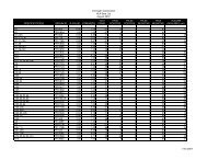
Table 1—Direct LanthaScreen on the PHERAstar: Statistics for PKC isoform titrations<br />
Protocol for PKC isoforms<br />
To demonstrate the functionality of Direct LanthaScreen technology on<br />
the PHERAstar, multiple PKC isoforms were titrated to determine optimal<br />
kinase concentrations for screening. The following protocol is an example<br />
of the conditions used to determine the EC₅₀ for one of the isoforms, PKCα.<br />
PKCα titration with Direct LanthaScreen technology<br />
A dilution series of PKCα, starting at a final concentration of 2.0 µg/ml,<br />
was incubated in the presence of 250 nM fluorescein-labeled PKC substrate<br />
and 20 µM ATP in a total volume of 10 µl in a black Corning® lowvolume<br />
384-well plate (Corning #3676). After a 90-minute incubation<br />
at room temperature, 10 µl of TR-FRET dilution buffer containing 2X<br />
EDTA (20 mM) and 2X Tb-PKC antibody (1.0 nM) was added and mixed<br />
to create a final volume of 20 µl per well, a final 1X EDTA concentration<br />
of 10 mM, and a final 1X antibody concentration of 0.5 nM. After a 60minute<br />
incubation at room temperature, the plate was read on the BMG<br />
LABTECH PHERAstar. Each data point represents the average of three<br />
wells. Figure 4 shows representative kinase titration curves for PKCα<br />
and PKCζ; Table 1 shows statistical data for all ten kinases tested.<br />
Figure 4—Direct LanthaScreen on the PHERAstar: PKCα and ζ titrations<br />
�� �� �� �� �� �� �� �� �� � �� � �� � �� � �� �<br />
PKCα PKCβI PKCβII PKCδ PKCε PKCη PKCγ PKCι PKCθ PKCς<br />
EC 50 (ng/ml) 0.43 0.27 0.22 0.07 0.03 0.04 0.18 0.13 0.16 0.12<br />
Min. value 0.44 0.43 0.79 .55 0.73 0.57 0.47 0.52 1.02 0.84<br />
Max. value 2.69 2.66 2.61 2.64 2.63 2.65 2.64 2.67 2.63 2.63<br />
Max-min 2.25 2.23 1.82 2.09 1.90 2.08 2.17 2.15 1.61 1.79<br />
Fold difference (max/min) 6.1 6.2 3.3 4.8 3.6 4.6 5.6 5.1 2.6 3.1<br />
Hill slope 1.1 0.89 1.01 0.70 0.93 1.10 0.87 0.73 0.96 0.83<br />
R² 0.991 0.996 0.988 0.979 0.988 0.993 0.994 0.988 0.976 0.982<br />
S:N 60 47 18 31 25 32 42 46 14 18<br />
Z’-factor value 0.94 0.91 0.78 0.87 0.85 0.88 0.91 0.91 0.74 0.79<br />
All ten PKC isoforms were titrated in a similar manner to the method described for PKCα. This data is meant to familiarize the reader with the relationship between Z’-factor values and other common<br />
statistical parameters. Regardless of “fold difference” or signal-to-noise ratios, all assays provided Z’-factor values far greater than 0.5. This is due to the use of FRET-based, ratiometric data analysis,<br />
which leads to low standard deviations of the replicates. Both Hill slope and R² measurements fall within acceptable limits. A Hill slope of 1.0 indicates the ideal binding curve; R² values approaching<br />
1.0 indicate the perfect curve fit.<br />
�������������<br />
�<br />
�<br />
�<br />
�<br />
���������<br />
��������<br />
�����������<br />
Ten PKC isoforms were tested for optimal kinase concentration in the Direct LanthaScreen<br />
format. This figure shows two representatives, alpha and zeta. Both kinases have EC₅₀ concentrations<br />
in the sub ng/ml range. See Table 1 for statistical details on all 10 kinases.
Data analysis<br />
Signal-to-noise ratio is calculated using the following formula:<br />
where µ p = mean of “positive control” (min ratio), µ n = mean of “negative<br />
control” (max ratio), and σ = the corresponding standard deviations.<br />
Z’-factor value is calculated using the following formula:<br />
where µ p = mean of “positive control” (min ratio), µ n = mean of “negative<br />
control” (max ratio), and σ = the corresponding standard deviations.<br />
Protocol for universal tyrosine kinase assay<br />
To demonstrate the functionality of a universal tyrosine kinase assay<br />
using Direct LanthaScreen, both fluorescein-poly-GAT (Glu, Ala,<br />
Tyr) and fluorescein-poly-GT (Glu, Tyr) substrates were used with terbium-labeled<br />
PY20 antibody. Figure 5 shows titration curves for two<br />
TK kinases using both substrates. Table 2 shows the EC₅₀ calculations<br />
derived from kinase titrations using both substrates.<br />
A dilution series of kinase was incubated with 400 nM fluoresceinlabeled<br />
substrate and 200 µM ATP in a total volume of 10 µl in a black<br />
Corning® low-volume 384-well plate (Corning #3676). After a 60-minute<br />
incubation at room temperature, 5 µl of TR-FRET dilution buffer<br />
containing 4X EDTA (60 mM) was added. Next, 5 µl of TR-FRET dilution<br />
buffer containing 4X antibody (8 nM) was added and mixed to create a<br />
final volume of 20 µl per well, a final 1X EDTA concentration of 15 mM,<br />
and a final 1X antibody concentration of 2 nM. After a 60-minute incubation<br />
at room temperature, the plate was read on the BMG LABTECH<br />
PHERAstar. Each data point represents the average of four wells.<br />
Figure 5—Kinase titrations for ZAP-70 and IGF-1R using both TK substrates<br />
�������������<br />
�������������<br />
���<br />
���<br />
���<br />
���<br />
���<br />
���<br />
���<br />
���<br />
���<br />
���<br />
���<br />
���<br />
���<br />
���<br />
���<br />
���<br />
�����������������������<br />
�����������������<br />
�� �� �� �� �� �� �� � �� � �� � �� � �� �<br />
������������������������<br />
������������������<br />
����������������<br />
Table 2—EC₅₀ calculations from kinase titration assays using either poly-<br />
GAT or poly-GT substrate<br />
�� �� �� �� �� �� �� �� �� � �� � �� � �� � �� �<br />
����������������<br />
���� ���� ���� ��� ��� ��� ��� ��� ���� ���� ���� ��� ��� ��� ��� ���� ���������������� ����������������<br />
������������� �������������<br />
���<br />
���<br />
���<br />
���<br />
���<br />
���<br />
���<br />
���<br />
���<br />
���<br />
�����������������������<br />
�����������������<br />
�����������������������<br />
�����������������<br />
A total of 24 tyrosine kinases were titrated in the presence of 200 µM ATP and either fluorescein-poly-GAT (Glu, Ala, Tyr) or fluorescein-poly-GT (Glu, Tyr) substrate. This figure shows representative<br />
results for ZAP-70 (A and B) and IGF-1R (C and D). As a trend, poly-GAT reactions yielded higher dynamic ranges, whereas poly-GT reactions required less kinase as determined by EC₅₀ calculations.<br />
Kinase<br />
EC₅₀ (ng/ml)<br />
EC₅₀ (ng/ml)<br />
Kinase<br />
polyGAT polyGT polyGAT polyGT<br />
ZAP-70 7.46 1.98 LynA 0.38 0.34<br />
TEC 5.02 14.70 LynB 1.01 0.08<br />
ErbB2 9.16 3.41 Abl1 4.73 0.39<br />
cKit 5.22 6.01 Arg 4.70 0.54<br />
cMet 1.25 0.71 FGFR1 0.45 0.29<br />
IGF1R 15.54 0.57 FGFR2 0.42 0.06<br />
FLT3 0.66 0.02 FGFR3 0.64 0.03<br />
PDGFRβ 3.40 1.84 FGFR4 0.97 0.02<br />
Lck 1.06 2.64 EGFR 15.55 9.74<br />
Fyn 7.48 0.53 CSF1R 0.37 0.30<br />
Yes 6.94 0.56 TrkA 0.08 0.10<br />
Src 0.75 0.16 EphB4 0.36 0.51<br />
Dynamic range: poly-GAT ~ 50 fold<br />
Dynamic range: poly-GT ~ 17 fold<br />
This table shows the trend for lower kinase concentrations needed (as shown by EC₅₀ values)<br />
using the poly-GT substrate. <strong>Note</strong>, however, that the poly-GT substrate generally leads to<br />
increased assay dynamic range.
<strong>Application</strong> <strong>Note</strong><br />
Results and conclusions<br />
Our results for the PKC isoform titrations demonstrate that Direct<br />
LanthaScreen PKC assays are sensitive in that they require sub ng/ml<br />
kinase concentrations. This is an important factor in keeping overall<br />
screening costs down. In addition, the assays are robust with Z’-factor<br />
values greater that 0.7 in all cases. These Z’-factor values provide confidence<br />
that the assay data is meaningful. It is important to stress this<br />
point: “Fold difference” (dynamic range, assay window) is not critical to<br />
assay robustness. Rather, the difference between positive and negative<br />
controls, combined with standard deviations of the same, lead to assay<br />
confidence as dictated by Z’-factor values.<br />
Our results for the TK assays show that use of a fluorescein-labeled<br />
substrate common for many TKs (poly-GT or poly-GAT) allows development<br />
of a “universal” TK assay using the Direct LanthaScreen format.<br />
For many of the TKs shown, EC 50 values are similar for both substrates.<br />
But for a number of kinases, especially the FGFRs, EC₅₀ values are much<br />
lower for the poly-GT substrate. This may indicate that the poly-GT<br />
substrate provides a more sensitive assay compared to the poly-GAT.<br />
However, on average, the poly-GAT substrate provided an increased<br />
dynamic range compared to poly-GT (~50 fold compared to ~17 fold).<br />
Therefore, depending on the kinase, the customer can choose which is<br />
more important: using less kinase or having increased dynamic range.<br />
Ordering information<br />
The terbium-based Direct LanthaScreen format offers several advantages<br />
over many other fluorescent assays, including traditional europium-based<br />
formats:<br />
1. The ratiometric nature of LanthaScreen eliminates well-to-well<br />
data variation.<br />
2. The time-resolved nature of LanthaScreen assays allows for the<br />
use of fluorescein without the associated drawbacks of compound<br />
interference.<br />
3. The ability to use fluorescein as an acceptor rather than APC simplifies<br />
assay development and reduces cost.<br />
4. Steric hindrance due to the biotin-streptavidin:APC complex is<br />
eliminated when using the relatively small fluorescein molecule.<br />
5. Fluorescein is very soluble, unlike some “stickier” red-shifted dyes.<br />
6. It is approximately 5–10 fold less expensive to label a peptide substrate<br />
with fluorescein than with a far-red dye such as Cy5.<br />
From an instrumentation point of view, BMG LABTECH provides an<br />
excellent platform to simplify LanthaScreen assay development. The<br />
PHERAstar provides the speed and sensitivity to truly take advantages<br />
of either LanthaScreen format described above. An optical module<br />
optimized for use with LanthaScreen assays is available from BMG<br />
LABTECH, making assay setup easy.<br />
Product Quantity Cat. no.<br />
Fluorescein PKC substrate, 1.0 mg/ml 1 mg PV3506<br />
Kinase Buffer, 5X (for PKC assays) 4 ml PV3189<br />
Kinase Buffer (for TK assays) 50 mM HEPES (pH 7.5), 10 mM MgCl₂,<br />
5 mM MnCl₂, 2 mM DTT, 200 µM Na₃VO₄, and 0.01% CHAPS<br />
LanthaScreen Tb-PKC Substrate Antibody<br />
N/A N/A<br />
25 µg PV3536<br />
100 µg PV3537<br />
PKCα 5 µg P2232<br />
LanthaScreen TR-FRET Dilution Buffer 200 ml PV3574<br />
ZAP-70 20 µg P2782<br />
LanthaScreen Tb-PY20 Antibody<br />
25 µg PV3528<br />
1 mg PV3529<br />
Fluorescein-poly-GAT (Glu, Ala, Tyr) 1 mg PV3611<br />
Fluorescein-poly-GT (Glu, Tyr) 1 mg PV3610<br />
Kinase Quench Buffer (500 mM EDTA) 1 ml P2825
<strong>Application</strong> <strong>Note</strong><br />
These products may be covered by one or more Limited Use Label Licenses (See the <strong>Invitrogen</strong> catalog or www.invitrogen.com).<br />
By use of these products you accept the terms and conditions of all applicable Limited Use Label Licenses.<br />
For research use only. Not intended for any animal or human therapeutic or diagnostic use.<br />
©2004 <strong>Invitrogen</strong> Corporation. All rights reserved. Reproduction forbidden without permission. Printed in the U.S.A.<br />
Corporate headquarters:<br />
1600 Faraday Avenue • Carlsbad, CA 92008 USA • Tel: 760 603 7200 • Fax: 760 602 6500 • Toll Free Tel: 800 955 6288 • E-mail: tech_service@invitrogen.com • www.invitrogen.com<br />
European headquarters:<br />
<strong>Invitrogen</strong> Ltd • Inchinnan Business Park • 3 Fountain Drive • Paisley PA4 9RF, UK • Tel: +44 (0) 141 814 6100 • Fax: +44 (0) 141 814 6260 • E-mail: eurotech@invitrogen.com<br />
O-13473-r1 US 1204