Redder is Better: Far-Red PolarScreen™ FP Kinase ... - Invitrogen
Redder is Better: Far-Red PolarScreen™ FP Kinase ... - Invitrogen
Redder is Better: Far-Red PolarScreen™ FP Kinase ... - Invitrogen

You also want an ePaper? Increase the reach of your titles
YUMPU automatically turns print PDFs into web optimized ePapers that Google loves.
Why <strong>is</strong> <strong><strong>Red</strong>der</strong> <strong>Better</strong>?<br />
<strong><strong>Red</strong>der</strong> <strong>is</strong> <strong>Better</strong>: <strong>Far</strong>-<strong>Red</strong> PolarScreen<br />
<strong>FP</strong> <strong>Kinase</strong> Assays<br />
Kevin L. Vedvik, Hildegard C. Eliason, Jennifer A. Fronczak,<br />
Mel<strong>is</strong>sa E. Krueger and Kurt W. Vogel<br />
<strong>Invitrogen</strong> Corporation • 501 Charmany Drive • Mad<strong>is</strong>on, WI 53719 • USA<br />
Compound interference <strong>is</strong> the bane of HTS. Two forms of interference that affect fluorescence polarization assays are compound fluorescence and<br />
light scatter. Compound fluorescence ar<strong>is</strong>es from the intrinsic fluorescence of some library compounds. Because compound libraries are typically<br />
screened at concentrations of 10 µM, and tracer concentrations are typically 1 nM, a library compound that has 1/10,000 th (or less) of the fluores-<br />
cent “brightness” of the tracer has the potential to interfere with the assay signal. Because most small molecules that fluoresce will emit a depolar-<br />
ized (low polarization) signal, th<strong>is</strong> can lead to false positive or false negative results, depending on the assay configuration.<br />
The ability of a molecule to absorb light in the “red” (longer wavelength) region of the spectrum depends largely on the<br />
degree of conjugation (adjacent double bonds and aromatic rings) within the molecule. In general, the more conjugation the<br />
molecule has, the better able it will be to absorb light in the red region of the spectrum. Because the ability to absorb light <strong>is</strong><br />
a prerequ<strong>is</strong>ite to fluorescence, it follows that in order to fluoresce at “redder” regions of the spectrum a molecule needs more<br />
conjugated double bonds. The high degree of conjugation necessary for “red” fluorescence <strong>is</strong> incompatible with “druglike”<br />
properties, and therefore there are, in general, fewer “red” fluorescent compounds in most libraries.<br />
The second interfering factor in <strong>FP</strong> assays ar<strong>is</strong>es from light scatter. Light scatter ar<strong>is</strong>es from precipitated compounds or other particulate matter<br />
(dust) in an assay well. Because light scatter <strong>is</strong> more efficient at lower (greener) wavelengths, and drops off in intensity relative to the 4 th power of<br />
the wavelength, scatter <strong>is</strong> less of an interference at longer wavelengths. Scattered light <strong>is</strong> often polarized (th<strong>is</strong> <strong>is</strong> the reason sunglasses are polar-<br />
ized—to block scattered light, or glare, from the horizon) and therefore interferes with an assay by giving an abnormally high polarization value.<br />
<strong>Invitrogen</strong> Corp • 1600 <strong>Far</strong>aday Avenue • Carlsbad, CA 92008 USA • Tel: 760 603 7200 • FAX: 760 602 6500 • E-mail: tech_service@invitrogen.com • www.invitrogen.com
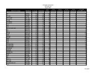
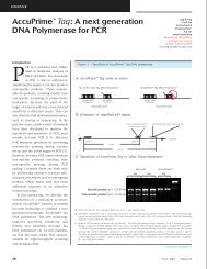
Figure 1—Assay Interference Decreases at Longer Wavelengths<br />
Compounds in LOPAC 1280 with greater than 50% signal intensity of 1 nM corresponding tracer<br />
Tracer λ (Ex / Em) # of Compounds<br />
Green 485 / 535 19<br />
<strong>Red</strong> 535 / 590 9<br />
<strong>Far</strong>-<strong>Red</strong> 590 / 650 3<br />
To determine an approximation of the amount of fluorescent interference that can be expected in a typical compound library at green,<br />
red, and far-red wavelengths, the LOPAC 1280 library was prepared at 10 µM in buffer and read in fluorescence intensity mode using filters<br />
typical to a green, red, or far-red tracer. Wells containing 1 nM of each tracer were placed on the assay plate so that signal intensity<br />
could be referenced to a typical tracer concentration.<br />
Relative Signal Intensity<br />
3.0<br />
2.5<br />
2.0<br />
1.5<br />
1.0<br />
0.5<br />
0.0<br />
Relative Interference from Scatter<br />
GW5047<br />
Green Tracer Green Scatter <strong>Red</strong> Tracer <strong>Red</strong> Scatter <strong>Far</strong> <strong>Red</strong> Tracer <strong>Far</strong> <strong>Red</strong> Scatter<br />
To demonstrate the effect of light scatter on assay interference, GW5047, a compound from the LOPAC Library prone to precipitation,<br />
was compared in signal intensity to 1 nM of green, red, and far-red tracers. The graph above illustrates negligible interference (<<br />
0.4%) seen from GW5047 when using a far-red tracer. The inset shows a close-up of a vial containing the precipitated compound.
Figure 2—Excitation / Em<strong>is</strong>sion Spectra of <strong>Far</strong>-<strong>Red</strong> Tracers<br />
100<br />
75<br />
50<br />
25<br />
0<br />
400 450 500 550 600 650 700<br />
Excitation/Em<strong>is</strong>sion spectra of commonly-used fluorescein and rhodamine-based tracers compared to the spectra of <strong>Invitrogen</strong>’s new<br />
<strong>Far</strong>-<strong>Red</strong> tracers. The excitation and em<strong>is</strong>sion maxima for the far-red tracer are red-shifted beyond 600 nm, allowing fluorescence polar-<br />
ization assays to be performed using a 610 nm excitation filter and 670 nm em<strong>is</strong>sion filters.<br />
Figure 3—<strong>FP</strong> <strong>Kinase</strong> Assay Schematic<br />
Inhibited Reaction Uninhibited Reaction<br />
<strong>Kinase</strong> Reaction<br />
<strong>Kinase</strong><br />
ATP<br />
<strong>Kinase</strong><br />
ATP<br />
+<br />
Inhibitor<br />
P<br />
P<br />
Fluorescein Rhodamine <strong>Far</strong> <strong>Red</strong><br />
P<br />
Wavelength (nm)<br />
Add Antibody<br />
and Tracer<br />
In a far-red <strong>FP</strong> kinase assay, kinase, substrate, and ATP are allowed to react in the presence of library compounds. After the reaction,<br />
antibody and far-red-labeled tracer are added. The amount of antibody that binds to the tracer <strong>is</strong> inversely related to the amount of phos-<br />
phorylated product present. Thus, library compounds that inhibit the reaction are identified as wells that have a high polarization value.<br />
ANTIBODY<br />
P<br />
Tracer<br />
ANTIBODY<br />
P<br />
Tracer<br />
750<br />
Detection<br />
P<br />
P<br />
P<br />
ANTIBODY<br />
P<br />
ANTIBODY<br />
P<br />
Low Polarization<br />
High Polarization
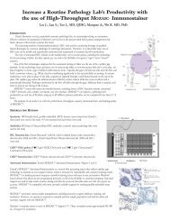
Figure 4—<strong>Far</strong> <strong>Red</strong> <strong>FP</strong> <strong>Kinase</strong> Kits Address a Range of <strong>Kinase</strong> Targets<br />
Polarization (mP)<br />
Polarization (mP)<br />
Polarization (mP)<br />
250<br />
225<br />
200<br />
175<br />
150<br />
125<br />
225<br />
200<br />
175<br />
150<br />
125<br />
100<br />
75<br />
250<br />
225<br />
200<br />
175<br />
150<br />
125<br />
100<br />
Rb ING <strong>Far</strong>-<strong>Red</strong> Assay (<strong>Kinase</strong>: CDK5/p35)<br />
10 -5 10 -4 10 -3 10 -2 10 -1 10 0 10 1<br />
[<strong>Kinase</strong>] (ng/well)<br />
PKC <strong>Far</strong>-<strong>Red</strong> Assay (<strong>Kinase</strong>: CHK1)<br />
10 -5 10 -4 10 -3 10 -2 10 -1 10 0<br />
[<strong>Kinase</strong>] (ng/well)<br />
CREBtide <strong>Far</strong>-<strong>Red</strong> Assay (<strong>Kinase</strong>: PKA)<br />
10 -2<br />
10 -1<br />
10 0 10 1 10 2 10 3<br />
[<strong>Kinase</strong>] (pg/well)<br />
Polarization (mP)<br />
Polarization (mP)<br />
300<br />
250<br />
200<br />
150<br />
100<br />
200<br />
150<br />
100<br />
50<br />
0<br />
PTK <strong>Far</strong>-<strong>Red</strong> Assay (<strong>Kinase</strong>: LynB)<br />
10 -5 10 -4 10 -3 10 -2 10 -1 10 0 10 1<br />
[<strong>Kinase</strong>] (ng/well)<br />
PDK1 <strong>Far</strong>-<strong>Red</strong> Assay (<strong>Kinase</strong>: PDK1)<br />
10 -4 10 -3 10 -2 10 -1 10 0 10 1<br />
[<strong>Kinase</strong>] (pg/well)<br />
Polarization (mP)<br />
Polarization (mP)<br />
250<br />
200<br />
150<br />
100<br />
50<br />
200<br />
150<br />
100<br />
50<br />
IκB-α pSer 32 <strong>Far</strong>-<strong>Red</strong> Assay (<strong>Kinase</strong>: CKII)<br />
10 -5 10 -4 10 -3 10 -2 10 -1 10 0 10 1<br />
[<strong>Kinase</strong>] (units/well)<br />
Crosstide <strong>Far</strong>-<strong>Red</strong> Assay (<strong>Kinase</strong>: Akt1/PKBα)<br />
10 -5 10 -4 10 -3 10 -2 10 -1 10 0<br />
[<strong>Kinase</strong>] (ng/well)<br />
Seven far-red kinase assays are available to address a wide variety of kinase targets. The plots in Figure 3 show the results from kinase<br />
titrations in 10 µL, 90 minute kinase reactions under conditions of non-limiting substrate and ATP. In general, picogram to nanogram<br />
quantities of kinase will phosphorylate sufficient peptide substrate to effect an assay window of between 125 and 250 mP.<br />
Figure 5—Inhibition of PDK1 by Staurosporine<br />
Polarization (mP)<br />
175<br />
150<br />
125<br />
100<br />
75<br />
50<br />
25<br />
0<br />
Inhibition of PDK1 with Staurosporine<br />
EC 50 = 9.4 nM<br />
0.1 1 10 100<br />
[Staurosporine] (nM)<br />
<strong>Far</strong>-<strong>Red</strong> kinase assays can be used for both high-throughput screening as well as follow-up of hits to determine accurate EC 50 values.
Conclusions<br />
As the size of compound libraries increases so does the cost of follow-up screening of false positive and false negative “hits” that are<br />
due to compound interference. The shift from “green” to “red” has been recognized as a valid strategy to overcome interference due to<br />
autofluorescence or light scatter due to precipitated compounds. <strong>Invitrogen</strong>’s new far-red PolarScreen assays employ a proprietary far-<br />
red fluorophore that gives excellent performance in fluorescence polarization assays. The fluorophore <strong>is</strong> highly water soluble and unlike<br />
cyanine-based fluorophores has a fluorescence lifetime that allows for large polarization shifts between free and bound tracer.<br />
839-0412392 041504