EasySelect™ Echo™-Adapted Pichia Expression Kit - Invitrogen
EasySelect™ Echo™-Adapted Pichia Expression Kit - Invitrogen
EasySelect™ Echo™-Adapted Pichia Expression Kit - Invitrogen

Create successful ePaper yourself
Turn your PDF publications into a flip-book with our unique Google optimized e-Paper software.
EasySelect Echo -<strong>Adapted</strong><br />
<strong>Pichia</strong> <strong>Expression</strong> <strong>Kit</strong><br />
For expression of the gene of interest in <strong>Pichia</strong><br />
pastoris using pPICZ-E and pPICZα-E with the<br />
Echo Cloning System<br />
Catalog nos. ET230-xx, ET231-xx, ET232-xx<br />
Version E<br />
07 September 2010<br />
25-0388<br />
A Limited Label License covers this product (see Purchaser Notification). By<br />
use of this product, you accept the terms and conditions of the Limited Label<br />
License.<br />
User Manual
INDIVIDUAL PICHIA EXPRESSION KIT LICENSE AGREEMENT<br />
The <strong>Pichia</strong> <strong>Expression</strong> <strong>Kit</strong> is based on the yeast <strong>Pichia</strong> pastoris. <strong>Pichia</strong> pastoris was developed into an expression system<br />
by scientists at Salk Institute Biotechnology/Industry Associates (SIBIA) for high-level expression of recombinant proteins.<br />
All patents for <strong>Pichia</strong> pastoris and licenses for its use as an expression system are owned by Research Corporation<br />
Technologies, Inc. Tucson, Arizona. <strong>Invitrogen</strong> has an exclusive license to sell the <strong>Pichia</strong> <strong>Expression</strong> <strong>Kit</strong> to scientists for<br />
research purposes only, under the terms described below. Use of <strong>Pichia</strong> pastoris by commercial corporations requires the<br />
user to obtain a commercial license as detailed below. Before using the <strong>Pichia</strong> <strong>Expression</strong> <strong>Kit</strong>, please read the following<br />
license a greement. If you do not agree to be bound by its terms, contact <strong>Invitrogen</strong> within 10 days for authorization to<br />
return the unused <strong>Pichia</strong> <strong>Expression</strong> <strong>Kit</strong> and to receive a full credit. If you do agree to the terms of this Agreement, please<br />
complete the User Registration Card and return it to <strong>Invitrogen</strong> before using the kit.<br />
INDIVIDUAL PICHIA EXPRESSION KIT LICENSE AGREEMENT<br />
<strong>Invitrogen</strong> Corporation (INVITROGEN) grants you a non-exclusive license to use the enclosed <strong>Pichia</strong> <strong>Expression</strong> <strong>Kit</strong><br />
(EXPRESSION KIT) for academic research or for evaluation purposes only. The EXPRESSION KIT is being transferred<br />
to you in furtherance of, and reliance on, such license. You may not use the EXPRESSION KIT, or the materials contained<br />
therein, for any commercial purpose without a license for such purpose from RESEARCH CORPORATION<br />
TECHNOLOGIES, INC., Tucson, Arizona. Commercial purposes include the use in or sale of expressed proteins as a<br />
commercial product, or use to facilitate or advance research or development of a commercial product. Commercial entities<br />
may conduct their evaluation for one year at which time this license automatically terminates. Commercial entities will be<br />
contacted by Research Corporation Technologies during the evaluation period regarding the purchase of a commercial<br />
license.<br />
Access to the EXPRESSION KIT must be limited solely to those officers, employees and students of your institution who<br />
need access thereto in order to perform the above-described research or evaluation. You must inform each of such officer,<br />
employee and student of the provisions of this Agreement and require them to agree, in writing, to be bound by the<br />
provisions of this Agreement. You may not distribute the EXPRESSION KIT to others, even those within your own<br />
institution. You may transfer modified, altered or original material from the EXPRESSION KIT to a third party following<br />
notification of INVITROGEN such that the recipient can be licensed. You may not assign, sub-license, rent lease or<br />
otherwise transfer this License or any of the rights or obligation hereunder, except as expressly permitted.<br />
This License is effective until terminated. You may terminate it at any time by destroying all <strong>Pichia</strong> expression products in<br />
your control. It will also terminate automatically if you fail to comply with the terms and conditions of the Agreement.<br />
You shall, upon termination of the License, destroy all <strong>Pichia</strong> <strong>Expression</strong> <strong>Kit</strong>s in your control, and so notify<br />
INVITROGEN in writing.<br />
This License Shall be governed in its interpretation and enforcement by the laws of the State of California..<br />
Technical Services<br />
<strong>Invitrogen</strong> provides Technical Services to all of our registered <strong>Pichia</strong> <strong>Expression</strong> <strong>Kit</strong> users. Please contact us if you need<br />
assistance with the <strong>Pichia</strong> <strong>Expression</strong> <strong>Kit</strong>.<br />
Corporate Headquarters: Japanese Headquarters European Headquarters:<br />
<strong>Invitrogen</strong> Corporation<br />
1600 Faraday Avenue<br />
Carlsbad, CA 92008 USA<br />
Tel: 1 760 603 7200<br />
Tel (Toll Free): 1 800 955 6288<br />
Fax: 1 760 602 6500<br />
E-mail: tech_service@invitrogen.com<br />
<strong>Invitrogen</strong> Japan<br />
LOOP-X Bldg. 6F<br />
3-9-15, Kaigan<br />
Minato-ku, Tokyo 108-0022<br />
Tel: 81 3 5730 6509<br />
Fax: 81 3 5730 6519<br />
E-mail: jpinfo@invitrogen.com<br />
<strong>Invitrogen</strong> Ltd<br />
3 Fountain Drive<br />
Inchinnan Business Park<br />
3 Fountain Drive<br />
Paisley PA4 9RF, UK<br />
Tel: +44 (0) 141 814 6100<br />
Fax: +44 (0) 141 814 6287<br />
E-mail: eurotech@invitrogen.com<br />
iii
Table of Contents<br />
Table of Contents....................................................................................................................................................v<br />
<strong>Kit</strong> Contents and Storage .......................................................................................................................................vi<br />
Product Qualification.............................................................................................................................................xi<br />
Accessory Products............................................................................................................................................. xiii<br />
Purchaser Notification ..........................................................................................................................................xv<br />
Using This Manual ........................................................................................................................................... xviii<br />
Introduction ..................................................................................................................1<br />
Overview of the Echo Cloning System ................................................................................................................1<br />
Overview of <strong>Pichia</strong> pastoris <strong>Expression</strong> System....................................................................................................4<br />
Methods ........................................................................................................................8<br />
Recombining Your Gene into pPICZ-E or pPICZα-E ...........................................................................................8<br />
Transforming the Recombination Reaction ............................................................................................................9<br />
<strong>Pichia</strong> Strains........................................................................................................................................................13<br />
<strong>Pichia</strong> Transformation ..........................................................................................................................................15<br />
EasyComp Transformation.................................................................................................................................19<br />
Determining the Mut Phenotype...........................................................................................................................22<br />
<strong>Expression</strong> in <strong>Pichia</strong> .............................................................................................................................................25<br />
Analysis by SDS-Polyacrylamide Gel Electrophoresis ........................................................................................30<br />
Optimization of <strong>Pichia</strong> Protein <strong>Expression</strong> ..........................................................................................................32<br />
Scale-up of <strong>Expression</strong> .........................................................................................................................................34<br />
Purification ...........................................................................................................................................................36<br />
<strong>Pichia</strong> Media Recipes ...........................................................................................................................................39<br />
Appendix.....................................................................................................................45<br />
Recipes..................................................................................................................................................................45<br />
Maps of pPICZ-E and pPICZα-E.........................................................................................................................46<br />
Features of pPICZ-E and pPICZα-E ....................................................................................................................48<br />
Map of pPICZ-E/Uni-lacZ....................................................................................................................................49<br />
Map of pPICZα-E/Uni-HSA ................................................................................................................................50<br />
Recombination and Integration in <strong>Pichia</strong> .............................................................................................................51<br />
Zeocin .................................................................................................................................................................53<br />
Direct PCR Screening of <strong>Pichia</strong> Clones ...............................................................................................................55<br />
Total DNA Isolation from <strong>Pichia</strong> .........................................................................................................................56<br />
Determination of Copy Number of Multiple Integrants .......................................................................................58<br />
Procedure for Total RNA Isolation from <strong>Pichia</strong>...................................................................................................60<br />
Technical Service..................................................................................................................................................61<br />
References.............................................................................................................................................................62<br />
v
<strong>Kit</strong> Contents and Storage<br />
Types of <strong>Kit</strong>s Several EasySelect Echo -adapted <strong>Pichia</strong> <strong>Expression</strong> <strong>Kit</strong>s, pPICZ-E and pPICZα-E<br />
Echo -adapted <strong>Expression</strong> Vector <strong>Kit</strong>s are available (see table below). This manual is<br />
included with all of these kits. Note that not all kits contain the reagents discussed in this<br />
manual (see page xviii for more details).<br />
vi<br />
<strong>Kit</strong> Reagents Supplied Catalog nos.<br />
EasySelect Echo -adapted <strong>Pichia</strong> pPICZ-E Echo<br />
<strong>Expression</strong> <strong>Kit</strong><br />
-adapted <strong>Expression</strong> Vector <strong>Kit</strong><br />
pPICZα-E Echo -adapted <strong>Expression</strong> Vector <strong>Kit</strong><br />
EasySelect <strong>Pichia</strong> Strains <strong>Kit</strong><br />
<strong>Pichia</strong> EasyComp ET230-02<br />
<strong>Kit</strong><br />
<strong>Pichia</strong> Media <strong>Kit</strong><br />
EasySelect Echo -adapted <strong>Pichia</strong><br />
<strong>Expression</strong> <strong>Kit</strong> with a choice of<br />
Donor Vector <strong>Kit</strong> and One Shot ®<br />
TOP10 Chemically Competent<br />
E. coli (see page xiii for more<br />
information on donor vectors)<br />
pPICZ-E Echo -adapted <strong>Expression</strong><br />
Vector <strong>Kit</strong><br />
pPICZα-E Echo -adapted<br />
<strong>Expression</strong> Vector <strong>Kit</strong><br />
pPICZ-E Echo -adapted <strong>Expression</strong><br />
Vector <strong>Kit</strong> with a choice of Donor<br />
Vector <strong>Kit</strong> and One Shot ® TOP10<br />
Chemically Competent E. coli (see<br />
page xiii for more information on<br />
donor vectors)<br />
pPICZα-E Echo -adapted<br />
<strong>Expression</strong> Vector <strong>Kit</strong> with a choice<br />
of Donor Vector <strong>Kit</strong> and One Shot ®<br />
TOP10 Chemically Competent<br />
E. coli (see page xiii for more<br />
information on donor vectors)<br />
pUni/V5-His-TOPO ® TA Cloning <strong>Kit</strong> ET230-10C<br />
pUniBlunt/V5-His-TOPO ® Cloning <strong>Kit</strong> ET230-20C<br />
pUni/V5-His A, B and C ET230-30C<br />
pUniD/V5-His-TOPO ® Cloning <strong>Kit</strong> ET230-40C<br />
pPICZ-E Vector<br />
<strong>Expression</strong> Control Vector<br />
Cre Recombinase and 10X Buffer<br />
5′ AOX1 Sequencing Primer<br />
Zeocin <br />
pPICZα-E Vector<br />
<strong>Expression</strong> Control Vector<br />
Cre Recombinase and 10X Buffer<br />
α-Factor Sequencing Primer<br />
Zeocin <br />
ET231-01<br />
ET232-01<br />
pUni/V5-His-TOPO ® TA Cloning <strong>Kit</strong> ET231-10C<br />
pUniBlunt/V5-His-TOPO ® Cloning <strong>Kit</strong> ET231-20C<br />
pUni/V5-His A, B and C ET231-30C<br />
pUniD/V5-His-TOPO ® Cloning <strong>Kit</strong> ET231-40C<br />
pUni/V5-His-TOPO ® TA Cloning <strong>Kit</strong> ET232-10C<br />
pUniBlunt/V5-His-TOPO ® Cloning <strong>Kit</strong> ET232-20C<br />
pUni/V5-His A, B and C ET232-30C<br />
pUniD/V5-His-TOPO ® Cloning <strong>Kit</strong> ET232-40C<br />
Continued on next page
<strong>Kit</strong> Contents and Storage, Continued<br />
Shipping and<br />
Storage<br />
pPICZ-E Echo <br />
Reagents<br />
The EasySelect Echo -adapted <strong>Pichia</strong> <strong>Expression</strong> <strong>Kit</strong> is shipped on dry ice except for<br />
the EasySelect <strong>Pichia</strong> strains and the <strong>Pichia</strong> Media <strong>Kit</strong>, which are shipped at room<br />
temperature. Each EasySelect Echo -adapted <strong>Pichia</strong> <strong>Expression</strong> <strong>Kit</strong> contains two<br />
vector kits (1 box for each vector), EasySelect <strong>Pichia</strong> Strains (Box 2), EasyComp <br />
<strong>Pichia</strong> <strong>Kit</strong> (Box 3), and <strong>Pichia</strong> Media <strong>Kit</strong>.<br />
The pPICZ-E and pPICZα-E Echo -adapted <strong>Expression</strong> <strong>Kit</strong>s are shipped on dry ice.<br />
Each of pPICZ-E and pPICZα-E Echo -<strong>Adapted</strong> <strong>Expression</strong> <strong>Kit</strong> contains the Echo -<br />
adapted expression vector, an expression control vector, a sequencing primer and<br />
Zeocin .<br />
Reagents Storage<br />
pPICZ-E Echo -adapted <strong>Expression</strong> <strong>Kit</strong> -20°C<br />
pPICZα-E Echo -adapted <strong>Expression</strong> <strong>Kit</strong><br />
-20°C<br />
EasySelect <strong>Pichia</strong> Strains <strong>Kit</strong> +4°C<br />
<strong>Pichia</strong> EasyComp <strong>Kit</strong> +4°C<br />
<strong>Pichia</strong> Media <strong>Kit</strong> Room temperature<br />
One Shot ® TOP10 Chemically Competent E. coli<br />
(Optional)<br />
-80°C<br />
The following items are supplied in the pPICZ-E Echo -adapted <strong>Expression</strong> <strong>Kit</strong>.<br />
Item Composition Amount<br />
pPICZ-E Supercoiled, lyophilized in TE, pH 8.0 20 µg<br />
pPICZ-E/Uni-lacZ<br />
expression control<br />
Supercoiled, lyophilized in TE, pH 8.0 20 µg<br />
5′ AOX1 sequencing primer Lyophilized in water 2 µg<br />
Cre Recombinase Check the label on the tube for exact<br />
concentration of the enzyme.<br />
Enzyme is supplied in:<br />
50 mM Tris-HCl, pH 8.0<br />
5 mM EDTA<br />
1 mM EGTA<br />
10 mM β-mercaptoethanol<br />
20% Glycerol<br />
10X Recombinase Buffer 500 mM Tris-HCl, pH 7.5<br />
100 mM MgCl2<br />
300 mM NaCl<br />
1.0 mg/ml BSA<br />
15 µl<br />
25 µl<br />
Zeocin 100 mg/ml 2 x 1.25 ml<br />
Continued on next page<br />
vii
<strong>Kit</strong> Contents and Storage, Continued<br />
pPICZα-E Echo <br />
Reagents<br />
The <strong>Pichia</strong><br />
EasyComp <strong>Kit</strong><br />
viii<br />
The following items are supplied in the pPICZα-E Echo -adapted <strong>Expression</strong> <strong>Kit</strong><br />
Item Composition Amount<br />
pPICZα-E Supercoiled, lyophilized in TE, pH 8.0 20 µg<br />
pPICZα-E/Uni-HSA<br />
expression control<br />
Supercoiled, lyophilized in TE, pH 8.0 20 µg<br />
α-Factor sequencing primer Lyophilized in water 2 µg<br />
Cre Recombinase Check the label on the tube for exact<br />
concentration of the enzyme.<br />
Enzyme is supplied in:<br />
50 mM Tris-HCl, pH 8.0<br />
5 mM EDTA<br />
1 mM EGTA<br />
10 mM β-mercaptoethanol<br />
20% Glycerol<br />
10X Recombinase Buffer 500 mM Tris-HCl, pH 7.5<br />
100 mM MgCl2<br />
300 mM NaCl<br />
1.0 mg/ml BSA<br />
15 µl<br />
25 µl<br />
Zeocin 100 mg/ml 2 x 1.25 ml<br />
This kit contains sufficient reagents for 6 preparations of competent cells. Each<br />
competent cell preparation yields enough cells for 20 transformations.<br />
Upon receipt, store the kit at +4°C. Solution II can also be stored at room temperature.<br />
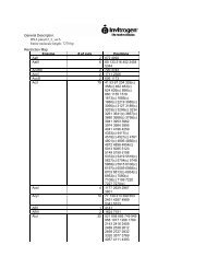
Component Description Quantity<br />
Solution I Sorbitol solution containing ethylene glycol and DMSO<br />
for the preparation of competent cells<br />
75 ml<br />
Solution II PEG solution for the transformation of competent cells 150 ml (2 x 75 ml)<br />
Solution III Salt solution for washing and plating transformed cells 150 ml (2 x 75 ml)<br />
Continued on next page
<strong>Kit</strong> Contents and Storage, Continued<br />
EasySelect <strong>Pichia</strong><br />
Strains <strong>Kit</strong><br />
The table below lists the genotype and phenotype of the different <strong>Pichia</strong> stabs. Store the<br />
stabs at +4°C.<br />
Strain Genotype Phenotype (<strong>Pichia</strong> only)<br />
X-33 wild-type Mut +<br />
GS115 his4 His - , Mut +<br />
KM71H arg4 aox1::ARG4 Mut S , Arg +<br />
GS115/pPICZ/lacZ his4 His - , Mut +<br />
GS115/Albumin his4 His - , Mut s<br />
TOP10F´ F´ {proAB, lacI q , lacZ∆M15, Tn10 (Tet R )} mcrA, ∆(mrrhsdRMS-mcrBC),<br />
φ80lacZ∆M15, ∆lacX74, , recA1, λ - araD139,<br />
∆(ara-leu)7697, galU, galK, rpsL(Str R ), endA1, nupG<br />
Media The following prepackaged media is included for your convenience. Instructions for<br />
use are provided on the package. Keep the media dry and store at room temperature.<br />
Sequence of<br />
Primers<br />
Media Amount Yield<br />
YP Base Medium 2 pouches 2 liters of YP medium<br />
YP Base Agar Medium 2 pouches 2 liters of YP agar medium<br />
Yeast Nitrogen Base 1 pouch 500 ml of 10X YNB<br />
The table below lists the sequence and pmoles of the primers included in this kit.<br />
Primer Sequence pmoles Supplied<br />
5′ AOX1 sequencing<br />
primer<br />
5’-GACTGGTTCCAATTGACAAGC-3’ 312 pmoles<br />
α-Factor sequencing<br />
primer<br />
5’-TACTATTGCCAGCAATTGCTGC-3’ 315 pmoles<br />
Continued on next page<br />
ix
<strong>Kit</strong> Contents and Storage, Continued<br />
One Shot ® TOP10<br />
Reagents<br />
(Optional)<br />
Genotype of<br />
TOP10<br />
x<br />
The table below describes the items included in the One Shot ® TOP10 Chemically<br />
Competent E. coli kit.<br />
Store at -80°C.<br />
Item Concentration Amount<br />
SOC Medium 2% Tryptone<br />
6 ml<br />
(may be stored at room 0.5% Yeast Extract<br />
temperature or at<br />
+4°C)<br />
10 mM NaCl<br />
2.5 mM KCl<br />
10 mM MgCl2<br />
10 mM MgSO4<br />
20 mM glucose<br />
TOP10 E. coli -- 11 x 50 µl<br />
pUC19 Control DNA 10 pg/µl in 5 mM Tris-HCl, 0.5 mM<br />
EDTA, pH 8<br />
50 µl<br />
TOP10: Use this strain for general cloning of your gene of interest. Note that this strain<br />
cannot be used for transformation and growth of the donor vectors.<br />
F- mcrA ∆(mrr-hsdRMS-mcrBC) Φ80lacZ∆M15 ∆lacX74 recA1 araD139 ∆(araleu)7697<br />
galU galK rpsL (Str R ) endA1 nupG
Product Qualification<br />
Vectors pPICZ-E, pPICZα-E and the control plasmids are qualified by restriction digest. The<br />
table below lists the restriction enzymes and the expected fragments.<br />
Restriction<br />
Enzyme<br />
pPICZ-E pPICZ-E/Uni-lacZ pPICZα-E pPICZα-E/Uni-HSA<br />
Bgl I 1777 bp, 1403 bp Not tested 1403 bp, 2042 bp Not tested<br />
EcoR I (linearizes) 3180 bp Not tested 3445 bp Not tested<br />
Hind III Not tested 3404 bp, 5148 bp Not tested 3404 bp, 4108 bp<br />
Not I Not tested 2133 bp, 6419 bp Not tested 2133 bp, 5379 bp<br />
Primers<br />
The sequencing primers are lot-qualified by DNA sequencing experiments using the<br />
dideoxy chain termination technique.<br />
Cre Recombinase Purity: >95% homogeneity<br />
Endonuclease activity: Negative<br />
Exonuclease activity: Negative<br />
Functional Assay: Cre recombinase is qualified using the assay on page 8 of this manual.<br />
The donor vector used is pUni/lacZ and the acceptor vector is pcDNA3.1-E. Five<br />
microliters of the recombination reaction is transformed into 50 µl One Shot ® TOP10<br />
Chemically Competent E. coli using the protocol on page 9. Twenty-five µl of the<br />
transformation reaction is plated on LB plates containing 50 µg/ml kanamycin (performed<br />
in duplicate). One microliter of Cre recombinase should yield > 500 blue, kanamycinresistant<br />
transformants.<br />
<strong>Pichia</strong><br />
EasyComp <strong>Kit</strong><br />
<strong>Pichia</strong> strains<br />
The <strong>Pichia</strong> EasyComp <strong>Kit</strong> is qualified by preparation of competent GS115 <strong>Pichia</strong> cells.<br />
50 µl of competent GS115 cells are transformed with 3 µg of linearized pPICZα A DNA.<br />
Transformation efficiency must be greater than 50 cfu/µg DNA.<br />
All buffers and solutions are tested for sterility.<br />
Each <strong>Pichia</strong> strain is qualified by recovery on YPD medium.<br />
Continued on next page<br />
xi
Product Qualification, Continued<br />
Zeocin Zeocin is lot-qualified by demonstrating that Low Salt LB medium containing 25 µg/ml<br />
Zeocin prevents growth of the E. coli strain, TOP10.<br />
Growth and<br />
<strong>Expression</strong> Media<br />
One Shot<br />
Competent E. coli<br />
xii<br />
<strong>Pichia</strong> growth and expression media are qualified by the ability to support growth of the<br />
GS115 <strong>Pichia</strong> strain.<br />
All competent cells are qualified as follows:<br />
• Cells are tested for transformation efficiency using the control plasmid included in<br />
the kit. Transformed cultures are plated on LB plates containing 100 µg/ml<br />
ampicillin and the transformation efficiency is calculated. Test transformations are<br />
performed in duplicate. Transformation efficiency should be ~1 x 10 9 cfu/µg DNA<br />
for chemically competent cells and >1 x 10 9 for electrocompetent cells.<br />
• To verify the absence of phage contamination, 0.5-1 ml of competent cells are<br />
added to LB top agar and poured onto LB plates. After overnight incubation, no<br />
plaques should be detected.<br />
• Untransformed cells are plated on LB plates 100 µg/ml ampicillin, 25 µg/ml<br />
streptomycin, 50 µg/ml kanamycin, or 15 µg/ml chloramphenicol to verify the<br />
absence of antibiotic-resistant contamination.
Accessory Products<br />
Additional<br />
Products<br />
Donor Vectors<br />
Many of the reagents in the EasySelect Echo -adapted <strong>Pichia</strong> <strong>Expression</strong> <strong>Kit</strong> and<br />
pPICZ-E or pPICZα-E Echo -adapted <strong>Expression</strong> Vector <strong>Kit</strong>s, as well as additional<br />
reagents that may be used with these kits, are available separately from <strong>Invitrogen</strong>.<br />
Ordering information is provided below.<br />
Product Amount Catalog no.<br />
One Shot ® PIR1 Chemically Competent E .coli 11 x 50 µl C1010-10<br />
One Shot ® PIR2 Chemically Competent E .coli 11 x 50 µl<br />
One Shot ® TOP10 Chemically Competent<br />
E. coli<br />
11 x 50 µl<br />
C1111-10<br />
C4040-10<br />
Cre Recombinase 10 reactions R100-10<br />
Kanamycin<br />
5 g 11815-024<br />
25 g 11815-032<br />
Zeocin 1 g R250-01<br />
5 g R250-05<br />
<strong>Pichia</strong> EasyComp Transformation <strong>Kit</strong> 1 kit K1730-01<br />
<strong>Pichia</strong> Protocols 1 book G100-01<br />
X-33 <strong>Pichia</strong> strain 1 stab C180-00<br />
KM71H <strong>Pichia</strong> strain 1 stab C182-00<br />
SMD1168H <strong>Pichia</strong> strain 1 stab C184-00<br />
GS115 <strong>Pichia</strong> strain 1 stab C181-00<br />
The table below lists a variety of donor vectors currently available from <strong>Invitrogen</strong> to<br />
facilitate cloning of your gene of interest for use with Echo Cloning System.<br />
Product Application Quantity Catalog no.<br />
pUniD/V5-His-TOPO ®<br />
Cloning <strong>Kit</strong><br />
pUni/V5-His-TOPO ® TA<br />
Cloning <strong>Kit</strong><br />
pUniBlunt/V5-His-TOPO ®<br />
Cloning <strong>Kit</strong><br />
Directional cloning of blunt<br />
PCR products<br />
10 reactions ET004-10<br />
Cloning A-tailed PCR products 10 reactions ET001-10<br />
Cloning blunt end products 10 reactions ET002-10<br />
pUni/V5-His A, B, and C Cloning DNA fragments using<br />
restriction enzymes<br />
10 reactions ET003-10<br />
Continued on next page<br />
xiii
Accessory Products, Continued<br />
Detection of<br />
Fusion Protein<br />
Purification of<br />
Fusion Protein<br />
xiv<br />
A number of antibodies and immunodetection kits are available from <strong>Invitrogen</strong> to<br />
detect expression of your fusion protein from the pPICZ-E or pPICZα-E vectors.<br />
Horseradish peroxidase (HRP)-or alkaline phosphatase (AP)-conjugated antibodies<br />
allow one-step detection in western blots using colorimetric or chemiluminescent<br />
detection methods. Sufficient antibody is provided for 25 westerns. The<br />
WesternBreeze kit contains enough reagents for 20 blots.<br />
Product Application Catalog no.<br />
Anti-V5 Antibody Detects the 14 amino acid epitope derived R960-25<br />
Anti-V5-HRP Antibody<br />
Anti-V5-AP Antibody<br />
from the P and V proteins of the<br />
paramyxovirus, SV5 (Southern et al., 1991)<br />
GKPINPLLGLDST<br />
R961-25<br />
R962-25<br />
Anti-His(C-term) Antibody Detects the C-terminal polyhistidine (6xHis) R930-25<br />
Anti-His(C-term)-HRP<br />
Antibody<br />
Anti-His(C-term)-AP Antibody<br />
WesternBreeze Chromogenic<br />
kits-αMouse<br />
WesternBreeze <br />
Chemiluminescent kitsαMouse<br />
tag (requires the free carboxyl group for<br />
detection) (Lindner et al., 1997):<br />
HHHHHH-COOH<br />
Chromogenic detection of proteins<br />
following Western transfer with an alkaline<br />
phosphatase substrate<br />
Chemiluminescent detection of proteins<br />
following Western transfer with an alkaline<br />
phosphatase substrate<br />
R931-25<br />
R932-25<br />
WB7103<br />
WB7104<br />
The polyhistidine (6xHis) tag allows purification of the recombinant fusion protein using<br />
metal-chelating resins such as ProBond . Ordering information for ProBond resin is<br />
provided below.<br />
Product Quantity Catalog no.<br />
ProBond Purification System 6 purifications K850-01<br />
ProBond Resin 50 ml R801-01<br />
150 ml R801-15<br />
Purification Columns 50 polypropylene columns R640-50
Purchaser Notification<br />
Limited Use Label<br />
License No. 74:<br />
<strong>Pichia</strong> Pastoris<br />
<strong>Expression</strong> System<br />
The <strong>Pichia</strong> <strong>Expression</strong> System is based on the yeast <strong>Pichia</strong> pastoris. <strong>Pichia</strong><br />
pastoris was developed into an expression system by scientists at Salk Institute<br />
Biotechnology/ Industry Associates (SIBIA) and Phillips Petroleum for highlevel<br />
expression of recombinant proteins. All patents for <strong>Pichia</strong> pastoris and<br />
licenses for its use as an expression system are owned by Research Corporation<br />
Technologies (RCT), Inc., Tucson, Arizona. Life Technologies has an exclusive<br />
license to sell <strong>Pichia</strong> expression kits and vectors to scientists for research<br />
purposes only, under the terms described below. Use of <strong>Pichia</strong> pastoris by<br />
commercial entities for any commercial purpose requires the user to obtain a<br />
commercial license as detailed below. Before using any <strong>Pichia</strong> expression<br />
product, please read the following license agreement. If you do not agree to be<br />
bound by its terms, contact Life Technologies within 10 days for authorization<br />
to return the unused <strong>Pichia</strong> expression products and to receive a full refund. If<br />
you do agree to the terms of this license agreement, please complete the User<br />
Registration Card and return it to Life Technologies before using the product.<br />
Life Technologies Corporation (”Life Technologies”) grants you a nonexclusive<br />
license to use the enclosed <strong>Pichia</strong> expression vectors (”<strong>Expression</strong><br />
Vector”) for academic research or for evaluation purposes only. The <strong>Expression</strong><br />
Vectors are being transferred to you in furtherance of, and reliance on, such<br />
license. You may not use the <strong>Expression</strong> Vectors for any commercial purpose<br />
without a license for such purpose from Research Corporation Technologies,<br />
Inc., Tucson, Arizona.<br />
Commercial purposes include: any use of <strong>Expression</strong> Products or <strong>Expression</strong><br />
Vectors in a Commercial Product; any use of <strong>Expression</strong> Products or <strong>Expression</strong><br />
Vectors in the manufacture of a Commercial Product; any sale of <strong>Expression</strong><br />
Products; any use of <strong>Expression</strong> Products or the <strong>Expression</strong> <strong>Kit</strong> to facilitate or<br />
advance research or development directed to a Commercial Product; and any<br />
use of <strong>Expression</strong> Products or the <strong>Expression</strong> <strong>Kit</strong> to facilitate or advance any<br />
research or development program the results of which will be directly applied<br />
to the development or manufacture of a Commercial Product. “<strong>Expression</strong><br />
Products” means products expressed with the <strong>Expression</strong> <strong>Kit</strong>, or with the use of<br />
any <strong>Pichia</strong> expression vectors (including the <strong>Expression</strong> Vector) or host strains.<br />
“Commercial Product” means any product intended for sale or commercial use.<br />
Commercial entities may conduct their evaluation for one year at which time<br />
this license automatically terminates. Commercial entities will be contacted by<br />
Research Corporation Technologies during the evaluation period regarding<br />
their desire for a commercial license.<br />
Continued on next page<br />
xv
Purchaser Notification, Continued<br />
Limited Use Label<br />
License No. 74:<br />
<strong>Pichia</strong> Pastoris<br />
<strong>Expression</strong><br />
System, continued<br />
Limited Use Label<br />
License<br />
No: 22 Vectors<br />
and Clones<br />
Encoding<br />
Histidine Hexamer<br />
xvi<br />
Commercial entities may conduct their evaluation for one year at which time<br />
this license automatically terminates. Commercial entities will be contacted by<br />
Research Corporation Technologies during the evaluation period regarding<br />
their desire for a commercial license.<br />
Access to the <strong>Expression</strong> <strong>Kit</strong> and Vector must be limited solely to those<br />
officers, employees and students of your institution who need access to perform<br />
the above-described research or evaluation. You must inform each such<br />
officer, employee and student of the provisions of this license agreement and<br />
require them to agree, in writing, to be bound by the provisions of this license<br />
agreement. You may not distribute any <strong>Expression</strong> Vector or host strain<br />
contained herein or in the <strong>Expression</strong> <strong>Kit</strong> to others, even those within your<br />
own institution. You may only transfer modified, altered, or original material<br />
from the <strong>Expression</strong> <strong>Kit</strong> or Vector to a third party following written<br />
notification of, and written approval from, Life Technologies so that the<br />
recipient can be licensed. You may not assign, sub-license, rent, lease or<br />
otherwise transfer this license agreement or any of the rights or obligation<br />
there under, except as expressly permitted by Life Technologies and RCT.<br />
This license agreement is effective until terminated. You may terminate it at<br />
any time by destroying all <strong>Pichia</strong> <strong>Expression</strong> products in your control. It will<br />
also terminate automatically if you fail to comply with the terms and<br />
conditions of the license agreement. You shall, upon termination of the license<br />
agreement, destroy all <strong>Pichia</strong> <strong>Expression</strong> products in your control, and so<br />
notify Life Technologies in writing.<br />
You may contact Research Corporation Technologies at the following address:<br />
Bennett Cohen, Ph.D., Research Corporation Technologies, 101 North Wilmot<br />
Road, Suite 600, Tucson, Arizona 85711-3335. Tel: 520-748-4443, Fax:<br />
520-748-0025.<br />
This product is licensed under U.S. Patent Nos. 5,284,933 and 5,310,663 and foreign<br />
equivalents from Hoffmann-LaRoche, Inc., Nutley, NJ and/or Hoffmann-LaRoche Ltd.,<br />
Basel, Switzerland and is provided only for use in research. Information about licenses<br />
for commercial use is available from QIAGEN GmbH, Max-Volmer-Str. 4, D-40724<br />
Hilden, Germany.<br />
Continued on next page
Purchaser Notification, Continued<br />
Limited Use Label<br />
License<br />
No: 119 Echo<br />
Cloning Products<br />
Limited Use Label<br />
License No: 5<br />
<strong>Invitrogen</strong><br />
Technology<br />
No license is conveyed to use this product with any recombination sites other than<br />
those purchased from Life Technologies Corporation or its authorized distributor. The<br />
buyer cannot modify the recombination sequence(s) contained in this product for any<br />
purpose.<br />
The purchase of this product conveys to the buyer the non-transferable right to use the<br />
purchased amount of the product and components of the product in research conducted by<br />
the buyer (whether the buyer is an academic or for-profit entity). The buyer cannot sell or<br />
otherwise transfer (a) this product (b) its components or (c) materials made using this<br />
product or its components to a third party or otherwise use this product or its components<br />
or materials made using this product or its components for Commercial Purposes. The<br />
buyer may transfer information or materials made through the use of this product to a<br />
scientific collaborator, provided that such transfer is not for any Commercial Purpose, and<br />
that such collaborator agrees in writing (a) not to transfer such materials to any third party,<br />
and (b) to use such transferred materials and/or information solely for research and not for<br />
Commercial Purposes. Commercial Purposes means any activity by a party for consideration<br />
and may include, but is not limited to: (1) use of the product or its components in<br />
manufacturing; (2) use of the product or its components to provide a service, information,<br />
or data; (3) use of the product or its components for therapeutic, diagnostic or prophylactic<br />
purposes; or (4) resale of the product or its components, whether or not such product or its<br />
components are resold for use in research. For products that are subject to multiple limited<br />
use label licenses, the terms of the most restrictive limited use label license shall control.<br />
Life Technologies Corporation will not assert a claim against the buyer of infringement of<br />
patents owned or controlled by Life Technologies Corporation which cover this product<br />
based upon the manufacture, use or sale of a therapeutic, clinical diagnostic, vaccine or<br />
prophylactic product developed in research by the buyer in which this product or its<br />
components was employed, provided that neither this product nor any of its components<br />
was used in the manufacture of such product. If the purchaser is not willing to accept the<br />
limitations of this limited use statement, Life Technologies is willing to accept return of the<br />
product with a full refund. For information about purchasing a license to use this product or<br />
the technology embedded in it for any use other than for research use please contact Out<br />
Licensing, Life Technologies, 5791 Van Allen Way, Carlsbad, California 92008; Phone<br />
(760) 603-7200 or e-mail: outlicensing@lifetech.com.<br />
xvii
Using This Manual<br />
xviii<br />
Important<br />
This manual is a comprehensive manual designed to support a variety of different<br />
<strong>Pichia</strong> kits. Some reagents discussed in this manual may not be included in the kit you<br />
purchased. In addition, you may have your own methods for transformation and<br />
expression in <strong>Pichia</strong>. Use the information from this manual according to your needs.
Introduction<br />
Overview of the Echo Cloning System<br />
Introduction<br />
The Echo <br />
Cloning System<br />
The Echo Cloning System allows direct recombination of your gene of interest<br />
downstream of an appropriate promoter for expression in the host system of choice.<br />
pPICZ-E and pPICZα E are members of the Echo Cloning System family of expression<br />
vectors and are specifically designed for expression in <strong>Pichia</strong> pastoris. The 5′AOX1<br />
promoter controls high-level inducible recombinant protein expression in any <strong>Pichia</strong><br />
pastoris strain of choice.<br />
The Echo Cloning System is based on the univector plasmid-fusion system (UPS) described<br />
by Elledge and coworkers to quickly and easily recombine a gene of interest into a<br />
series of recipient (acceptor) vectors (Liu et al., 1998; Liu et al., 1999). The system consists<br />
of the univector (donor) vector containing the gene of interest and recipient<br />
(acceptor) vector containing various regulatory sequences for expression in the host of<br />
choice. The Echo System utilizes the cre-lox site-specific recombination system of<br />
bacteriophage P1 (Abremski et al., 1983; Sternberg et al., 1981). The product of the cre<br />
gene is a site-specific recombinase that catalyzes conservative recombination between two<br />
34 bp loxP sequences or, a loxP and a loxH sequence to resolve P1 dimers generated by<br />
replication of circular lysogens. It does not catalyze recombination between two loxH<br />
sequences.<br />
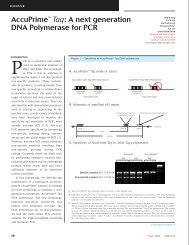
Plasmid Fusion The donor (pUni) vector and the acceptor vector (i.e. pPICZ-E or pPICZα-E) each<br />
contain a single lox site. The donor vector and the acceptor vector each contain a loxP<br />
site. You may insert your gene of interest into the donor vector via the TOPO ® Cloning<br />
method or traditional restriction enzyme-mediated cloning. pPICZ-E and pPICZα-E<br />
contain the appropriate transcription regulatory sequences to control expression of your<br />
gene of interest in <strong>Pichia</strong> and a unique loxP site located downstream of these sequences.<br />
By mixing the donor vector containing the gene of interest with pPICZ-E or pPICZα-E<br />
in the presence of Cre recombinase, a plasmid fusion is created that expresses the gene of<br />
interest in <strong>Pichia</strong> pastoris. A generic diagram is shown below.<br />
R6Kg ori<br />
pUC ori<br />
Kan R<br />
pUni<br />
(2.3 kb + gene)<br />
Promoter<br />
loxP<br />
X<br />
lox*<br />
Amp R<br />
gene<br />
pAcceptor<br />
(2.5 to 5.8 kb)<br />
Cre<br />
recombinase<br />
pUC ori<br />
lox* = loxP or loxH depending on acceptor vector<br />
Promoter<br />
lox*<br />
Amp R<br />
gene<br />
Recombinant<br />
Plasmid<br />
(4.8 kb + gene to<br />
8.1 kb + gene)<br />
loxP<br />
Kan R<br />
R6Kg<br />
Continued on next page<br />
1
Overview of Echo Cloning System, Continued<br />
loxP or loxH Sites The sequence of the loxP site is shown below. The loxP site consists of a 34 bp sequence<br />
containing two 13 bp inverted repeats (see underlined bases) separated by an 8 bp spacer<br />
(Hoess et al., 1982). The inverted repeats may form a stem and loop structure that may<br />
reduce expression of the gene of interest in some cases. A variation of the loxP site (loxH,<br />
see below) was created to eliminate the formation of a stem and loop structure and improve<br />
expression. We have not observed any differences in expression levels in constructs<br />
containing a loxP or a loxH site. Mutated bases are shown in boldface. Cre-mediated<br />
recombination can still occur between a loxP and a loxH site although the efficiency may<br />
be slightly reduced.<br />
• loxP: ATA ACT TCG TAT AGC ATA CAT TAT ACG AAG TTA T<br />
• loxH: ATT ACC TCA TAT AGC ATA CAT TAT ACG AAG TTA T<br />
Cre Recombinase Cre recombinase (MW = 35 kDa) is a site-specific recombinase that binds to specific<br />
sequences (loxP and loxH sites), brings together the target sites, cleaves them, and<br />
covalently attaches to the DNA. Recombination occurs following two pairs of strand<br />
exchanges and ligation of the DNAs in a novel (recombinant) form. A nucleophilic<br />
hydroxylated tyrosine initiates the DNA cleavage event by attack on a specific<br />
phosphodiester bond followed by the covalent attachment of the recombinase to the target<br />
sequence through a phosphoamino acid bond (Abremski & Hoess, 1992; Argos et al.,<br />
1986). The reaction does not require any host factors or ATP, but does require Mg 2+ or<br />
spermidine for activity (Abremski et al., 1983). Recombination between two supercoiled<br />
substrates, each containing a loxP or loxH site, results in a supercoiled dimer. The extent<br />
of the reaction is 10-20% and appears to be stoichiometric (Abremski & Hoess, 1984;<br />
Abremski et al., 1983).<br />
Selection of<br />
Recombinants<br />
2<br />
By fusing the two plasmids, kanamycin resistance is now linked to the pUC origin of<br />
replication. The recombination reaction is transformed into TOP10 E. coli and<br />
recombinants are selected by plating the transformation reaction onto plates containing<br />
kanamycin. Because the donor plasmid carries the R6Kγ origin of replication, it will not<br />
propagate in E. coli strains such as TOP10 which do not carry the pir gene. In addition, the<br />
acceptor vector, which carries the ampicillin resistance gene will not be selected. Therefore<br />
every colony that is selected on kanamycin will represent a recombined fusion plasmid.<br />
Continued on next page
Overview of Echo Cloning System, Continued<br />
pPICZ-E and<br />
pPICZα-E<br />
Selection of<br />
Vector<br />
pPICZ-E and pPICZα-E (~ 3 kb) are derived from pPICZB and pPICZα-B, respectively.<br />
They are designed for high-level recombinant protein expression in <strong>Pichia</strong> pastoris. The<br />
vectors contain the following elements:<br />
• 5′ fragment containing the AOX1 promoter for tightly regulated, methanol-induced<br />
expression of the gene of interest (Ellis et al., 1985: Koutz et al., 1989: Tschopp et<br />
al., 1987a)<br />
• A loxP site for plasmid fusion<br />
• Saccharomycese cerevisiae α-factor secretion signal sequence (pPICZα-E) for<br />
secretion of recombinant proteins in the medium<br />
• Zeocin resistance gene for selection in both E. coli and <strong>Pichia</strong> (Baron et al., 1992:<br />
Drocourt et al., 1990)<br />
• The pUC origin for high copy replication and maintenance of the plasmid in E. coli<br />
For a map and a description of the features of pPICZ-E or pPICZα-E, refer to the<br />
Appendix, pages 46-48.<br />
Other Echo -adapted acceptor vectors are available separately and are provided with<br />
their own manuals. For more information on other available acceptor vectors, visit our<br />
Web site (www.invitrogen.com) or call Technical Service (see page 61).<br />
To utilize the strong, highly-inducible PAOX1 promoter (see next page) for expression of<br />
your protein, there are two expression vectors included in this kit. One vector, pPICZ-E,<br />
is for intracellular expression while the other vector, pPICZα-E, is for secreted<br />
expression. All vectors contain the Zeocin resistance gene for positive selection in<br />
E. coli and <strong>Pichia</strong>. See pages 46-48 for more information on these vectors.<br />
3
Overview of <strong>Pichia</strong> pastoris <strong>Expression</strong> System<br />
Review Articles<br />
General<br />
Characteristics of<br />
<strong>Pichia</strong> pastoris<br />
Similarity to<br />
Saccharomyces<br />
<strong>Pichia</strong> pastoris as<br />
a Methylotrophic<br />
Yeast<br />
Two Alcohol<br />
Oxidase Proteins<br />
4<br />
The information presented here is designed to give you a concise overview of the <strong>Pichia</strong><br />
pastoris expression system. It is by no means exhaustive. For further information, read<br />
the articles cited in the text along with recent review articles (Higgins & Cregg, 1998),<br />
(Buckholz & Gleeson, 1991; Cregg & Higgins, 1995; Cregg et al., 1993; Nico-Farber et<br />
al., 1995; Sreekrishna et al., 1988; Wegner, 1990). A general review of foreign gene<br />
expression in yeast is also available (Romanos et al., 1992)<br />
As a eukaryote, <strong>Pichia</strong> pastoris has many of the advantages of higher eukaryotic expression<br />
systems such as protein processing, protein folding, and posttranslational modification,<br />
while being as easy to manipulate as E. coli or Saccharomyces cerevisiae. It is faster,<br />
easier, and less expensive to use than other eukaryotic expression systems such as<br />
baculovirus or mammalian tissue culture, and generally gives higher expression levels. As a<br />
yeast, it shares the advantages of molecular and genetic manipulations with Saccharomyces<br />
and has the added advantage of 10- to 100-fold higher heterologous protein expression<br />
levels. These features make <strong>Pichia</strong> very useful as a protein expression system.<br />
Many of the techniques developed for Saccharomyces may be applied to <strong>Pichia</strong><br />
including transformation by complementation, gene disruption, and gene replacement.<br />
In addition, the genetic nomenclature used for Saccharomyces has been applied to<br />
<strong>Pichia</strong>. For example, histidinol dehydrogenase is encoded by the HIS4 gene in both<br />
Saccharomyces and <strong>Pichia</strong>. There is also cross-complementation between gene products<br />
in both Saccharomyces and <strong>Pichia</strong>. Several wild-type genes from Saccharomyces<br />
complement comparable mutant genes in <strong>Pichia</strong>. Genes such as HIS4, LEU2, ARG4,<br />
TRP1, and URA3 all complement their respective mutant genes in <strong>Pichia</strong>.<br />
<strong>Pichia</strong> pastoris is a methylotrophic yeast, capable of utlilizing methanol as its sole<br />
carbon source. Alcohol oxidase catalyses the oxidation of methanol to formaldehyde and<br />
hydrogen peroxide using molecular oxygen. To avoid hydrogen peroxide toxicity,<br />
methanol metabolism takes place within a specialized cell organelle, the peroxisome,<br />
which sequesters toxic by-products away from the rest of the cell. Alcohol oxidase has a<br />
poor affinity for O2, and <strong>Pichia</strong> compensates by generating large amounts of the enzyme.<br />
The promoter regulating the production of alcohol oxidase is used to drive recombinant<br />
protein expression in <strong>Pichia</strong>.<br />
Two genes in <strong>Pichia</strong> pastoris code for alcohol oxidase-AOX1 and AOX2. The majority of<br />
alcohol oxidase activity in the cell is attributable to the product of the AOX1 gene.<br />
<strong>Expression</strong> of the AOX1 gene is tightly regulated and induced by methanol to very high<br />
levels, typically > 30% of the total soluble protein in cells grown with methanol. The AOX1<br />
gene has been isolated and a plasmid-borne version of the AOX1 promoter is used to drive<br />
expression of the gene of interest (Ellis et al., 1985; Koutz et al., 1989; Tschopp et al.,<br />
1987a). While AOX2 is about 97% homologous to AOX1, growth on methanol is much<br />
slower than with AOX1. This slow growth on methanol allows isolation of Mut S strains (see<br />
below) (aox1) (Cregg et al., 1989; Koutz et al., 1989).<br />
Continued on next page
Overview of <strong>Pichia</strong> pastoris <strong>Expression</strong> System, Continued<br />
<strong>Expression</strong><br />
Phenotype of aox1<br />
Mutants<br />
Intracellular and<br />
Secretory Protein<br />
<strong>Expression</strong><br />
Posttranslational<br />
Modifications<br />
<strong>Expression</strong> of the AOX1 gene is controlled at the level of transcription. In methanolgrown<br />
cells approximately 5% of the total polyA + RNA is from the AOX1 gene. The<br />
regulation of the AOX1 gene is a two step process of repression/derepression and<br />
induction mechanism. Briefly, growth on glucose represses transcription, even in the<br />
presence of the inducer methanol. For this reason, growth on glycerol is recommended<br />
for optimal induction with methanol. Note: Growth on glycerol alone (derepression) is<br />
not sufficient to generate even minute levels of expression from the AOX1 gene. The<br />
inducer, methanol, is necessary for detectable levels of AOX1 expression (Ellis et al.,<br />
1985; Koutz et al., 1989; Tschopp et al., 1987a).<br />
Loss of the AOX1 gene results in a strain that is phenotypically Mut S (Methanol<br />
utilization slow, also referred to as Mut - in the past). The Mut S designation is chosen to<br />
accurately describe the phenotype of these mutants. This results in a reduction in the cells<br />
ability to metabolize methanol and they exhibit poor growth on methanol medium. Mut +<br />
(Methanol utilization plus) refers to the wild type ability of strains to metabolize<br />
methanol as the sole carbon source. These two phenotypes are used when evaluating<br />
<strong>Pichia</strong> transformants for integration of your gene (Experimental Outline, page 7).<br />
Heterologous expression in <strong>Pichia</strong> pastoris can be either intracellular or secreted.<br />
Secretion requires the presence of a signal sequence on the expressed protein to target it<br />
to the secretory pathway. The native secretion signal present on some heterologous<br />
proteins and several different secretion signal sequences have been used with varied<br />
success. Saccharomyces cerevisiae α factor secretion signal sequence has been used<br />
most successfully (Cregg et al., 1993; Scorer et al., 1993)<br />
The major advantages of expressing recombinant proteins as secreted proteins are:<br />
• <strong>Pichia</strong> pastoris secretes very low levels of native proteins<br />
• Very low amount of protein is present in the minimal <strong>Pichia</strong> growth medium<br />
The secreted recombinant protein usually comprises the vast majority of the total protein<br />
in the medium and serves as the first step in purification of the protein (Barr et al., 1992).<br />
Note: If there are recognized glycosylation sites (Asn-X-Ser/Thr) in your protein's<br />
primary sequence, glycosylation may occur at these sites.<br />
Unlike Saccharomyces cerevisiae, <strong>Pichia</strong> does not hyperglycosylate the secreted<br />
proteins. Both Saccharomyces cerevisiae and <strong>Pichia</strong> pastoris have a majority of N-linked<br />
glycosylation of the high-mannose type; however, the length of the oligosaccharide<br />
chains added posttranslationally to proteins in <strong>Pichia</strong> (average 8-14 mannose residues per<br />
side chain) is much shorter than those in Saccharomyces cerevisiae (50-150 mannose<br />
residues) (Grinna & Tschopp, 1989; Tschopp et al., 1987b) Very little O-linked<br />
glycosylation has been observed in <strong>Pichia</strong>.<br />
In addition, Saccharomyces cerevisiae core oligosaccharides have terminal α1,3 glycan<br />
linkages whereas <strong>Pichia</strong> pastoris does not. It is believed that the α1,3 glycan linkages in<br />
glycosylated proteins produced from Saccharomyces cerevisiae are primarily responsible<br />
for the hyper-antigenic nature of these proteins making them particularly unsuitable for<br />
therapeutic use. Although not yet proven, this is predicted to be less of a problem for<br />
glycoproteins generated in <strong>Pichia</strong> pastoris, because it may resemble the glycoprotein<br />
structure of higher eukaryotes (Cregg et al., 1993).<br />
Continued on next page<br />
5
Overview of <strong>Pichia</strong> pastoris <strong>Expression</strong> System, Continued<br />
Transformation<br />
and Integration<br />
<strong>Expression</strong> and<br />
Scale-up<br />
Purification<br />
6<br />
Two different phenotypic classes of recombinant strains can be generated: Mut + and<br />
MutS . MutS refers to the "Methanol utilization slow" phenotype caused by the loss of<br />
alcohol oxidase activity encoded by the AOX1 gene. A strain with a MutS phenotype has<br />
a mutant aox1 locus, but is wild type for AOX2. This results in a slow growth phenotype<br />
on methanol medium. <strong>Pichia</strong> strains X-33 and GS115 are Mut + while KM71H is MutS .<br />
Transformation of X-33 or GS115 with plasmid DNA linearized in the 5´ AOX1 region<br />
will yield Mut + transformants, while KM71H will yield only MutS transformants. Both<br />
Mut + and MutS recombinants are useful to have as one phenotype may favor better<br />
expression of your protein than the other. You should test between 6-10 recombinants<br />
per phenotype because the site of recombination may affect expression. There is no way<br />
to predict beforehand which construct or isolate will better express your protein. For<br />
more information on recombination in <strong>Pichia</strong>, please see page 51.<br />
Once you have successfully cloned your gene behind the AOX1 promoter, you will then<br />
linearize your plasmid to permit recombination when the plasmid is transformed into<br />
<strong>Pichia</strong>.<br />
After isolating your <strong>Pichia</strong> recombinants, you will then test expression of both Mut + and<br />
Mut S recombinants. This will involve growing a small culture of each recombinant,<br />
inducing with methanol, and taking time points. If looking for intracellular expression,<br />
analyze the cell pellet from each time point by SDS polyacrylamide gel electrophoresis<br />
(SDS-PAGE). If looking for secreted expression, analyze both the cell pellet and<br />
supernatant from each time point. We recommend that you analyze your SDS-PAGE gels<br />
by both Coomassie staining and western blot (for proteins expressed at low levels). We<br />
also suggest checking for protein by functional assay if one is available.<br />
Choose the <strong>Pichia</strong> recombinant strain that best expresses your protein and optimize<br />
induction based on the suggestions on pages 32. Once expression is optimized, scale-up<br />
your expression protocol to produce more protein for purification.<br />
The donor vector contains a C-terminal tag consisting of a V5 epitope and a polyhistidine<br />
tag. If the donor vector is recombined with the acceptor vector (pPICZ-E or pPICZα-E)<br />
such that the C-terminal tag is maintained, then both pPICZ-E or pPICZα-E will contain<br />
the V5 epitope and the polyhistidine tag. The polyhistidine tag facilitates purification by<br />
binding to divalent cations like Ni2+ usually present in metal-binding resins such as<br />
ProBond . We recommend that you use the ProBond Purification System (Catalog no.<br />
K850-01) to purify fusion proteins expressed using pPICZ-E or pPICZα-E. Preliminary<br />
preparation steps are described on pages 36-37.<br />
If you are using a metal-chelating resin other than ProBond , follow the manufacturer’s<br />
recommendations for fusion proteins expressed in yeast.<br />
Continued on next page
Overview of <strong>Pichia</strong> pastoris <strong>Expression</strong> System, Continued<br />
Experimental<br />
Outline<br />
The table below describes the general steps needed to recombine, transform, and express<br />
your protein of interest.<br />
Step Action Page<br />
1 Perform the recombination reaction using your donor vector and<br />
pPICZ-E or pPICZα-E.<br />
8<br />
2 Transform the recombination reaction into competent TOP10 E. coli. 9<br />
3 Select transformants on LB plates containing 50 µg/ml kanamycin. 9<br />
4 Analyze transformants by restriction digestion. 10<br />
5 Select the correct clone and linearize the construct with appropriate<br />
restriction enzyme.<br />
6 Transform your construct into appropriate competent <strong>Pichia</strong> host strain<br />
(X-33 or GS115 for Mut + , and KM71H for Mut s ) using your<br />
transformation method of choice. Select transformants on medium<br />
containing Zeocin .<br />
7 Select 6-10 clones for small scale expression and analyze the<br />
expression of your recombinant protein by western blot analysis or<br />
functional assay.<br />
8 Choose the highest expressers for large-scale expression in shake flask<br />
or fermenter.<br />
9 Purify your protein using metal-chelating resin (Probond ) or any other<br />
method of choice.<br />
17<br />
18-21<br />
25<br />
34<br />
36<br />
7
8<br />
Methods<br />
Recombining Your Gene into pPICZ-E or pPICZα-E<br />
Introduction<br />
Preparation and<br />
Maintenance of<br />
pPICZ-E or<br />
pPICZα-E<br />
Before Starting<br />
Recombination<br />
Reaction<br />
You will need a plasmid preparation of your donor vector containing the gene of interest<br />
in addition to the pPICZ-E or pPICZα-E vector. Review the information below and on the<br />
next page before performing the recombination reaction.<br />
To prepare pPICZ-E or pPICZα-E for use, add 20 µl sterile water to prepare a 1 µg/µl<br />
stock solution. You can further dilute a small aliquot of plasmid or use the stock solution<br />
as is. Store the stock solution at -20°C when you are finished.<br />
If you wish to propagate the pPICZ-E or pPICZα-E plasmid or prepare plasmid DNA,<br />
you may transform the plasmid into One Shot ® TOP10 Chemically Competent E. coli as<br />
described on page 9 or TOP10F′ provided in the kit. Use10-100 ng of plasmid DNA for<br />
transformation and select transformants on low salt LB plates containing 25 µg/ml<br />
Zeocin . Be sure to prepare a glycerol stock of your plasmid-containing TOP10 strain<br />
for long-term storage (see page 12).<br />
You will need the following reagents and equipment.<br />
• 100 ng of your donor vector construct<br />
• 100 ng of pPICZ-E or pPICZα-E (included in the kit)<br />
• Microcentrifuge tubes<br />
• Heat blocks set at 37°C and 65°C<br />
• Ice bucket with ice<br />
• Cre recombinase (included in the kit)<br />
• 10X Recombinase Buffer (included in the kit)<br />
1. Set up each 20 µl recombination reaction on ice as follows:<br />
Donor vector (100 ng) x µl<br />
pPICZ-E or pPICZα-E (100 ng) y µl<br />
10X Recombinase Buffer 2 µl<br />
Deionized water add to a total volume of 19 µl<br />
Cre Recombinase 1 µl<br />
Final Volume 20 µl<br />
2. Incubate at 37°C for 20 minutes.<br />
3. Incubate at 65°C for 5 minutes to inactivate the recombinase.<br />
4. Place the tube on ice and proceed to Transformation, next page. If you run out of<br />
time, you may store the recombination reaction at +4°C or -20°C overnight. Longer<br />
storage times have not been tested.
Transforming the Recombination Reaction<br />
Introduction<br />
Materials Supplied<br />
by the User<br />
Important<br />
Preparing for<br />
Transformation<br />
One Shot ®<br />
Transformation<br />
Reaction<br />
Once you have performed the recombination reaction, you are ready to transform your<br />
E. coli host. We recommend using TOP10 E. coli for transformation, but other strains are<br />
suitable. E. coli strains should be endonuclease A deficient (endA) and recombination<br />
deficient (recA) to ensure quality plasmid preparations and reduce the chances of nonspecific<br />
recombination, respectively.<br />
In addition to general microbiological supplies (i.e. plates, spreaders), you will need the<br />
following reagents and equipment.<br />
• 42°C water bath<br />
• LB plates containing 50 µg/ml kanamycin (see Important, below)<br />
• LB medium and SOB medium (see page 45 for recipe)<br />
• 37°C shaking and non-shaking incubator<br />
It is important to select the fusion plasmid using kanamycin. The donor vector contains<br />
the R6Kγ origin that can only be maintained in E. coli strains containing the pir gene.<br />
After recombination between the donor vector and acceptor vector, the kanamycin<br />
resistance gene (from the donor vector) in the fusion plasmid is linked to the pUC origin<br />
(from pPICZ-E or pPICZα-E). The fusion plasmid can be maintained in E. coli strains<br />
that do not contain the pir gene (i.e. TOP10). By selecting for kanamycin resistance, you<br />
ensure that only colonies containing the fusion plasmid are selected.<br />
This transformation protocol is for use with the One Shot ® TOP10 Chemically Competent<br />
E. coli available with the kit. Follow the manufacturer’s protocol if you are using other<br />
competent cells.<br />
For each transformation, you will need one vial of One Shot ® TOP10 Chemically<br />
Competent E. coli and two selective plates. Perform the following steps before beginning.<br />
1. Equilibrate a water bath to 42°C.<br />
2. Thaw the vial of SOC medium from the kit and bring to room temperature.<br />
3. Warm LB plates containing 50 µg/ml kanamycin at 37°C for 30 minutes.<br />
4. Thaw on ice 1 vial of One Shot ® cells for each transformation.<br />
1. Add 5 µl of the recombination reaction (step 4, page 8) to a vial of One Shot ® TOP10<br />
E. coli and mix by stirring with a pipette tip. Do not mix by pipetting up and down.<br />
2. Heat-shock the cells for 30 seconds at 42°C without shaking.<br />
3. Immediately transfer the tubes to ice.<br />
4. Add 500 µl of room temperature SOC medium.<br />
5. Cap the tube tightly and shake the tube horizontally at 37°C for 45 minutes.<br />
6. Spread 50 µl from each transformation onto a prewarmed LB plate containing<br />
50 µg/ml kanamycin. Pellet the remaining cells, resuspend the cell pellet in 50 µl<br />
SOC and plate. Incubate plates overnight at 37°C.<br />
7. An efficient recombination reaction will produce hundreds of colonies. Pick five<br />
colonies for analysis.<br />
Continued on next page<br />
9
Transforming the Recombination Reaction, Continued<br />
Analyzing Positive<br />
Clones<br />
Sequencing Your<br />
Construct in<br />
pPICZ-E<br />
10<br />
801<br />
861<br />
1. Culture the 5 colonies (see previous page) overnight in 2-5 ml LB or SOB medium<br />
containing 50 µg/ml kanamycin.<br />
2. Isolate plasmid DNA using your method of choice. If you need ultra-pure plasmid<br />
DNA for automated or manual sequencing, we recommend the S.N.A.P. MiniPrep<br />
<strong>Kit</strong> (10-15 µg DNA, Catalog no. K1900-01) or the S.N.A.P. MidiPrep <strong>Kit</strong> (10-<br />
200 µg DNA, Catalog no. K1910-01).<br />
3. Analyze the plasmids by restriction analysis. Use an enzyme (or enzymes) that cut<br />
once in the donor vector and once in the acceptor vector to yield two fragments that<br />
are distinguishable from one another. Note that other strategies are possible.<br />
4. (Optional) To sequence the fusion plasmid to confirm the fusion junctions, use the<br />
5′ AOX1 primer and the Uni1 Forward primer. Refer to the diagrams below and on the<br />
next page for the sequence around the pPICZ-E or pPICZα-E loxP site. Refer to the<br />
donor vector manual for the sequence around the donor vector loxP site.<br />
If you need help with setting up restriction enzyme digests or DNA sequencing, refer to<br />
general molecular biology texts (Ausubel et al., 1994; Sambrook et al., 1989)<br />
The sequence surrounding your insert is shown below. Unique restriction sites are labeled<br />
to indicate the cleavage site. Note that the complete sequence of pPICZ-E is available for<br />
downloading from our Web site: (www.invitrogen.com) or from Technical Service (see<br />
page 61).<br />
CCCTGTCTTA AACCTTTTTT TTTATCATCA TTATTAGCTT ACTTTCATAA TTGCGACTGG<br />
TTCCAATTGA CAAGCTTTTG ATTTTAACGA CTTTTAACGA CAACTTGAGA AGATCAAAAA<br />
921 ACAACTAATT ATTCGAAACG AGGAATTC ATA ACT TCG TAT AGC ATA CAT TAT ACG<br />
976<br />
AAG TTA T<br />
loxP site<br />
Age I<br />
Sfu I<br />
donor<br />
vector<br />
5´ end of AOX1 mRNA<br />
Gene of<br />
interest<br />
EcoR I<br />
C-terminal tag<br />
(optional)<br />
loxP site<br />
Uni1 Forward priming site<br />
donor vector<br />
5´ AOX1 priming site<br />
ACCGGTCT TGCTAGATTC TAATCAAGAG GATGTCAGAA TGCCATTTGC<br />
Continued on next page
Transforming the Recombination Reaction, Continued<br />
Sequencing Your<br />
Construct in<br />
pPICZα-E<br />
The sequence surrounding your insert is shown below. Unique restriction sites are labeled<br />
to indicate the cleavage site. Note that the complete sequence of pPICZα-E is available for<br />
downloading from our Web site: (www.invitrogen.com) or from Technical Service (see<br />
page 61).<br />
5´ end of AOX1 mRNA 5´ AOX1 priming site<br />
811 AACCTTTTTT TTTATCATCA TTATTAGCTT ACTTTCATAA TTGCGACTGG TTCCAATTGA<br />
871 CAAGCTTTTG ATTTTAACGA CTTTTAACGA CAACTTGAGA AGATCAAAAA ACAACTAATT<br />
931 ATTCGAAACG ATG AGA TTT CCT TCA ATT TTT ACT GCT GTT TTA TTC GCA GCA<br />
Met Arg Phe Pro Ser Ile Phe Thr Ala Val Leu Phe Ala Ala<br />
983 TCC TCC GCA TTA GCT GCT CCA GTC AAC ACT ACA ACA GAA GAT GAA ACG<br />
Ser Ser Ala Leu Ala Ala Pro Val Asn Thr Thr Thr Glu Asp Glu Thr<br />
1031 GCA CAA ATT CCG GCT GAA GCT GTC ATC GGT TAC TCA GAT TTA GAA GGG<br />
Ala Gln Ile Pro Ala Glu Ala Val Ile Gly Tyr Ser Asp Leu Glu Gly<br />
a-factor signal sequence<br />
1079 GAT TTC GAT GTT GCT GTT TTG CCA TTT TCC AAC AGC ACA AAT AAC GGG<br />
Asp Phe Asp Val Ala Val Leu Pro Phe Ser Asn Ser Thr Asn Asn Gly<br />
a-factor priming site<br />
1127 TTA TTG TTT ATA AAT ACT ACT ATT GCC AGC ATT GCT GCT AAA GAA GAA<br />
Leu Leu Phe Ile Asn Thr Thr Ile Ala Ser Ile Ala Ala Lys Glu Glu<br />
Kex2 signal cleavage<br />
EcoR I<br />
loxP site<br />
1175 GGG GTA TCT CTC GAG AAA AGA GAG GCT GAA GCT GAATTC ATA ACT TCG TAT<br />
Gly Val Ser Leu Glu Lys Arg Glu Ala Glu Ala<br />
Uni1 Forward<br />
Ste13 signal cleavage<br />
priming site<br />
1226 AGC ATA CAT TAT ACG AAG TTA T donor<br />
vector<br />
Gene of<br />
interest<br />
C-terminal tag<br />
(optional)<br />
donor<br />
vector<br />
Age I<br />
loxP site ACCGGTCTTG CTAGATTCTA ATCAAGAGGA TGTCAGAATG CCATTTGCCT<br />
Continued on next page<br />
11
Transforming the Recombination Reaction, Continued<br />
Fusion Plasmid<br />
Analysis<br />
Preparing Plasmid<br />
DNA and Glycerol<br />
Stock for Long-<br />
Term Storage<br />
12<br />
It should be clear from restriction analysis that you have a dimer plasmid consisting of the<br />
donor vector and pPICZ-E or pPICZα-E. Occasionally, trimers will result. Trimers usually<br />
consist of two donor vector molecules and one acceptor molecule, but they usually express<br />
as well as the dimer product.<br />
In theory, trimers may result from two sequential fusion events or a single fusion event<br />
between a pre-existing monomeric substrate and a dimeric substrate. The production of<br />
trimers can be eliminated if gel-purified monomeric supercoiled DNA is used in the<br />
recombination reaction.<br />
Once you have identified the correct clone, isolate plasmid DNA using your method of<br />
choice for transformation of your construct into <strong>Pichia</strong> (see page 15-21).<br />
We recommend to make a glycerol stock for long term storage.<br />
1. Streak out the original colony on LB plates containing 50 µg/ml kanamycin to isolate<br />
single colonies.<br />
2. Select a single colony and inoculate into 1-2 ml of LB containing 50 µg/ml kanamycin.<br />
3. Grow overnight until culture is saturated.<br />
4. Mix 0.85 ml of culture with 0.15 ml of sterile glycerol and transfer to a cryovial.<br />
5. Store at -80°C. (You may also want to store a stock of plasmid DNA at -20°C.)
<strong>Pichia</strong> Strains<br />
Introduction<br />
Genotypes of<br />
<strong>Pichia</strong> Strains<br />
Construction of<br />
KM71H<br />
Important<br />
After isolating the plasmid DNA of your fusion construct, you are ready to transfom this<br />
construct into <strong>Pichia</strong> for expression of the recombinant protein. This section provides<br />
information on <strong>Pichia</strong> strains, initiation of <strong>Pichia</strong> culture, growth characteristics and<br />
storage of <strong>Pichia</strong> pastoris. You should be familiar with basic sterile microbiological,<br />
molecular biology and protein chemistry techniques before attempting to grow and<br />
manipulate <strong>Pichia</strong>. Some general references to consult are Guide to Yeast Genetics and<br />
Molecular Biology, (Guthrie & Fink, 1991), Current Protocols in Molecular Biology,<br />
(Ausubel et al., 1994), Molecular Cloning: A Laboratory Manual, (Sambrook et al.,<br />
1989), Protein Methods, (Bollag & Edelstein, 1991) and Guide to Protein Purification,<br />
(Deutscher, 1990).<br />
X-33 is a wild-type <strong>Pichia</strong> strain that is useful for selection on Zeocin and large-scale<br />
growth. It will grow in YPD and in minimal media.<br />
The <strong>Pichia</strong> host strain GS115 has a mutation in the histidinol dehydrogenase gene (his4)<br />
that prevents it from synthesizing histidine. GS115 will grow on complex medium such<br />
as YPD (also known as YEPD) and on minimal media supplemented with histidine.<br />
The parent strain of KM71H has a mutation in the argininosuccinate lyase gene (arg4)<br />
that prevents the strain from growing in the absence of arginine. The wild-type ARG4<br />
gene was used to disrupt AOX1, creating KM71H, a MutS , Arg + strain.<br />
The ARG4 gene (~2 kb) was inserted into the cloned, wild-type AOX1 gene between the<br />
BamH I site (codons 15/16 of AOX1) and the Sal I site (codons 227/228 of AOX1). ARG4<br />
replaces codons 16 through 227 of AOX1. This construct was transformed into the parent<br />
strain of KM71 (arg4 his4) and Arg+ transformants were isolated and analyzed for the<br />
Mut S phenotype. Genetic analysis of Arg+ transformants showed that the wild-type AOX1<br />
gene was replaced by the aox1::ARG4 construct. To create KM71H, KM71 was transformed<br />
with a gene fragment encoding the HIS4 gene and a His + convertant was isolated.<br />
The advantage of using KM71H is that there is no need to screen for the Mut phenotype<br />
on methanol minimal medium. All transformants will be Mut S . Secondly, since the AOX1<br />
locus was not completely deleted, it is theoretically possible to replace aox1::ARG4 with<br />
your construct by gene replacement. The phenotype of this strain would be Mut S Arg - .<br />
This means the recombinant strain would require arginine in the medium to grow.<br />
Unfortunately, simple inclusion of arginine does not totally alleviate the effects of the<br />
arg4 mutation, and arg4 strains do not grow well on minimal medium supplemented with<br />
arginine. Therefore, we do not recommend that you generate transformants in KM71H by<br />
replacing the aox1::ARG4 construct.<br />
Continued on next page<br />
13
<strong>Pichia</strong> Strains, Continued<br />
Growth of <strong>Pichia</strong><br />
Strains<br />
Growth on<br />
Methanol<br />
Initiating <strong>Pichia</strong><br />
culture<br />
Storage of <strong>Pichia</strong><br />
Strains<br />
14<br />
The growth temperature of <strong>Pichia</strong> pastoris is 28-30°C for liquid cultures, plates, and<br />
stabs. Growth above 32°C during induction can be detrimental to protein expression and<br />
can even lead to cell death. Other important facts:<br />
• Doubling time of log phase Mut + or MutS <strong>Pichia</strong> in YPD is ~2 hours<br />
• Mut + and MutS strains do not differ in growth rates unless grown on methanol<br />
• Doubling time of log phase Mut + <strong>Pichia</strong> in methanol medium (MM) is 4-6 hours<br />
• Doubling time of log phase MutS <strong>Pichia</strong> in MM is ~18 hours<br />
• One OD600 = ~5 x 10⁷ cells/ml<br />
Note that growth characteristics may vary depending on the recombinant protein<br />
expressed<br />
When plates or medium containing methanol are used as growth medium, it is advisable<br />
to add methanol every day to compensate for loss because of evaporation or<br />
consumption.<br />
• For plates add 100 µl of 100% methanol to the lid of the inverted plate.<br />
• For liquid medium add 100% methanol to a final concentration of 0.5%.<br />
Some researchers have had success adding methanol to 1% every day for MutS strains<br />
and up to 3% for Mut + without any negative effect to their liquid culture<br />
To initiate a culture of <strong>Pichia</strong> from the stab provided with the kit, streak a small amount<br />
from the stab on YPD plate (see page 41) and incubate at 30°C. Once growth is<br />
established, you may pick a single colony and grow it in the appropriate medium for<br />
downstream applications. Remember to prepare frozen stocks for long-term storage of<br />
all <strong>Pichia</strong> strains included in this kit (see below).<br />
To store cells for weeks to months, use YPD medium and YPD agar slants (see<br />
page 41).<br />
• Streak each strain for single colonies on YPD.<br />
• Transfer one colony to a YPD stab and grow for 2 days at 30°C.<br />
• The cells can be stored on YPD for several weeks at +4°C.<br />
To store cells for months to years, store frozen at -80°C.<br />
• Culture a single colony of each strain overnight in YPD.<br />
• Harvest the cells and suspend in YPD containing 15% glycerol at a final OD600 of<br />
50-100 (approximately 2.5-5.0 x 10⁹ cells/ml).<br />
• Cells are frozen in liquid nitrogen or a dry ice/ethanol bath and then stored at -80°C.<br />
After extended storage at +4°C or -80°C, it is recommended that Zeo R transformants be<br />
checked for correct phenotype and protein expression.
<strong>Pichia</strong> Transformation<br />
Introduction Once you have created the pPICZ-E or pPICZα-E fusion plasmid and have verified its<br />
integrity, you are ready to transform the fusion plasmid into a <strong>Pichia</strong> host strain of your<br />
choice.<br />
Zeocin Selection We generally use 100 µg/ml Zeocin to select for transformants when using the X-33<br />
<strong>Pichia</strong> strain. If you are transforming the fusion plasmid into another <strong>Pichia</strong> strain, note<br />
that selection conditions may vary. We recommend performing a dose response curve to<br />
determine the appropriate concentration of Zeocin to use for selection of transformants<br />
in your strain.<br />
Method of<br />
Transformation<br />
Important<br />
His4 Host Strains<br />
We do not recommend spheroplasting for transformation of <strong>Pichia</strong> with plasmids<br />
containing the Zeocin resistance marker. Spheroplasting involves removal of the cell<br />
wall to allow DNA to enter the cell. Cells must first regenerate the cell wall before they<br />
are able to express the Zeocin resistance gene. For this reason, plating spheroplasts<br />
directly onto selective medium containing Zeocin does not yield any transformants.<br />
We recommend electroporation for transformation of <strong>Pichia</strong> with pPICZ-E or pPICZα-E.<br />
Electroporation yields 10 3 to 10 4 transformants per µg of linearized DNA and does not<br />
destroy the cell wall of <strong>Pichia</strong>. If you do not have access to an electroporation device,<br />
use the <strong>Pichia</strong> EasyComp Transformation <strong>Kit</strong> included in the kit (see page 19). The<br />
<strong>Pichia</strong> EasyComp Transformation <strong>Kit</strong> provides reagents to prepare 6 preparations of<br />
competent cells. Each preparation will yield enough competent cells for 20<br />
transformations<br />
Since pPICZ-E or pPICZα-E do not contain the HIS4 gene, integration can only occur at<br />
the AOX1 locus. Vector linearized within the 5´ AOX1 region will integrate by gene<br />
insertion into the host 5´ AOX1 region. Therefore, the <strong>Pichia</strong> host that you use will<br />
determine whether the recombinant strain will be Mut + or Mut S . Use a <strong>Pichia</strong> host that<br />
contains the native AOX1 gene (e.g. X-33, GS115, SMD1168H) to generate a Mut +<br />
recombinant strain and a <strong>Pichia</strong> host that has a disrupted AOX1 gene (i.e. KM71H) to<br />
generate a Mut S recombinant strain. Information on <strong>Pichia</strong> recombination is provided on<br />
page 51.<br />
Host strains containing the his4 allele (e.g. GS115) and transformed with the pPICZ-E or<br />
pPICZα-E vectors require histidine when grown in minimal media. Add histidine to a<br />
final concentration of 0.004% to ensure growth of your transformants.<br />
Continued on next page<br />
15
<strong>Pichia</strong> Transformation, Continued<br />
16<br />
The pPICZ-E or pPICZα-E vectors do not contain a yeast origin of replication.<br />
Transformants can only be isolated if recombination occurs between the plasmid and the<br />
<strong>Pichia</strong> genome.<br />
<strong>Pichia</strong> Controls pPICZ-E/Uni-lacZ and pPICZα-E/Uni-HSA are control plasmids included in the kit to<br />
check for transformation and expression in <strong>Pichia</strong> pastoris. The lacZ gene was cloned<br />
into a donor vector, pUni/V5-His-Gene-TOPO ® . The resulting vector was recombined<br />
with pPICZ-E to create pPICZ-E/Uni-lacZ, which can be used as an intracellular<br />
expression control after transformation into a <strong>Pichia</strong> host.<br />
The human serum albumin (HSA) gene was cloned into a donor vector, pUni/V5-His-<br />
Gene-TOPO ® . The resulting vector was recombined with pPICZα-E to create pPICZα-<br />
E/Uni-HSA/V5-His, which can be used as a secreted expression control. For a map and<br />
more details on pPICZ-E/Uni-lacZ and pPICZα-E/Uni-HSA controls, see pages 49-50.<br />
Use the control plasmids pPICZ-E/Uni-lacZ or pPICZα-E/Uni-HSA to perform a control<br />
transformation using any method of choice in a <strong>Pichia</strong> host strain. We have used X-33 as<br />
the host strain. Linearize the plasmid using Pme I to promote integration (see next page).<br />
If you wish to propagate the supercoiled plasmids, you may transform the plasmid into<br />
TOP10 E. coli (see page 8).<br />
Before Starting<br />
GS115/pPICZ/lacZ (Mut + ) and GS115/Albumin (Mut s ) are control strains included in the<br />
kit to check the Mut phenotype. Cloning the lacZ gene into the pPICZ vector and<br />
transforming the resulting construct into GS115 creating the GS115/pPICZ/lacZ (Mut + )<br />
strain. This strain expresses β-galactosidase and can also be used as a control for<br />
intracellular expression. GS115/Albumin is Mut s and is also a control for secreted<br />
expression. The gene for serum albumin was cloned with its native secretion signal and<br />
then transformed into GS115 to create GS115/Albumin. If you decide to use<br />
GS115/pPICZ/lacZ or GS115/Albumin as a control, then you need to initiate a culture<br />
from the stab (see page 14).<br />
We recommend you use pPICZ-E/Uni-lacZ and pPICZα-E/Uni-HSA as transformation<br />
and expression controls, and GS115/pPICZ/lacZ and GS115/Albumin as controls for<br />
determining the Mut phenotype (see page 22) and confirming expression conditions.<br />
The following reagents are needed for linearizing your construct, transforming <strong>Pichia</strong><br />
and selecting transformants on Zeocin .<br />
• 10-50 µg pPICZ-E or pPICZα-E fusion plasmid and appropriate expression control<br />
plasmids<br />
• Appropriate restriction enzymes and buffer<br />
• Phenol/chloroform<br />
• 3M sodium acetate<br />
• Agarose gel apparatus and Electroporation device with 0.2 cm cuvettes<br />
• 80% and 100% ethanol<br />
• YPD Medium and YPDS plates containing the appropriate concentration of Zeocin <br />
• 50 ml conical polypropylene tubes<br />
• 1 liter cold (+4°C) sterile water (place on ice the day of the experiment)<br />
• 25 ml cold (+4°C) sterile 1 M sorbitol (place on ice the day of the experiment)<br />
Continued on next page
<strong>Pichia</strong> Transformation, Continued<br />
Linearizing Your<br />
pPICZ-E or<br />
pPICZα-E<br />
Construct<br />
To promote integration, we recommend that you linearize your pPICZ-E or pPICZα-E<br />
fusion plasmid within the 5′ AOX1 region. The table below lists unique sites that may be<br />
used to linearize pPICZ-E or pPICZα-E prior to transformation. Other restriction sites<br />
are possible. Be sure that your insert does not contain the restriction site you wish to use<br />
to linearize your vector.<br />
Enzyme Restriction Site (bp) Supplier<br />
Sac I 209 New England Biolabs<br />
Pme I 414 New England Biolabs<br />
BstX I 707 New England Biolabs<br />
Restriction Digest 1. Digest ~10-50 µg of plasmid DNA with one of the enzymes listed above. We<br />
recommend to use more DNA as you will lose some DNA during purification after<br />
the restriction digestion and will be helpful if you decide to do multiple<br />
transformations with varying concentrations of Zeocin .<br />
2. Check a small aliquot of your digest by agarose gel electrophoresis for complete<br />
linearization.<br />
3. If the vector is completely linearized, heat inactivate or add EDTA to stop the<br />
reaction, phenol/chloroform extract once, and ethanol precipitate using 1/10 volume<br />
3 M sodium acetate and 2.5 volumes of 100% ethanol. You can also use any resin<br />
based purification methods to purify the linearized vector.<br />
4. Centrifuge the solution to pellet the DNA, wash the pellet with 80% ethanol, air-dry,<br />
and resuspend in 10 µl sterile, deionized water. Use immediately or store at -20°C.<br />
Preparing<br />
<strong>Pichia</strong> for<br />
Electroporation<br />
Follow the procedure below to prepare your <strong>Pichia</strong> pastoris strain for electroporation.<br />
1. Grow 5 ml of your <strong>Pichia</strong> pastoris strain in YPD in a 50 ml conical tube at 30°C<br />
overnight.<br />
2. Inoculate 500 ml of fresh medium in a 2 liter flask with 0.1-0.5 ml of the overnight<br />
culture. Grow overnight again to an OD600 = 1.3-1.5.<br />
3. Centrifuge the cells at 1500 x g for 5 minutes at +4°C. Resuspend the pellet with<br />
500 ml of ice-cold (0°C), sterile water.<br />
4. Centrifuge the cells as in Step 3, then resuspend the pellet with 250 ml of ice-cold<br />
(0°C), sterile water.<br />
5. Centrifuge the cells as in Step 3, then resuspend the pellet in 20 ml of ice-cold<br />
(0°C) 1 M sorbitol.<br />
6. Centrifuge the cells as in Step 3, then resuspend the pellet in 1 ml of ice-cold (0°C)<br />
1 M sorbitol for a final volume of approximately 1.5 ml. Keep the cells on ice and<br />
use that day. Do not store cells.<br />
Continued on next page<br />
17
<strong>Pichia</strong> Transformation, Continued<br />
Transformation by<br />
Electroporation<br />
Isolation of MulticopyRecombinants<br />
in vivo<br />
Analyzing <strong>Pichia</strong><br />
Transformants<br />
18<br />
1. Mix 80 µl of the cells from Step 6 (above) with 5-10 µg of linearized DNA (in 5-<br />
10 µl sterile water) and transfer them to an ice-cold 0.2 cm electroporation cuvette.<br />
2. Incubate the cuvette with the cells on ice for 5 minutes.<br />
3. Pulse the cells according to the parameters for yeast (Saccharomyces cerevisiae) as<br />
suggested by the manufacturer of the specific electroporation device being used.<br />
4. Immediately add 500 µl of ice-cold 1 M sorbitol to the cuvette. Transfer the cuvette<br />
contents to a sterile 15 ml tube. Rinse the cuvette with additional 500 µl of ice-cold 1<br />
M sorbitol and transfer the contents to the 15 ml tube as above.<br />
5. Let the tube incubate at 30°C without shaking for 1 to 2 hours.<br />
6. Spread 50-200 µl each on separate, labeled YPDS plates containing the appropriate<br />
concentration of Zeocin . Note: Inclusion of sorbitol in YPD plates stabilizes<br />
electroporated cells as they appear to be somewhat osmotically sensitive<br />
7. Incubate plates for 2 to 7 days at 30°C until colonies form.<br />
8. Pick 10-20 colonies and purify (streak for single colonies) on fresh YPD or YPDS<br />
plates containing the appropriate concentration of Zeocin .<br />
A quick, direct way to select multi-copy recombinants is to plate the transformation mix<br />
on increasing concentrations of Zeocin .<br />
1. Prepare YPDS plates containing 100, 500, 1000, and 2000 µg/ml Zeocin <br />
2. Plate 100 to 200 µl of the transformation mix on each plate and incubate at 30°C for 2<br />
to 7 days.<br />
3. Test transformants for the Mut phenotype (page 22) and expression of your protein<br />
(page 25)<br />
Generally, several hundred Zeocin -resistant colonies are generated using the protocol<br />
on the previous page. If more colonies are needed, the protocol may be modified as<br />
described below. Note that you will need twenty 150 mm plates with YPDS agar<br />
containing the appropriate concentration of Zeocin .<br />
1. Set up two transformations per construct and follow Steps 1 through 5 of the<br />
Transformation by Electroporation protocol, see above.<br />
2. After 1 hour in 1 M sorbitol at 30°C (Step 5, see above), add 1 ml YPD medium to<br />
each tube.<br />
3. Shake (~200 rpm) the cultures at 30°C.<br />
4. After 1 hour, take one of the tubes and plate out all of the cells by spreading 200 µl<br />
on 150 mm plates containing the appropriate concentration of Zeocin .<br />
5. (Optional) Continue incubating the other culture for three more hours (for a total of<br />
four hours) and then plate out all of the cells by spreading 200 µl on 150 mm plates<br />
containing the appropriate concentration of Zeocin .<br />
5. Incubate plates for 2 to 7 days at 30°C until colonies form.<br />
Select 6-10 of your Zeocin -resistant <strong>Pichia</strong> transformants and confirm the Mut<br />
phenotype as described on page 22. You can also analyze for the presence of insert using<br />
PCR (page 55), or for copy number using Southern analysis (page 60).
EasyComp Transformation<br />
Introduction<br />
Required<br />
Reagents and<br />
Equipment<br />
The <strong>Pichia</strong> EasyComp <strong>Kit</strong> produces chemically competent <strong>Pichia</strong> cells and is included<br />
to provide a rapid and convenient alternative to electroporation. However, because of the<br />
low transformation efficiency (3 µg plasmid DNA yields about 50 colonies), it is very<br />
difficult to isolate multi-copy integrants. If multi-copy integrants are desired, use<br />
electroporation (page 16) for best results. Do not use cells prepared using the<br />
EasyComp protocol for electroporation as cells are prepared differently for each<br />
method.<br />
• 30°C rotary shaking incubator<br />
• YPD (Yeast Extract Peptone Dextrose) medium (see Recipes, page 41)<br />
• 50 ml, sterile conical tubes<br />
• Centrifuge suitable for 50 ml conical tubes (floor or table-top)<br />
• 1.5 ml sterile screw-cap microcentrifuge tubes<br />
• Styrofoam box or paper towels<br />
Before Beginning Streak a YPD plate with your <strong>Pichia</strong> pastoris strain such that isolated, single colonies<br />
will grow. Incubate the plate at 28-30°C for 2 days.<br />
Equilibrate Solution I to room temperature.<br />
Preparing<br />
Competent Cells<br />
1. Inoculate 10 ml of YPD with a single colony of your <strong>Pichia</strong> strain. Grow overnight<br />
at 28-30°C in a shaking incubator (250-300 rpm).<br />
2. Dilute cells from the overnight culture to an OD600 of 0.1-0.2 in 10 ml of YPD.<br />
Grow the cells at 28-30°C in a shaking incubator until the OD600 reached 0.6-1.0.<br />
This will take approximately 4 to 6 hours.<br />
3. Pellet the cells by centrifugation at 500 x g for 5 minutes at room temperature.<br />
Discard the supernatant.<br />
4. Resuspend the cell pellet in 10 ml of Solution I. No incubation time is required.<br />
5. Pellet the cells by centrifugation at 500 x g for 5 minutes at room temperature.<br />
Discard the supernatant.<br />
6. Resuspend the cell pellet in 1 ml of Solution I. The cells are now competent.<br />
7. Aliquot 50 to 200 µl of competent cells into labeled 1.5 ml sterile screw-cap<br />
microcentrifuge tubes.<br />
Note: 50 µl of cells are used for each transformation. Cells can be thawed and<br />
refrozen several times without significant loss in transformation efficiency.<br />
8. At this point, the cells may be kept at room temperature, used directly for<br />
transformation or frozen for future use. To slowly freeze the cells, place tubes in a<br />
Styrofoam box or wrap in several layers of paper towels and place in a -80°C<br />
freezer. Do not snap-freeze the cells in liquid nitrogen.<br />
9. Proceed to the transformation procedure.<br />
Higher transformation efficiencies are obtained with frozen versus freshly prepared cells.<br />
You may choose to use some of the cells immediately following preparation and freeze<br />
the remaining cells in small aliquots.<br />
Continued on next page<br />
19
EasyComp Transformation, Continued<br />
Transformation<br />
Required<br />
Reagents and<br />
Equipment<br />
20<br />
The following protocol can be used to transform either freshly prepared or frozen competent<br />
<strong>Pichia</strong> cells. Transformation efficiency may vary with each strain and vector used.<br />
• 30°C incubator<br />
• Water baths or heat blocks at 30°C and 42°C<br />
• Microcentrifuge at room temperature<br />
• YPDS with 100 µg/ml Zeocin plates (see Recipes, page 41)<br />
Before Beginning If you see a precipitate in Solution II, warm the solution at 37°C until the precipitate<br />
dissolves. To prevent formation of a precipitate, store Solution II at room temperature.<br />
Equilibrate Solution III to room temperature.<br />
Equilibrate the appropriate number and type of plates to room temperature. You will<br />
need one plate for each transformation.<br />
You may want to include controls. We recommend a no DNA and a plasmid only<br />
control.<br />
Transformation<br />
Protocol<br />
1. For each transformation, thaw one tube of competent cells at room temperature and<br />
aliquot 50 µl into a sterile microcentrifuge tube. If transforming fresh cells, use 50 µl<br />
of cells from Preparation of Competent Cells, Step 7, previous page.<br />
2. Add 3 µg of linearized <strong>Pichia</strong> expression vector DNA to the competent cells.<br />
Note: Using greater than 3 µg of DNA may increase transformation efficiencies in<br />
some cases. The volume of DNA should not exceed 5 µl. Linearized DNA can be<br />
used directly from a restriction digest reaction without affecting transformation<br />
efficiency. Phenol chloroform extraction and ethanol precipitation are not necessary.<br />
3. Add 1 ml of Solution II to DNA/cell mixture. Mix by vortexing or flicking the tube.<br />
4. Incubate the transformation reactions for 1 hour at 30°C in a water bath or incubator.<br />
Mix the transformation reaction every 15 minutes by vortexing or flicking the tube.<br />
Failure to mix the transformation reaction every 15 minutes will result in decreased<br />
transformation efficiency.<br />
5. Heat shock the cells in a 42°C heat block or water bath for 10 minutes.<br />
6. Split the cells into 2 microcentrifuge tubes (approximately 525 µl per tube) and add<br />
1 ml of YPD medium to each tube.<br />
7. Incubate the cells at 30°C for 1 hour to allow expression of Zeocin resistance.<br />
8. Pellet the cells by centrifugation at 3000 x g for 5 minutes at room temperature.<br />
Discard the supernatant.<br />
9. Resuspend each tube of cells in 500 µl of Solution III and combine the cells into one<br />
tube.<br />
10. Pellet the cells by centrifugation at 3000 x g for 5 minutes at room temperature.<br />
Discard the supernatant.<br />
11. Resuspend the cell pellet in 100 to 150 µl of Solution III.<br />
12. Plate the entire transformation on appropriate selection plates using a sterile<br />
spreader. Incubate the plates for 2 to 4 days at 30°C. Each transformation should<br />
yield approximately 50 colonies.<br />
Continued on next page
EasyComp Transformation, Continued<br />
Analyzing <strong>Pichia</strong><br />
Transformants<br />
Troubleshooting<br />
Select 6-10 of your Zeocin -resistant <strong>Pichia</strong> transformants and confirm the Mut<br />
phenotype as described on page 22. You may also wish to analyze for the presence of<br />
insert using PCR (page 55).<br />
The table below provides solutions to possible problems you may encounter when<br />
preparing and transforming competent <strong>Pichia</strong> pastoris cells.<br />
Problem Probable Cause Possible Solution<br />
Low efficiency of<br />
transformation<br />
The pH of Solution I or<br />
Solution III may have<br />
drifted. The pH of both<br />
solutions should be 8.0<br />
Transformation<br />
reaction not mixed<br />
during incubation<br />
Incubation time is too<br />
short or temperature is<br />
too low<br />
Cell density is too low<br />
(OD600
Determining the Mut Phenotype<br />
Introduction<br />
Screening for Mut +<br />
in X-33 and GS115<br />
Mut S in KM71H<br />
22<br />
Important<br />
Preparation<br />
Mut + in GS115 or<br />
X-33<br />
To verify the Mut phenotype of your Zeocin -resistant transformants, refer to the<br />
guidelines below. Note: KM71H recombinants do not need to be screened for their Mut<br />
phenotype as they all will be Mut S .<br />
Transformation of X-33 or GS115 with linearized constructs favor single crossover<br />
recombination at the AOX1 locus and result in Mut + transformants. However, with the<br />
presence of the AOX1 sequences in the plasmid, there is a chance that recombination will<br />
also occur in the 3´AOX1 region, disrupting the wild-type AOX1 gene and creating Mut S<br />
transformants. Testing on MDH and MMH plates will allow you to confirm the Mut +<br />
phenotype (see below).<br />
All Zeo R transformants in KM71H will be Mut S because of the disruption of the AOX1<br />
gene (aox1::ARG4). There is no need to test recombinants for the Mut phenotype.<br />
Transformants need to be purified on minimal plates to ensure pure clonal isolates before<br />
either testing for expression (see page 28) or confirming integration by PCR (page 55).<br />
Host strains containing the his4 allele (e.g. GS115) and transformed with the pPICZ-E or<br />
pPICZα-E vectors require histidine when grown in minimal media. Add histidine to a<br />
final concentration of 0.004% to ensure growth of your transformant.<br />
We recommend purifying your ZeoR transformants to ensure isolation of a pure clonal<br />
isolates. This is done by streaking for single colonies on YPD or minimal plates with<br />
histidine. You may do this before or after testing for the Mut phenotype.<br />
The following media (see page 42-43) and materials can be prepared several days in<br />
advance and stored at +4°C:<br />
• Minimal Dextrose with histidine (MDH) agar plates, 1 liter<br />
• Minimal Methanol with histidine (MMH) agar plates, 1 liter<br />
• Sterile toothpicks and Scoring Templates (see page 24).<br />
Use the plates containing the ZeoR transformants and confirm the Mut + phenotype as<br />
described below. Note: Instructions are for GS115 strains. These strains require histidine<br />
for growth. X-33 does not require histidine for growth, so you may leave it out of the<br />
medium.<br />
1. Pick one colony using a sterile toothpick and streak/patch one ZeoR transformant in<br />
a regular pattern first on an MMH plate and then on an MDH plate.<br />
3. Use a new toothpick for each transformant and continue until 10 transformants<br />
have been patched (1 plate).<br />
4. To differentiate Mut + from Mut s , make one patch for each of the controls (GS115/<br />
Albumin Mut s and GS115/pPICZ/lacZ Mut + ) onto the MDH and MMH plates.<br />
5. Incubate the plates at 30°C for 2 days.<br />
6. After >2 days at 30°C, score the plates (see page 24). Mut + strains will grow<br />
normally on both plates, while Mut S strains will grow normally on the MDH plate<br />
but show little or no growth on the MMH plate.<br />
Continued on next page
Determining the Mut Phenotype, Continued<br />
Replica-Plating<br />
Procedure<br />
Screening by<br />
Functional Assay<br />
Multiple<br />
Integration Events<br />
The Next Step<br />
This procedure gives a lower rate of misclassifications, but it increases the overall<br />
Mut + /MutS screening procedure by 2 days. You will need equipment to replica-plate.<br />
1. Using sterile toothpicks, patch 10 ZeoR transformants on an MDH plate.<br />
2. Incubate the plate at 28-30°C for 2 days.<br />
3. After 2 days, replica-plate the patches from the MDH plate onto fresh MMH and<br />
MDH plates to screen for MutS transformants.<br />
4. Incubate the replica plates at 28-30°C for 2 days.<br />
5. After 2 days at 28-30°C, score the replica plates. Look for patches that grow<br />
normally on the MDH replica plate but show little or no growth on the MMH<br />
replica plate.<br />
Some researchers have used a functional assay to directly screen for high expressing<br />
<strong>Pichia</strong> recombinant clones without first screening for Mut S or Mut + phenotypes. If you<br />
elect to screen directly for high-expressing recombinants, be sure to also check the Mut<br />
phenotype. This will help you optimize expression of your recombinant clone.<br />
<strong>Pichia</strong> pastoris is capable of integrating multiple copies of transforming DNA via<br />
recombination into the genome at sites of sequence homology (see page 52 for figure).<br />
Although the exact mechanism of multiple integration events is not fully understood,<br />
such events are reasonably common among selected transformants.<br />
Successful expression of the gene of interest to useful levels may depend upon the<br />
generation of a recombinant strain that contains multiple copies integrated at the AOX1<br />
locus. In addition to simply screening expression levels among several MutS or Mut +<br />
recombinants via SDS-PAGE analysis, it may be desirable to determine the existence of<br />
strains that have multiple integrants in the MutS or Mut + recombinant strain.<br />
See page 58 for methods to determine copy number.<br />
After confirming the Mut phenotype, you may proceed to small-scale expression<br />
(page 25) to test for expression of your gene.<br />
Continued on next page<br />
23
Determining the Mut Phenotype, Continued<br />
Scoring Templates<br />
24<br />
1 2 3 4<br />
1 2 3 4<br />
5 6 7 8 9 10<br />
5 6 7 8 9 10<br />
11 12 13 14 15 16 17 18<br />
11 12 13 14 15 16 17 18<br />
19 20 21 22 23 24 25 26<br />
19 20 21 22 23 24 25 26<br />
27 28 29 30 31 32 33 34<br />
27 28 29 30 31 32 33 34<br />
35 36 37 38 39 40 41 42<br />
35 36 37 38 39 40 41 42<br />
43 44 45 46 47 48<br />
43 44 45 46 47 48<br />
49 50 51 52<br />
49 50<br />
51 52
<strong>Expression</strong> in <strong>Pichia</strong><br />
Introduction<br />
The primary purpose of small-scale expression is to identify/confirm a recombinant<br />
<strong>Pichia</strong> clone that is expressing the correct protein. Small-scale expression conditions may<br />
not be optimal for your protein. For this reason, the method you choose for detection<br />
(e.g. SDS-PAGE, western, or functional assay) may be an important factor in<br />
determining the success of expression. If your method of detection does not reveal any<br />
expression, you may want to consider using a more sensitive method.<br />
Once a positive clone has been identified, large-scale expression can be carried out in<br />
shake flask or fermentation, and expression conditions can be optimized.<br />
Control Strains Use the positive control strains, pPICZ-E/Uni-lacZ or pPICZα-E/Uni-HSA transformed<br />
into a <strong>Pichia</strong> host, which you made in the previous section (see page 16), to test for<br />
expression. <strong>Expression</strong> in shake flasks is detectable after 48 hours for lacZ and reaches a<br />
maximum at 96 hours (4 days). β-galactosidase (116 kDa) can be detected by Coomassie<br />
Blue staining of the SDS-PAGE gel or performing the β-galactosidase assay (β-Gal Assay<br />
<strong>Kit</strong>, Catalog no. K1455-01). Note: β-galactosidase does not contain a C-terminal tag and<br />
cannot be detected using either anti-V5 or anti-His(C-term) antibodies. If the expression<br />
level is low, then use anti-β-galactosidase antibody to detect expression.<br />
HSA (69 kDa) expression starts as early as 24 hours in the medium and reaches a<br />
maximum by 96 hours as determined by Coomassie ® Blue staining of the SDS/PAGE gel.<br />
If you need to do a western analysis for detection, then use anti-V5 or anti-His(C-term)<br />
antibody.<br />
Detection<br />
You may also use the strains, GS115/pPICZ/lacZ or GS115/Albumin as positive controls<br />
for intracellular and secreted expression, respectively. GS115/pPICZ/lacZ expresses<br />
β-galactosidase (121 kDa) fused at the C-terminus to the myc epitope and the polyhistidine<br />
tag. GS115/Albumin secretes albumin (67 kDa) into the medium.<br />
Once you have obtained Zeocin -resistant transformants, it is not necessary to maintain<br />
your recombinant <strong>Pichia</strong> clone in medium containing Zeocin for expression studies.<br />
Zeocin is only required for initial screening and selection of recombinant clones.<br />
We recommend that you use the following techniques to assay expression of your<br />
protein.<br />
Technique Method of Detection Sensitivity<br />
SDS-PAGE<br />
(Coomassie-stained)<br />
SDS-PAGE (Silverstained)<br />
Visualization by eye Can detect as little as 100 ng in a<br />
single band<br />
Visualization by eye Can detect as little as 2 ng in a<br />
single band<br />
Western Analysis Antibody to your particular protein Can detect as little as 1-10 pg,<br />
Anti-V5 antibodies (see the next page)<br />
Anti-His(C-term) antibodies (see the<br />
next page)<br />
depending on detection method<br />
(alkaline phosphatase, horseradish<br />
peroxidase, radiolabeled antibody)<br />
Functional assay Varies depending on assay Varies depending on assay<br />
25
<strong>Expression</strong> in <strong>Pichia</strong>, Continued<br />
Media<br />
26<br />
Important<br />
Proteases<br />
Aeration<br />
Host strains containing the his4 allele (e.g. GS115) and transformed with the pPICZ-E or<br />
pPICZα-E vectors require histidine when grown in minimal media. Add histidine to a<br />
final concentration of 0.004% to ensure growth of your transformant. Complex media<br />
such as YPD and BMGY already contain histidine.<br />
You will need either BMGY/BMMY (buffered complex glycerol or methanol medium),<br />
BMGH/BMMH (buffered minimal glycerol or methanol medium containing histidine) or<br />
MGYH/MMH (minimal glycerol or minimal methanol medium containing histidine) for<br />
expression (see Recipes, pages 43-44). BMGH, BMMH, BMGY, and BMMY are<br />
usually used for the expression of secreted proteins, particularly if pH is important for the<br />
activity of your protein. Unlike MGYH and MMH, they are all buffered media. Because<br />
these media are buffered with phosphate buffer, a wide range of pH values may be used<br />
to optimize production of your protein. BMGY/BMMY contain yeast extract and peptone<br />
to stabilize secreted proteins and prevent or decrease proteolysis of secreted proteins.<br />
Inclusion of yeast extract and peptone allow better growth and biomass accumulation.<br />
There are some proteins specifically susceptible to proteases that have optimal activity at<br />
neutral pH. If this is the case, use MGYH or MMH media for expression. As <strong>Pichia</strong><br />
expression progresses in an unbuffered medium such as MMH, the pH drops to 3 or<br />
below, inactivating many neutral pH proteases (Brierley et al., 1994). <strong>Pichia</strong> is resistant<br />
to low pH, so the low pH will not affect growth. In contrast, it has been reported that by<br />
including 1% Casamino acids (Difco) and buffering the medium at pH 6.0, extracellular<br />
proteases were inhibited, increasing the yield of mouse epidermal growth factor (Clare et<br />
al., 1991b).<br />
If your secreted protein of interest is susceptible to neutral pH proteases you may prefer<br />
to do your expressions in an unbuffered medium (MMH). If your secreted protein of<br />
interest is not susceptible to proteases at neutral pH, then you can use BMMY for initial<br />
expression. Try expression in an unbuffered medium if the expressed protein is degraded.<br />
The most important parameter for efficient expression in <strong>Pichia</strong> is adequate aeration<br />
during methanol induction. As a general rule when inducing expression, never allow<br />
cultures to be more than 10-30% of your total flask volume. It is strongly recommended<br />
that baffled flasks be used. See page 44 for suppliers of baffled flasks. Cover the flasks<br />
with cheesecloth (2-3 layers) or another loose fitting cover. Never use tight fitting covers.<br />
(Aeration is not as critical when generating biomass before induction.)<br />
Kinetics of Growth Note that while Mut + and Mut S strains will grow at essentially the same rate in YPD or<br />
glycerol media, Mut + will grow faster than Mut S when both are grown on methanol<br />
because of the presence of the AOX1 gene product.<br />
Temperature and<br />
Shaking<br />
All expression is done at 30°C, in a shaking incubator. It is critical that the temperature<br />
does not exceed 30°C. If your incubator temperature fluctuates, set the temperature at<br />
28°C. If using a floor shaking incubator, shake at 225-250 rpm. If using a table-top<br />
shaker that sits inside an incubator, shake at 250-300 rpm.<br />
Continued on next page
<strong>Expression</strong> in <strong>Pichia</strong>, Continued<br />
Signal Sequence<br />
Processing<br />
Optimizing Signal<br />
Cleavage<br />
Before Starting<br />
Guidelines for<br />
<strong>Expression</strong><br />
The processing of the α-factor signal sequence in pPICZα−Ε occurs in two steps:<br />
1. The preliminary cleavage of the signal sequence by the KEX2 gene product, with the<br />
final Kex2 cleavage occurring between arginine and glutamine in the sequence Glu-<br />
Lys-Arg * Glu-Ala-Glu-Ala, where * is the site of cleavage.<br />
2. The Glu-Ala repeats are further cleaved by the STE13 gene product.<br />
In Saccharomyces cerevisiae, it has been noted that the Glu-Ala repeats are not necessary<br />
for cleavage by Kex2, but cleavage after Glu-Lys-Arg may be more efficient when<br />
followed by Glu-Ala repeats. A number of amino acids are tolerated at site X instead of<br />
Glu in the sequence Glu-Lys-Arg-X. These amino acids include the aromatic amino<br />
acids, small amino acids, and histidine. Proline, however, will inhibit Kex2 cleavage. For<br />
more information on Kex2 cleavage, see (Brake et al., 1984).<br />
There are some cases where Ste13 cleavage of Glu-Ala repeats is not efficient, and Glu-<br />
Ala repeats are left on the N-terminus of the expressed protein of interest. This is<br />
generally dependent on the protein of interest.<br />
When performing your expression, it is important to run the proper controls so that you<br />
will be able to interpret your expression results. The expression controls which should be<br />
used are:<br />
pPICZα-E/Uni-HSA in <strong>Pichia</strong> strain of your choice Secretion control<br />
pPICZ-E/Uni-lacZ in <strong>Pichia</strong> strain of your choice Intracellular control<br />
GS115/pPICZ/lacZ Mut + Mut + -Intracellular control<br />
GS115/Albumin Mut s Mut s -Secretion control<br />
Appropriate <strong>Pichia</strong> strain /Vector (no insert) Background control<br />
Since recombination can occur in many different ways that can effect expression (clonal<br />
variation), we recommend you screen 6-10 verified recombinant clones for expression<br />
levels. Start with fresh colonies as colony viability drops over time. (You may also start<br />
the cultures with a small sample from a frozen glycerol stock that was generated from a<br />
single colony).<br />
The following steps should be viewed as guidelines and are presented to get you started<br />
with expression. You may have to change the conditions to optimize expression for your<br />
particular protein. Use bottom or side baffled flasks whenever possible (available in 50 -<br />
2000 ml sizes). If you are analyzing a number of recombinants, you can try 50 ml conical<br />
tubes. Be sure that the medium is well-aerated by increasing the rate of shaking or<br />
placing the tubes at an angle in the shaker.<br />
Continued on next page<br />
27
<strong>Expression</strong> in <strong>Pichia</strong>, Continued<br />
Mut + Intracellular<br />
or Secreted<br />
28<br />
To evaluate expression, we recommend including the control strains discussed earlier (see<br />
page 16). Include X-33, GS115, or KM71H transformed with the parent vector as a<br />
control for background expression.<br />
1. Inoculate a single colony into 25 ml of MGYH, BMGH, or BMGY in a 250 ml<br />
baffled flask. Grow at 28-30°C in a shaking incubator (250-300 rpm) until culture<br />
reaches log phase (OD600 = 2-6) (approximately 16-18 hours).<br />
2. Harvest the cells by centrifuging at 1500-3000 x g for 5 minutes at room<br />
temperature. Decant supernatant and resuspend cell pellet to an OD600 of 1.0 in<br />
MMH, BMMH, or BMMY medium to induce expression (approximately 100-<br />
200 ml).<br />
3. Place culture in a 1 liter baffled flask. Cover the flask with 2 layers of sterile gauze<br />
or cheesecloth and return to incubator to continue growth.<br />
4. Add 100% methanol to a final concentration of 0.5% methanol every 24 hours to<br />
maintain induction.<br />
5. At each of the time points (below), transfer 1 ml of the expression culture to a 1.5 ml<br />
microcentrifuge tube. These samples will be used to analyze expression levels and<br />
determine the optimal time post-induction to harvest. Centrifuge at maximum speed<br />
in a tabletop microcentrifuge for 2-3 minutes at room temperature.<br />
Time points (hours): 0, 6, 12, 24 (1 day), 36, 48 (2 days), 60, 72 (3 days), 84, and 96<br />
(4 days).<br />
6. For secreted expression, transfer the supernatant to a separate tube. Store the<br />
supernatant and the cell pellets at -80°C until ready to assay. Freeze quickly in liquid<br />
N2 or a dry ice/alcohol bath.<br />
For intracellular expression, decant the supernatant and store just the cell pellets at<br />
-80°C until ready to assay. Freeze quickly in liquid N2 or a dry ice/alcohol bath.<br />
7. Analyze the supernatants and cell pellets for protein expression by Coomassiestained<br />
SDS-PAGE and western blot or functional assay (see page 30).<br />
Continued on next page
<strong>Expression</strong> in <strong>Pichia</strong>, Continued<br />
Mut S Intracellular<br />
or Secreted<br />
To evaluate expression, we recommend including the control strains discussed earlier<br />
(see page 16). Include X-33, GS115 or KM71H transformed with the parent vector as a<br />
control for background expression.<br />
1. Using a single colony, inoculate 100 ml of MGYH, BMGH, or BMGY in a 1 liter<br />
baffled flask. Grow at 28-30°C in a shaking incubator (250-300 rpm) until the<br />
culture reaches an OD600 = 2-6 (approximately 16-18 hours.)<br />
2. Harvest the cells by centrifuging at 1500-3000 x g for 5 minutes at room<br />
temperature. To induce expression, decant the supernatant, and resuspend cell pellet<br />
in MMH, BMMH, or BMMY medium using 1/5 to 1/10 of the original culture<br />
volume (approximately 10-20 ml).<br />
3. Place in a 100 ml baffled flask. Cover the flask with 2 layers of sterile gauze or<br />
cheesecloth and return to incubator to continue to grow.<br />
4. Add 100% methanol to a final concentration of 0.5% every 24 hours to maintain<br />
induction.<br />
5. At each of the time points (below) transfer 1 ml of the expression culture to a<br />
1.5 ml microcentrifuge tube. These samples will be used to analyze expression<br />
levels and determine the optimal time post-induction to harvest. Centrifuge at<br />
maximum speed in a tabletop microcentrifuge for 2-3 minutes at room temperature.<br />
Time points (hours): 0, 24 (1 day), 48 (2 days), 72 (3 days), 96 (4 days), 120<br />
(5 days), and 144 (6 days).<br />
6. For secreted expression, transfer the supernatant to a separate tube. Store the<br />
supernatant and the cell pellets at -80°C until ready to assay. Freeze quickly in<br />
liquid N2 or a dry ice/alcohol bath.<br />
For intracellular expression, decant the supernatant, and store just the cell pellets at<br />
-80°C until ready to assay. Freeze quickly in liquid N2 or a dry ice/alcohol bath.<br />
7. Analyze the cell pellets for protein expression by Coomassie-stained SDS-PAGE<br />
and western blot or functional assay (see Analysis by SDS-Polyacrylamide Gel<br />
Electrophoresis, next page).<br />
The C-terminal tag will add 2.5 kDa to the size of your protein and the α-factor signal<br />
sequence will add 9.3 kDa to the size of the protein, if it is not processed. Be sure to<br />
account for any additional amino acids that are in between the signal sequence site and<br />
the N-terminus of your protein, and also the end of your protein and the C-terminal tag.<br />
29
Analysis by SDS-Polyacrylamide Gel Electrophoresis<br />
Introduction<br />
Polyacrylamide<br />
Gel<br />
Electrophoresis<br />
Preparing<br />
Samples<br />
30<br />
This section provides guidelines to prepare and analyze your samples using SDS<br />
polyacrylamide gel electrophoresis.<br />
To facilitate separation and visualization of your recombinant protein by polyacrylamide<br />
gel electrophoresis, a wide range of pre-cast NuPAGE ® and Tris-Glycine polyacrylamide<br />
gels are available from <strong>Invitrogen</strong>. The NuPAGE ® Gel System avoids the protein<br />
modifications associated with Laemmli-type SDS-PAGE, ensuring optimal separation for<br />
protein analysis. In addition, <strong>Invitrogen</strong> also carries a large selection of molecular weight<br />
protein standards and staining kits. For more information about the appropriate gels,<br />
standards, and stains to use to visualize your recombinant protein, refer to our Web site<br />
(www.invitrogen.com) or call Technical Service (see page 61).<br />
You will need to prepare Breaking Buffer (see page 44) and have acid-washed 0.5 mm<br />
glass beads on hand.<br />
Preparation of cell pellets (Intracellular and Secreted <strong>Expression</strong>):<br />
1. Thaw cell pellets quickly and place on ice.<br />
2. For each 1 ml sample, add 100 µl Breaking Buffer to the cell pellet and resuspend.<br />
3. Add an equal volume of acid-washed glass beads (size 0.5 mm). Estimate equal<br />
volume by displacement.<br />
4. Vortex 30 seconds, then incubate on ice for 30 seconds. Repeat for a total of 8 cycles.<br />
5. Centrifuge at maximum speed for 10 minutes at +4°C. Transfer the clear<br />
supernatant to a fresh microcentrifuge tube.<br />
6. Take 50 µl of supernatant (cell lysate) and mix with 50 µl 2X SDS-PAGE Gel<br />
Loading buffer (Sample Buffer).<br />
7. Boil for 10 minutes and load 10-20 µl per well. Thickness of the gel and number of<br />
wells will determine volume loaded. Remaining sample may be stored at -20°C for<br />
western blots, if necessary. Cell lysates may be stored at -80°C for further analysis.<br />
Preparation of supernatant (Secreted <strong>Expression</strong> only):<br />
1. Thaw supernatants and place on ice.<br />
2. Mix 50 µl of the supernatant with 50 µl of SDS-PAGE Gel Loading buffer.<br />
3. Boil 10 minutes, then load 10-30 µl onto the gel. Remaining sample may be stored<br />
at -20°C for western blots, if necessary. Supernatants may be stored at -80°C for<br />
further analysis.<br />
4. If no protein is seen by Coomassie or by western blot, then concentrate the<br />
supernatant 5-10 fold and analyze samples again by western blot. Centricon and<br />
Centriprep filters (Amicon) are very useful for this purpose.<br />
Continued on next page
Analysis by SDS-Polyacrylamide Gel Electrophoresis,<br />
Continued<br />
Protein<br />
Concentration<br />
Controls<br />
RECOMMENDATION<br />
Western Blot<br />
Analysis<br />
Analysis of<br />
Protein<br />
<strong>Expression</strong><br />
Lowry, BCA (Pierce) or Bradford protein determinations can be performed to quantify<br />
the amounts of protein in the cell lysates and medium supernatants. In general, <strong>Pichia</strong><br />
cell lysates contain 5-10 µg/µl protein. <strong>Pichia</strong> medium supernatants will vary in protein<br />
concentration primarily due to the amount of your secreted protein. <strong>Pichia</strong> secretes very<br />
few native proteins. If the protein concentration of the medium is > 50 µg/ml, 10 µl of<br />
medium will give a faint band on a Coomassie-stained SDS-PAGE gel.<br />
Include the following samples as controls on your SDS-PAGE:<br />
• Molecular weight standards appropriate for your desired protein<br />
• A sample of your protein as a standard (if available)<br />
• A sample of X-33, GS115, or KM71H with the parent plasmid transformed into it.<br />
Inclusion of this sample shows the background of native <strong>Pichia</strong> proteins that are<br />
present intracellularly and will help you differentiate your protein from background<br />
• Analyze the lacZ and HSA controls also as they should indicate any problems with<br />
the media or expression conditions<br />
In addition to Coomassie-stained SDS-PAGE, we strongly recommend performing a<br />
western blot or another more sensitive assay to detect your protein. Visualization of<br />
the expressed protein will depend on several factors, including its expression level, its<br />
solubility, its molecular weight, and whether it will be masked by an abundant cellular<br />
protein of the same size.<br />
To detect expression of your recombinant fusion protein by western blot analysis, you<br />
may use the Anti-V5 antibodies or the Anti-His(C-term) antibodies available from<br />
<strong>Invitrogen</strong> (see page xiii for ordering information) or an antibody to your protein of<br />
interest. The ready-to-use WesternBreeze Chromogenic <strong>Kit</strong>s and WesternBreeze <br />
Chemiluminescent <strong>Kit</strong>s are available from <strong>Invitrogen</strong> to facilitate detection of antibodies<br />
by colorimetric or chemiluminescent methods. The Positope Control Protein from<br />
<strong>Invitrogen</strong> can be used a s a positive control for detection of fusion proteins containing a<br />
V5 epitope and a polyhistidine tag. For more information, refer to our Web site<br />
(www.invitrogen.com) or call Technical Service (see page 61).<br />
The Coomassie-stained SDS-PAGE gel should reveal the induction over time of your<br />
recombinant protein co-migrating with your standard. If you are satisfied with the level<br />
of expression, try a test purification (page 36) or scale-up expression (page 34).<br />
If there is no recombinant protein visible, then perform either a western blot or a<br />
functional assay if available.<br />
If you detect low expression of your recombinant protein, see Optimization of <strong>Pichia</strong><br />
Protein <strong>Expression</strong>, page 31, for guidelines to optimize expression.<br />
Test your expression conditions with the control strains included in the kit (see page 25).<br />
If there is no indication of expression at all, use PCR to analyze your recombinants for<br />
the correctly sized PCR product (page 55). If you find that you have recombinants,<br />
perform a northern analysis to see if and how much full-length mRNA is induced (see<br />
page 60).<br />
31
Optimization of <strong>Pichia</strong> Protein <strong>Expression</strong><br />
Introduction<br />
Proteolysis or<br />
Degradation<br />
Low Secreted<br />
<strong>Expression</strong> Levels<br />
Low <strong>Expression</strong><br />
Levels<br />
32<br />
Based on available data, there is approximately a 50 to 75% chance of expressing your<br />
protein of interest in <strong>Pichia</strong> pastoris at reasonable levels. The biggest hurdle seems to be<br />
generating initial success-i.e. expression of your protein at any level. While there are<br />
relatively few examples of expression of >10 grams/liter, there are many examples of<br />
expression in the >1 gram/liter range, making the <strong>Pichia</strong> pastoris expression system one<br />
of the most productive eukaryotic expression systems available. Likewise, there are<br />
several examples of proteins that have been successfully expressed in <strong>Pichia</strong> pastoris that<br />
were completely unsuccessful in baculovirus or Saccharomyces cerevisiae, suggesting<br />
that the <strong>Pichia</strong> pastoris system is an important alternative to have available. If you obtain<br />
no or low protein expression in your initial expression experiment, use the following<br />
guidelines to optimize expression.<br />
• Do a time course study of expression. Check to see if there is a time point that yields<br />
a larger percentage of full-length protein.<br />
• If secreting your protein, check to see if your protein is susceptible to neutral pH<br />
proteases by expressing in unbuffered medium (MMH). In addition, try 1%<br />
Casamino acids with buffered medium to inhibit extracellular proteases.<br />
• Check cell pellet to see if overall expression is low or if the protein did not secrete.<br />
If it did not secrete, try a different signal sequence (e. g. a native or α-factor signal<br />
sequence).<br />
• Concentrate your supernatant by ammonium sulfate precipitation or ultrafiltration<br />
(see page 35).<br />
• For Mut + , induce expression with a higher density culture.<br />
• Look for multi-copy recombinants (i.e. jackpot clones) by dot blot (see page 57).<br />
There are quite a few examples of increasing the expression levels of a particular<br />
protein by increasing the gene dosage. See Clare, 1991a; Clare et al., 1991b;<br />
Romanos et al., 1991).<br />
• Check both Mut + and MutS recombinants for increased expression. Some proteins<br />
express better in one type of genetic background than another.<br />
• If secreting your protein, try intracellular expression. The protein may not be<br />
processed correctly and fail to secrete. Be sure you check your cell pellets for<br />
evidence of expression. If you are having problems with intracellular expression, try<br />
secreting your protein. It probably will glycosylate which may be desirable or not. If<br />
glycosylation is undesirable, oligosaccharides can be removed with Peptide:N-<br />
Glycosidase F (New England BioLabs).<br />
• Scale up to fermentation (page 35). <strong>Pichia</strong> is a yeast and is particularly well suited to<br />
fermentation.<br />
Continued on next page
Optimization of <strong>Pichia</strong> Protein <strong>Expression</strong>, Continued<br />
No <strong>Expression</strong><br />
Hyperglycosylation<br />
Be sure to try some of the things listed above as no expression can be the same thing as<br />
very low expression. If none of these things improve protein expression, use PCR to<br />
check for insertion of your gene into the <strong>Pichia</strong> genome (page 55). If your gene is<br />
present, perform a northern blot analysis to check for transcription of your gene. There is<br />
a protocol in the Appendix for RNA isolation from <strong>Pichia</strong> (see page 60).<br />
If you see premature transcriptional termination, check the AT content of your gene. In<br />
Saccharomyces, there are a few consensus sequences which promote premature termination.<br />
One of these, TTTTTATA, resembles a sequence in HIV-1 gp120, ATTATTTTAT<br />
AAA, which when expressed in <strong>Pichia</strong> gave premature termination of the mRNA. When<br />
this sequence was changed, longer transcripts were found (Scorer et al., 1993).<br />
If your protein is hyperglycosylated:<br />
• Try intracellular expression as your protein will not go through the secretion<br />
pathway and therefore, not be modified.<br />
• Try deglycosylating the protein with Peptide:N-Glycosidase F or other enzymes.<br />
33
Scale-up of <strong>Expression</strong><br />
Introduction<br />
Mut + Intracellular<br />
or Secreted<br />
Mut S Intracellular<br />
or Secreted<br />
34<br />
Once expression is optimized, the expression protocol can be scaled up to produce more<br />
protein by increasing the culture volume using larger baffled flasks or fermentation. Use<br />
the guidelines below to scale-up your expression protocol.<br />
1. Using a single colony, inoculate 25 ml of MGYH, BMGH, or BMGY in a 250 ml<br />
baffled flask. Grow at 28-30°C in a shaking incubator (250-300 rpm) until culture<br />
reaches an OD600 = 2-6 (approximately 16-18 hours).<br />
2. Use this 25 ml culture to inoculate 1 liter of MGYH, BMGH, or BMGY in a 3 or 4<br />
liter baffled flask and grow at 28-30°C with vigorous shaking (250-300 rpm) until<br />
the culture reaches log phase growth (OD600 = 2-6).<br />
3. Harvest the cells using sterile centrifuge bottles by centrifuging at 1500-3000 x g<br />
for 5 minutes. To induce expression, decant the supernatant and resuspend cell<br />
pellet to an OD600 = 1.0 (2-6 liters) in MMH, BMMH, or BMMY medium to start<br />
induction.<br />
4. Aliquot the culture between several 3 or 4 liter baffled flask. Cover the flasks with<br />
2 layers of sterile gauze or cheesecloth and return to incubator. Continue to grow at<br />
28-30°C with shaking.<br />
5. Add 100% methanol to 0.5% every 24 hours until the optimal time of induction is<br />
reached as determined from the time course study.<br />
6. Harvest cells by centrifuging at 1500-3000 x g for 5 minutes at room temperature.<br />
For intracellular expression, decant the supernatant. Cells can be processed<br />
immediately or stored at -80°C until ready for use.<br />
For secreted expression, save the supernatant, chill to +4°C, and concentrate it<br />
if desired (see next page). Proceed directly to purification (page 36) or store the<br />
supernatant at -80°C until ready to process further.<br />
1. Using a single colony, inoculate 10 ml of MGYH, BMGH, or BMGY in a 100 ml<br />
baffled flask. Grow at 28-30°C in a shaking incubator (250-300 rpm) to an<br />
OD600 = 2-6 (approximately 16-18 hours).<br />
2. Use this 10 ml culture to inoculate 1 liter of MGYH, BMGH, or BMGY in a 3 or 4<br />
liter baffled flask and grow at 28-30°C with vigorous shaking (250-300 rpm) until<br />
the culture reaches log phase growth (OD600 = 2-6).<br />
3. Harvest the cells by centrifuging at 1500-3000 x g for 5 minutes. To induce<br />
expression, decant the supernatant and resuspend cell pellet in 1/5 to 1/10 of the<br />
original culture volume of MMH, BMMH, or BMMY medium (~ 100-200 ml).<br />
4. Place the culture in a 1 liter baffled flask. Cover the flask with 2 layers of sterile gauze<br />
or cheesecloth and return to incubator. Continue to grow at 28-30°C with shaking.<br />
5. Add 100% methanol to 0.5% every 24 hours until the optimal induction time is<br />
reached.<br />
6. Harvest cells by centrifuging at 1500-3000 x g for 5 minutes at room temperature.<br />
For intracellular expression, decant the supernatant and store the cell pellets at -80°C<br />
until ready to process.<br />
For secreted expression, save the supernatant, chill to +4°C, and concentrate it<br />
if desired (see next page). Proceed directly to purification (page 36) or store the<br />
supernatant at -80°C until ready to process further.<br />
Continued on next page
Scale-up of <strong>Expression</strong>, Continued<br />
Concentration of<br />
Proteins<br />
Cell Lysis<br />
Fermentation<br />
To increase the amount of cells for Mut S recombinants, increase the number of flasks, put<br />
200-300 ml in a 3 liter flask, or try fermentation.<br />
Proteins secreted into the media are usually > 50% homogeneous and will require some<br />
additional purification (see page 35). It is optimal to concentrate the protein if the<br />
expression level is not particularly high. There are several general methods to<br />
concentrate proteins secreted from <strong>Pichia</strong>. A general guide to protein techniques is<br />
Protein Methods (Bollag & Edelstein, 1991). Some general methods include:<br />
• Ammonium sulfate precipitation<br />
• Centrifuge concentrator for small volumes (e.g. Centricon or Centriprep devices<br />
available from Amicon)<br />
• Pressurized cell concentrators for large volumes (Amicon ultrafiltration devices)<br />
• Lyophilization<br />
A general procedure for cell lysis using glass beads is provided on the next page. There is<br />
also a cell lysis protocol in Current Protocols in Molecular Biology, page 13.13.4.<br />
(Ausubel et al., 1994) and in Guide to Protein Purification (Deutscher, 1990). We also<br />
recommend lysis by French Press (follow the manufacturer's suggestions for yeast).<br />
Basic guidelines are available for fermentation of <strong>Pichia</strong> from <strong>Invitrogen</strong>. We recommend<br />
that only those with fermentation experience or those who have access to people with<br />
experience attempt fermentation. Call Technical Service (see page 61) for more<br />
information or you may download the manual from our Web site at<br />
(www.invitrogen.com).<br />
35
Purification<br />
Introduction<br />
ProBond Resin<br />
Binding Capacity<br />
of ProBond <br />
36<br />
Important<br />
Ordering<br />
Information<br />
In this section, you will grow and induce a 10-200 ml culture of your <strong>Pichia</strong> transformant<br />
for trial purification on a metal-chelating resin such as ProBond . You may harvest the<br />
cells and store them at -80°C until you are ready to purify your fusion protein, or you<br />
may proceed directly with protein purification. This section only describes the<br />
preparation of cell lysates and sample application onto ProBond . For instructions<br />
on how to prepare and use ProBond resin, refer to the ProBond Purification manual.<br />
We recommend that you use the ProBond Purification System (Catalog no. K850-01) to<br />
purify fusion proteins expressed from pPICZ-E or pPICZα-E. Note that instructions for<br />
equilibration of and chromatography on ProBond resin are contained in the<br />
ProBond Purification <strong>Kit</strong>. You may download the manual from our Web site<br />
(www.invitrogen.com) or call Technical Service (see page 61).<br />
If you are using a metal-chelating resin other than ProBond , follow the manufacturer's<br />
recommendations to purify fusion proteins expressed in bacteria or yeast.<br />
One milliliter of ProBond resin binds at least 1 mg of recombinant protein. This amount<br />
can vary depending on the protein.<br />
Throughout the following protocol, be sure to keep the cell lysate and fractions on ice.<br />
Small-scale purifications using the 2 ml ProBond columns and buffers can be<br />
performed at room temperature on the bench top. For large scale purifications, all<br />
reagents must be kept at +4°C.<br />
ProBond resin is available precharged and prepacked in 2 ml columns or in bulk<br />
quantities. For ordering information, refer to page xiv.<br />
Continued on next page
Purification, Continued<br />
Preparation of Cell<br />
Lysates<br />
Sample<br />
Application<br />
(Native<br />
Conditions)<br />
Sample Application<br />
(Denaturing<br />
Conditions)<br />
Express your protein using a small-scale culture (10-20 ml for Mut S strains; 100-200 ml<br />
for Mut + ) and the optimal conditions for expression (if determined). Once your protein is<br />
expressed, follow the protocol below to prepare a cell lysate for chromatography on<br />
ProBond .<br />
Prepare Breaking Buffer (BB) as described in the Recipes, page 44. Do not add 1 mM<br />
EDTA to the breaking buffer as it will interfere with binding to Probond .<br />
1. Wash cells once in BB by resuspending them and centrifuging 5-10 minutes at<br />
3000 x g at +4°C.<br />
2. Resuspend the cells to an OD600 of 50-100 in BB.<br />
3. Add an equal volume of acid-washed glass beads (0.5 mm). Estimate volume by<br />
displacement.<br />
4. Vortex the mixture for 30 seconds, then incubate on ice for 30 seconds. Repeat 7<br />
more times. Alternating vortexing with cooling keeps the cell extracts cold and<br />
reduces denaturation of your protein.<br />
5. Centrifuge the sample at +4°C for 5-10 minutes at 12,000 x g.<br />
6. Transfer the clear supernatant to a fresh container and analyze for your protein. The<br />
total protein concentration should be around 2-3 mg/ml.<br />
7. Save the pellet and extract with 6 M urea or 1% Triton X-100 to check for insoluble<br />
protein.<br />
For sample application onto ProBond , you will need Native Binding Buffer, pH 7.8 and<br />
a 2 ml ProBond column, pre-equilibrated using native conditions.<br />
1. Combine 1 ml (2-3 mg/ml total protein) of <strong>Pichia</strong> lysate with 7 ml Native Binding<br />
Buffer.<br />
2. Take a pre-equilibrated ProBond column and resuspend the resin in 4 ml of the<br />
diluted lysate from Step 1.<br />
3. Seal the column and batch-bind by rocking gently at room temperature for<br />
10 minutes.<br />
4. Let the resin settle by gravity or low speed centrifugation (800 x g) and carefully<br />
remove the supernatant. Save the supernatant to check for unbound protein.<br />
5. Repeat Steps 2 through 4 with the remaining 4 ml of diluted lysate. Proceed to<br />
Column Washing and Elution Under Native Conditions in the ProBond <br />
Purification manual. Use the recommendations noted for bacterial cell lysates.<br />
Use the protocol above except pre-equilibrate the ProBond column using Denaturing<br />
Binding Buffer and combine 1 ml of the <strong>Pichia</strong> cell lysate with 7 ml of the Denaturing<br />
Binding Buffer.<br />
Continued on next page<br />
37
Purification, Continued<br />
Analysis of<br />
Purification<br />
Scale-up<br />
38<br />
We have observed that some <strong>Pichia</strong> proteins may be retained on the ProBond column<br />
using native purification conditions. Optimization of the purification (see ProBond <br />
Purification manual) or using denaturing purification may remove these non-specific<br />
<strong>Pichia</strong> proteins.<br />
Be sure to save all fractions, washes, and flow-through for analysis by SDS-PAGE. You<br />
may need to use western blot analysis to detect your protein if expression is low or not<br />
enough protein was loaded onto the column. Refer to the ProBond Purification System<br />
manual for a guide to troubleshoot chromatography.<br />
You may find it necessary to scale-up your purification to obtain sufficient amounts of<br />
purified protein. Adjust the pH and NaCl concentration of your lysate with 1/10 volume<br />
of 10X Stock Solution B (ProBond Purification <strong>Kit</strong>) before adding it to the column.<br />
The pH should be greater than or equal to 7.5 and the NaCl concentration should be<br />
~500 mM. Using 10X Stock Solution B to adjust the pH and the ionic strength keeps the<br />
total volume small for sample application.
<strong>Pichia</strong> Media Recipes<br />
Using <strong>Pichia</strong><br />
Media<br />
The table below is designed to help you decide which <strong>Pichia</strong> medium to use for a<br />
particular application.<br />
Medium Description Application<br />
YPD or YEPD Rich, complex broth General growth and storage<br />
YPDS + Zeocin YPD with sorbitol and<br />
Zeocin <br />
Selection of <strong>Pichia</strong> ZeoR transformants<br />
MGYH or MGY Minimal medium containing Intracellular <strong>Expression</strong>:<br />
glycerol and/or histidine Generation of biomass prior to<br />
methanol induction<br />
MDH or MD Minimal medium containing Determination of Mut<br />
glucose and/or histidine phenotype<br />
MMH or MM Minimal medium containing Determination of Mut<br />
methanol and/or histidine phenotype<br />
Intracellular expression of<br />
desired protein<br />
BMGH or BMG Buffered minimal medium Secreted <strong>Expression</strong>: Used to<br />
containing glycerol and/or control the pH of the medium<br />
histidine<br />
and generate biomass<br />
BMMH or BMM Buffered minimal medium Secreted <strong>Expression</strong>: Used to<br />
containing methanol and/or control the pH of the medium<br />
histidine<br />
and induce expression of the<br />
desired protein<br />
BMGY Buffered complex medium Secreted <strong>Expression</strong>: Used to<br />
containing glycerol<br />
control the pH of the medium,<br />
decrease protease activity, and<br />
generate biomass.<br />
BMMY Buffered complex medium Secreted <strong>Expression</strong>: Used to<br />
containing methanol<br />
control the pH of the medium,<br />
decrease protease activity, and<br />
induce expression.<br />
Continued on next page<br />
39
<strong>Pichia</strong> Media Recipes, Continued<br />
Stock Solutions 10X YNB (13.4% Yeast Nitrogen Base with Ammonium Sulfate without amino<br />
acids)<br />
Dissolve 134 g of yeast nitrogen base (YNB) with ammonium sulfate and without amino<br />
acids in 1000 ml of water and filter sterilize. Heat the solution to dissolve YNB<br />
completely in water. Store at +4°C. Alternatively, use 34 g of YNB without ammonium<br />
sulfate and amino acids and 100 g of ammonium sulfate. The shelf life of this solution is<br />
approximately one year. If you are using the YNB pouch included in the kit, follow the<br />
directions on the pouch. Note: <strong>Pichia</strong> cells exhibit optimal growth with higher YNB<br />
concentrations, therefore, the amount of YNB used in this kit is twice as concentrated as<br />
YNB formulations for Saccharomyces.<br />
500X B (0.02% Biotin)<br />
Dissolve 20 mg biotin in 100 ml of water and filter sterilize. Store at +4°C. The shelf life<br />
of this solution is approximately one year.<br />
100X H (0.4% Histidine)<br />
Dissolve 400 mg of L-histidine in 100 ml of water. Heat the solution, if necessary, to no<br />
greater than 50°C in order to dissolve. Filter sterilize and store at +4°C. The shelf life of<br />
this solution is approximately one year.<br />
10X D (20% Dextrose)<br />
Dissolve 200 g of D-glucose in 1000 ml of water. Autoclave for 15 minutes or filter<br />
sterilize. The shelf life of this solution is approximately one year.<br />
10X M (5% Methanol)<br />
Mix 5 ml of methanol with 95 ml of water. Filter sterilize and store at +4°C. The shelf<br />
life of this solution is approximately two months.<br />
10X GY (10% Glycerol)<br />
Mix 100 ml of glycerol with 900 ml of water. Sterilize either by filtering or autoclaving.<br />
Store at room temperature. The shelf life of this solution is greater than one year.<br />
1 M potassium phosphate buffer, pH 6.0:<br />
Combine 132 ml of 1 M K2HPO4, 868 ml of 1 M KH2PO4 and confirm that the pH = 6.0<br />
± 0.1 (if the pH needs to be adjusted, use phosphoric acid or KOH). Sterilize by<br />
autoclaving and store at room temperature. The shelf life of this solution is greater than<br />
one year.<br />
40<br />
Continued on next page
<strong>Pichia</strong> Media Recipes, Continued<br />
YPD or YEPD Yeast Extract Peptone Dextrose Medium (1 liter)<br />
1% yeast extract<br />
2% peptone<br />
2% dextrose (glucose)<br />
Note: If you are using the YP Base Medium or the YP Base Agar medium pouches<br />
included with the Original <strong>Pichia</strong> <strong>Expression</strong> <strong>Kit</strong>, follow the directions on the pouch.<br />
1. Dissolve 10 g yeast extract and 20 g of peptone in 900 ml of water. Note: Add 20 g<br />
of agar if making YPD slants or plates.<br />
2. Autoclave for 20 minutes on liquid cycle.<br />
3. Add 100 ml of 10X D.<br />
The liquid medium is stored at room temperature. YPD slants or plates are stored at<br />
+4°C. The shelf life is several months.<br />
YPD (+ Zeocin )<br />
Yeast Extract Peptone Dextrose Medium (1 liter)<br />
1% yeast extract<br />
2% peptone<br />
2% dextrose (glucose)<br />
+ 2% agar<br />
+ 100 µg/ml Zeocin <br />
1. Dissolve the following in 900 ml of water: 10 g yeast extract<br />
20 g of peptone<br />
2. Include 20 g of agar if making YPD slants or plates.<br />
3. Autoclave for 20 minutes on liquid cycle.<br />
4. Cool solution to ~60°C and add 100 ml of 10X D. Add 1.0 ml of 100 mg/ml<br />
Zeocin , if desired.<br />
Liquid medium without Zeocin can be stored at room temperature. Medium containing<br />
Zeocin should be stored at +4°C in the dark. YPD slants or plates are stored at +4°C.<br />
The shelf life of medium is several months. Medium containing Zeocin has a shelf life<br />
of one to two weeks.<br />
Continued on next page<br />
41
<strong>Pichia</strong> Media Recipes, Continued<br />
YPDS + Zeocin <br />
Agar<br />
42<br />
Yeast Extract Peptone Dextrose Medium (1 liter)<br />
1% yeast extract<br />
2% peptone<br />
2% dextrose (glucose)<br />
1 M sorbitol<br />
2% agar<br />
100 µg/ml Zeocin <br />
1. Dissolve the following in 900 ml of water: 10 g yeast extract<br />
182.2 g sorbitol<br />
20 g of peptone<br />
2. Add 20 g of agar.<br />
3. Autoclave for 20 minutes on liquid cycle.<br />
4. Add 100 ml of 10X D.<br />
5. Cool solution to ~60°C and add 1.0 ml of 100 mg/ml Zeocin .<br />
Store YPDS slants or plates containing Zeocin at +4°C in the dark. The shelf life is one<br />
to two weeks.<br />
MGY and MGYH Minimal Glycerol Medium + Histidine (1 liter)<br />
1.34% YNB<br />
1% glycerol<br />
4 x 10⁻⁵ % biotin<br />
+ 0.004% histidine<br />
1. Combine aseptically 800 ml autoclaved water with 100 ml of 10X YNB, 2 ml of<br />
500X B, and 100 ml of 10X GY.<br />
2. For growth of his4 strains in this medium, a version can be made that contains<br />
histidine (called MGYH) by adding 10 ml of 100X H stock solution.<br />
Store at +4°C. The shelf life of this solution is approximately two months.<br />
MD and MDH Minimal Dextrose Medium + Histidine (1 liter)<br />
1.34% YNB<br />
4 x 10⁻⁵ % biotin<br />
2% dextrose<br />
1. For medium, autoclave 800 ml of water for 20 minutes on liquid cycle.<br />
2. Cool to about 60°C and then add:<br />
100 ml of 10X YNB<br />
2 ml of 500X B<br />
100 ml of 10X D<br />
3. To make MDH, add 10 ml of 100X H stock solution. Mix and store at +4°C.<br />
4. For plates, add 15 g agar to the water in Step 1 and proceed.<br />
5. If preparing plates, pour the plates immediately. MD stores well for several months<br />
at +4°C.<br />
Continued on next page
<strong>Pichia</strong> Media Recipes, Continued<br />
MM and MMH Minimal Methanol + Histidine (1 liter)<br />
1.34% YNB<br />
4 x 10⁻⁵ % biotin<br />
0.5% methanol<br />
1. For medium, autoclave 800 ml of water for 20 minutes on liquid cycle.<br />
2. Cool autoclaved water to 60°C and add:<br />
100 ml of 10X YNB<br />
2 ml of 500X B<br />
100 ml of 10X M<br />
3. To make MMH, add 10 ml of 100X H stock solution. Mix and store at +4°C.<br />
4. For plates, add 15 g agar to the water in Step 1 and proceed.<br />
5. After mixing, pour the plates immediately. MM and MMH stores well for several<br />
months at +4°C.<br />
BMGH and BMMH Buffered Minimal Glycerol<br />
Buffered Minimal Methanol (1 liter)<br />
100 mM potassium phosphate, pH 6.0<br />
1.34% YNB<br />
4 x 10⁻⁵ % biotin<br />
1% glycerol or 0.5% methanol<br />
1. Autoclave 690 ml water for 20 minutes on liquid cycle.<br />
2. Cool to room temperature, then add the following and mix well:<br />
100 ml 1 M potassium phosphate buffer, pH 6.0<br />
100 ml 10X YNB<br />
2 ml 500X B<br />
100 ml 10X GY<br />
3. For BMMH, add 100 ml 10X M instead of glycerol.<br />
4. To add histidine, add 10 ml of 100X H stock solution. Mix and store at +4°C.<br />
5. Store media at +4°C. The shelf life of this solution is approximately two months.<br />
Continued on next page<br />
43
<strong>Pichia</strong> Media Recipes, Continued<br />
BMGY and BMMY Buffered Glycerol-complex Medium<br />
Buffered Methanol-complex Medium (1 liter)<br />
1% yeast extract<br />
2% peptone<br />
100 mM potassium phosphate, pH 6.0<br />
1.34% YNB<br />
4 x 10⁻⁵ % biotin<br />
1% glycerol or 0.5% methanol<br />
1. Dissolve 10 g of yeast extract, 20 g peptone in 700 ml water.<br />
2. Autoclave 20 minutes on liquid cycle.<br />
3. Cool to room temperature, then add the following and mix well:<br />
100 ml 1 M potassium phosphate buffer, pH 6.0<br />
100 ml 10X YNB<br />
2 ml 500X B<br />
100 ml 10X GY<br />
4. For BMMY, add 100 ml 10X M instead of glycerol.<br />
5. Store media at +4°C. The shelf life of this solution is approximately two months.<br />
Breaking Buffer<br />
Vendors for<br />
Baffled Flasks<br />
44<br />
50 mM sodium phosphate, pH 7.4<br />
1 mM PMSF (phenylmethylsulfonyl fluoride or other protease inhibitors)<br />
1 mM EDTA (Omit EDTA if using this buffer for purification on metal chelating resins)<br />
5% glycerol<br />
1. Prepare a stock solution of your desired protease inhibitors and store appropriately.<br />
Follow manufacturer's recommendations.<br />
2. For 1 liter, dissolve the following in 900 ml deionized water:<br />
6 g sodium phosphate (monobasic)<br />
372 mg EDTA<br />
50 ml glycerol<br />
3. Use NaOH to adjust pH and bring up the volume to 1 liter. Store at +4°C.<br />
4. Right before use, add the protease inhibitors.<br />
Bellco (1-800-257-7043) has a wide variety of baffled flasks from 50 to 2000 ml.<br />
Wheaton (1-609-825-1100) only sells side baffle flasks.
Recipes<br />
Low Salt LB<br />
Medium with<br />
Zeocin <br />
LB (Luria-Bertani)<br />
Medium<br />
SOB Medium (with<br />
Kanamycin)<br />
Appendix<br />
10 g Tryptone<br />
5 g NaCl<br />
5 g Yeast Extract<br />
1. Combine the dry reagents above and add deionized, distilled water to 950 ml.<br />
Adjust pH to 7.5 with 1N NaOH. Bring the volume up to 1 liter. For plates, add<br />
15 g/L agar before autoclaving.<br />
2. Autoclave on liquid cycle at 15 psi and 121°C for 20 minutes.<br />
3. Allow the medium to cool to at least 55°C before adding the Zeocin to 25 µg/ml<br />
final concentration.<br />
4. Store plates at +4°C in the dark. Plates containing Zeocin are stable for up to 2<br />
weeks.<br />
Composition:<br />
1.0% Tryptone<br />
0.5% Yeast Extract<br />
1.0% NaCl<br />
pH 7.0<br />
1. For 1 liter, dissolve 10 g tryptone, 5 g yeast extract, and 10 g NaCl in 950 ml<br />
deionized water.<br />
2. Adjust the pH of the solution to 7.0 with NaOH and bring the volume up to 1 liter.<br />
3. Autoclave on liquid cycle for 20 minutes at 15 psi. Allow solution to cool to 55°C<br />
and add antibiotic if needed.<br />
4. Store at room temperature or at +4°C.<br />
SOB (per liter)<br />
2% Tryptone<br />
0.5% Yeast Extract<br />
0.05% NaCl<br />
2.5 mM KCl<br />
10 mM MgCl2 1. Dissolve 20 g tryptone, 5 g yeast extract, and 0.5 g NaCl in 950 ml deionized water.<br />
2. Make a 250 mM KCl solution by dissolving 1.86 g of KCl in 100 ml of deionized<br />
water. Add 10 ml of this stock KCl solution to the solution in Step 1.<br />
3. Adjust pH to 7.5 with 5 M NaOH and add deionized water to 1 liter.<br />
4. Autoclave this solution, cool to ~55°C, and add 10 ml of sterile 1 M MgCl2. You may<br />
also add kanamycin to 50 µg/ml.<br />
5. Store at +4°C. Medium is stable for only ~1 month.<br />
45
Maps of pPICZ-E and pPICZα-E<br />
Map of<br />
pPICZ-E<br />
46<br />
The figure below summarizes the features of the pPICZ-E vector. The complete sequence<br />
for pPICZ-E is available for downloading from our Web site (www.invitrogen.com)<br />
or from Technical Service (see page 61). Details of the sequences surrounding the loxP<br />
site in pPICZ-E may be found on page 10.<br />
Comments for pPICZ-E<br />
3180 nucleotides<br />
5´ AOX1<br />
Bgl II<br />
Sfu I<br />
EcoR I<br />
pPICZ-E<br />
3.2 kb<br />
pUC ori<br />
5´ AOX1 promoter region: bases 1-941<br />
5´ AOX1 priming site: bases 855-875<br />
loxP site: bases 949-982<br />
AOX1 transcription termination region: bases 983-1269<br />
TEF1 promoter: bases 1270-1678<br />
EM7 promoter: bases 1683-1749<br />
Zeocin resistance gene: bases 1750-2124<br />
CYC1 transcription termination region: bases 2125-2442<br />
pUC origin: bases 2450-3179<br />
loxP<br />
Age I<br />
AOX1 TT PTEF1<br />
cyc1 TT<br />
BamH I<br />
Zeocin<br />
PEM7<br />
Continued on next page
Maps of pPICZ-E and pPICZα-E, Continued<br />
Map of<br />
pPICZα-E<br />
The figure below summarizes the features of the pPICZα-E vector. The complete<br />
sequence for pPICZα-E is available for downloading from our Web site<br />
(www.invitrogen.com) or from Technical Service (see page 61). Details of the<br />
sequences surrounding the loxP site in pPICZα-E may be found on page 11.<br />
Comments for pPICZa-E<br />
3445 nucleotides<br />
5´ AOX1<br />
Bgl II<br />
EcoR I<br />
a-factor loxP Age I<br />
pPICZa-E<br />
3.4 kb<br />
pUC ori<br />
5´ AOX1 promoter region: bases 1-941<br />
5´ AOX1 priming site: bases 855-875<br />
a-factor signal sequence: bases 941-1207<br />
a-factor priming site: bases 1144-1164<br />
loxP site: bases 1214-1247<br />
3´ AOX1 priming site: bases 1275-1295<br />
AOX1 transcription termination region: bases 1248-1534<br />
TEF1 promoter: bases:1535-1943<br />
EM7 promoter: bases: 1948-2014<br />
Zeocin resistance gene: bases: 2015-2389<br />
CYC1 transcription termination region: bases: 2390-2707<br />
pUC origin: bases: 2715-3444<br />
AOX1 TT PTEF1<br />
CYC1 TT<br />
BamH I<br />
Zeocin<br />
PEM7<br />
Continued on next page<br />
47
Features of pPICZ-E and pPICZα-E<br />
Features The important elements of pPICZ-E (3.2 kb) and pPICZα-E (3.4 kb) are described in<br />
the table below. All features have been functionally tested.<br />
48<br />
Feature Benefit<br />
5´ AOX1 A 942 bp fragment containing the AOX1 promoter<br />
that allows methanol-inducible, high-level<br />
expression in <strong>Pichia</strong>.<br />
Targets plasmid integration to the AOX1 locus.<br />
Native Saccharomyces cerevisiae<br />
α-factor secretion signal in<br />
pPICZα-E<br />
Allows for efficient secretion of most proteins from<br />
<strong>Pichia</strong>.<br />
loxP site Allows recombination between the donor vector<br />
and pPICZ-E or pPICZα-E acceptor vector ((Hoess<br />
et al., 1982).<br />
AOX1 Transcription Termination<br />
(TT)<br />
TEF1 promoter<br />
(GenBank accession numbers<br />
D12478, D01130)<br />
EM7 (synthetic prokaryotic<br />
promoter)<br />
Sh ble gene (Streptoalloteichus<br />
hindustanus ble gene)<br />
CYC1 transcription termination<br />
region<br />
(GenBank accession number<br />
M34014)<br />
Native transcription termination and<br />
polyadenylation signal from AOX1 gene (~260 bp)<br />
that permits efficient 3´ mRNA processing,<br />
including polyadenylation, for increased mRNA<br />
stability.<br />
Transcription elongation factor 1 gene promoter<br />
from Saccharomyces cerevisiae that drives<br />
expression of the Sh ble gene in <strong>Pichia</strong>, conferring<br />
Zeocin resistance.<br />
Constitutive promoter that drives expression of the<br />
Sh ble gene in E. coli, conferring Zeocin <br />
resistance.<br />
Zeocin resistance gene.<br />
3´ end of the Saccharomyces cerevisiae CYC1 gene<br />
that allows efficient 3´ mRNA processing of the<br />
Sh ble gene for increased stability.<br />
pUC origin Allows high copy replication and maintenance of<br />
the plasmid in E. coli.<br />
Sac I<br />
Unique restriction sites that permit linearization of<br />
Pme I<br />
BstX I<br />
the vectors at the AOX1 locus for efficient<br />
integration into the <strong>Pichia</strong> genome.
Map of pPICZ-E/Uni-lacZ<br />
Description pPICZ-E/Uni-lacZ is a 8.5 kb control vector expressing a β-galactosidase protein<br />
(116 kDa). The lacZ gene was amplified and cloned into a donor vector, pUni/V5-His-<br />
Gene-TOPO ® . The resulting vector was recombined with pPICZ-E using Cre<br />
recombinase to create pPICZ-E/Uni-lacZ. Note: pUniV5/Gene-TOPO ® is similar to<br />
pUni/V5-His-TOPO ® TA except that it contains additional DNA between the TOPO ®<br />
Cloning site and the V5 epitope.<br />
Map of pPICZ-<br />
E/Uni-lacZ<br />
The figure below summarizes the features of pPICZ-E/Uni-lacZ vector. The complete<br />
nucleotide sequence for pPICZ-E/Uni-lacZ is available for downloading from our<br />
Web site (www.invitrogen.com) or by contacting Technical Service (see page 61).<br />
Comments for pPICZ-E/Uni-lacZ<br />
8550 nucleotides<br />
5´ AOX1 promoter region: bases 1-942<br />
5´ AOX1 priming site: bases 855-875<br />
loxP site: bases 949-982<br />
lacZ ORF: bases 1003-4062<br />
BGH polyadenylation region: bases 4222-4430<br />
T7 transcriptional termination region: bases 4445-4573<br />
Kanamycin resistance gene: bases 4752-5546 (C)<br />
kan promoter region: bases 5547-5684 (C)<br />
R6Kg origin: bases 6018-6290<br />
loxP site: bases 6319-6352<br />
AOX1 transcription termination region: bases 6353-6639<br />
TEF1 promoter: bases 6640-7048<br />
EM7 promoter: bases 7053-7101<br />
Zeocin resistance gene: bases 7120-7494<br />
CYC1 transcription termination region: bases 7495-7812<br />
pUC origin: bases 7822-8496<br />
(C) = complementary strand<br />
cyc1 TT<br />
Zeocin<br />
1<br />
pUC ori<br />
PEM7<br />
5´ AOX1<br />
pPICZ-E/Uni-lacZ<br />
8.5 kb<br />
Ori<br />
PTEF1<br />
loxP<br />
lacZ<br />
AOX1 TT<br />
BGHpA<br />
loxP<br />
T7 Term<br />
Kanamycin<br />
R6Kg ori<br />
49
Map of pPICZα-E/Uni-HSA<br />
Description pPICZα-E/Uni-HSA is a 7.5 kb control vector expressing a human serum albumin<br />
(HSA) protein (69 kDa). The HSA gene was amplified and cloned into a donor vector,<br />
pUni/V5-His-Gene-TOPO ® . The resulting vector was recombined with pPICZα-E<br />
using Cre recombinase to create pPICZα-E/Uni-HSA Note: pUniV5/Gene-TOPO ® is<br />
similar to pUni/V5-His-TOPO ® TA except that it contains additional DNA between the<br />
TOPO ® Cloning site and the V5 epitope.<br />
Map of pPICZα-<br />
E/Uni-HSA<br />
50<br />
Comments for pPICZa-E/Uni-HSA<br />
7512 nucleotides<br />
The figure below summarizes the features of pPICZα-E/Uni-HSA vector. The<br />
complete nucleotide sequence for pPICZα-E/Uni-HSA is available for<br />
downloading from our Web Site (www.invitrogen.com) or by contacting Technical<br />
Service (see page 61).<br />
5´ AOX1 promoter region: bases 1-941<br />
5´ AOX1 priming site: bases 855-875<br />
a-factor signal sequence: bases 942-1207<br />
a-factor priming site: bases 1144-1164<br />
loxP site: bases 1214-1247<br />
HSA gene: bases 1268-3024<br />
V5 epitope: bases 3035-3076<br />
6xHis tag: bases 3086-3103<br />
BGH polyadenylation region: bases 3184-3392<br />
T7 transcriptional termination region: bases 3407-3535<br />
Kanamycin resistance gene: bases 3714-4508 (C)<br />
kan promoter region: bases 4509-4646 (C)<br />
R6Kg origin: bases 4827-5266<br />
loxP site: bases 5281-5314<br />
AOX1 transcription termination region: bases 5315-5601<br />
TEF1 promoter: bases 5602-6010<br />
EM7 promoter: bases 6015-6081<br />
Zeocin resistance gene: bases 6082-6456<br />
CYC1 transcription termination region: bases 6457-6774<br />
pUC origin: bases 6785-7511<br />
(C) =complementary strand<br />
1<br />
pUC ori<br />
cyc1 TT<br />
Zeocin<br />
5´ AOX1<br />
pPICZa-E/Uni-HSA<br />
7.5 kb<br />
Ori<br />
a-factor<br />
PEM7<br />
loxP<br />
PTEF1<br />
HSA V5-His BGHpA<br />
AOX1 TT<br />
loxP<br />
T7 Term<br />
R6Kgori<br />
Kanamycin
Recombination and Integration in <strong>Pichia</strong><br />
Introduction<br />
Gene Insertion at<br />
AOX1 or<br />
aox1::ARG4<br />
Like Saccharomyces cerevisiae, linear DNA can generate stable transformants of <strong>Pichia</strong><br />
pastoris via homologous recombination between the transforming DNA and regions of<br />
homology within the genome (Cregg et al., 1985; Cregg et al., 1989). Such integrants<br />
show extreme stability in the absence of selective pressure even when present as multiple<br />
copies. Note that single crossover events (insertions) are much more likely to happen<br />
than double crossover events (replacements). Multiple insertion events occur<br />
spontaneously at about 1-10% of the single insertion events.<br />
Gene insertion events at the AOX1 (X-33 or GS115) or aox1::ARG4 (KM71H) loci arise<br />
from a single crossover event between the loci and either of the two AOX1 regions on the<br />
pPICZ-E or pPICZα-E vectors: the AOX1 promoter or the AOX1 transcription<br />
termination region (TT). This results in the insertion of one or more copies of the vector<br />
upstream or downstream of the AOX1 or the aox1::ARG4 genes. The phenotype of such a<br />
transformant is Mut + (X-33 or GS115) or MutS (KM71H). By linearizing the<br />
recombinant vector at a restriction enzyme site located in the 5´ AOX1 regions,<br />
Mut + or MutS recombinants can be conveniently generated depending on the host<br />
strain used.<br />
The figure below shows the result of an insertion of the plasmid 5´ to the intact AOX1<br />
locus (Mut + ) and the gain of PAOX1, your gene of interest, and the Zeocin resistance<br />
gene. This also occurs with non-linearized plasmid and plasmid that religates, although at<br />
a lower frequency.<br />
Zeocin<br />
5´ AOX1<br />
Gene of Interest<br />
TT<br />
5´ AOX1 or aox1::ARG4 TT 3´<br />
<strong>Pichia</strong> Genome (HIS4 or his4)<br />
5´ PAOX1 Gene of Interest TT Zeocin 5´ AOX1 or aox1::ARG4 TT 3´<br />
<strong>Expression</strong> Cassette<br />
Continued on next page<br />
51
Recombination and Integration in <strong>Pichia</strong>, Continued<br />
Multiple Gene<br />
Insertion Events<br />
52<br />
Multiple gene insertion events at a single locus in a cell do occur spontaneously with a<br />
low, but detectable frequency-between 1 and 10% of all selected Zeo R transformants.<br />
Because of the low frequency of multiple gene insertion events, you will need to screen<br />
hundreds to thousands of Zeocin -resistant transformants to locate these "jack-pot"<br />
clones. We recommend that you use electroporation to generate ZeoR transformants for<br />
screening.<br />
Multi-copy events can occur as gene insertions either at the AOX1 or the aox1::ARG4<br />
loci. This results in a Mut + phenotype in X-33 or GS115 and a MutS phenotype in<br />
KM71H. Multiple gene insertion events can be detected by quantitative dot blot analysis,<br />
Southern blot analysis, and differential hybridization. See page 18 for a protocol to<br />
screen for multiple inserts.<br />
((<br />
<strong>Expression</strong><br />
Cassette 1<br />
(( Zeocin<br />
(<br />
Zeocin<br />
5´ PAOX1<br />
((<br />
5´ PAOX1<br />
Gene of Interest<br />
TT<br />
Gene of Interest TT<br />
<strong>Expression</strong> Cassette 1<br />
Zeocin<br />
3' AOX1 5´ PAOX1 Gene of Interest TT<br />
<strong>Expression</strong> Cassette 2<br />
2nd Insertion Event<br />
3' 5´ AOX1 AOX1 or aox1::ARG4 TT 3´<br />
3rd Insertion Event, etc.<br />
5´ AOX1 or aox1::ARG4 TT 3´
Zeocin <br />
Zeocin Zeocin is a member of the bleomycin/phleomycin family of antibiotics isolated from<br />
Streptomyces. Antibiotics in this family are broad spectrum antibiotics that act as strong<br />
anti-bacterial and anti-tumor drugs. They show strong toxicity against bacteria, fungi<br />
(including yeast), plants, and mammalian cells (Baron et al., 1992; Drocourt et al., 1990;<br />
Mulsant et al., 1988; Perez et al., 1989)<br />
The Zeocin resistance protein has been isolated and characterized (Calmels et al., 1991;<br />
Drocourt et al., 1990). This protein, the product of the Sh ble gene (Streptoalloteichus<br />
hindustanus bleomycin gene), is a 13.7 kDa protein that binds Zeocin and inhibits its<br />
DNA strand cleavage activity. <strong>Expression</strong> of this protein in eukaryotic and prokaryotic<br />
hosts confers resistance to Zeocin .<br />
Molecular Weight,<br />
Formula, and<br />
Structure<br />
Applications of<br />
Zeocin <br />
The formula for Zeocin is C60H89N21O21S3 and the molecular weight is 1,535. The<br />
diagram below shows the structure of Zeocin .<br />
H<br />
H 2N<br />
H 2N<br />
H<br />
O<br />
CONH2<br />
H2<br />
N<br />
N N<br />
N<br />
N<br />
++<br />
Cu<br />
CH 3<br />
O<br />
H<br />
N CH3 H<br />
O<br />
N<br />
HO<br />
O<br />
N<br />
NH N<br />
O<br />
N<br />
N<br />
H<br />
CH3 HO CH3 S<br />
O<br />
NH2<br />
O<br />
H<br />
O<br />
HOHO<br />
HO<br />
OH<br />
OH<br />
OH<br />
O<br />
O<br />
O<br />
R =<br />
HN<br />
MW = 1,535<br />
Zeocin is used for selection in mammalian cells (Mulsant et al., 1988); plants (Perez et<br />
al., 1989); yeast (Baron et al., 1992); and prokaryotes (Drocourt et al., 1990). Suggested<br />
concentrations of Zeocin for selection in <strong>Pichia</strong> and E. coli are listed below:<br />
Organism Zeocin Concentration and Selective Medium<br />
E. coli 25-50 µg/ml in Low Salt LB medium * (see page 45<br />
for a recipe)<br />
<strong>Pichia</strong> 100-1000 µg/ml (varies with strain and medium)<br />
* Efficient selection requires that the concentration of NaCl be no more than 5 g/l (< 90 mm)<br />
S<br />
H<br />
N<br />
O<br />
NH<br />
R<br />
NH 2<br />
Continued on next page<br />
53
Zeocin , Continued<br />
Handling Zeocin • High salt and acidity or basicity inactivate Zeocin ; therefore, we recommend that<br />
you reduce the salt in bacterial medium and adjust the pH to 7.5 to keep the drug<br />
active (see Low Salt LB Medium, page 45). Note that the salt concentration and pH<br />
do not need to be adjusted when preparing tissue culture medium containing Zeocin .<br />
• Store Zeocin at -20°C and thaw on ice before use.<br />
• Zeocin is light sensitive. Store drug, plates, and medium containing drug in the dark.<br />
• Wear gloves, a laboratory coat, and safety glasses or goggles when handling solutions<br />
containing Zeocin .<br />
• Zeocin is toxic. Do not ingest or inhale solutions containing the drug.<br />
• Store tissue culture medium containing Zeocin at +4°C in the dark. Medium<br />
containing Zeocin is stable for 1-2 months.<br />
54
Direct PCR Screening of <strong>Pichia</strong> Clones<br />
Introduction<br />
Before Starting<br />
A simple protocol has been reported in the literature to directly test <strong>Pichia</strong> clones for<br />
insertion of your gene by PCR (Linder et al., 1996) Briefly, the cells are lysed by a<br />
combination of enzyme, freezing, and heating treatment. The genomic DNA can be used<br />
directly as a PCR template.<br />
You will need the following reagents and equipment on hand:<br />
• A culture or single colony of a <strong>Pichia</strong> transformant<br />
• 1.5 ml microcentrifuge tube<br />
• 5 U/µl solution of Lyticase (Sigma)<br />
• 30°C water bath or heat block<br />
• Liquid nitrogen<br />
• Reagents for PCR<br />
Procedure 1. Place 10 µl of a <strong>Pichia</strong> pastoris culture into a 1.5 ml microcentrifuge tube. For<br />
relatively dense cultures, dilute 1 µl of the culture into 9 µl water. Alternatively,<br />
pick a single colony and resuspend in 10 µl of water.<br />
2. Add 5 µl of a 5 U/µl solution of lyticase and incubate at 30°C for 10 minutes.<br />
3. Freeze the sample at -80°C for 10 minutes or immerse in liquid nitrogen for<br />
1 minute.<br />
4. Set up a 50 µl PCR for a hot start:<br />
10X Reaction Buffer 5 µl<br />
25 mM MgCl2 5 µl<br />
25 mM dNTPs 1 µl<br />
5´ AOX1 primer (10 pmol/µl) 1 µl<br />
3´ AOX1 primer (10 pmol/µl) 1 µl<br />
Sterile water 27 µl<br />
Cell lysate 5 µl<br />
Total Volume 45 µl<br />
5. Mix the solution and overlay with 20 µl of mineral oil.<br />
6. Place the solution in the thermocyler and incubate at 95°C for 5 minutes.<br />
7. Add 5 µl of a 0.16 U/µl solution of Taq polymerase (0.8 units).<br />
8. Cycle 30 times using the following parameters:<br />
Step Temperature Time<br />
Denaturation 95°C 1 minute<br />
Annealing 54°C 1 minute<br />
Extension 72°C 1 minute<br />
Include a final extension of 7 minutes.<br />
9. Analyze a 10 µl aliquot by agarose gel electrophoresis.<br />
55
Total DNA Isolation from <strong>Pichia</strong><br />
Introduction<br />
Solutions<br />
56<br />
The protocol below allows you to isolate DNA from the desired recombinant which is<br />
suitable for Southern blot analysis, dot/slot blot analysis or genomic PCR. See Current<br />
Protocols in Molecular Biology, pages 13.11.1 to 13.11.4 (Ausubel et al., 1994), Guide<br />
to Yeast Genetics and Molecular Biology, pages 322-323 (Strathern & Higgins, 1991), or<br />
(Holm et al., 1986).<br />
You will need to make the following solutions. There is not enough of some of these<br />
reagents in the kit to perform this experiment<br />
• Minimal Medium (MD, MGYH)<br />
• Sterile water<br />
• SCED (1 M sorbitol, 10 mM sodium citrate, pH 7.5, 10 mM EDTA, 10 mM DTT)<br />
• Zymolyase, 3 mg/ml stock solution in water (Seikagaku America, Inc., 1-800-237-<br />
4512)<br />
• 1% SDS in water<br />
• 5 M potassium acetate, pH 8.9<br />
• TE buffer, pH 7.4 (10 mM Tris-HCl, pH 7.4, 1 mM EDTA, pH 8.0)<br />
• 7.5 M ammonium acetate, pH 7.5<br />
• Phenol:chloroform (1:1 v/v)<br />
Preparation 1. Grow at 30°C the recombinant strain and the parent strain to an OD600 of 5-10 in 10<br />
ml of minimal media such as MD or MDH. Note: his4 strains require histidine for<br />
growth.<br />
2. Collect the cells by centrifugation at 1500 x g for 5-10 minutes at room<br />
temperature.<br />
3. Wash the cells with 10 ml sterile water by centrifugation as in Step 2.<br />
Spheroplasting<br />
and Lysis<br />
1. Resuspend the cells in 2 ml of SCED buffer, pH 7.5. Make this solution fresh.<br />
2. Add 0.1-0.3 mg of Zymolyase (mix well before adding to the cells). Incubate at<br />
37°C for 50 minutes to achieve < 80% spheroplasting.<br />
3. Add 2 ml of 1% SDS, mix gently and set on ice (0 to +4°C) for 5 minutes.<br />
4. Add 1.5 ml of 5 M potassium acetate, pH 8.9, and mix gently.<br />
5. Centrifuge at 10,000 x g for 5-10 minutes at +4°C and save the supernatant.<br />
Continued on next page
Total DNA Isolation from <strong>Pichia</strong>, Continued<br />
DNA Precipitation 1. Transfer the supernatant from Step 5 above and add 2 volumes of ethanol to the<br />
supernatant. Incubate at room temperature for 15 minutes.<br />
2. Centrifuge at 10,000 x g for 20 minutes at +4°C.<br />
3. Resuspend the pellet gently in 0.7 ml of TE buffer, pH 7.4 and transfer to a<br />
microcentrifuge tube.<br />
4. Gently extract with an equal volume of phenol:chloroform (1:1 v/v) followed by an<br />
equal volume of chloroform:isoamyl alcohol (24:1). Split the aqueous layer into two<br />
microcentrifuge tubes.<br />
5. Add 1/2 volume of 7.5 M ammonium acetate, pH 7.5, and 2 volumes of ethanol to<br />
each tube. Place on dry ice for 10 minutes or at -20°C for 60 minutes.<br />
6. Centrifuge at 10,000 x g for 20 minutes at +4°C and wash the pellets once with 1 ml<br />
of 70% ethanol. Air dry the pellets and resuspend each in 50 µl of TE buffer, pH 7.5.<br />
Determine the concentration of the DNA sample. The two samples can be stored<br />
separately or combined and stored at -20°C until ready for use.<br />
57
Determination of Copy Number of Multiple Integrants<br />
Introduction<br />
Quantitative Dot<br />
Blot Solutions<br />
Quantitative Dot<br />
Blot Procedure<br />
58<br />
You may wish to determine the actual number of gene copies in your <strong>Pichia</strong> recombinant<br />
using either quantitative dot blots or Southern hybridization. ((Brierley et al., 1994; Clare<br />
et al., 1991a; Romanos et al., 1991; Scorer et al., 1993; Scorer et al., 1994)). This<br />
requires isolation of genomic DNA from <strong>Pichia</strong> recombinants transformed with the<br />
parent vector (0 copies of your gene), pPICZ-E or pPICZα-E containing 1 copy of your<br />
gene (single copy control), and the <strong>Pichia</strong> recombinants containing multiple copies of<br />
your gene. Use the protocol detailed on the previous page to isolate genomic DNA or<br />
you may use any method of your choice.<br />
You will need the following solutions, 10-15 ml of each for each dot blot.<br />
• 50 mM EDTA, 2.5% β-mercaptoethanol pH 9<br />
• 1 mg/ml Zymolyase 100T in water (Seikagaku America, Inc., 1-800-237-4512)<br />
• 0.1N NaOH, 1.5 M NaCl<br />
• 2 x SSC<br />
• 3MM paper<br />
The following protocol is a summary of a rapid DNA dot blot technique to detect<br />
multiple integrants (Romanos, et al., 1991). It is very important to spot equivalent<br />
numbers of cells onto filters in order to quantify copy number. Genomic DNA may also<br />
be isolated and spotted directly onto nitrocellulose or nylon, fixed, and analyzed.<br />
1. Grow Mut + or MutS transformants in individual wells of a 96-well microtiter plate<br />
in 200 µl of YPD broth at 30°C until all wells have approximately the same density.<br />
Alternatively, individual transformants may be grown in culture tubes and the<br />
absorbance at 600 nm normalized with the addition of medium.<br />
2. Filter 50 µl of each sample onto a nitrocellulose or nylon filter placed into a dot<br />
(slot) blot apparatus using multi-channel pipettor. Air dry filters.<br />
3. Lyse the cells on the filter with four solutions as follows: soak two sheets of 3 MM<br />
paper in a tray with 10-15 ml of 50 mM EDTA, 2.5% β-mercaptoethanol pH 9.<br />
After the paper is uniformly soaked, place the nitrocellulose filter face down on the<br />
treated 3MM paper. Incubate for 15 minutes at room temperature.<br />
4. Remove the nitrocellulose filter from the 3MM paper and replace the 3MM paper<br />
with two new sheets. Soak with 10-15 ml of 1 mg/ml Zymolyase 100T as described<br />
in Step 3. Place the nitrocellulose filter face down on the 3MM paper and incubate<br />
for 4 hours at 37°C.<br />
5. Remove the nitrocellulose filter from the paper and replace the paper with two new<br />
sheets. Soak with 10-15 ml of 0.1 N NaOH, 1.5 M NaCl. Place the nitrocellulose<br />
filter face down on the paper and incubate for 5 minutes at room temperature.<br />
6. Remove the nitrocellulose filter and replace with two new 3MM sheets. Soak with<br />
10-15 ml of 2 x SSC. Place the nitrocellulose filter face down on the 3MM paper<br />
and incubate for 5 minutes at room temperature. Repeat.<br />
7. Bake the nitrocellulose filters at 80°C or UV-crosslink DNA to nylon. The filters<br />
may be probed with a nonradioactive-labeled or random-primed, 32P-labeled probe<br />
complementary to your gene.<br />
Multi-copy integrants can be identified by a strong hybridization signal relative to the<br />
single copy control and can then be quantified by densitometry of the film or blot, or by<br />
using a β-scanner (if radiolabeled).<br />
Continued on next page
Determination of Copy Number of Multiple Integrants,<br />
Continued<br />
Southern Blot<br />
Analysis<br />
Controls<br />
General<br />
Guidelines<br />
For a detailed description of this technique as applied to <strong>Pichia</strong> pastoris, see (Clare, et<br />
al., 1991a). It is very important to digest your DNA with the right restriction enzyme(s)<br />
to generate a blot of digested and gel-separated genomic DNA. We recommend that you<br />
use a restriction enzyme that cuts outside of the expression cassette. This will generate a<br />
restriction fragment whose size reflects the number of multimers. For example, if a<br />
1.2 kb fragment is cloned into pichia vector and transformed into <strong>Pichia</strong>, DNA from this<br />
recombinant can be digested with Nhe I (which does not cut within the vector or the<br />
insert). The size of the fragment will increase in additions of 4.7 kb over the size of the<br />
fragment containing 1 copy of the gene.<br />
Alternatively, digestion of DNA from <strong>Pichia</strong> recombinants containing multiple copies<br />
will produce a band that will vary in intensity depending on the number of copies of your<br />
gene. It is very important to include a control to show the intensity of a single copy gene.<br />
The band intensities can be relatively quantified using densitometry to estimate gene<br />
dosage.<br />
It is very important to include DNA from the host strain alone (X-33, GS115 or<br />
KM71H), the host strain transformed with the parent vector, and the host strain<br />
transformed with a vector containing one copy of your gene.<br />
• Use standard procedures and solutions for Southern blotting as outlined in<br />
Molecular Cloning: A Laboratory Manual (Sambrook, et al., 1989), pages 9.31-<br />
9.58.<br />
• Isolate genomic DNA and quantify using fluorometry. Be sure to eliminate RNA. It<br />
is very important to load the same amount of DNA into each lane in order to<br />
accurately determine copy number if you are using relative intensities.<br />
• Probe your Southern blot with a fragment complementary to your gene<br />
59
Procedure for Total RNA Isolation from <strong>Pichia</strong><br />
Introduction This protocol is designed to isolate 60-300 µg total RNA (Schmitt et al., 1990) from<br />
<strong>Pichia</strong> which is suitable for mRNA isolation using <strong>Invitrogen</strong>'s FastTrack ® 2.0 or Micro<br />
FastTrack 2.0 mRNA Isolation <strong>Kit</strong>. If you wish to use another protocol, you should<br />
scale-up the reaction to yield about 2 mg of total RNA per time point. The mRNA is for<br />
northern blot analysis of <strong>Pichia</strong> recombinants to determine if the gene of interest is being<br />
induced and transcribed. RNA isolation should be done from induced cultures using an<br />
uninduced culture as a negative control.<br />
Solutions<br />
60<br />
You will need the following solutions. Remember to use DEPC-treated water and to use<br />
equipment free of RNase.<br />
MGYH or BMGY medium DEPC-treated water<br />
AE buffer (50 mM sodium acetate, pH 5.3, 1 mM EDTA) Buffered phenol<br />
10% SDS in DEPC treated water Phenol:chloroform (1:1)<br />
Chloroform:isoamyl alcohol (24:1)<br />
3 M sodium acetate, pH 5.3<br />
65°C water bath<br />
Growth of Cells 1. Grow up two cultures (100-200 ml in MGYH or BMGY), but induce only one of<br />
them. Use the same protocol for induction that you used in the <strong>Expression</strong> section.<br />
2. Take 10 ml time points at 1, 2, 3, 4, and 6 days.<br />
3. Harvest the cells from each time point by centrifugation at 1500 x g for 10 minutes<br />
at room temperature.<br />
4. Resuspend cell pellet in 400 µl AE buffer and transfer to a microcentrifuge tube.<br />
Lysis of Cells 1. Add 40 µl 10% SDS and vortex for ~ 20 seconds.<br />
2. Add an equal volume (450-500 µl) of buffer saturated phenol and vortex for<br />
~20 seconds.<br />
3. Incubate at 65°C for 4 minutes.<br />
4. Incubate in a dry ice/ethanol bath until crystals show (~1 minute). Centrifuge at<br />
maximum speed for 2 minutes at +4°C.<br />
5. Transfer aqueous phase to new centrifuge tube and add an equal volume of<br />
phenol/chloroform and vortex for ~20 seconds. Centrifuge at maximum speed for 2<br />
minutes at +4°C.<br />
6. Remove upper phase to a new tube and add 40 µl of 3 M sodium acetate, pH 5.3<br />
and 2.5 volumes of 100% ethanol (-20°C). Centrifuge at maximum speed for 15<br />
minutes at +4°C. Remove ethanol.<br />
7. Wash pellet with 80% ethanol and air dry briefly. Resuspend total RNA in 20 µl<br />
DEPC-treated water and store at -80°C. Yield is 60-300 µg total RNA.<br />
mRNA Isolation<br />
and Northern<br />
Analysis<br />
See (Ausubel et al., 1994) for a protocol for mRNA isolation and northern analysis. The<br />
FastTrack ® 2.0 mRNA <strong>Kit</strong> (Catalog no K1593-02; 6 reactions) is designed to isolate<br />
mRNA from 0.2 to 1 mg total RNA. The Micro-FastTrack 2.0 <strong>Kit</strong> (Catalog no. K1520-<br />
02; 20 reactions) is designed to isolate mRNA from ~100 µg total RNA. You will need<br />
approximately 1-5 µg mRNA per time point.
Technical Service<br />
Web<br />
Resources<br />
Contact Us<br />
Corporate Headquarters:<br />
<strong>Invitrogen</strong> Corporation<br />
1600 Faraday Avenue<br />
Carlsbad, CA 92008 USA<br />
Tel: 1 760 603 7200<br />
Tel (Toll Free): 1 800 955 6288<br />
Fax: 1 760 602 6500<br />
E-mail:<br />
tech_service@invitrogen.com<br />
Material Data<br />
Safety Sheets<br />
(MSDSs)<br />
Limited<br />
Warranty<br />
Visit the <strong>Invitrogen</strong> Web site at www.invitrogen.com for:<br />
• Technical resources, including manuals, vector maps and sequences,<br />
application notes, MSDSs, FAQs, formulations, citations, handbooks, etc.<br />
• Complete technical service contact information<br />
• Access to the <strong>Invitrogen</strong> Online Catalog<br />
• Additional product information and special offers<br />
For more information or technical assistance, call, write, fax, or email. Additional<br />
international offices are listed on our Web page (www.invitrogen.com).<br />
Japanese Headquarters:<br />
<strong>Invitrogen</strong> Japan<br />
LOOP-X Bldg. 6F<br />
3-9-15, Kaigan<br />
Minato-ku, Tokyo 108-0022<br />
Tel: 81 3 5730 6509<br />
Fax: 81 3 5730 6519<br />
E-mail:<br />
jpinfo@invitrogen.com<br />
European Headquarters:<br />
<strong>Invitrogen</strong> Ltd<br />
Inchinnan Business Park<br />
3 Fountain Drive<br />
Paisley PA4 9RF, UK<br />
Tel: +44 (0) 141 814 6100<br />
Tech Fax: +44 (0) 141 814 6117<br />
E-mail:<br />
eurotech@invitrogen.com<br />
MSDSs are available on our Web site at www.invitrogen.com. On the home<br />
page, click on Technical Resources and follow instructions on the page to<br />
download the MSDS for your product.<br />
<strong>Invitrogen</strong> is committed to providing our customers with high-quality goods and services.<br />
Our goal is to ensure that every customer is 100% satisfied with our products and our<br />
service. If you should have any questions or concerns about an <strong>Invitrogen</strong> product or<br />
service, contact our Technical Service Representatives.<br />
<strong>Invitrogen</strong> warrants that all of its products will perform according to specifications stated<br />
on the certificate of analysis. The company will replace, free of charge, any product that<br />
does not meet those specifications. This warranty limits <strong>Invitrogen</strong> Corporation’s liability<br />
only to the cost of the product. No warranty is granted for products beyond their listed<br />
expiration date. No warranty is applicable unless all product components are stored in<br />
accordance with instructions. <strong>Invitrogen</strong> reserves the right to select the method(s) used to<br />
analyze a product unless <strong>Invitrogen</strong> agrees to a specified method in writing prior to<br />
acceptance of the order.<br />
<strong>Invitrogen</strong> makes every effort to ensure the accuracy of its publications, but realizes that<br />
the occasional typographical or other error is inevitable. Therefore <strong>Invitrogen</strong> makes no<br />
warranty of any kind regarding the contents of any publications or documentation. If you<br />
discover an error in any of our publications, please report it to our Technical Service<br />
Representatives.<br />
<strong>Invitrogen</strong> assumes no responsibility or liability for any special, incidental, indirect<br />
or consequential loss or damage whatsoever. The above limited warranty is sole and<br />
exclusive. No other warranty is made, whether expressed or implied, including any<br />
warranty of merchantability or fitness for a particular purpose.<br />
61
References<br />
Abremski, K. & Hoess, R. (1984). Bacteriophage P1 Site-Specific Recombination. Purification and Properties of the<br />
Cre Recombinase Protein. J. Biol. Chem. 259, 1509-1514.<br />
Abremski, K., Hoess, R. & Sternberg, N. (1983). Studies on the Properties of P1 Site-Specific Recombination:<br />
Evidence for Topologically Unlinked Products Following Recombination. Cell 32, 1301-1311.<br />
Abremski, K. E. & Hoess, R. H. (1992). Evidence for a Second Conserved Arginine Residue in the Integrase<br />
Family of Recombination Proteins. Protein Eng. 5, 87-91.<br />
Argos, P., Landy, A., Abremski, K., Egan, J. B., Haggard-Ljungquist, E., Hoess, R. H., Kahn, M. L., Kalionis, B.,<br />
Narayana, S. V. L., Pierson III, L. S., Sternberg, N. & Leong, J. M. (1986). The Integrase Family of Site-<br />
Specific Recombinases: Regional Similarities and Global Diversity. EMBO J. 5, 433-440.<br />
Ausubel, F. M., Brent, R., Kingston, R. E., Moore, D. D., Seidman, J. G., Smith, J. A. & Struhl, K. (1994). Current<br />
Protocols in Molecular Biology, Greene Publishing Associates and Wiley-Interscience, New York.<br />
Baron, M., Reynes, J. P., Stassi, D. & Tiraby, G. (1992). A Selectable Bifunctional β-Galactosidase::Phleomycinresistance<br />
Fusion Protein as a Potential Marker for Eukaryotic Cells. Gene 114, 239-243.<br />
Barr, K. A., Hopkins, S. A. & Sreekrishna, K. (1992). Protocol for Efficient Secretion of HSA Developed from<br />
<strong>Pichia</strong> pastoris. Pharm. Eng. 12(2), 48-51.<br />
Bollag, D. M. & Edelstein, S. J. (1991). Protein Methods, Wiley-Liss, New York.<br />
Brierley, R. A., Davis, G. R. & Holtz, G. C. (1994). Production of Insulin-Like Growth Factor-1 in Methylotrophic<br />
Yeast Cells. United States Patent 5,324,639.<br />
Buckholz, R. G. & Gleeson, M. A. G. (1991). Yeast Systems for the Commercial Production of Heterologous<br />
Protein. Bio/Technology 9, 1067-1072.<br />
Calmels, T., Parriche, M., Burand, H. & Tiraby, G. (1991). High Efficiency Transformation of Tolypocladium<br />
geodes Conidiospores to Phleomycin Resistance. Curr. Genet. 20, 309-314.<br />
Clare, J. J., Rayment, F. B., Ballantine, S. P., Sreekrishna, K. & Romanos, M. A. (1991a). High-level <strong>Expression</strong> of<br />
Tetanus Toxin Fragment c in <strong>Pichia</strong> pastoris Strains Containing Multiple Tandem Integrations of the Gene.<br />
Bio/Technology 9, 455-460.<br />
Clare, J. J., Romanos, M. A., Rayment, F. B., Rowedder, J. E., Smith, M. A., Payne, M. M., Sreekrishna, K. &<br />
Henwood, C. A. (1991b). Production of Epidermal Growth Factor in Yeast: High-Level Secretion Using <strong>Pichia</strong><br />
pastoris Strains Containing Multiple Gene Copies. Gene 105, 205-212.<br />
Cregg, J. M., Barringer, K. J. & Hessler, A. Y. (1985). <strong>Pichia</strong> pastoris as a Host System for Transformations. Mol.<br />
Cell. Biol. 5, 3376-3385.<br />
Cregg, J. M. & Higgins, D. R. (1995). Production of Foreign Proteins in the Yeast <strong>Pichia</strong> pastoris. Canadian J.<br />
Botany Supp. 73, 5981-5987.<br />
Cregg, J. M., Madden, K. R., Barringer, K. J., Thill, G. & Stillman, C. A. (1989). Functional Characterization of the<br />
Two Alcohol Oxidase Genes from the Yeast, <strong>Pichia</strong> pastoris. Mol. Cell. Biol. 9, 1316-1323.<br />
Cregg, J. M., Vedvick, T. S. & Raschke, W. C. (1993). Recent Advances in the <strong>Expression</strong> of Foreign Genes in<br />
<strong>Pichia</strong> pastoris. Bio/Technology 11, 905-910.<br />
Deutscher, M. P., Ed. (1990). Guide to Protein Purification. Vol. 182. Methods in Enzymology. Edited by Abelson,<br />
J. N. & Simon, M. I. San Diego, CA.: Academic Press.<br />
Drocourt, D., Calmels, T. P. G., Reynes, J. P., Baron, M. & Tiraby, G. (1990). Cassettes of the Streptoalloteichus<br />
hindustanus ble Gene for Transformation of Lower and Higher Eukaryotes to Phleomycin Resistance. Nucleic<br />
Acids Res. 18, 4009.<br />
62<br />
Continued on next page
References, Continued<br />
Ellis, S. B., Brust, P. F., Koutz, P. J., Waters, A. F., Harpold, M. M. & Gingeras, T. R. (1985). Isolation of Alcohol<br />
Oxidase and Two other Methanol Regulatable Genes from the Yeast, <strong>Pichia</strong> pastoris. Mol. Cell. Biol. 5(5),<br />
1111-1121.<br />
Grinna, L. S. & Tschopp, J. F. (1989). Size Distribution and General Structural Features of N-Linked<br />
Oligosaccharides from the Methylotrophic Yeast, <strong>Pichia</strong> pastoris. Yeast 5, 107-115.<br />
Guthrie, C. & Fink, G. R., Eds. (1991). Guide to Yeast Genetics and Molecular Biology. Vol. 194. Methods in<br />
Enzymology. Edited by Abelson, J. N. & Simon, M. I. San Diego, CA: Academic Press.<br />
Higgins, D. R. & Cregg, J. M., Eds. (1998). <strong>Pichia</strong> Protocols. Vol. 103. Methods in Molecular Biology. Edited by<br />
Walker, J. M. Totowa, NJ: Humana Press.<br />
Hoess, R. H., Ziese, M. & Sternberg, N. (1982). P1 Site-Specific Recombination: Nucleotide Sequence of the<br />
Recombining Sites. Proc. Natl. Acad. Sci USA 79, 3398-3402.<br />
Holm, C., Meeks-Wagner, D. W., Fangman, W. L. & Botstein, D. (1986). A Rapid, Efficient Method for Isolating<br />
DNA from Yeast. Gene 42, 169-173.<br />
Koutz, P. J., Davis, G. R., Stillman, C., Barringer, K., Cregg, J. M. & Thill, G. (1989). Structural Comparison of the<br />
<strong>Pichia</strong> pastoris Alcohol Oxidase Genes. Yeast 5, 167-177.<br />
Linder, S., Schliwa, M. & Kube-Granderath, E. (1996). Direct PCR Screening of <strong>Pichia</strong> pastoris Clones.<br />
BioTechniques 20, 980-982.<br />
Lindner, P., Bauer, K., Krebber, A., Nieba, L., Kremmer, E., Krebber, C., Honegger, A., Klinger, B., Mocikat, R. &<br />
Pluckthun, A. (1997). Specific Detection of His-tagged Proteins With Recombinant Anti-His Tag scFv-<br />
Phosphatase or scFv-Phage Fusions. BioTechniques 22, 140-149.<br />
Liu, Q., Li, M. Z., Leibham, D., Cortez, D. & Elledge, S. (1998). The Univector Plasmid-Fusion System, a Method<br />
for Rapid construction of Recombinant DNA Without Restriction Enzymes. Current Biology 8, 1300-1309.<br />
Liu, Q., Li, M. Z., Liu, D. & Elledge, S. J. (1999). Rapid Construction of Recombinant DNA by the Univector<br />
Plasmid-Fusion System. Methods in Enzymology, in press.<br />
Mulsant, P., Tiraby, G., Kallerhoff, J. & Perret, J. (1988). Phleomycin Resistance as a Dominant Selectable Marker<br />
in CHO Cells. Somat. Cell Mol. Genet. 14, 243-252.<br />
Nico-Farber, K., Harder, W., Ab, G. & Veenhuis, M. (1995). Review: Methylotrophic Yeasts as Factories for the<br />
Production of Foreign Proteins. Yeast 11, 1331-1344.<br />
Perez, P., Tiraby, G., Kallerhoff, J. & Perret, J. (1989). Phleomycin Resistance as a Dominant Selectable Marker for<br />
Plant Cell Transformation. Plant Mol. Biol. 13, 365-373.<br />
Romanos, M. A., Clare, J. J., Beesley, K. M., Rayment, F. B., Ballantine, S. P., Makoff, A. J., Dougan, G.,<br />
Fairweather, N. F. & Charles, I. G. (1991). Recombinant Bordetella pertussis Pertactin p69 from the Yeast<br />
<strong>Pichia</strong> pastoris High Level Production and Immunological Properties. Vaccine 9, 901-906.<br />
Romanos, M. A., Scorer, C. A. & Clare, J. J. (1992). Foreign Gene <strong>Expression</strong> in Yeast: A Review. Yeast 8, 423-<br />
488.<br />
Sambrook, J., Fritsch, E. F. & Maniatis, T. (1989). Molecular Cloning: A Laboratory Manual. Second edit, Cold<br />
Spring Harbor Laboratory Press, Plainview, New York.<br />
Schmitt, M. E., Brown, T. A. & Trumpower, B. L. (1990). A Rapid and Simple Method for Preparation of RNA<br />
from Saccharomyces cerevisiae. Nucleic Acids Res. 18(10), 3091.<br />
Scorer, C. A., Buckholz, R. G., Clare, J. J. & Romanos, M. A. (1993). The Intracellular Production and Secretion of<br />
HIV-1 Envelope Protein in the Methylotrophic Yeast <strong>Pichia</strong> pastoris. Gene 136, 111-119.<br />
Continued on next page<br />
63
References, Continued<br />
Scorer, C. A., Clare, J. J., McCombie, W. R., Romanos, M. A. & Sreekrishna, K. (1994). Rapid Selection Using<br />
G418 of High Copy Number Transformants of <strong>Pichia</strong> pastoris for High-level Foreign Gene <strong>Expression</strong>.<br />
Bio/Technology 12, 181-184.<br />
Southern, J. A., Young, D. F., Heaney, F., Baumgartner, W. & Randall, R. E. (1991). Identification of an Epitope<br />
on the P and V Proteins of Simian Virus 5 That Distinguishes Between Two Isolates with Different Biological<br />
Characteristics. J. Gen. Virol. 72, 1551-1557.<br />
Sreekrishna, K., Potenz, R. H. B., Cruze, J. A., McCombie, W. R., Parker, K. A., Nelles, L., Mazzaferro, P. K.,<br />
Holden, K. A., Harrison, R. G., Wood, P. J., Phelps, D. A., Hubbard, C. E. & Fuke, M. (1988). High Level<br />
<strong>Expression</strong> of Heterologous Proteins in Methylotrophic Yeast <strong>Pichia</strong> pastoris. J. Basic Microbiol. 28(4), 265-<br />
278.<br />
Sternberg, N., Hamilton, D., Austin, S., Yarmolinsky, M. & Hoess, R. (1981). Site-Specific Recombination and its<br />
Role in the Life Cycle of P1. CSH Symp. Quant. Biol. 45, 297-309.<br />
Strathern, J. N. & Higgins, D. R. (1991). Recovery of Plasmids from Yeast into Escherichia coli: Shuttle Vectors.<br />
In Guide to Yeast Genetics and Molecular Biology (Guthrie, C. & Fink, G. R., eds.), Vol. 194. Academic Press,<br />
San Diego, CA.<br />
Tschopp, J. F., Brust, P. F., Cregg, J. M., Stillman, C. & Gingeras, T. R. (1987a). <strong>Expression</strong> of the lacZ Gene from<br />
Two Methanol Regulated Promoters in <strong>Pichia</strong> pastoris. Nucleic Acids Res. 15(9), 3859-3876.<br />
Tschopp, J. F., Sverlow, G., Kosson, R., Craig, W. & Grinna, L. (1987b). High Level Secretion of Glycosylated<br />
Invertase in the Methylotrophic Yeast <strong>Pichia</strong> pastoris. Bio/Technology 5, 1305-1308.<br />
Wegner, G. H. (1990). Emerging Applications of the Methylotrophic Yeasts. FEMS Microbiology Reviews 87, 279-<br />
284.<br />
©2000-2006 <strong>Invitrogen</strong> Corporation. All rights reserved.<br />
For research use only. Not intended for any animal or human therapeutic or diagnostic use.<br />
64
Corporate Headquarters<br />
<strong>Invitrogen</strong> Corporation<br />
1600 Faraday Avenue<br />
Carlsbad, CA 92008<br />
T: 1 760 603 7200<br />
F: 1 760 602 6500<br />
E: tech.service@invitrogen.com<br />
For country-specific contact information visit our web site at www.invitrogen.com