KRN2011 - Johannes Gutenberg-Universität Mainz
KRN2011 - Johannes Gutenberg-Universität Mainz
KRN2011 - Johannes Gutenberg-Universität Mainz

You also want an ePaper? Increase the reach of your titles
YUMPU automatically turns print PDFs into web optimized ePapers that Google loves.
different structures, one of which is a transient structure<br />
that transform with time (and after annealing to<br />
440 K) into the more stable structure, exhibiting a (1 5)<br />
unit cell. We tentatively ascribe this transition to the<br />
deprotonation step (see Supplementary Figure S2 and<br />
Table S1). Upon annealing this structure to 520 K, the<br />
ordered structure vanishes, leaving behind an unordered<br />
structure of probably fragmented molecules.<br />
This structure remains on the surface for all temperatures<br />
accessible. Most importantly, no structure is<br />
found that resembles the molecular row structure as<br />
observed before in the cases of DIBA and DCBA. This is<br />
a further confirmation for the specifity of the above<br />
shown covalent linking.<br />
Next, we have changed the substitution position<br />
from 2,5 to 3,5 with the objective to create a zigzag<br />
structure after covalent linking. As 3,5-diiodobenzoic<br />
acid was not available for this study, 3,5-diiodosalicylic<br />
acid is used instead, having a pK a of 2.07. This low pK a<br />
value suggests the molecules to be already deprotonated<br />
at RT. The as-deposited structure is shown in<br />
Figure 3a, revealing a commensurate (1 1) superstructure<br />
with an apparent height of 0.6 nm, which<br />
agrees with upright-standing molecules. Annealing<br />
this sample to 580 K again results in a distinctly<br />
different molecular pattern, as shown in Figure 3b. In<br />
this image, molecular features of differing shape are<br />
observed. The apparent height of these structures is on<br />
the order of 0.35 nm, suggesting flat-lying molecules in<br />
this case. Among them, zigzag structures are, indeed,<br />
obtained, as marked by a rectangle in the center of the<br />
image. An enlarged view is given in Figure 3c along<br />
with a model of covalently linked poly(metaphenyl)<br />
molecules forming a zigzag row. The observed transition<br />
from standing molecules at RT to lying molecular<br />
structures upon annealing can be readily understood<br />
by considering the position of the carboxylate groups.<br />
In the case of individual DISA molecules, all molecules<br />
can arrange with the carboxylate groups pointing<br />
toward the calcite surface, resulting in a layer of upright-standing<br />
molecules. After reaction, however, the<br />
carboxylate groups point in opposite directions, which<br />
prevents concerted binding of these groups to the<br />
surface, explaining the transition from standing to flatlying<br />
molecules. Besides zigzag structures, DISA can<br />
form other patterns, which occur when the molecules<br />
link with a different angle, as illustrated in Figure 3d.<br />
This fact readily explains the less ordered appearance<br />
of DISA upon annealing as compared to the structures<br />
observed after annealing of DIBA, as only straight lines<br />
are possible in the latter case. The structural change<br />
from straight lines as observed for 2,5-substituted DIBA<br />
and DCBA to aggregates with a kinked linking in the<br />
case of 3,5-substituted DISA constitutes a further<br />
strong confirmation of the above-drawn conclusion<br />
of covalent linking.<br />
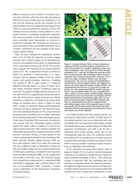
Figure 4. Covalent linking of IBA. (a) Upon moderate annealing<br />
of the IBA-covered sample to a temperature of<br />
520 K, two domains are revealed, exhibiting a large-scale<br />
moiré modulation. Inset: Close-up of one of the domains,<br />
exhibiting a (8 1) unit cell. (b) Molecular structure of the<br />
IBA-covered substrate after annealing to 580 K showing<br />
molecular rows running along the [421] direction. Two or<br />
more rows align, resulting in double rows and larger<br />
arrangements. The rows are composed of individually resolved<br />
features that are aligned side-by-side. Ellipses having<br />
the size of the expected reaction product, BPDCA, are<br />
superimposed onto the row. (c) Close-up of a single molecular<br />
row with superimposed BPCDA model molecules.<br />
Due to a tip-convolution effect, the row is imaged with a<br />
“halo”, which is a known artifact in AFM imaging. Along the<br />
[010] direction, lines are drawn that superimpose with the<br />
bright calcite features (protruding oxygen atoms of one<br />
type of carbonate group). Along the [421] direction, the<br />
lines are superimposed onto the less bright features<br />
(protruding oxygen atoms of the opposite type of carbonate<br />
group). (d) Structural model showing the flat-lying<br />
molecules with the carboxylate groups adsorbed on top of<br />
two calcium ions of the substrate.<br />
As a further evidence for the coupling reaction, we<br />
next discuss reducing the number of halide atoms at<br />
the phenyl ring from two to one. Molecules with only<br />
one halide atom are expected to form dimers instead<br />
of extended wires upon covalent linking. To follow this<br />
approach, 4-iodobenzoic acid with a pKa of 4.02 is<br />
deposited onto freshly cleaved calcite. Due to the<br />
comparatively high pKa value, the molecules remain<br />
protonated at RT, as is evident from a further structural<br />
transition observed upon moderate annealing to 520 K<br />
before inducing covalent coupling at higher temperatures<br />
as explained in the following.<br />
At RT, extended two-dimensional islands with an<br />
apparent height of 0.5 nm and an internal hexagonal<br />
ordering are revealed (see Supplementary Figure S3).<br />
Upon moderate annealing at 520 K, a first structural<br />
change is observed. After this annealing step, two<br />
domains of extended islands are observed; however,<br />
the apparent height increases to 0.8 nm (see Figure 4a).<br />
A highly ordered structure can be resolved within the<br />
domains (see inset in Figure 4a), revealing a (8 1) unit<br />
KITTELMANN ET AL. VOL. 5 ’ NO. 10 ’ 8420–8425 ’ 2011<br />
www.acsnano.org<br />
ARTICLE<br />
8423