Shoulder - Latarjet Protocol - Brigham and Women's Hospital
Shoulder - Latarjet Protocol - Brigham and Women's Hospital
Shoulder - Latarjet Protocol - Brigham and Women's Hospital

You also want an ePaper? Increase the reach of your titles
YUMPU automatically turns print PDFs into web optimized ePapers that Google loves.
Department of Rehabilitation Services<br />
Physical Therapy<br />
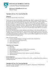
Anterior Stabilization of the <strong>Shoulder</strong>: <strong>Latarjet</strong> <strong>Protocol</strong><br />
<strong>Shoulder</strong> instability may be caused from congenital deformity, recurrent overuse activity,<br />
<strong>and</strong>/or traumatic dislocation. Surgical stabilization of the glenohumeral joint is indicated<br />
after conservative treatment fails <strong>and</strong> recurrent instability/subluxation continues. A<br />
number of different surgical procedures may be indicated in this situation, often divided<br />
into soft tissue or bony procedures.<br />
<strong>Shoulder</strong> Instability – Soft Tissue:<br />
Surgical reconstruction targeting the glenohumeral joint’s soft tissues for shoulder<br />
instability, typically involves labral repairs, the most common being the Bankart repair. A<br />
Bankart lesion typically occurs from an anterior-inferior dislocation of the humerus,<br />
tearing the labrum from it’s attachment to the glenoid, thereby detaching the inferior<br />
gleno-humeral ligament (IGHL). Surgical management of this revolves around labral<br />
repair to reattach the IGHL under appropriate tension. This may be accomplished<br />
either arthroscopically or through an open approach. 1 Most traumatic glenohumeral<br />
dislocations may not only cause a Bankart lesion, but may create impression fractures in<br />
the postero-superior humeral head termed Hill-Sachs lesions. 2 An adverse effect from this<br />
procedure includes suturing the capsule too tightly, causing a shortening of the capsule,<br />
<strong>and</strong> thus decreasing the external rotation allowed at the glenohumeral joint. Other<br />
complications are extremely rare, but may include axillary nerve damage, subscapularis<br />
rupture (seen only in open repairs performed with subscapularis detachment <strong>and</strong> repair),<br />
<strong>and</strong> recurrent instability. If there is bony deficiency in the glenoid, which represents 20%<br />
or more of the antero-inferior glenoid, it is biomechanically impossible to restore the<br />
same stability <strong>and</strong> is therefore more prone to recurrent instability <strong>and</strong> failure.<br />
<strong>Shoulder</strong> Instability – Bony Deficiency:<br />
In cases where significant bony deficiency is present (where greater than 20% of the<br />
glenoid’s surface area is missing) addressing only the soft tissue issues during the<br />
surgical procedure may lead to eventual recurrence of instability. Bony deficiency can<br />
result from congenital deformity, trauma, or recurrent dislocation. These lesions are not<br />
well visualized on plain films <strong>and</strong> are best seen on 3-dimensional CT scan (see Fig. 1).<br />
Anterior Stabilization of the <strong>Shoulder</strong>: <strong>Latarjet</strong> <strong>Protocol</strong><br />
Copyright © 2009 The <strong>Brigham</strong> <strong>and</strong> <strong>Women's</strong> <strong>Hospital</strong>, Inc. Department of Rehabilitation Services. All<br />
rights reserved.<br />
1
Figure 1: A 3D CT reconstruction of the scapula. The blue segment illustrates the bony<br />
deficit of the glenoid.<br />
When bony lesions reach critical dimensions, reconstruction of this deficit using autograft<br />
bone yields the best surgical results. The <strong>Latarjet</strong> procedure is the most popular <strong>and</strong><br />
highly effective, transferring the distal coracoid into the bony defect. 3<br />
Surgical Technique<br />
A deltopectoral approach is used to expose the coracoid process. The corcoacromial<br />
ligament <strong>and</strong> the pectoralis minor attachment are divided, where as coracobrachialis <strong>and</strong><br />
the short head of the biceps origins remain intact. The coracoid is osteotomized at its’<br />
“knee” yielding bony graft approximately 1.5 cm in length. Great care is taken to avoid<br />
damage to the soft tissues <strong>and</strong> musculotaneous nerve in the surrounding area.<br />
With the arm in external rotation, the subscapularis muscle is either split along its’ length<br />
or detached from the lesser tuberosity <strong>and</strong> the joint is exposed. The graft is shaped <strong>and</strong><br />
contoured to fill the defect <strong>and</strong> is secured with screw fixation placed at the antero-inferior<br />
glenoid. With the corachobracialis <strong>and</strong> biceps still attached to the coracoid, they now<br />
serve as a dynamic sling further stabilizing the glenohumeral joint. 2-5 The subscapularis<br />
split is then repaired. The final bony reconstruction is illustrated in Figure 2.<br />
Anterior Stabilization of the <strong>Shoulder</strong>: <strong>Latarjet</strong> <strong>Protocol</strong><br />
Copyright © 2009 The <strong>Brigham</strong> <strong>and</strong> <strong>Women's</strong> <strong>Hospital</strong>, Inc. Department of Rehabilitation Services. All<br />
rights reserved.<br />
2
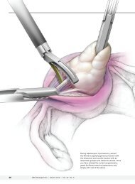
Figure 2: Illustration of the coracoid transfer to correct the inferior glenoid bony<br />
deficiency.<br />
Potential Complications<br />
There are several possible complications that could occur after a <strong>Latarjet</strong> procedure.<br />
Considering the coracoid osteotomy, there is a risk for non-union of the transferred<br />
coracoid process, which occurs typically in 3% of patients. 2 In a long-term follow-up by<br />
Banas et al, 82% had bony union <strong>and</strong> 14% had fibrous union of the coracoid <strong>and</strong> glenoid.<br />
4 Despite the bony union, many patients continued to experience discomfort years postoperatively<br />
<strong>and</strong> underwent another procedure to extract the screws. Screw loosening <strong>and</strong><br />
screw breakage are other possible reasons a patient may undergo a screw removal<br />
procedure. A follow-up performed by Banas et al, found 14% of shoulders required<br />
secondary operations, 4% for stabilization, <strong>and</strong> 10% for screw removal secondary to<br />
discomfort. Current research is evaluating the optimal screw placement during the<br />
procedure to reduce loosening, breakage, <strong>and</strong> discomfort. 2-4 Other complications,<br />
including musculocutaneous nerve palsy <strong>and</strong> subscapularis dysfunction, are reported but<br />
rare events.<br />
Following a <strong>Latarjet</strong> procedure, the most functional limitation reported is a decrease in<br />
external rotation range of motion. Although some patients may return to overhead<br />
throwing sports, most do not regain full external rotation. 2-5 According to Hovelius <strong>and</strong><br />
colleagues, the mean loss of external rotation was 7.4 degrees in adduction <strong>and</strong> 8 degrees<br />
in abduction. The complications of rotator cuff tendonitis <strong>and</strong> limitation in external<br />
rotation can be reduced with proper progression in rehabilitation. 6<br />
Anterior Stabilization of the <strong>Shoulder</strong>: <strong>Latarjet</strong> <strong>Protocol</strong><br />
Copyright © 2009 The <strong>Brigham</strong> <strong>and</strong> <strong>Women's</strong> <strong>Hospital</strong>, Inc. Department of Rehabilitation Services. All<br />
rights reserved.<br />
3
Rehabilitation Considerations<br />
One must recall that the purpose of the <strong>Latarjet</strong> procedure is to reinstate anterior stability<br />
to the glenohumeral joint. While this is primarily a bony procedure, specific attention<br />
must be directed towards the soft tissues which play a critical role in maintaining<br />
stability. Early post-operative therapy must protect the repair of the subscapularis as well<br />
as the developing bony union of the coracoid process.<br />
Since it will take approximately 6-8 weeks to form an osseous union of the newly<br />
reconstructed glenoid, the biceps <strong>and</strong> coracobrachialis attachment to the coracoid needs<br />
to be protected during the initial postoperative period. Aggressive shoulder extension <strong>and</strong><br />
combined extension <strong>and</strong> external rotation stretching is not indicated. Once strengthening<br />
commences, a gradual progressed program of biceps <strong>and</strong> coracobrachialis strengthening<br />
needs to be followed to minimize undue stress <strong>and</strong> tension on their muscular origins.<br />
In addition, isolated external rotation range of motion needs to be gradually regained after<br />
surgery to allow the anterior capsule <strong>and</strong> subscapularis to heal appropriately. 7 For that<br />
reason, external rotation range of motion is advanced in a protected fashion, with early<br />
emphasis on external rotation work being done in an open packed position (i.e. scapular<br />
plane at about 45 degrees of abduction) <strong>and</strong> then progressed to positions that gradually<br />
tension the subscapularis (i.e. full adduction <strong>and</strong> then at 90 degrees of abduction <strong>and</strong><br />
above). Please refer to protocol below for more detail. (In the case of a subscapularis take<br />
down <strong>and</strong> repair, external rotation gains need to be progressed slower <strong>and</strong> one should<br />
avoid aggressive external rotation stretching <strong>and</strong> internal rotation strengthening until the<br />
subscapularis is well healed. In these cases it is helpful to get a ‘safe zone’ of initial<br />
external rotation range of motion from the referring surgeon, as determined from<br />
intraoperative inspection from either the operative note or discussion with surgeon.)<br />
Due to the surgical technique <strong>and</strong> early immobilization required to promote healing, the<br />
subscapularis may not only be impacted in terms of length, but in terms of force<br />
production <strong>and</strong> proprioception. Hence, specific subscapularis proprioception <strong>and</strong><br />
strengthening needs to be incorporated to enhance subscapularis function postoperatively.<br />
The clinician needs to tailor the rehabilitation program to address the unique structure of<br />
the subscapularis to enhance both the upper <strong>and</strong> lower subscapularis fibers. This is<br />
warranted due to the fact that the subscapularis is innervated by both the upper <strong>and</strong> lower<br />
subscapular nerves, along with the presence of two different muscular fiber alignments;<br />
hence, its action has been described as being like that of two different muscles depending<br />
upon the functional activity. 8 The upper fibers are primarily aligned in a horizontal<br />
fashion <strong>and</strong> the lower fibers are arranged in more of an oblique alignment. One must<br />
therefore be selective in the rehabilitation protocol to maximally stimulate the appropriate<br />
portion of the subscapularis with the correct exercise.<br />
Anterior Stabilization of the <strong>Shoulder</strong>: <strong>Latarjet</strong> <strong>Protocol</strong><br />
Copyright © 2009 The <strong>Brigham</strong> <strong>and</strong> <strong>Women's</strong> <strong>Hospital</strong>, Inc. Department of Rehabilitation Services. All<br />
rights reserved.<br />
4
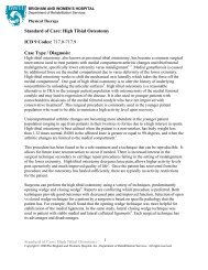
Anterior Stabilization of the <strong>Shoulder</strong>: <strong>Latarjet</strong> <strong>Protocol</strong><br />
The intent of this protocol is to provide the clinician with a guideline of the postoperative<br />
rehabilitation course of a patient that has undergone a <strong>Latarjet</strong> procedure for<br />
anterior stabilization. It is no means intended to be a substitute for one’s clinical<br />
decision making regarding the progression of a patient’s post-operative course based<br />
on their physical exam/findings, individual progress, <strong>and</strong>/or the presence of postoperative<br />
complications. If a clinician requires assistance in the progression of a postoperative<br />
patient they should consult with the referring Surgeon.<br />
Progression to the next phase based on Clinical Criteria <strong>and</strong>/or Time Frames as<br />
Appropriate.<br />
Phase I – Immediate Post Surgical Phase (approximately Weeks 1- 3)<br />
Goals:<br />
• Minimize shoulder pain <strong>and</strong> inflammatory response<br />
• Protect the integrity of the surgical repair<br />
• Achieve gradual restoration of passive range of motion (PROM)<br />
• Enhance/ensure adequate scapular function<br />
Precautions/Patient Education:<br />
• No active range of motion (AROM) of the operative shoulder<br />
• No excessive external rotation range of motion (ROM) / stretching. Stop at first<br />
end feel felt<br />
• Remain in sling, only removing for showering. Shower with arm held at side<br />
• No lifting of objects with operative shoulder<br />
• Keep incisions clean <strong>and</strong> dry<br />
• Patient education regarding limited use of upper extremity despite the potential<br />
lack of or minimal pain or other symptoms<br />
Activity:<br />
• Arm in sling except when performing distal upper extremity exercises<br />
• (PROM)/Active-Assisted Range of Motion (AAROM)/ (AROM) elbow <strong>and</strong><br />
wrist/h<strong>and</strong><br />
• Begin shoulder PROM (do not force any painful motion)<br />
• Forward flexion <strong>and</strong> elevation to tolerance<br />
• Abduction in the plane of the scapula to tolerance<br />
• Internal rotation (IR) to 45 degrees at 30 degrees of abduction<br />
• External rotation (ER) in the plane of the scapula from 0-25 degrees; begin at<br />
30-40 degrees of abduction; respect anterior capsule tissue integrity with ER<br />
Anterior Stabilization of the <strong>Shoulder</strong>: <strong>Latarjet</strong> <strong>Protocol</strong><br />
Copyright © 2009 The <strong>Brigham</strong> <strong>and</strong> <strong>Women's</strong> <strong>Hospital</strong>, Inc. Department of Rehabilitation Services. All<br />
rights reserved.<br />
5
ange of motion; (seek guidance from intraoperative measurements of external<br />
rotation ROM)<br />
• Scapular clock exercises progressed to scapular isometric exercises<br />
• Ball squeezes<br />
• Sleep with sling supporting operative shoulder, place a towel under the elbow to<br />
prevent shoulder hyperextension<br />
• Frequent cryotherapy for pain <strong>and</strong> inflammation<br />
• Patient education regarding posture, joint protection, positioning, hygiene, etc.<br />
Milestones to progress to phase II:<br />
• Appropriate healing of the surgical repair<br />
• Adherence to the precautions <strong>and</strong> immobilization guidelines<br />
• Achieved at least 100 degrees of passive forward elevation <strong>and</strong> 30 degrees of<br />
passive external rotation at 20 degrees abduction<br />
• Completion of phase I activities without pain or difficulty<br />
Phase II – Intermediate Phase/ROM (approximately Week 4-9)<br />
Goals:<br />
• Minimize shoulder pain <strong>and</strong> inflammatory response<br />
• Protect the integrity of the surgical repair<br />
• Achieve gradual restoration of (AROM)<br />
• To be weaned from the sling by the end of week 4-5<br />
• Begin light waist level activites<br />
Precautions:<br />
• No active movement of shoulder till adequate PROM with good mechanics<br />
• No lifting with affected upper extremity<br />
• No excessive external rotation ROM / stretching<br />
• Do not perform activities or strengthening exercises that place an excessive load<br />
on the anterior capsule of the shoulder joint (i.e. no pushups, pec flys, etc..)<br />
• Do not perform scaption with internal rotation (empty can) during any stage of<br />
rehabilitation due to the possibility of impingement<br />
Early Phase II (approximately week 4):<br />
• Progress shoulder PROM (do not force any painful motion)<br />
• Forward flexion <strong>and</strong> elevation to tolerance<br />
• Abduction in the plane of the scapula to tolerance<br />
• IR to 45 degrees at 30 degrees of abduction<br />
• ER to 0-45 degrees; begin at 30-40 degrees of abduction; respect anterior<br />
capsule tissue integrity with ER range of motion; seek guidance from<br />
intraoperative measurements of external rotation ROM)<br />
Anterior Stabilization of the <strong>Shoulder</strong>: <strong>Latarjet</strong> <strong>Protocol</strong><br />
Copyright © 2009 The <strong>Brigham</strong> <strong>and</strong> <strong>Women's</strong> <strong>Hospital</strong>, Inc. Department of Rehabilitation Services. All<br />
rights reserved.<br />
6
• Glenohumeral joint mobilizations as indicated (Grade I, II) when ROM is<br />
significantly less than expected. Mobilizations should be done in directions of<br />
limited motion <strong>and</strong> only until adequate ROM is gained.<br />
• Address scapulothoracic <strong>and</strong> trunk mobility limitations. Scapulothoracic <strong>and</strong><br />
thoracic spine joint mobilizations as indicated (Grade I, II, III) when ROM is<br />
significantly less than expected. Mobilizations should be done in directions of<br />
limited <strong>and</strong> only until adequate ROM is gained.<br />
• Begin incorporating posterior capsular stretching as indicated<br />
• Cross body adduction stretch<br />
• Side lying internal rotation stretch (sleeper stretch)<br />
• Continued Cryotherapy for pain <strong>and</strong> inflammation<br />
• Continued patient education: posture, joint protection, positioning, hygiene, etc.<br />
Late Phase II (approximately Week 6):<br />
• Progress shoulder PROM (do not force any painful motion)<br />
• Forward flexion, elevation, <strong>and</strong> abduction in the plane of the scapula to<br />
tolerance<br />
• IR as tolerated at multiple angles of abduction<br />
• ER to tolerance; progress to multiple angles of abduction once >/= 35 degrees<br />
at 0-40 degrees of abduction<br />
• Glenohumeral <strong>and</strong> scapulothoracic joint mobilizations as indicated (Grade I-IV as<br />
appropriate)<br />
• Progress to AA/AROM activities of the shoulder as tolerated with good shoulder<br />
mechanics (i.e. minimal to no scapulathoracic substitution with up to 90-110<br />
degrees of elevation.)<br />
• Begin rhythmic stabilization drills<br />
• ER/IR in the scapular plane<br />
• Flexion/extension <strong>and</strong> abduction/adduction at various angles of elevation<br />
• Continue AROM elbow, wrist, <strong>and</strong> h<strong>and</strong><br />
• Strengthen scapular retractors <strong>and</strong> upward rotators<br />
• Initiate balanced AROM / strengthening program<br />
o Initially in low dynamic positions<br />
o Gain muscular endurance with high repetition of 30-50, low resistance 1-3<br />
lbs)<br />
o Exercises should be progressive in terms of muscle dem<strong>and</strong> / intensity,<br />
shoulder elevation, <strong>and</strong> stress on the anterior joint capsule<br />
o Nearly full elevation in the scapula plane should be achieved before<br />
beginning elevation in other planes<br />
o All activities should be pain free <strong>and</strong> without substitution patterns<br />
o Exercises should consist of both open <strong>and</strong> closed chain activities<br />
o No heavy lifting or plyometrics should be performed at this time<br />
� Initiate full can scapular plane raises to 90 degrees with good<br />
mechanics<br />
Anterior Stabilization of the <strong>Shoulder</strong>: <strong>Latarjet</strong> <strong>Protocol</strong><br />
Copyright © 2009 The <strong>Brigham</strong> <strong>and</strong> <strong>Women's</strong> <strong>Hospital</strong>, Inc. Department of Rehabilitation Services. All<br />
rights reserved.<br />
7
� Initiate ER/IR strengthening using exercise tubing at 0° of<br />
abduction (use towel roll)<br />
� Initiate sidelying ER with towel roll<br />
� Initiate manual resistance ER supine in scapular plane (light<br />
resistance)<br />
� Initiate prone rowing at 30/45/90 degrees of abduction to neutral<br />
arm position<br />
• Continued cryotherapy for pain <strong>and</strong> inflammation<br />
• Continued patient education: posture, joint protection, positioning, hygiene, etc.<br />
Milestones to progress to phase III:<br />
• Passive forward elevation at least 155 degrees<br />
• Passive external rotation within 8-10 degrees of contralateral side at 20 degrees<br />
abduction<br />
• Passive external rotation at least 75 degrees at 90 degrees abduction<br />
• Active forward elevation at least 145 degrees with good mechanics<br />
• Appropriate scapular posture at rest <strong>and</strong> dynamic scapular control with ROM <strong>and</strong><br />
functional activities<br />
• Completion of phase II activities without pain or difficulty<br />
Phase III - Strengthening Phase (approximately Week 10 – Week 15)<br />
Goals:<br />
• Normalize strength, endurance, neuromuscular control<br />
• Return to chest level full functional activities<br />
• Gradual <strong>and</strong> planned buildup of stress to anterior joint capsule<br />
Precautions:<br />
• Do not overstress the anterior capsule with aggressive overhead activities /<br />
strengthening<br />
• Avoid contact sports/activities<br />
• Do not perform strengthening or functional activities in a given plan until the<br />
patient has near full ROM <strong>and</strong> strength in that plane of movement<br />
• Patient education regarding a gradual increase to shoulder activities<br />
Activity:<br />
• Continue A/PROM as needed/indicated<br />
• Initiate biceps curls with light resistance, progress as tolerated<br />
• Initiate gradually progressed strengthening for pectoralis major <strong>and</strong> minor; avoid<br />
positions that excessively stress the anterior capsule<br />
• Progress subscapularis strengthening to focus on both upper <strong>and</strong> lower segments<br />
o Push up plus (wall, counter, knees on the floor, floor)<br />
o Cross body diagonals with resistive tubing<br />
o IR resistive b<strong>and</strong> (0, 45, 90 degrees of abduction<br />
o Forward punch<br />
Anterior Stabilization of the <strong>Shoulder</strong>: <strong>Latarjet</strong> <strong>Protocol</strong><br />
Copyright © 2009 The <strong>Brigham</strong> <strong>and</strong> <strong>Women's</strong> <strong>Hospital</strong>, Inc. Department of Rehabilitation Services. All<br />
rights reserved.<br />
8
Milestones to progress to phase IV:<br />
• Passive forward elevation WNL<br />
• Passive external rotation at all angles of abduction WNL<br />
• Active forward elevation WNL with good mechanics<br />
• Appropriate rotator cuff <strong>and</strong> scapular muscular performance for chest level<br />
activities<br />
• Completion of phase III activities without pain or difficulty<br />
Phase IV - Overhead Activities Phase / Return to activity phase<br />
(approximately Week 16-20)<br />
Goals:<br />
• Continue stretching <strong>and</strong> PROM as needed/indicated<br />
• Maintain full non-painful AROM<br />
• Return to full strenuous work activities<br />
• Return to full recreational activities<br />
Precautions:<br />
• Avoid excessive anterior capsule stress<br />
• With weight lifting, avoid tricep dips, wide grip bench press, <strong>and</strong> no military press<br />
or lat pulls behind the head. Be sure to “always see your elbows”<br />
• Do not begin throwing, or overhead athletic moves until 4 months post-op or<br />
cleared by MD<br />
Activity:<br />
• Continue all exercises listed above<br />
o Progress isotonic strengthening if patient demonstrates no compensatory<br />
strategies, is not painful, <strong>and</strong> has no residual soreness<br />
• Strengthening overhead if ROM <strong>and</strong> strength below 90 degree elevation is good<br />
• Continue shoulder stretching <strong>and</strong> strengthening at least four times per week<br />
• Progressive return to upper extremity weight lifting program emphasizing the<br />
larger, primary upper extremity muscles (deltoid, latissimus dorsi, pectoralis<br />
major)<br />
o Start with relatively light weight <strong>and</strong> high repetitions (15-25)<br />
• May do pushups as long as the elbows do not flex past 90 degrees<br />
• May initiate plyometrics/interval sports program if appropriate/cleared by PT <strong>and</strong><br />
MD<br />
• Can begin generalized upper extremity weight lifting with low weight, <strong>and</strong> high<br />
repetitions, being sure to follow weight lifting precautions.<br />
• May initiate pre injury level activities/ vigorous sports if appropriate / cleared by<br />
MD<br />
Anterior Stabilization of the <strong>Shoulder</strong>: <strong>Latarjet</strong> <strong>Protocol</strong><br />
Copyright © 2009 The <strong>Brigham</strong> <strong>and</strong> <strong>Women's</strong> <strong>Hospital</strong>, Inc. Department of Rehabilitation Services. All<br />
rights reserved.<br />
9
Milestones to return to overhead work <strong>and</strong> sport activities:<br />
• Clearance from MD<br />
• No complaints of pain or instability<br />
• Adequate ROM for task completion<br />
• Full strength <strong>and</strong> endurance of rotator cuff <strong>and</strong> scapular musculature for task<br />
completion<br />
• Regular completion of continued home exercise program<br />
Authors: Reviewers:<br />
Ashley Burns, PT Laurence D. Higgins, MD<br />
Reg B. Wilcox III, PT Joel Fallano, PT<br />
3/2009 Ken Shannon, PT<br />
References<br />
1. Jones D WJ. <strong>Shoulder</strong> instability. In: Chapman MW, Lane JM, Mann RA, Marder RA,<br />
McLain RF, Rab GT, Szabo RM, Vince KG. Chapman's Orthopaedic Surgery. Vol 2. 3rd<br />
ed. Lippincott Williams <strong>and</strong> Wilkins.<br />
2. Yoneda M, Hayashida K, Wakitani S, Nakagawa S, Fukushima S. Bankart procedure<br />
augmented by coracoid transfer for contact athletes with traumatic anterior shoulder<br />
instability. Am J Sports Med. 1999; 27(1):21-26.<br />
3. Matthes G, Horvath V, Seifert J, et al. Oldie but goldie: Bristow-latarjet procedure for<br />
anterior shoulder instability. J Orthop Surg (Hong Kong). 2007; 15(1):4-8.<br />
4. Banas MP, Dalldorf PG, Sebastianelli WJ, DeHaven KE. Long-term followup of the<br />
modified bristow procedure. Am J Sports Med. 1993; 21(5):666-671.<br />
5. Schauder KS, Tullos HS. Role of the coracoid bone block in the modified bristow<br />
procedure. Am J Sports Med. 1992; 20(1):31-34.<br />
6. Hovelius L, S<strong>and</strong>strom B, Saebo M. One hundred eighteen bristow-latarjet repairs for<br />
recurrent anterior dislocation of the shoulder prospectively followed for fifteen years:<br />
Study II-the evolution of dislocation arthropathy. J <strong>Shoulder</strong> Elbow Surg. 2006;<br />
15(3):279-289.<br />
7. Hall CM BL. Therapeutic Exercise: Moving Toward Function. 2nd ed. ed.<br />
Philadelphia: Lippincott Williams <strong>and</strong> Wilkins; 2005:787.<br />
8. Decker MJ, Tokish JM, Ellis HB, Torry MR, Hawkins RJ. Subscapularis muscle<br />
activity during selected rehabilitation exercises. Am J Sports Med. 2003; 31(1):126-134.<br />
Anterior Stabilization of the <strong>Shoulder</strong>: <strong>Latarjet</strong> <strong>Protocol</strong><br />
Copyright © 2009 The <strong>Brigham</strong> <strong>and</strong> <strong>Women's</strong> <strong>Hospital</strong>, Inc. Department of Rehabilitation Services. All<br />
rights reserved.<br />
10