Aspectos anatómicos de la semilla de papaya - Sociedad ...
Aspectos anatómicos de la semilla de papaya - Sociedad ...
Aspectos anatómicos de la semilla de papaya - Sociedad ...

Create successful ePaper yourself
Turn your PDF publications into a flip-book with our unique Google optimized e-Paper software.
<strong>Aspectos</strong> <strong>anatómicos</strong> <strong>de</strong> <strong>la</strong> semil<strong>la</strong> <strong>de</strong> <strong>papaya</strong><br />
(Carica <strong>papaya</strong> L.)<br />
Anatomical aspects of <strong>papaya</strong><br />
(Carica <strong>papaya</strong> L.) seed<br />
reSumeN<br />
arlette ivoNNe gil 1,3<br />
Diego miraNDa 2<br />
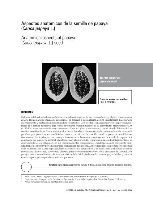
Frutos <strong>de</strong> <strong>papaya</strong> con semil<strong>la</strong>s.<br />
Foto: D. Miranda L.<br />
Debido a <strong>la</strong> falta <strong>de</strong> estudios <strong>anatómicos</strong> en semil<strong>la</strong>s <strong>de</strong> especies <strong>de</strong> interés económico, y el poco conocimiento<br />
<strong>de</strong> este tópico para los ingenieros agrónomos, se procedió a <strong>la</strong> realización <strong>de</strong> una investigación base para su<br />
entendimiento y posterior ampliación en futuros estudios. Con este fin se analizaron diversos aspectos <strong>anatómicos</strong><br />
<strong>de</strong> <strong>la</strong> semil<strong>la</strong> <strong>de</strong> <strong>papaya</strong>, para lo cual se tomaron frutos inmaduros <strong>de</strong> 60 días y frutos maduros entre 120<br />
y 150 días, entre madurez fisiológica y comercial, en una p<strong>la</strong>ntación sembrada con el híbrido Tainung-1. Las<br />
semil<strong>la</strong>s extraídas <strong>de</strong> los frutos mencionados fueron llevadas al <strong>la</strong>boratorio y a<strong>de</strong>cuadas mediante <strong>la</strong> técnica <strong>de</strong><br />
parafina, para posteriormente realizar los cortes en micrótomo <strong>de</strong> rotación con el propósito <strong>de</strong> <strong>de</strong>scribir anatómicamente<br />
los tejidos y estructuras que <strong>la</strong>s componen, bajo microscopía óptica. La semil<strong>la</strong> <strong>de</strong> <strong>papaya</strong> está<br />
compuesta por <strong>la</strong> cubierta seminal, el endospermo y el embrión. Por tratarse <strong>de</strong> una semil<strong>la</strong> bitegumentada, se<br />
observaron <strong>la</strong> testa y el tegmen con sus correspondientes componentes. El endospermo está compuesto principalmente<br />
<strong>de</strong> lípidos y proteínas agrupados en granos <strong>de</strong> aleurona. Los cotiledones están compuestos a<strong>de</strong>más<br />
<strong>de</strong> <strong>la</strong> epi<strong>de</strong>rmis, por cuatro capas celu<strong>la</strong>res internas y en <strong>la</strong> zona radicu<strong>la</strong>r se pudo apreciar el esbozo <strong>de</strong> raíces<br />
secundarias. Este estudio tuvo como objetivo generar conocimiento acerca <strong>de</strong> <strong>la</strong> anatomía <strong>de</strong> <strong>la</strong> semil<strong>la</strong> <strong>de</strong><br />
<strong>papaya</strong>, para el entendimiento <strong>de</strong> aspectos básicos <strong>de</strong> <strong>la</strong> fisiología <strong>de</strong> semil<strong>la</strong>s como vigor, viabilidad y <strong>la</strong>tencia<br />
en esta especie, previo para futuras investigaciones.<br />
pa<strong>la</strong>bras c<strong>la</strong>ve adicionales: híbrido Tainung-1, testa, endospermo, embrión, granos <strong>de</strong> aleurona.<br />
1 Facultad <strong>de</strong> Ciencias Agropecuarias, Universidad <strong>de</strong> Cundinamarca, Fusagasugá (Colombia).<br />
2 Departamento <strong>de</strong> Agronomía, Facultad <strong>de</strong> Agronomía, Universidad Nacional <strong>de</strong> Colombia, Bogotá (Colombia).<br />
3 Autor para correspon<strong>de</strong>ncia. arlettegil@hotmail.com<br />
REVISTA COLOMBIANA DE CIENCIAS HORTÍCOLAS - Vol. 2 - No.2 - pp. 145-156, 2008
146<br />
abStraCt<br />
GIL/MIRANDA<br />
Due to the <strong>la</strong>ck of the anatomical studies in seeds of the species of economical interest and a few knowledge<br />
of this matter by the agronomists, the investigation as a base for un<strong>de</strong>rstanding of this problem and further<br />
<strong>de</strong>velopment in future studies was done. With this purpose, several anatomical aspects of <strong>papaya</strong> seed were<br />
analyzed from immature fruits of 60 days age and mature fruits of 120 and 150 days between physiological<br />
and commercial maturity were taken from Tainung-1 hybrid p<strong>la</strong>ntation. The seeds extracted from fruits were<br />
transported to the <strong>la</strong>boratory and a<strong>de</strong>quated with paraffin technique to make the microtome sections in<br />
or<strong>de</strong>r to <strong>de</strong>scribe anatomically the tissues and structures un<strong>de</strong>r optical microscopy. Papaya seed is composed<br />
of the seed coat, endosperm, and embryo. As a bitegumental seed, the testa and tegmen were observed with<br />
their own parts. The endosperm is composed mainly of lipids and proteins grouped in aleurone grains. The<br />
cotyledons are composed by the epi<strong>de</strong>rmis and four-<strong>la</strong>yer inner cells and, in the radicle zone, initiation of the<br />
secondary roots was observed. The objective of this study was to generate knowledge about the anatomy of<br />
<strong>papaya</strong> seed for un<strong>de</strong>rstanding the basic aspects of seed physiology, such as vigour, viability, and dormancy in<br />
this species, prior to future researches.<br />
additional key words: Tainung-1 hybrid, testa, endosperm, embryo, aleurone grains.<br />
Fecha <strong>de</strong> recepción: 30-07-2008 Aprobado para publicación: 02-12-2008<br />
La <strong>papaya</strong> (Carica <strong>papaya</strong> L.) es consi<strong>de</strong>rada <strong>de</strong> origen<br />
Americano, específicamente <strong>de</strong> Centroamérica,<br />
entre México y Costa Rica (León, 1987). La<br />
i<strong>de</strong>ntificación <strong>de</strong> <strong>la</strong>s distintas características morfológicas<br />
y fisiológicas es un paso inicial importante<br />
en el estudio <strong>de</strong>l <strong>de</strong>sarrollo <strong>de</strong> <strong>la</strong>s semil<strong>la</strong>s en<br />
<strong>la</strong>s diversas especies (Ren y Bewley, 1998). El megasporangio<br />
(óvulo) es <strong>la</strong> estructura a partir <strong>de</strong> <strong>la</strong><br />
cual se forman <strong>la</strong>s semil<strong>la</strong>s. En <strong>la</strong>s angiospermas<br />
está formado por uno o dos tegumentos, ocasionalmente<br />
por tres, y muy raramente por cuatro.<br />
En <strong>la</strong> semil<strong>la</strong> madura, los tegumentos que ro<strong>de</strong>an<br />
al óvulo se transforman en <strong>la</strong> cubierta seminal<br />
(Niembro, 1988; Reiser y Fischer, 1993).<br />
Las semil<strong>la</strong>s <strong>de</strong> <strong>la</strong>s angiospermas pue<strong>de</strong>n ser divididas<br />
en tres partes, <strong>de</strong> origen genéticamente diferente:<br />
el embrión, el endospermo y <strong>la</strong> cubierta<br />
seminal. El cigoto, a partir <strong>de</strong>l cual se <strong>de</strong>sarrol<strong>la</strong><br />
el embrión, combina los genotipos <strong>de</strong>l haploi<strong>de</strong><br />
masculino y el gameto femenino. Después <strong>de</strong> <strong>la</strong><br />
REV. COLOMB. CIENC. HORTIC.<br />
iNtroDuCCióN<br />
fertilización, durante el <strong>de</strong>sarrollo <strong>de</strong>l embrión<br />
y <strong>la</strong> subsiguiente maduración <strong>de</strong> <strong>la</strong> semil<strong>la</strong>, los<br />
integumentos experimentan cambios morfológicos<br />
y se convertirán en <strong>la</strong> testa, <strong>la</strong> cual posee el<br />
genotipo materno (Fosket, 1994; León-Kloosterziel<br />
et al., 1994). Debido a que el embrión está<br />
encerrado por el endospermo, el cual a su vez<br />
está <strong>de</strong>ntro <strong>de</strong>l integumento <strong>de</strong>l óvulo, estas tres<br />
estructuras <strong>de</strong>ben coordinar su <strong>de</strong>sarrollo para<br />
producir una semil<strong>la</strong> madura <strong>de</strong> tamaño a<strong>de</strong>cuado<br />
(García et al., 2003).<br />
Cuando <strong>la</strong> semil<strong>la</strong> proviene <strong>de</strong> un primordio seminal<br />
con un solo tegumento, es unitegumentada<br />
y su cubierta seminal se <strong>de</strong>nomina testa.<br />
Igualmente si el primordio posee dos tegumentos,<br />
los tejidos <strong>de</strong>rivados <strong>de</strong>l tegumento externo<br />
constituyen <strong>la</strong> testa y los <strong>de</strong>rivados <strong>de</strong>l tegumento<br />
interno conforman el tegmen, cuando <strong>la</strong><br />
semil<strong>la</strong> es bitegumentada (Becerra y Chaparro,<br />
1999).
El endospermo es un tejido que se origina a partir<br />
<strong>de</strong> <strong>la</strong> fusión <strong>de</strong> uno <strong>de</strong> los núcleos espermáticos<br />
<strong>de</strong>l grano <strong>de</strong> polen con dos núcleos po<strong>la</strong>res<br />
<strong>de</strong>l saco embrionario, dando como resultado un<br />
tejido triploi<strong>de</strong> (Becerra y Chaparro, 1999). El<br />
endospermo acumu<strong>la</strong> <strong>la</strong> mayor parte <strong>de</strong> <strong>la</strong>s reservas<br />
nutritivas en <strong>la</strong> mayoría <strong>de</strong> los casos cuando<br />
los cotiledones no lo hacen (Esau, 1985; Besnier,<br />
1988). Algunas semil<strong>la</strong>s pue<strong>de</strong>n presentar un endospermo<br />
muy <strong>de</strong>lgado, limitado a una o varias<br />
célu<strong>la</strong>s <strong>de</strong> grosor, o bien pue<strong>de</strong>n estar provistas<br />
<strong>de</strong> uno masivo, parenquimatoso (Fahn, 1985).<br />
El endospermo es único en diversos aspectos <strong>de</strong><br />
su <strong>de</strong>sarrollo, biología celu<strong>la</strong>r y bioquímica. Su<br />
aparente simplicidad reflejada en <strong>la</strong> presencia <strong>de</strong><br />
unos cuantos tipos <strong>de</strong> célu<strong>la</strong>s diferenciadas, ha<br />
permitido estudios <strong>de</strong>tal<strong>la</strong>dos en histodiferenciación<br />
y biosíntesis <strong>de</strong> ciertos metabolitos almacenados<br />
(Lopes y Larkins, 1993).<br />
El encerramiento <strong>de</strong>l embrión y <strong>de</strong>l endospermo<br />
en integumentos <strong>de</strong> origen estrictamente maternos,<br />
ha llevado a interacciones entre los tejidos<br />
<strong>de</strong> diferentes contribuciones parentales en <strong>la</strong> protección<br />
<strong>de</strong>l embrión y <strong>de</strong>l endospermo, y en el<br />
control <strong>de</strong> <strong>la</strong> protrusión radicu<strong>la</strong>r <strong>de</strong> <strong>la</strong>s semil<strong>la</strong>s.<br />
Tales interacciones influencian <strong>la</strong> germinación,<br />
<strong>la</strong>tencia, calidad <strong>de</strong> <strong>la</strong> semil<strong>la</strong>, en particu<strong>la</strong>r vigor<br />
y longevidad (Debeaujon et al., 2000).<br />
El embrión <strong>de</strong> <strong>la</strong>s angiospermas consta <strong>de</strong> un<br />
breve eje que lleva uno o dos cotiledones u hojas<br />
embrionales. El punto <strong>de</strong> fijación <strong>de</strong> los cotiledones<br />
al eje, <strong>de</strong>signado como nudo cotiledonar,<br />
divi<strong>de</strong> al eje en dos regiones: basal o hipocotilo<br />
que termina en una raíz embrionaria o radícu<strong>la</strong>,<br />
y <strong>la</strong> región superior o epicotilo que pue<strong>de</strong> presentar<br />
una yema <strong>de</strong>nominada plúmu<strong>la</strong> (Becerra y<br />
Chaparro, 1999).<br />
El objetivo <strong>de</strong> esta investigación consistió en<br />
generar un conocimiento básico acerca <strong>de</strong> <strong>la</strong> estructura<br />
<strong>de</strong> <strong>la</strong> semil<strong>la</strong> <strong>de</strong> <strong>papaya</strong>, con el fin <strong>de</strong><br />
servir como base para futuros estudios en el área<br />
<strong>de</strong> anatomía y fisiología vegetal.<br />
A S p E C TO S A N AT ó M I C O S D E L A S E M I L L A D E pA pAyA<br />
materialeS y métoDoS<br />
En <strong>la</strong> finca Santa Helena propiedad <strong>de</strong>l grupo<br />
Grajales S.A., se realizó el muestreo <strong>de</strong> frutos<br />
<strong>de</strong> <strong>papaya</strong> <strong>de</strong>l híbrido Tainung-1 cuyo origen es<br />
taiwanés, con el fin <strong>de</strong> extraer sus semil<strong>la</strong>s. La<br />
p<strong>la</strong>ntación se encuentra ubicada en el municipio<br />
<strong>de</strong> Roldanillo (Valle <strong>de</strong>l Cauca, Colombia), a una<br />
altura <strong>de</strong> 935 msnm, temperatura <strong>de</strong> 24,2°C,<br />
precipitación anual <strong>de</strong> 1.015 mm y humedad re<strong>la</strong>tiva<br />
<strong>de</strong> 71,1%.<br />
Se tomaron frutos inmaduros, <strong>de</strong> 60 d <strong>de</strong> edad,<br />
que contenían rudimentos <strong>de</strong> semil<strong>la</strong>s (primordios<br />
seminales) y frutos maduros (madurez fisiológica<br />
y comercial) entre 120 y 150 d. Las semil<strong>la</strong>s<br />
(figura 1) se extrajeron <strong>de</strong> los frutos y fueron<br />
acondicionadas según <strong>la</strong> metodología <strong>de</strong> Johansen<br />
(1940), en <strong>la</strong> cual el material vegetal (semil<strong>la</strong>)<br />
fue incluido en bloques <strong>de</strong> parafina para posteriormente<br />
proce<strong>de</strong>r a los cortes en micrótomo <strong>de</strong><br />
rotación, con el fin <strong>de</strong> estudiar su anatomía bajo<br />
microscopio óptico. Para el teñido <strong>de</strong>l material se<br />
utilizó Fast Green y safranina.<br />
reSultaDoS y DiSCuSióN<br />
primordios seminales<br />
En el corte transversal medio realizado a un fruto<br />
inmaduro <strong>de</strong> 60 d, se observó que <strong>la</strong>s semil<strong>la</strong>s<br />
en formación (primordios seminales) se encontraban<br />
adheridas a <strong>la</strong>s pare<strong>de</strong>s internas <strong>de</strong>l fruto<br />
por medio <strong>de</strong>l funículo, lo que se conoce como<br />
p<strong>la</strong>centación parietal (figura 2). Esta característica<br />
ayuda a c<strong>la</strong>sificar a <strong>la</strong> <strong>papaya</strong> <strong>de</strong>ntro <strong>de</strong>l or<strong>de</strong>n<br />
<strong>de</strong> <strong>la</strong>s parietales, lo que ratifica lo afirmado<br />
por Font Quer (1965). Al interior <strong>de</strong>l funículo se<br />
observaron <strong>de</strong> seis a siete capas <strong>de</strong> célu<strong>la</strong>s muy<br />
a<strong>la</strong>rgadas, correspondientes al haz conductor,<br />
con unas pocas diferenciadas en xilema, <strong>de</strong> engrosamientos<br />
anu<strong>la</strong>res. En este estado, <strong>la</strong>s célu<strong>la</strong>s<br />
<strong>de</strong>l rudimento presentaban <strong>la</strong> apariencia <strong>de</strong><br />
célu<strong>la</strong>s embrionarias, con pare<strong>de</strong>s <strong>de</strong>lgadas y nú-<br />
Vol. 2 - No.2 - 2008<br />
147
148<br />
GIL/MIRANDA<br />
cleos prominentes, como evi<strong>de</strong>ncia <strong>de</strong> <strong>la</strong> división<br />
activa a <strong>la</strong> que se encontraban sometidas.<br />
Stadler et al. (2005) mostraron en semil<strong>la</strong>s <strong>de</strong><br />
Arabidopsis thaliana que el integumento externo<br />
es una extensión simplástica <strong>de</strong>l floema <strong>de</strong>l<br />
funículo, sugiriendo que los nutrientes al final<br />
<strong>de</strong> este pue<strong>de</strong>n ser <strong>de</strong>scargados a través <strong>de</strong> este<br />
tejido. El transporte <strong>de</strong> sacarosa en el funículo<br />
indica que esta es una vía común para <strong>la</strong> <strong>de</strong>scarga<br />
<strong>de</strong> nutrientes a <strong>la</strong>s semil<strong>la</strong>s en <strong>de</strong>sarrollo<br />
(Skinner et al., 2004). La asimi<strong>la</strong>ción <strong>de</strong> carbono<br />
es suplida hacia el embrión en <strong>de</strong>sarrollo. Los haces<br />
vascu<strong>la</strong>res están conectados al óvulo a través<br />
<strong>de</strong>l funículo al final <strong>de</strong>l tejido nuce<strong>la</strong>r. Los tejidos<br />
vascu<strong>la</strong>res no se extien<strong>de</strong>n más allá <strong>de</strong>l funículo<br />
<strong>de</strong>ntro <strong>de</strong>l óvulo (Im<strong>la</strong>u et al., 1999).<br />
La epi<strong>de</strong>rmis que recubría el funículo y <strong>la</strong> parte<br />
externa <strong>de</strong> <strong>la</strong> semil<strong>la</strong> correspondía a <strong>la</strong> continuación<br />
<strong>de</strong> <strong>la</strong> epi<strong>de</strong>rmis interna <strong>de</strong>l fruto, constituida<br />
por célu<strong>la</strong>s <strong>de</strong> forma rectangu<strong>la</strong>r, un poco<br />
más altas que anchas, con <strong>la</strong> pared externa algo<br />
arqueada, a modo <strong>de</strong> <strong>la</strong>s célu<strong>la</strong>s epi<strong>de</strong>rmales típicas.<br />
Al analizar <strong>la</strong> parte interna <strong>de</strong>l rudimento,<br />
se observaron célu<strong>la</strong>s un poco más anchas, muy<br />
juntas, que se unían sin <strong>de</strong>jar espacios entre sí. La<br />
cubierta seminal, que es <strong>la</strong> estructura <strong>de</strong> protección<br />
<strong>de</strong> <strong>la</strong> semil<strong>la</strong> contra los agentes externos, se<br />
<strong>de</strong>riva <strong>de</strong> estos tegumentos dispuestos <strong>de</strong> forma<br />
concéntrica (figura 3). Las seis capas subyacentes<br />
a <strong>la</strong> epi<strong>de</strong>rmis, constituyentes <strong>de</strong>l tegumento<br />
externo, midieron 7 μm <strong>de</strong> espesor, presentando<br />
forma ap<strong>la</strong>nada y disposición regu<strong>la</strong>r. Hacia <strong>la</strong><br />
parte interna se observaron unas 10 capas celu<strong>la</strong>res,<br />
correspondientes al tegumento interno, con<br />
pare<strong>de</strong>s angu<strong>la</strong>res <strong>de</strong> formas y tamaños diversos,<br />
cuyo espesor fue <strong>de</strong> 7,5 μm.<br />
Los óvulos son el sitio <strong>de</strong> los procesos esenciales<br />
para <strong>la</strong> reproducción sexual <strong>de</strong> <strong>la</strong>s p<strong>la</strong>ntas, incluidas<br />
<strong>la</strong> formación <strong>de</strong>l megagametofito, fertilización,<br />
embriogénesis y, finalmente, <strong>la</strong> formación<br />
<strong>de</strong> <strong>la</strong> semil<strong>la</strong>. En Arabidopsis, los óvulos se inician<br />
en los primordios con una forma a<strong>la</strong>rgada a partir<br />
<strong>de</strong> <strong>la</strong> p<strong>la</strong>centa <strong>de</strong> <strong>la</strong> superficie interna <strong>de</strong> los<br />
REV. COLOMB. CIENC. HORTIC.<br />
carpelos (Skinner et al., 2004). Los integumentos<br />
interno y externo surgen <strong>de</strong> cada primordio<br />
<strong>de</strong>l óvulo, con su región <strong>de</strong> origen <strong>de</strong>finiendo <strong>la</strong><br />
cha<strong>la</strong>za, <strong>la</strong> cual separa <strong>la</strong> nuce<strong>la</strong> apical <strong>de</strong>l funículo.<br />
Los dos integumentos crecen para cubrir<br />
y encapsu<strong>la</strong>r <strong>la</strong> nuce<strong>la</strong>, teniendo una pequeña<br />
abertura, el micrópilo. El funículo provee <strong>la</strong> conducción<br />
<strong>de</strong> nutrientes al óvulo en <strong>de</strong>sarrollo y al<br />
embrión, y <strong>de</strong>termina parcialmente <strong>la</strong> posición<br />
<strong>de</strong>l micrópilo. Los integumentos son requeridos<br />
para encerrar el saco embrionario, contribuyendo<br />
al posicionamiento <strong>de</strong>l óvulo para posteriormente<br />
formar <strong>la</strong> cubierta seminal. La nuce<strong>la</strong><br />
provee el inicio celu<strong>la</strong>r para <strong>la</strong> diferenciación <strong>de</strong>l<br />
megasporocito, el cual, por procesos <strong>de</strong> mitosis<br />
y meiosis, produce un megagametofito l<strong>la</strong>mado<br />
saco embrionario (Webb y Gunning, 1990).<br />
Semil<strong>la</strong> madura<br />
Al observar una vista parcial <strong>de</strong>l corte transmediano<br />
<strong>de</strong> estas semil<strong>la</strong>s, se diferenciaron c<strong>la</strong>ramente<br />
tres zonas <strong>de</strong> tejidos: <strong>la</strong> cubierta seminal,<br />
el endospermo y el embrión (figura 4).<br />
La elongación celu<strong>la</strong>r en <strong>la</strong> cubierta seminal está<br />
dirigida por el crecimiento <strong>de</strong>l endospermo y es<br />
el mayor responsable en el control <strong>de</strong>l tamaño<br />
<strong>de</strong> <strong>la</strong> semil<strong>la</strong>. La proliferación celu<strong>la</strong>r ocurre en<br />
<strong>la</strong> cubierta seminal <strong>de</strong>spués <strong>de</strong> <strong>la</strong> fertilización<br />
y también pue<strong>de</strong> cumplir un papel importante<br />
en el tamaño <strong>de</strong> <strong>la</strong> semil<strong>la</strong> (Garcia et al., 2005;<br />
Ingouff et al., 2006; Schruff et al., 2006). Sin embargo,<br />
es <strong>de</strong>sconocido si <strong>la</strong> proliferación celu<strong>la</strong>r<br />
es regu<strong>la</strong>da en <strong>la</strong> cubierta seminal en respuesta<br />
a <strong>la</strong> fertilización. A<strong>de</strong>más <strong>la</strong>s interacciones involucran<br />
<strong>la</strong> proliferación celu<strong>la</strong>r y <strong>la</strong> elongación en<br />
<strong>la</strong> cubierta seminal, y <strong>la</strong> evi<strong>de</strong>ncia ha sido obtenida<br />
por <strong>la</strong> interacción <strong>de</strong> <strong>la</strong> diferenciación entre<br />
el endospermo y <strong>la</strong> cubierta seminal en cebada<br />
(Hor<strong>de</strong>um vulgare L.) y maíz (Zea mays L.) (Chaudhury<br />
y Berger, 2001).<br />
La cubierta seminal estuvo constituida por cuatro<br />
capas <strong>de</strong> coloración diferencial, <strong>de</strong>nominadas exotesta,<br />
mesotesta, endotesta y tegmen (figura 5).
La zona más externa consistió en varias capas <strong>de</strong><br />
tejido <strong>de</strong> pare<strong>de</strong>s <strong>de</strong>lgadas y transparentes, con<br />
forma rectangu<strong>la</strong>r y dispuestas en empalizada,<br />
elongadas radialmente y organizadas sin <strong>de</strong>jar<br />
espacios entre sí, recordando <strong>la</strong> organización típica<br />
<strong>de</strong> <strong>la</strong>s capas epi<strong>de</strong>rmales. Estas capas constituyeron<br />
<strong>la</strong> sarcotesta, que correspon<strong>de</strong> a <strong>la</strong> exotesta<br />
jugosa que recubre a <strong>la</strong>s semil<strong>la</strong>s <strong>de</strong> <strong>papaya</strong>.<br />
Hacia el interior se encontraron hasta 15 capas<br />
<strong>de</strong> célu<strong>la</strong>s escleróticas cuboi<strong>de</strong>s o elongadas tangencialmente,<br />
que constituyeron <strong>la</strong> parte dura y<br />
oscura <strong>de</strong> <strong>la</strong> cubierta seminal. Estas célu<strong>la</strong>s poseían<br />
lúmenes gran<strong>de</strong>s que al unirse <strong>de</strong>jaban pequeños<br />
espacios entre sí, dando <strong>la</strong> apariencia <strong>de</strong><br />
una mal<strong>la</strong> con unida<strong>de</strong>s <strong>de</strong> forma estrel<strong>la</strong>da. Al<br />
final <strong>de</strong> <strong>la</strong>s aristas formadas por <strong>la</strong>s pare<strong>de</strong>s, en<br />
<strong>la</strong> conjunción <strong>de</strong> tres célu<strong>la</strong>s, se formaba un espacio<br />
en forma <strong>de</strong> triángulo. Esta capa formaba<br />
<strong>la</strong> parte rugosa <strong>de</strong> <strong>la</strong> semil<strong>la</strong>, que es posible observar<br />
una vez se ha removido <strong>la</strong> exotesta. El número<br />
<strong>de</strong> capas celu<strong>la</strong>res que <strong>la</strong> constituía <strong>de</strong>terminaba<br />
los valles y los picos en <strong>la</strong>s rugosida<strong>de</strong>s, y<br />
conformaban <strong>la</strong> mesotesta externa <strong>de</strong> <strong>la</strong> semil<strong>la</strong>.<br />
Inmediatamente por <strong>de</strong>bajo <strong>de</strong> esta capa, se encontró<br />
otra <strong>de</strong> unas 25 fi<strong>la</strong>s <strong>de</strong> célu<strong>la</strong>s también<br />
esclerenquimáticas, <strong>de</strong> entre 0,5 a 1 μm <strong>de</strong> alto<br />
por 1 μm <strong>de</strong> ancho, con un grosor <strong>de</strong> 16 μm. Las<br />
célu<strong>la</strong>s fueron <strong>de</strong> forma ovoi<strong>de</strong>, más anchas que<br />
altas y se unían entre sí sin <strong>de</strong>jar espacios. En<br />
conjunto esta capa se <strong>de</strong>nomina mesotesta interna<br />
(figura 6).<br />
La capa subyacente o endotesta estuvo constituida<br />
por una fi<strong>la</strong> <strong>de</strong> célu<strong>la</strong>s esclerenquimáticas redon<strong>de</strong>adas,<br />
con <strong>la</strong> pared tangencial interna ligeramente<br />
engrosada y lignificada, con una altura <strong>de</strong><br />
3 μm y ancho <strong>de</strong> 1,5 μm. Debajo <strong>de</strong> esta capa fue<br />
posible observar el exotegmen y el mesotegmen,<br />
que estaban formados por célu<strong>la</strong>s entrecruzadas.<br />
La capa más interna <strong>de</strong> <strong>la</strong> cubierta seminal, correspondiente<br />
al endotegmen, estuvo constituida<br />
por una fi<strong>la</strong> <strong>de</strong> célu<strong>la</strong>s rectangu<strong>la</strong>res, <strong>de</strong> 0,7<br />
μm <strong>de</strong> alto por 2 μm <strong>de</strong> ancho, cuyo contenido<br />
celu<strong>la</strong>r estaba constituido por taninos. Seguidamente<br />
se observó <strong>la</strong> cutícu<strong>la</strong> nuce<strong>la</strong>r, en contacto<br />
con el endospermo <strong>de</strong> <strong>la</strong> semil<strong>la</strong> (figura 7).<br />
A S p E C TO S A N AT ó M I C O S D E L A S E M I L L A D E pA pAyA<br />
Durante el <strong>de</strong>sarrollo <strong>de</strong> <strong>la</strong> semil<strong>la</strong>, cada integumento<br />
se encamina hacia un dramático proceso<br />
<strong>de</strong> diferenciación para formar <strong>la</strong> cubierta seminal<br />
madura: algunos <strong>de</strong> los tejidos que son generados<br />
en los estados tempranos e intermedios<br />
<strong>de</strong>l <strong>de</strong>sarrollo <strong>de</strong> <strong>la</strong> semil<strong>la</strong> <strong>de</strong>saparecen en los estados<br />
tardíos en semil<strong>la</strong>s <strong>de</strong> Brassicaceae (Beeckman<br />
et al., 2000; Western et al., 2000; Windsor<br />
et al., 2000). Durante el proceso <strong>de</strong> diferenciación,<br />
el tegumento interno llega a comprimirse<br />
e impregnarse con pigmentos que le imparten<br />
<strong>la</strong> característica <strong>de</strong> color marrón, sin embargo,<br />
los mecanismos molecu<strong>la</strong>res bioquímicos base<br />
<strong>de</strong> este proceso no son <strong>de</strong>l todo entendibles<br />
(Nakaune et al., 2005).<br />
El endospermo se observó <strong>de</strong> consistencia carnosa<br />
y suave, externo al embrión. Cada <strong>la</strong>do <strong>de</strong> este<br />
tuvo cerca <strong>de</strong> 16 capas <strong>de</strong> célu<strong>la</strong>s parenquimáticas.<br />
Las célu<strong>la</strong>s <strong>de</strong> <strong>la</strong>s seis más externas fueron<br />
<strong>de</strong> forma rectangu<strong>la</strong>r, mientras que <strong>la</strong>s capas<br />
internas estuvieron constituidas por célu<strong>la</strong>s isodiamétricas<br />
<strong>de</strong> forma poliédrica. Los lípidos y <strong>la</strong>s<br />
proteínas son <strong>la</strong>s sustancias <strong>de</strong> reserva almacenadas<br />
en el endospermo <strong>de</strong> <strong>la</strong> semil<strong>la</strong> <strong>de</strong> <strong>papaya</strong>.<br />
Los lípidos se pudieron observar como oleosomas,<br />
mientras que <strong>la</strong>s proteínas se encontraron<br />
almacenadas en pequeñas estructuras l<strong>la</strong>madas<br />
cuerpos proteínicos o granos <strong>de</strong> aleurona, <strong>la</strong>s cuales<br />
presentan gran similitud en tamaño y forma<br />
con los granos <strong>de</strong> almidón (figura 8). Las pare<strong>de</strong>s<br />
celu<strong>la</strong>res en este tejido <strong>de</strong> reserva son <strong>de</strong>lgadas, <strong>de</strong><br />
acuerdo a lo reportado por Niembro (1988).<br />
El endospermo es un tejido único. Debido a su<br />
rol central en <strong>la</strong> nutrición y protección <strong>de</strong>l embrión,<br />
el <strong>de</strong>sarrollo <strong>de</strong>l endospermo está sujeto a<br />
los procesos adaptativos, los cuales conllevan a<br />
<strong>la</strong> evolución <strong>de</strong>l origen parental <strong>de</strong>pendiente <strong>de</strong><br />
<strong>la</strong> regu<strong>la</strong>ción genética (Baroux et al., 2007). El<br />
endospermo es un tejido efímero restringido al<br />
periodo <strong>de</strong> producción <strong>de</strong> semil<strong>la</strong> y germinación.<br />
Es mayormente consumido tanto por el embrión<br />
en <strong>de</strong>sarrollo o <strong>de</strong>spués <strong>de</strong> <strong>la</strong> germinación <strong>de</strong> <strong>la</strong><br />
semil<strong>la</strong>. La inusual constitución genética <strong>de</strong>l endospermo<br />
pue<strong>de</strong> ser vista <strong>de</strong>s<strong>de</strong> <strong>la</strong> contribución<br />
Vol. 2 - No.2 - 2008<br />
149
150<br />
GIL/MIRANDA<br />
parental como una fuente <strong>de</strong> heterocigosis proveyendo<br />
vigor poliploi<strong>de</strong> -un estado genético beneficioso<br />
para <strong>la</strong> alta tasa <strong>de</strong> proliferación y metabolismo<br />
<strong>de</strong>l endospermo (Lopes y Larkins, 1993)-.<br />
A<strong>de</strong>más <strong>de</strong> su función como tejido <strong>de</strong> reserva, el<br />
endospermo ha <strong>de</strong>mostrado ejercer control sobre<br />
<strong>la</strong> germinación, al secretar enzimas que aflojan <strong>la</strong><br />
pared celu<strong>la</strong>r <strong>de</strong>bilitando <strong>la</strong> resistencia mecánica<br />
para facilitar <strong>la</strong> protrusión radicu<strong>la</strong>r (Penfield et<br />
al., 2006).<br />
Haig y Westoby (1989) proponen que <strong>la</strong> triplodía<br />
y el origen biparental <strong>de</strong>l endospermo pue<strong>de</strong><br />
ser el resultado <strong>de</strong> conflictos genéticos parentales<br />
sobre <strong>la</strong> localización <strong>de</strong> <strong>la</strong> fuente a partir <strong>de</strong>l<br />
esporofito materno a su progenie. La doble fertilización<br />
pudo primero evolucionar para transmitir<br />
los intereses <strong>de</strong>l padre en promocionar su tasa<br />
<strong>de</strong> crecimiento en <strong>la</strong> progenie, mientras que el<br />
doble genoma materno pue<strong>de</strong> ser visto como una<br />
respuesta evolutiva para reforzar los intereses <strong>de</strong><br />
<strong>la</strong> madre en <strong>la</strong> repartición equitativa <strong>de</strong> recursos<br />
entre hermanos.<br />
Las semil<strong>la</strong>s acumu<strong>la</strong>n lípidos con el fin <strong>de</strong> suplir<br />
los requerimientos energéticos para el crecimiento<br />
<strong>de</strong> <strong>la</strong> plántu<strong>la</strong> <strong>de</strong>spués <strong>de</strong> <strong>la</strong> germinación. Tales<br />
lípidos son generalmente acumu<strong>la</strong>dos como triacilgliceroles<br />
(TAG) en compartimientos esféricos<br />
conocidos como esferosomas, oleosomas o más<br />
comúnmente, cuerpos lipídicos. Esos organelos<br />
emergen <strong>de</strong>l retículo endop<strong>la</strong>smático, el cual es<br />
responsable <strong>de</strong> <strong>la</strong> síntesis <strong>de</strong> TAG (Siloto et al.,<br />
2006). La glicólisis es una vía ubicua esencial para<br />
<strong>la</strong> producción <strong>de</strong> aceite en semil<strong>la</strong>s en <strong>de</strong>sarrollo<br />
<strong>de</strong> Arabidopsis thaliana y cultivos oleaginosos.<br />
Una importante función metabólica <strong>de</strong>l <strong>de</strong>sarrollo<br />
<strong>de</strong> <strong>la</strong> semil<strong>la</strong> <strong>de</strong> esta especie, es <strong>la</strong> <strong>de</strong>posición<br />
<strong>de</strong> sustancias <strong>de</strong> reserva -aceite en <strong>la</strong> forma <strong>de</strong><br />
triacilgliceroles, pero también proteínas y oligo<br />
y polisacáridos (Andre et al., 2007)-. En semil<strong>la</strong>s<br />
no endospérmicas, como Brassica spp, los cuerpos<br />
lipídicos se encuentran en los cotiledones y el eje<br />
embrionario. En Ricinus communis y otras semil<strong>la</strong>s<br />
REV. COLOMB. CIENC. HORTIC.<br />
endospérmicas, los cuerpos lipídicos pue<strong>de</strong>n ser<br />
encontrados en el endospermo (Anil et al., 2003).<br />
El embrión estuvo limitado por <strong>la</strong> proto<strong>de</strong>rmis, <strong>la</strong><br />
cual estaba formada por célu<strong>la</strong>s entre 0,7 a 1,2 μm<br />
<strong>de</strong> alto por 3,0 a 3,5 μm <strong>de</strong> ancho. Las célu<strong>la</strong>s proto<strong>de</strong>rmales<br />
fueron homogéneas y se unían entre<br />
sí, sin <strong>de</strong>jar espacios. Por <strong>de</strong>bajo <strong>de</strong> <strong>la</strong> proto<strong>de</strong>rmis,<br />
en el p<strong>la</strong>no medio <strong>de</strong>l embrión, se encontraron <strong>de</strong><br />
ocho a nueve capas <strong>de</strong> célu<strong>la</strong>s, que constituían el<br />
parénquima fundamental cortical. Las célu<strong>la</strong>s se<br />
observaron <strong>de</strong> forma cuadrangu<strong>la</strong>r (<strong>de</strong> 0,8 a 1,2<br />
μm <strong>de</strong> alto por 1,2 a 1,4 μm <strong>de</strong> ancho), agrupadas<br />
sin <strong>de</strong>jar espacios intercelu<strong>la</strong>res y formando columnas.<br />
Al interior se encontraron <strong>de</strong> siete a ocho<br />
capas <strong>de</strong> procambium a cada <strong>la</strong>do <strong>de</strong>l embrión.<br />
Las célu<strong>la</strong>s midieron <strong>de</strong> 2,0 a 2,8 μm <strong>de</strong> alto por<br />
0,6 a 1,0 μm <strong>de</strong> ancho. La parte interna estuvo<br />
constituida por cinco capas <strong>de</strong> célu<strong>la</strong>s, que midieron<br />
entre 1,7 y 2,0 μm <strong>de</strong> alto por 2,0 a 2,4 μm <strong>de</strong><br />
ancho, <strong>la</strong>s cuales se <strong>de</strong>nominan parénquima fundamental<br />
medu<strong>la</strong>r (figura 9).<br />
Durante <strong>la</strong> embriogénesis, se establece un p<strong>la</strong>n<br />
para el establecimiento <strong>de</strong>l cuerpo <strong>de</strong> <strong>la</strong> p<strong>la</strong>nta<br />
que consiste en el meristemo aéreo, cotiledones,<br />
hipocotilo, raíz y meristemo radicu<strong>la</strong>r a lo <strong>la</strong>rgo<br />
<strong>de</strong>l eje apical-basal y un arreglo concéntrico <strong>de</strong><br />
<strong>la</strong> epi<strong>de</strong>rmis, el tejido subepi<strong>de</strong>rmal y el cilindro<br />
central vascu<strong>la</strong>r a lo <strong>la</strong>rgo <strong>de</strong>l eje radial -para<br />
establecer esta organización, <strong>la</strong>s célu<strong>la</strong>s <strong>de</strong>l embrión<br />
necesitan especializarse y <strong>de</strong>ben diferenciarse<br />
(Laux et al., 2004)-.<br />
En el ápice <strong>de</strong>l embrión se encontró <strong>la</strong> túnica, que<br />
es el promeristemo que origina primero a <strong>la</strong> proto<strong>de</strong>rmis<br />
y posteriormente a <strong>la</strong> epi<strong>de</strong>rmis <strong>de</strong> <strong>la</strong><br />
p<strong>la</strong>nta, <strong>de</strong> acuerdo con Barton y Poethig (1993).<br />
Estas célu<strong>la</strong>s se dividieron <strong>de</strong> forma anticlinal y<br />
en <strong>la</strong>s semil<strong>la</strong>s <strong>de</strong> <strong>papaya</strong> estuvo constituida por<br />
una so<strong>la</strong> capa. Debajo <strong>de</strong> <strong>la</strong> túnica se encontró el<br />
cuerpo o corpus, que es el promeristemo <strong>de</strong>l cual<br />
se originan los <strong>de</strong>más órganos <strong>de</strong> <strong>la</strong> p<strong>la</strong>nta. Las<br />
célu<strong>la</strong>s que lo constituyeron fueron en su mayo-
ía <strong>de</strong> forma redon<strong>de</strong>ada, con cuatro a seis capas<br />
<strong>de</strong> célu<strong>la</strong>s (figura 10).<br />
El crecimiento <strong>de</strong>l ápice <strong>de</strong>l embrión pue<strong>de</strong> ser<br />
visto en términos <strong>de</strong> tres parámetros celu<strong>la</strong>res.<br />
El primero se manifiesta como una po<strong>la</strong>ridad regional<br />
que pue<strong>de</strong> distinguirse por los p<strong>la</strong>nos <strong>de</strong><br />
división celu<strong>la</strong>r en diferentes capas en el meristemo<br />
apical. En <strong>la</strong>s capas superficiales que constituyen<br />
<strong>la</strong> túnica, <strong>la</strong>s divisiones celu<strong>la</strong>res son<br />
predominantemente anticlinales (nuevas pare<strong>de</strong>s<br />
celu<strong>la</strong>res se forman perpendicu<strong>la</strong>rmente a <strong>la</strong><br />
superficie <strong>de</strong>l eje). En contraste, <strong>la</strong>s célu<strong>la</strong>s más<br />
internas constituyen el cuerpo o corpus y experimentan<br />
divisiones en una variedad <strong>de</strong> p<strong>la</strong>nos<br />
(Szymkowiak y Sussex, 1996).<br />
Los cotiledones fueron ovados y <strong>de</strong> posición recta,<br />
p<strong>la</strong>nos y <strong>de</strong>lgados. A<strong>de</strong>más <strong>de</strong> <strong>la</strong> epi<strong>de</strong>rmis abaxial<br />
y adaxial, poseían cuatro capas celu<strong>la</strong>res internas,<br />
cuyo grosor fue <strong>de</strong> 14 μm, en don<strong>de</strong> se observó<br />
cierta organización <strong>de</strong> <strong>la</strong>s capas parenquimáticas<br />
internas: <strong>la</strong> primera <strong>de</strong> el<strong>la</strong>s en empalizada, <strong>la</strong><br />
subyacente estuvo constituida por célu<strong>la</strong>s ovoi<strong>de</strong>s<br />
<strong>de</strong> gran tamaño alternando con grupos <strong>de</strong> tres<br />
célu<strong>la</strong>s <strong>de</strong> forma simi<strong>la</strong>r pero <strong>de</strong> menor tamaño,<br />
y <strong>la</strong>s dos últimas se constituían <strong>de</strong> célu<strong>la</strong>s más o<br />
menos cuadrangu<strong>la</strong>res, por <strong>de</strong>bajo <strong>de</strong> <strong>la</strong>s cuales se<br />
encontró <strong>la</strong> epi<strong>de</strong>rmis abaxial (figura 11).<br />
En dicotiledóneas, los cotiledones son órganos<br />
<strong>la</strong>terales y el meristemo apical está en una posición<br />
central, mientras que en monocotiledóneas<br />
el cotiledón es terminal y el meristemo apical está<br />
en una posición <strong>la</strong>teral. Los cotiledones se forman<br />
durante <strong>la</strong> embriogénesis y también están <strong>de</strong>finidos<br />
como epígeos si emergen por encima <strong>de</strong>l suelo,<br />
se expan<strong>de</strong>n <strong>de</strong>spués <strong>de</strong> <strong>la</strong> germinación y llegan a<br />
ser fotosintéticos, o hipógeos, si ellos permanecen<br />
por <strong>de</strong>bajo <strong>de</strong>l suelo, no se expan<strong>de</strong>n y permanecen<br />
no fotosintéticos. Los cotiledones pue<strong>de</strong>n ser<br />
estructuras efímeras, persistiendo so<strong>la</strong>mente por<br />
unos pocos días <strong>de</strong>spués <strong>de</strong> <strong>la</strong> emergencia, o pue<strong>de</strong>n<br />
ser retenidos por <strong>la</strong> p<strong>la</strong>nta a través <strong>de</strong> su ciclo<br />
<strong>de</strong> vida (Chandler, 2008).<br />
A S p E C TO S A N AT ó M I C O S D E L A S E M I L L A D E pA pAyA<br />
La po<strong>la</strong>ridad <strong>de</strong> los cotiledones y <strong>la</strong>s hojas es<br />
establecida para ambos órganos con respecto al<br />
meristemo apical, siendo <strong>la</strong> superficie adaxial <strong>la</strong><br />
más cercana al eje <strong>de</strong> <strong>la</strong> p<strong>la</strong>nta o al meristemo<br />
apical, y <strong>de</strong>rivándose a partir <strong>de</strong>l dominio central<br />
apical <strong>de</strong>l embrión, y el <strong>la</strong>do abaxial, opuesto<br />
y generado por el dominio periférico <strong>de</strong>l<br />
embrión. Las superficies adaxiales y abaxiales<br />
<strong>de</strong> los cotiledones son morfológicamente distintas<br />
con respecto al tamaño celu<strong>la</strong>r y <strong>de</strong>nsidad<br />
estomática: <strong>la</strong> epi<strong>de</strong>rmis adaxial posee célu<strong>la</strong>s<br />
<strong>de</strong> tamaño uniforme y tiene una superficie ap<strong>la</strong>nada<br />
con una baja <strong>de</strong>nsidad estomática, mientras<br />
que <strong>la</strong> superficie abaxial tiene entremezc<strong>la</strong>das<br />
célu<strong>la</strong>s gran<strong>de</strong>s y pequeñas, una superficie<br />
áspera y alta <strong>de</strong>nsidad estomática (Siegfried et<br />
al., 1999).<br />
En <strong>la</strong> zona radicu<strong>la</strong>r fue posible observar el esbozo<br />
<strong>de</strong> raíces secundarias <strong>de</strong> forma semicircu<strong>la</strong>r<br />
(figura 12). Las raíces <strong>la</strong>terales se originan por<br />
divisiones en el periciclo, dando como resultado<br />
una masa pequeña <strong>de</strong> célu<strong>la</strong>s, que crecen atravesando<br />
<strong>la</strong> endo<strong>de</strong>rmis, el córtex, <strong>la</strong> epi<strong>de</strong>rmis,<br />
para brotar como una raíz joven. La epi<strong>de</strong>rmis<br />
(rizo<strong>de</strong>rmis) estuvo conformada por un estrato<br />
<strong>de</strong> célu<strong>la</strong>s estrechamente unidas. En el córtex,<br />
<strong>la</strong>s célu<strong>la</strong>s fueron <strong>de</strong> forma a<strong>la</strong>rgada, <strong>de</strong> pare<strong>de</strong>s<br />
<strong>de</strong>lgadas, como <strong>la</strong>s célu<strong>la</strong>s parenquimáticas. En<br />
<strong>la</strong> endo<strong>de</strong>rmis, <strong>la</strong>s célu<strong>la</strong>s se observaron muy<br />
unidas, sin <strong>de</strong>jar espacios. El periciclo constó <strong>de</strong><br />
una so<strong>la</strong> capa <strong>de</strong> célu<strong>la</strong>s <strong>de</strong> pare<strong>de</strong>s <strong>de</strong>lgadas.<br />
En A. thaliana, <strong>la</strong>s raíces <strong>la</strong>terales se forman a<br />
partir <strong>de</strong> <strong>la</strong>s célu<strong>la</strong>s <strong>de</strong>l periciclo adyacentes a los<br />
polos <strong>de</strong>l xilema. El <strong>de</strong>sarrollo <strong>de</strong> <strong>la</strong>s raíces <strong>la</strong>terales<br />
está regu<strong>la</strong>do antagónicamente por auxinas<br />
y citoquininas. Mientras que es ampliamente<br />
conocido cómo <strong>la</strong>s auxinas promueven el <strong>de</strong>sarrollo<br />
<strong>de</strong> raíces <strong>la</strong>terales, el mecanismo represor<br />
<strong>de</strong> <strong>la</strong>s citoquininas aún no es c<strong>la</strong>ro. Elevados niveles<br />
<strong>de</strong> citoquininas <strong>de</strong>sorganizan <strong>la</strong> iniciación<br />
<strong>de</strong> raíces <strong>la</strong>terales y el patrón <strong>de</strong> división regu<strong>la</strong>r<br />
que caracteriza el <strong>de</strong>sarrollo <strong>la</strong>teral <strong>de</strong> raíces en<br />
Arabidopsis (Lap<strong>la</strong>ze et al., 2007).<br />
Vol. 2 - No.2 - 2008<br />
151
152<br />
GIL/MIRANDA<br />
a b C<br />
Figura 1. Tipo <strong>de</strong> semil<strong>la</strong>s utilizadas para los cortes en micrótomo. A, primordios seminales <strong>de</strong> 60 días; B, semil<strong>la</strong> <strong>de</strong> fruto <strong>de</strong><br />
120 días; C, 150 días.<br />
Figura 2. Panorámica <strong>de</strong> los primordios seminales (ps) en<br />
<strong>papaya</strong>. Funículo (f), mesocarpo <strong>de</strong>l fruto (mf).<br />
aumento <strong>de</strong> 4X.<br />
REV. COLOMB. CIENC. HORTIC.<br />
mf ps f<br />
C em en<br />
Figura 4. Vista parcial <strong>de</strong> semil<strong>la</strong> <strong>de</strong> <strong>papaya</strong> <strong>de</strong> 120 días.<br />
Cubierta seminal (C), endospermo (En), embrión<br />
(Em). Aumento <strong>de</strong> 10X.<br />
f e n te ti<br />
Figura 3. primordio seminal con capas celu<strong>la</strong>res concéntricas<br />
correspondientes a los tegumentos. Epi<strong>de</strong>rmis (e),<br />
funículo (f), nuce<strong>la</strong> (n), tegumento externo (te),<br />
tegumento interno (ti). Aumento <strong>de</strong> 10X.<br />
tg et mt et<br />
Figura 5. Componentes <strong>de</strong> <strong>la</strong> cubierta seminal <strong>de</strong> <strong>la</strong><br />
semil<strong>la</strong> <strong>de</strong> <strong>papaya</strong>. Exotesta (ex), mesotesta (mt),<br />
endotesta (et) y tegmen (tg). Aumento <strong>de</strong> 10X.
Las raíces <strong>la</strong>terales se originan a partir <strong>de</strong> un pequeño<br />
número <strong>de</strong> célu<strong>la</strong>s diferenciadas situadas<br />
en <strong>la</strong> periferia <strong>de</strong> los tejidos vascu<strong>la</strong>res. En Arabidopsis,<br />
<strong>la</strong>s raíces <strong>la</strong>terales son <strong>de</strong>rivadas a partir<br />
<strong>de</strong> <strong>la</strong>s célu<strong>la</strong>s <strong>de</strong>l periciclo adyacentes a los<br />
polos xilemáticos, l<strong>la</strong>madas célu<strong>la</strong>s fundadoras<br />
<strong>de</strong>l periciclo (Casimiro et al., 2001). Dichas célu<strong>la</strong>s<br />
experimentan un programa <strong>de</strong>finido <strong>de</strong> divisiones<br />
y expansiones para formar un primordio<br />
<strong>la</strong>teral radicu<strong>la</strong>r. El primer paso en el <strong>de</strong>sarrollo<br />
<strong>la</strong>teral <strong>de</strong> raíces ocurre en <strong>la</strong>s tres fi<strong>la</strong>s <strong>de</strong> célu<strong>la</strong>s<br />
adyacentes al periciclo. Una división anticlinal<br />
po<strong>la</strong>rizada tiene lugar en dos célu<strong>la</strong>s fundadoras<br />
por fi<strong>la</strong> conllevando a <strong>la</strong> formación <strong>de</strong> dos pequeñas<br />
célu<strong>la</strong>s hijas encerradas por dos célu<strong>la</strong>s<br />
más gran<strong>de</strong>s (estado I). Divisiones anticlinales,<br />
expansión celu<strong>la</strong>r y divisiones periclinales dan<br />
lugar a un primordio <strong>la</strong>teral radicu<strong>la</strong>r <strong>de</strong> cuatro<br />
capas (estado IV). Más divisiones y expansión<br />
resultan en <strong>la</strong> formación <strong>de</strong> un estado complejo<br />
(estado VI) cuya organización es simi<strong>la</strong>r al meristemo<br />
<strong>de</strong> <strong>la</strong> raíz primaria. La expansión celu<strong>la</strong>r<br />
conlleva a <strong>la</strong> emergencia <strong>de</strong> primordios <strong>la</strong>terales<br />
radicu<strong>la</strong>res a partir <strong>de</strong> <strong>la</strong> raíz primaria (estado<br />
VIII) (Lap<strong>la</strong>ze et al., 2007).<br />
A S p E C TO S A N AT ó M I C O S D E L A S E M I L L A D E pA pAyA<br />
CoNCluSioNeS<br />
La semil<strong>la</strong> <strong>de</strong> <strong>papaya</strong> está constituida por tres<br />
partes principales: <strong>la</strong> cubierta seminal, el endospermo<br />
y el embrión.<br />
Por ser bitegumentada, <strong>la</strong> cubierta seminal <strong>de</strong> <strong>la</strong><br />
semil<strong>la</strong> <strong>de</strong> <strong>papaya</strong> posee testa y tegmen. La testa<br />
se compone <strong>de</strong> <strong>la</strong> sarcotesta o exotesta jugosa,<br />
<strong>la</strong> cual recubre <strong>la</strong> mesotesta, compuesta a su vez<br />
por <strong>la</strong> mesotesta externa e interna, y <strong>la</strong> endotesta,<br />
que correspon<strong>de</strong> a <strong>la</strong> parte más interna. El tegmen<br />
está compuesto por tres partes <strong>de</strong>nominadas exotegmen,<br />
mesotegmen y endotegmen, en estrecho<br />
contacto con el endospermo <strong>de</strong> <strong>la</strong> semil<strong>la</strong>.<br />
El endospermo <strong>de</strong> <strong>la</strong> semil<strong>la</strong> <strong>de</strong> <strong>papaya</strong> está constituido<br />
principalmente por lípidos y proteínas,<br />
almacenados en estructuras <strong>de</strong>nominadas granos<br />
<strong>de</strong> aleurona.<br />
En el embrión se encontró que los cotiledones<br />
están compuestos por <strong>la</strong> epi<strong>de</strong>rmis adaxial y<br />
abaxial, a<strong>de</strong>más <strong>de</strong> cuatro capas celu<strong>la</strong>res internas<br />
or<strong>de</strong>nadas, y en <strong>la</strong> zona radicu<strong>la</strong>r se observó<br />
el esbozo <strong>de</strong> raíces secundarias <strong>la</strong>terales.<br />
agraDeCimieNtoS<br />
A William López, <strong>la</strong>boratorista <strong>de</strong> <strong>la</strong> sección <strong>de</strong> Histotecnia, Departamento <strong>de</strong> Biología, Universidad<br />
Nacional <strong>de</strong> Colombia, por su co<strong>la</strong>boración para <strong>la</strong> realización <strong>de</strong> los cortes en micrótomo y el teñido<br />
<strong>de</strong>l material vegetal presentado en este artículo.<br />
A <strong>la</strong> profesora Martha <strong>de</strong> Valencia, Departamento <strong>de</strong> Biología, Universidad Nacional <strong>de</strong> Colombia,<br />
por su ayuda en <strong>la</strong> i<strong>de</strong>ntificación <strong>de</strong> algunas estructuras anatómicas <strong>de</strong>l material vegetal.<br />
Vol. 2 - No.2 - 2008<br />
153
154<br />
GIL/MIRANDA<br />
mti mte ex<br />
Figura 6. Detalle <strong>de</strong> <strong>la</strong> exotesta (ex), mesotesta externa<br />
(mte) y mesotesta interna (mti) en <strong>la</strong> semil<strong>la</strong> <strong>de</strong><br />
<strong>papaya</strong>. Aumento <strong>de</strong> 45X.<br />
Figura 8. Granos <strong>de</strong> aleurona (ga) en el endospermo<br />
<strong>de</strong> semil<strong>la</strong> <strong>de</strong> <strong>papaya</strong>. Aumento <strong>de</strong> 45X.<br />
tun<br />
Figura 10. Detalle <strong>de</strong>l ápice <strong>de</strong>l embrión mostrando <strong>la</strong> túnica<br />
(tun) y el cuerpo (cuer). Aumento <strong>de</strong> 45X.<br />
REV. COLOMB. CIENC. HORTIC.<br />
ed<br />
cuer<br />
ga<br />
en cn etg<br />
mtg<br />
et<br />
extg<br />
Figura 7. Endotesta (et), exotegmen (extg), mesotegmen (mtg),<br />
endotegmen (etg) y cutícu<strong>la</strong> nuce<strong>la</strong>r (cn) en <strong>la</strong> semil<strong>la</strong><br />
<strong>de</strong> <strong>papaya</strong>. Endospermo (En). Aumento <strong>de</strong> 45X.<br />
co pr pm p pc<br />
Figura 9. vista parcial <strong>de</strong>l embrión <strong>de</strong> <strong>la</strong> semil<strong>la</strong> <strong>de</strong><br />
<strong>papaya</strong>. Proto<strong>de</strong>rmis (pr), parénquima cortical<br />
(pc), parénquima medu<strong>la</strong>r (pm), procambium (p),<br />
cotiledón (co). Aumento <strong>de</strong> 10X.<br />
Figura 11. vista parcial <strong>de</strong> <strong>la</strong>s capas celu<strong>la</strong>res <strong>de</strong> los<br />
cotiledones (co). Aumento <strong>de</strong> 45X.<br />
rs<br />
cr pr cc e<br />
Figura 12. Esbozo <strong>de</strong> raíces<br />
secundarias<br />
(rs). Endo<strong>de</strong>rmis<br />
(ed), córtex (cr),<br />
periciclo (pr),<br />
cilindro central<br />
(cc), epi<strong>de</strong>rmis (e).<br />
Aumento <strong>de</strong> 10X.<br />
co
Andre, C.; J.R. Froehlich; M.R. Moll y C. Benning. 2007.<br />
A heteromeric p<strong>la</strong>stidic pyruvate kinase complex<br />
involved in seed oil biosynthesis in Arabidopsis.<br />
P<strong>la</strong>nt Cell 19, 2006-2022.<br />
Anil, V.S.; A.C. Harmon y K.S. Rao. 2003. Temporal association<br />
of Ca 2+ -<strong>de</strong>pen<strong>de</strong>nt protein kinase with<br />
oilbodies during seed <strong>de</strong>velopment in Santalum album<br />
L.: Its biochemical characterization and significance.<br />
P<strong>la</strong>nt Cell Physiol.44, 367-376.<br />
Baroux, C.; A. Pecinka; J. Fuchs; I. Schubert y S. Grossnik<strong>la</strong>u.<br />
2007. The triploid endosperm genome of<br />
Arabidopsis adopts a peculiar, parental-dosage-<strong>de</strong>pen<strong>de</strong>nt<br />
chromatin organization. P<strong>la</strong>nt Cell 19,<br />
1782-1794.<br />
Barton, M.K. y R.S. Poethig. 1993. Formation of the<br />
shoot apical meristem in Arabidopsis thaliana: an<br />
analysis of <strong>de</strong>velopment in the wild type and in<br />
the shoot meristemless mutant. Development 119,<br />
823-831.<br />
Becerra, N. y M. Chaparro. 1999. Morfología y anatomía<br />
vegetal. Departamento <strong>de</strong> Biología, Facultad<br />
<strong>de</strong> Ciencias, Universidad Nacional <strong>de</strong> Colombia,<br />
Bogotá.<br />
Beeckman, T.; R. <strong>de</strong> Rycke; R. Viane y R. Inze. 2000.<br />
Histological study of seed coat <strong>de</strong>velopment in<br />
Arabidopsis thaliana. J. P<strong>la</strong>nt Res. 113, 139-148.<br />
Besnier, F. 1988. Semil<strong>la</strong>s: biología y tecnología. Ediciones<br />
Mundi-Prensa, Madrid.<br />
Casimiro, I.; A. Marchant; R. Bhalerao; T. Beeckman; S.<br />
Dhoge; R. Swarup; N. Graham; D. Inze; G. Sandberg;<br />
P. Casero y M. Bennet. 2001. Auxin transport<br />
promotes Arabidopsis <strong>la</strong>teral root initiation. P<strong>la</strong>nt<br />
Cell 13, 843-852.<br />
Chandler, J.W. 2008. Cotyledon organogénesis. J. Exp.<br />
Bot. 59(11), 2917-2931.<br />
Chaudhury, A.M. y F. Berger. 2001. Maternal control of<br />
seed <strong>de</strong>velopment. Cell Dev. Biol. 12, 381-386.<br />
Debeaujon, I.; K.M. León-Kloosterziel y M. Koornneef.<br />
2000. Influence of the testa on seed dormancy, germination,<br />
and longevity in Arabidopsis. P<strong>la</strong>nt Physiol.<br />
122, 403-413.<br />
Esau, K. 1985. Anatomía vegetal. Ediciones Omega, Barcelona,<br />
España. pp. 641-659.<br />
Fahn, A. 1985. Anatomía vegetal. Ediciones Pirámi<strong>de</strong>,<br />
Madrid. pp. 528-554.<br />
A S p E C TO S A N AT ó M I C O S D E L A S E M I L L A D E pA pAyA<br />
reFereNCiaS bibliogrÁFiCaS<br />
Font Quer, P. 1965. Diccionario <strong>de</strong> botánica. Editorial<br />
Labor, Barcelona, España.<br />
Fosket, D.E. 1994. P<strong>la</strong>nt growth and <strong>de</strong>velopment: a molecu<strong>la</strong>r<br />
approach. Aca<strong>de</strong>mic Press, San Diego, CA.<br />
Garcia. D.; J. Fitzgerald y F. Berger. 2005. Maternal control<br />
of integument cell elongation and zygotic control<br />
of endosperm growth are coordinated to <strong>de</strong>termine<br />
seed size in Arabidopsis. P<strong>la</strong>nt Cell 17, 52-60.<br />
García, D.; V, Saingery; P, Chambrier; U, Mayer; G, Jürgens<br />
y F. Berger. 2003. Arabidopsis haiku mutants<br />
reveal new controls of seed size by endosperm.<br />
P<strong>la</strong>nt Physiol. 131, 1661-1670.<br />
Haig, D. y M. Westoby. 1989. Parent-specific gene expression<br />
and the triploid endosperm. Amer. Nat.<br />
134, 147-155.<br />
Im<strong>la</strong>u, A.; E. Truernit y N. Sauer. 1999. Cell-to-cell and<br />
long-distance trafficking of the green fluorescent<br />
protein in the phloem and symp<strong>la</strong>stic unloading of<br />
the protein into sink tissues. P<strong>la</strong>nt Cell 11, 309-322.<br />
Ingouff, M; P.E. Jullien y F. Berger. 2006. The female gametophyte<br />
and the endosperm control cell proliferation<br />
and differentiation of the seed coat in Arabidopsis.<br />
P<strong>la</strong>nt Cell 18, 3491-3501.<br />
Johansen, D.A. 1940. P<strong>la</strong>nt microtechnique. Mc Graw-<br />
Hill Book Company, New York, NY.<br />
Lap<strong>la</strong>ze, L.; E. Benkova; I. Casimiro; L. Maes; S. Vanneste;<br />
R. Swarup; D. Weijers; V. Calvo; B. Parizot;<br />
M.B. Herrera-Rodríguez; R. Offringa; N. Graham,<br />
P. Doumas; J. Friml; D. Bogusz; T. Beeckman y M.<br />
Bennet. 2007. Cytokinins act directly on <strong>la</strong>teral<br />
root foun<strong>de</strong>r cells to inhibit root initiation. P<strong>la</strong>nt<br />
Cell 19, 3889-3900.<br />
Laux, T.; T. Würschum y H. Breuninger. 2004. Genetic<br />
regu<strong>la</strong>tion of embryonic pattern formation. P<strong>la</strong>nt<br />
Cell 16, S190-S202.<br />
León, J. 1987. Botánica <strong>de</strong> los cultivos tropicales. Segunda<br />
edición. Instituto Interamericano <strong>de</strong> Cooperación<br />
para <strong>la</strong> Agricultura (IICA), San José. pp. 375-<br />
379.<br />
Léon-Kloosterziel, K.M.; C.J. Keijzer y M. Koornneef.<br />
1994. A seed shape mutant of Arabidopsis that is<br />
affected in integument <strong>de</strong>velopment. P<strong>la</strong>nt Cell 6,<br />
385-392.<br />
Lopes, M.A. y B.A. Larkins. 1993. Endosperm origin, <strong>de</strong>velopment<br />
and function. P<strong>la</strong>nt Cell 5, 1383-1399.<br />
Vol. 2 - No.2 - 2008<br />
155
156<br />
GIL/MIRANDA<br />
Nakaune, S.; K. Yamada; M. Nondo; T. Kato; T. Satoshi;<br />
M. Nishimura y I. Hara-Nishimura. 2005. A vacuo<strong>la</strong>r<br />
processing enzyme, VPE, is involved in seed<br />
coat formation at the early stage of seed <strong>de</strong>velopment.<br />
P<strong>la</strong>nt Cell 17, 876-887.<br />
Niembro, A. 1988. Semil<strong>la</strong>s <strong>de</strong> árboles y arbustos: ontogenia<br />
y estructura. Limusa, México.<br />
Penfield, S.; Y. Li; A. Gilday; S. Graham e I. Graham.<br />
2006. Arabidopsis ABA INSENSITIVE4 regu<strong>la</strong>tes lipid<br />
mobilization in the embryo and reveals repression<br />
of seed germination by the endosperm. P<strong>la</strong>nt<br />
Cell 18, 1887-1899.<br />
Reiser, L. y R. Fischer. 1993. The ovule and the embryo<br />
sac. P<strong>la</strong>nt Cell 5, 1291-1301.<br />
Ren, C. y J.D. Bewley. 1998. Seed <strong>de</strong>velopment, testa<br />
structure and precocious germination of Chinese<br />
cabbage (Brassica rapa subsp. pekinensis). Seed Sci.<br />
Res. 8, 385-397.<br />
Schruff, M.; M. Spielman; S. Tiwari; S. Adams; N. Fenby<br />
y R. Scott. 2006. The AUXIN RESPONSE FACTOR<br />
2 gene of Arabidopsis links auxin signalling, cell<br />
division, and the size of seeds and other organs.<br />
Development 133, 251-261.<br />
Siegfried, K.R.; Y. Ehed; S.F. Baum; D. Otsuga; G. Drews<br />
y J. Bowman. 1999. Members of the YABBY gene<br />
family specify abaxial cell fate in Arabidopsis. Development<br />
126, 4117-4128.<br />
REV. COLOMB. CIENC. HORTIC.<br />
Siloto, R.; K. Find<strong>la</strong>y; A. Lopez-Vil<strong>la</strong>lobos; E. Yeung; C.<br />
Nykiforuk y M. Moloney. 2006. The accumu<strong>la</strong>tion<br />
of oleosins <strong>de</strong>termines the size of seed oilbodies in<br />
Arabidopsis. P<strong>la</strong>nt Cell 18, 1961-1974.<br />
Skinner, D.; T. Hill y C. Gasser. 2004. Regu<strong>la</strong>tion of<br />
Ovule Development. P<strong>la</strong>nt Cell 16, S32-S45.<br />
Stadler, R.; C. Lauterbach y N. Sauer. 2005. Cell-to-cell<br />
movement of green fluorescent protein reveals<br />
post-phloem transport in the outer integument<br />
and i<strong>de</strong>ntifies symp<strong>la</strong>stic domains in Arabidopsis<br />
seeds and embryos. P<strong>la</strong>nt Physiol. 139, 701-712.<br />
Szymkowiak, E.J. e I.M. Sussex. 1996. What chimeras<br />
can tell us about p<strong>la</strong>nt <strong>de</strong>velopment. Annu. Rev.<br />
P<strong>la</strong>nt Physiol. P<strong>la</strong>nt Mol. Biol. 47, 351-376.<br />
Webb, M. y B. Gunning. 1990. Embryo sac <strong>de</strong>velopment<br />
in Arabidopsis thaliana. 1. Megasporogenesis, including<br />
the microtubu<strong>la</strong>r cytoskeleton. Sex. P<strong>la</strong>nt<br />
Reprod. 3, 244-256.<br />
Western, T.L.; D.J. Skinner y G.W. Haughn. 2000. Differentiation<br />
of muci<strong>la</strong>ge secretory cells of the Arabidopsis<br />
seed coat. P<strong>la</strong>nt Physiol. 122, 345-355.<br />
Windsor, J.B.; V. Symonds; J. Men<strong>de</strong>nhall y A.M. Lloyd.<br />
2000. Arabidopsis seed coat <strong>de</strong>velopment: Morphological<br />
differentiation of the outer integument.<br />
P<strong>la</strong>nt J. 22, 483-493.