Les mécanismes de repliement des protéines solubles
Les mécanismes de repliement des protéines solubles
Les mécanismes de repliement des protéines solubles

Create successful ePaper yourself
Turn your PDF publications into a flip-book with our unique Google optimized e-Paper software.
B A<br />
S E Biotechnol. Agron. Soc. Environ. 2000 4 (2), 71–81<br />
<strong>Les</strong> <strong>mécanismes</strong> <strong>de</strong> <strong>repliement</strong> <strong>de</strong>s <strong>protéines</strong> <strong>solubles</strong><br />
Nora Benhabilès (1), Annick Thomas (1), Robert Brasseur (2)<br />
(1) U410. INSERM. Institut fédératif <strong>de</strong> Recherche Xavier Bichat. Paris. France.<br />
(2) Centre <strong>de</strong> Biophysique moléculaire numérique. Faculté universitaire <strong>de</strong>s Sciences agronomiques <strong>de</strong> Gembloux. Passage<br />
<strong>de</strong>s Déportés, 2. B–5030 Gembloux. Belgique. E-mail : brasseur.r@fsagx.ac.be<br />
Reçu le 19 janvier 2000, accepté le 20 mars 2000.<br />
La fonction d’une protéine est portée par sa structure tridimensionnelle. <strong>Les</strong> règles <strong>de</strong> traduction <strong>de</strong> l’information encodée dans<br />
l’ADN en une séquence d’aci<strong>de</strong>s aminés sont connues. Par contre les règles qui permettent à une séquence d’aci<strong>de</strong>s aminés<br />
<strong>de</strong> se replier en une structure tridimensionnelle univoque sont actuellement inconnues. La rapidité <strong>de</strong>s phénomènes mis en<br />
jeu rend l’étu<strong>de</strong> expérimentale difficile par les métho<strong>de</strong>s biophysiques. Différentes approches théoriques ont été élaborées<br />
avec plus ou moins <strong>de</strong> succès. Aucune actuellement ne décrit <strong>de</strong> façon correcte les <strong>mécanismes</strong> mis en jeu. Nous présentons<br />
ici les différentes hypothèses émises dans la littérature concernant les étapes précoces du <strong>repliement</strong> <strong>de</strong>s <strong>protéines</strong> <strong>solubles</strong>.<br />
Mots-clés. Repliement <strong>de</strong>s <strong>protéines</strong>, structure primaire, structure secondaire, structure tertiaire, hydrophobicité, relations<br />
structure-fonction.<br />
Mechanisms of folding of soluble proteins. The function of a protein is carried out by its three-dimensional structure. The<br />
rules for translation of the information enco<strong>de</strong>d by DNAinto an amino acid sequence are well known. On the other hand, the<br />
rules governing the folding of an amino acid sequence into an univocal three-dimensional structure are still unknown. The speed<br />
at which this phenomenon takes place makes the experimental investigations difficult using biophysical methods. Various<br />
theoretical approaches have been set up with more or less success. None, nowadays, <strong>de</strong>scribes in a right way, the mechanisms<br />
involved. We present here the various hypotheses found in the literature about the early steps of the folding of soluble proteins.<br />
Keywords. Protein folding, primary structure, secondary structure, tertiary structure, hydrophobicity, structure-function<br />
relationships.<br />
1. INTRODUCTION<br />
<strong>Les</strong> cellules sont en gran<strong>de</strong> partie constituées <strong>de</strong> <strong>protéines</strong><br />
qui représentent environ la moitié <strong>de</strong> leur poids sec.<br />
Une protéine est un polymère linéaire combinant 20<br />
aci<strong>de</strong>s aminés naturels, aux propriétés chimiques diff érentes.<br />
Ces polymères complexes ont la charge d’assurer<br />
<strong>de</strong>s rôles variés et vitaux dans tous les organismes :<br />
– un rôle structural (comme l’actine, le collagène,<br />
l’élastine, etc.) ;<br />
– un rôle <strong>de</strong> reconnaissance moléculaire et <strong>de</strong> catalyse<br />
(systèmes antigène-anticorps, hormones, récepteurs<br />
membranaires, enzymes, etc.).<br />
La fonction <strong>de</strong> chaque protéine est portée par sa<br />
structure tridimensionnelle qui résulte <strong>de</strong> l’enchaînement<br />
<strong>de</strong>s aci<strong>de</strong>s aminés et <strong>de</strong> la nature <strong>de</strong> chaque aci<strong>de</strong><br />
aminé. Schématiquement, la séquence puis la structure<br />
3-D d’une protéine sont l’aboutissement <strong>de</strong> la<br />
réplication, la transcription et la traduction du génome.<br />
Dans la cellule, les chaînes polypeptidiques naissantes<br />
sont synthétisées <strong>de</strong> façon séquentielle par le ribosome<br />
(Moore, 1997) <strong>de</strong> l’extrémité N vers l’extrémité C.<br />
C’est l’étape <strong>de</strong> traduction <strong>de</strong> l’information génétique<br />
contenue dans l’ADN. Ensuite, en un temps qui varie<br />
<strong>de</strong> quelques microsecon<strong>de</strong>s à quelques secon<strong>de</strong>s, la<br />
protéine se replie (seule ou grâce à l’ai<strong>de</strong> <strong>de</strong> <strong>protéines</strong><br />
chaperonnes) en une structure tridimensionnelle.<br />
Si les règles <strong>de</strong> traduction qui permettent <strong>de</strong> passer<br />
<strong>de</strong> l’ADN à la structure primaire <strong>de</strong> la protéine sont<br />
connues, les <strong>mécanismes</strong> expliquant le <strong>repliement</strong> <strong>de</strong>s<br />
<strong>protéines</strong> divergent et décrivent <strong>de</strong> façon imprécise<br />
différentes étapes qui mènent à la structure native. En<br />
e ffet, la rapidité <strong>de</strong>s phénomènes <strong>de</strong> <strong>repliement</strong><br />
(Karplus et al., 1995 ; Baldwin, 1996a) rend leur étu<strong>de</strong><br />
expérimentale difficile par <strong>de</strong>s métho<strong>de</strong>s <strong>de</strong> biophysique<br />
(Kataoka, Goto, 1996 ; Plaxco, Dobson, 1996).<br />
D’autre part, le fait que les travaux expérimentaux<br />
soient menés essentiellement in vitro (Deleu et al.,<br />
1998), pose le problème <strong>de</strong> définir en quoi le<br />
<strong>repliement</strong> généré in vitro ressemble ou diffère du<br />
<strong>repliement</strong> in vivo. Par exemple, que la ribonucléase<br />
se renature spontanément dans un tube à essai<br />
(Anfinsen et al., 1961) ne signifie pas pour autant que<br />
les gradients ioniques et la machinerie cellulaire ne<br />
soient pas impliqués dans son <strong>repliement</strong> in vivo.
72 Biotechnol. Agron. Soc. Environ. 2000 4 (2), 71–81 N. Benhabilès, A. Thomas, R. Brasseur<br />
Le <strong>repliement</strong> <strong>de</strong>s <strong>protéines</strong> peut être analysé <strong>de</strong><br />
<strong>de</strong>ux façons distinctes (Freedman, 1992) : l’une informative,<br />
contenue dans le co<strong>de</strong> <strong>de</strong> la séquence et l’autre<br />
mécanique, avec la <strong>de</strong>scription <strong>de</strong> la cinétique et <strong>de</strong> la<br />
thermodynamique <strong>de</strong>s processus qui décrivent l’évolution<br />
<strong>de</strong> la protéine au cours du <strong>repliement</strong>. Nous<br />
insisterons sur la partie “mécanique” du <strong>repliement</strong>.<br />
De nombreuses informations ont pu être déduites<br />
<strong>de</strong>s expériences menées in vitro sur les <strong>protéines</strong>. Elles<br />
sont d’une importance considérable pour comprendre<br />
les règles principales du <strong>repliement</strong>.<br />
Selon Anfinsen (1973) “toute l’information<br />
nécessaire pour obtenir la conformation native d’une<br />
protéine dans un environnement donné est contenue<br />
dans l’enchaînement <strong>de</strong>s aci<strong>de</strong>s aminés”. Ceci suggère<br />
que le <strong>repliement</strong> soit sous contrôle thermodynamique<br />
(Anfinsen, Scheraga, 1975). Tanford (1970) a analysé<br />
les constantes apparentes <strong>de</strong> vitesse et montre que le<br />
<strong>repliement</strong> <strong>de</strong>s petites <strong>protéines</strong> constituées <strong>de</strong> monodomaine<br />
est sous contrôle thermodynamique.<br />
Le nombre <strong>de</strong> conformations qu’une chaîne polypeptidique<br />
peut adopter <strong>de</strong>vrait être égal à m n, avec n<br />
le nombre d’aci<strong>de</strong>s aminés contenus dans la séquence<br />
et m le nombre <strong>de</strong> structures possibles pour chaque aci<strong>de</strong><br />
aminé (13 structures secondaires différentes au minimum).<br />
Une recherche aléatoire <strong>de</strong> la structure d’énerg i e<br />
la plus basse à travers toutes les structures p o s s i b l e s<br />
nécessiterait un temps gigantesque non compatible avec<br />
celui mesuré i n vivo ou i n v i t ro pour que la protéine se<br />
replie. C’est le paradoxe <strong>de</strong> Levinthal (Levinthal, 1968).<br />
Wetlaufer (1973) a calculé qu’une chaîne polypeptidique<br />
<strong>de</strong> 100 aci<strong>de</strong>s aminés prendrait 10 85 secon<strong>de</strong>s (soit<br />
3,17.10 75 siècles) pour explorer toutes les configurations<br />
possibles. On en déduit qu’une recherche<br />
simplement aléatoire est impossible. En suggérant<br />
l’existence d’un contrôle cinétique, Levinthal propose<br />
la formation simultanée <strong>de</strong> petits noyaux structurés<br />
dans plusieurs régions d’une même chaîne polypeptidique<br />
qui initieraient et accélèreraient le <strong>repliement</strong>.<br />
<strong>Les</strong> <strong>de</strong>ux hypothèses, thermodynamique et cinétique,<br />
ont longtemps été sujettes à controverses<br />
(Finkelstein, Ptitsyn, 1987 ; Camacho, Thirumalai,<br />
1993), certains auteurs comme Anfinsen affirmaient<br />
que le <strong>repliement</strong> ne pouvait être que thermodynamique<br />
alors que d’autres, comme Levintha (1968) ou<br />
Wetlaufer (1973), proposaient un contrôle cinétique.<br />
Dans les <strong>de</strong>ux cas, les interactions à moyennes et<br />
longues distances peuvent initier les processus <strong>de</strong><br />
<strong>repliement</strong>, en réduisant le nombre <strong>de</strong> conformations<br />
possibles. <strong>Les</strong> contraintes stéréochimiques, en limitant<br />
le nombre <strong>de</strong> voies <strong>de</strong> <strong>repliement</strong> pour chaque séquence,<br />
semblent jouer le même rôle. Ces évènements resteraient<br />
compatibles avec le paradoxe <strong>de</strong> Levinthal (Ptitsyn,<br />
Finkelstein, 1980 ; Chothia, 1992 ; Finkelstein, Ptitsyn,<br />
1987 ; Blun<strong>de</strong>ll, Johnson, 1993 ; Alexandrov, Go,<br />
1994 ; Orengo et al., 1994 ; Wang, 1996).<br />
Il semble que l’on puisse poser l’hypothèse que les<br />
<strong>mécanismes</strong> qui dirigent le <strong>repliement</strong> <strong>de</strong>s <strong>protéines</strong><br />
globulaires sont sous contrôle mixte : cinétique et<br />
thermodynamique (Camacho, Thirumalai 1993 ; Guo,<br />
Thirumalai, 1996).<br />
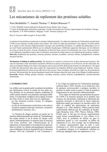
Le postulat <strong>de</strong> Levinthal peut aussi être illustré en<br />
termes <strong>de</strong> paysages énergétiques (Energy Landscape)<br />
par le concept <strong>de</strong> <strong>repliement</strong> en entonnoir (Folding<br />
F u n n e l) proposé par le groupe <strong>de</strong> Wolynes (Bryngelson<br />
e t a l ., 1995 ; Socci e t a l ., 1998) et repris plus récemment<br />
par Chan et Dill (1998). Dans ce concept, les auteurs<br />
décrivent le comportement thermodynamique et<br />
cinétique <strong>de</strong> la transformation d’un ensemble <strong>de</strong><br />
structures correpondant à l’état dénaturé d’une<br />
protéine vers son état natif unique (Figure 1). Tandis<br />
que le <strong>repliement</strong> <strong>de</strong> la structure progresse vers l’état<br />
natif, le nombre <strong>de</strong> conformations à explorer diminue.<br />
Le processus qui conduit la structure <strong>de</strong> l’état initial<br />
(structure aléatoire) vers l’état final <strong>de</strong> la protéine (état<br />
natif) est exergonique.<br />
Une gran<strong>de</strong> variété <strong>de</strong> comportements <strong>de</strong> <strong>repliement</strong><br />
émerge <strong>de</strong> ce paysage énergétique (Figure 1), elle<br />
dépend <strong>de</strong>s paramètres énergétiques et <strong>de</strong>s conditions<br />
expérimentales (Wolynes et al., 1995) : Wolynes et al.<br />
ont suggéré que la vitesse du <strong>repliement</strong> est diminuée<br />
Enat/4<br />
Enat/2<br />
E, Énergie<br />
Enat,<br />
(Énergie <strong>de</strong> la<br />
structure native<br />
Début <strong>de</strong> la formation <strong>de</strong> l’hélice<br />
et du collapsus<br />
Entropie<br />
États du<br />
globule fondu<br />
Structure-native<br />
Q = 0.27<br />
Q = 0.6<br />
Q = 0.71<br />
Intermédiaires <strong>de</strong><br />
<strong>repliement</strong> discrets<br />
Q, fraction <strong>de</strong>s<br />
contacts natifs<br />
retrouvés<br />
Région <strong>de</strong> l’état<br />
<strong>de</strong> transition<br />
(Q = 0.6)<br />
F i g u re 1 . Repliement en entonnoir schématique d’une<br />
protéine. La largeur <strong>de</strong> l’entonnoir représente l’entropie, la<br />
p r o f o n d e u r, l’énerg i e — “Folding funnel” of a pro t e i n .Wi d t h<br />
and <strong>de</strong>pth respectively represent entropy and energy.<br />
1.0
<strong>Les</strong> <strong>mécanismes</strong> <strong>de</strong> <strong>repliement</strong> <strong>de</strong>s <strong>protéines</strong> <strong>solubles</strong> 73<br />
par la présence <strong>de</strong> puits intermédiaires sur la surface<br />
énergétique. Chaque puits correspond à un minimum<br />
énergétique local peuplé par une population d’intermédiaires<br />
“stables”. <strong>Les</strong> auteurs décrivent que la <strong>de</strong>scente<br />
vers le fond <strong>de</strong> l’entonnoir s’accompagne d’une<br />
diminution <strong>de</strong> l’entropie <strong>de</strong> la chaîne polypeptidique.<br />
Plus la pente est rai<strong>de</strong>, plus le <strong>repliement</strong> sera rapi<strong>de</strong>.<br />
On admet à l’heure actuelle que le <strong>repliement</strong> <strong>de</strong> la<br />
structure 3-D <strong>de</strong> la plupart <strong>de</strong>s <strong>protéines</strong> est sous<br />
contrôle thermodynamique et que l’état natif est atteint<br />
grâce à la formation d’intermédiaires partiellement<br />
structurés dont la formation est sous contrôle cinétique<br />
(Sali et al., 1994a ; Ballew et al., 1996a).<br />
2. QUELS SONT LES DIFFÉRENTS MODÈLES<br />
DE CHEMINS DE REPLIEMENT ?<br />
On distingue schématiquement :<br />
– <strong>Les</strong> modèles construits à partir <strong>de</strong> considérations<br />
théoriques (Karplus, Weaver, 1976; Bryngelson,<br />
Wolynes, 1987; Karplus, Weaver, 1994; Dill et al.,<br />
1995; Karplus, Sali, 1995). Avec les nouvelles<br />
approches théoriques sur les <strong>mécanismes</strong> du<br />
<strong>repliement</strong>, expérimentateurs et théoriciens ont mis<br />
en commun leurs informations pour proposer <strong>de</strong>s<br />
modèles. <strong>Les</strong> théoriciens travaillent sur <strong>de</strong>s modèles<br />
avec <strong>de</strong>s fonctions <strong>de</strong> potentiels simples (Levitt,<br />
Warshel, 1975 ; Taketomi et al., 1988 ; Skolnick,<br />
Kolinski, 1990 ; Godzik e t a l ., 1992 ; Dagget,<br />
Levitt, 1993 ; Kolinski, Skolnick, 1994 ; Sali et al.,<br />
1994b). <strong>Les</strong> résultats obtenus sont prometteurs<br />
(pour une revue voir Pan<strong>de</strong> et al. (1998)).<br />
– <strong>Les</strong> modèles fondés sur <strong>de</strong>s observations expérimentales<br />
(Baldwin, 1975 ; Kim, Baldwin, 1982 ; Guo,<br />
Thirumalai, 1995 ; Daggett et al., 1996 ; Viguera<br />
et al., 1996 ; Fersht, 1997; Matagne, Dobson, 1998)<br />
sont proposés sur base d’étu<strong>de</strong>s menées en<br />
enzymologie, biochimie, biologie moléculaire et<br />
biophysique.<br />
2.1. Quels sont les modèles <strong>de</strong> <strong>repliement</strong> proposés<br />
dans la littérature ?<br />
Un certain nombre <strong>de</strong> modèles seront présentés<br />
brièvement afin <strong>de</strong> faire le point sur les différents<br />
<strong>mécanismes</strong> qui sont proposés :<br />
Le modèle <strong>de</strong> nucléation-con<strong>de</strong>nsation (Zimm, Bragg,<br />
1 9 5 9 ; Lifson, Roig, 1961 ; We t l a u f e r, 1973 ; Matheson,<br />
Scheraga, 1978 ; Fersht, 1995a ; Guo, Thirumalai,<br />
1997). Il inclut une étape <strong>de</strong> nucléation suivie par une<br />
propagation rapi<strong>de</strong> <strong>de</strong> la structure. L’étape limitante<br />
est le processus <strong>de</strong> nucléation. Ce modèle, sensé<br />
r e p r é s e n t e rles premiers résultats cinétiques, prend en<br />
compte la coopérativité du <strong>repliement</strong>.<br />
Le modèle <strong>de</strong> diff u s i o n - c o l l i s i o n, proposé par Karplus<br />
et Weaver (1976). La nucléation intervient simultanément<br />
en plusieurs points <strong>de</strong> la chaîne polypeptidique<br />
et génère <strong>de</strong>s structures qui diffusent puis coalescent<br />
jusqu’à former <strong>de</strong>s microstructures ayant une<br />
conformation native. Ces microstructures ont une durée<br />
<strong>de</strong> vie contrôlée par la diffusion, et le <strong>repliement</strong> d’une<br />
chaîne <strong>de</strong> 100 ou 200 aci<strong>de</strong>s aminés peut avoir lieu en<br />
moins d’une secon<strong>de</strong>. Selon ce modèle, le <strong>repliement</strong><br />
intervient en passant par plusieurs étapes <strong>de</strong> diffusioncollision.<br />
Le statut du modèle <strong>de</strong> diffusion-collision a<br />
été réévalué à la lumière <strong>de</strong> données expérimentales<br />
récentes (Karplus, Weaver, 1994).<br />
Le <strong>repliement</strong> séquentiel et hiérarchique. Plusieurs<br />
segments <strong>de</strong> structures sont formés et assemblés à diff ér<br />
e n t s niveaux suivant un chemin <strong>de</strong> <strong>repliement</strong> unique<br />
(Baldwin, 1975 ; Kim, Baldwin, 1982). Schulz (1977)<br />
a proposé une hiérarchie dans le <strong>repliement</strong> <strong>de</strong>s <strong>protéines</strong><br />
correspondant aux structures primaire – secondaire –<br />
t e r t i a i r e– et éventuellement quaternaire. Dans ce modèle,<br />
le premier évènement, la nucléation, est suivi par la<br />
formation <strong>de</strong>s structures secondaires qui s’associent<br />
pour former <strong>de</strong>s superstructures secondaires, puis <strong>de</strong>s<br />
domaines et éventuellement un monomère actif. L’ a s s ociation<br />
entre les sous-unités <strong>de</strong>s <strong>protéines</strong> oligomériques<br />
se ferait à l’étape finale du processus <strong>de</strong> <strong>repliement</strong>.<br />
Un modèle modulable <strong>de</strong> <strong>repliement</strong> a été proposé<br />
en considérant la structure 3-D <strong>de</strong>s <strong>protéines</strong>.<br />
Wetlaufer (1981) considère les domaines comme <strong>de</strong>s<br />
unités <strong>de</strong> <strong>repliement</strong> (Folding Units) ; il suggére que<br />
les sous-domaines pourraient se replier <strong>de</strong> façon<br />
indépendante en formant, comme intermédiaires <strong>de</strong><br />
<strong>repliement</strong>, <strong>de</strong>s modules structuraux qui s’assemblent<br />
pour donner la structure native (Chothia, 1984).<br />
Le modèle Framework (Ptitsyn, 1991) pose comme<br />
hypothèse que la structure secondaire est formée à une<br />
étape précoce du <strong>repliement</strong> (avant la structure 3-D).<br />
Le modèle d’effondrement (collapse) hydrophobe<br />
implique que le premier évènement du <strong>repliement</strong> <strong>de</strong>s<br />
<strong>protéines</strong> est un collapse hydrophobe ayant lieu avant<br />
la formation <strong>de</strong>s structures secondaires (Levitt,<br />
Warshel, 1975 ; Dill 1990). Ce collapse (Kauzmann,<br />
1959) conduirait au <strong>repliement</strong> <strong>de</strong>s <strong>protéines</strong> et à la<br />
stabilisation <strong>de</strong> la structure 3-D. L’enfouissement <strong>de</strong>s<br />
résidus hydrophobes peut avoir lieu dans les étapes<br />
précoces du <strong>repliement</strong>.<br />
Le modèle <strong>de</strong> la fermeture éclair h y d ro p h o b e<br />
(Hydrophobic Zipper), proposé par Dill et al. (1993)<br />
suggère que la formation <strong>de</strong> segments <strong>de</strong> structures<br />
secondaires est simultanée à la formation du collapse<br />
hydrophobe.
74 Biotechnol. Agron. Soc. Environ. 2000 4 (2), 71–81 N. Benhabilès, A. Thomas, R. Brasseur<br />
Le modèle du Jigsaw Puzzle (Harrison, Durbin,<br />
1 9 8 5 ) : le <strong>repliement</strong> est considéré comme un<br />
assemblage en puzzle avec existence <strong>de</strong> plusieurs<br />
chemins menant à une solution unique. <strong>Les</strong><br />
intermédiaires du lysozyme se réarrangeraient pour<br />
générer un globule fondu à partir duquel la protéine<br />
native se formerait en une ou plusieurs étapes<br />
séquentielles, en suivant le Jigsaw Puzzle. Ce modèle<br />
présente <strong>de</strong>s similarités avec celui <strong>de</strong> Karplus et<br />
Weaver (1994). L’i<strong>de</strong>ntification d’intermédiaires<br />
représente une <strong>de</strong>scription cinétique plutôt qu’une<br />
<strong>de</strong>scription structurale, chaque intermédiaire étant<br />
constitué d’espèces hétérogènes. Le modèle du Jigsaw<br />
P u z z l e apporte un éclairage nouveau car on a<br />
longtemps postulé (Baldwin, 1975 ; Kim, Baldwin,<br />
1982 ; Jaenicke 1987) que le <strong>repliement</strong> <strong>de</strong>s <strong>protéines</strong><br />
suivait un chemin unique en passant par la création<br />
séquentielle d’intermédiaires instables le long <strong>de</strong> ce<br />
chemin.<br />
Ces différents modèles ont été construits à partir <strong>de</strong><br />
résultats expérimentaux. Comme on peut le constater,<br />
il n’y a pas <strong>de</strong> consensus sur les modèles <strong>de</strong> <strong>repliement</strong><br />
proposés dans la littérature. On peut simplement dire<br />
qu’à la lumière <strong>de</strong> telle ou telle expérience, un modèle<br />
paraît plus crédible qu’un autre pour illustrer le ou les<br />
chemins <strong>de</strong> <strong>repliement</strong>.<br />
2.2. Que se passe-t-il pendant le <strong>repliement</strong> ?<br />
<strong>Les</strong> étu<strong>de</strong>s cinétiques ont permis <strong>de</strong> montrer l’existence<br />
<strong>de</strong> structures intermédiaires instables (Wetlaufer,<br />
R i s t o w, 1973 ; Creighton, 1984 ; Sancho, Fersht,<br />
1992 ; Weissman, Kim, 1992 ; Pecorari et al., 1993 ;<br />
Fersht, 1995c). Néanmoins, il faut se repérer dans un<br />
schéma à un ou plusieurs chemins <strong>de</strong> <strong>repliement</strong> et<br />
puis, comme ces espèces transitoires forment <strong>de</strong>s états<br />
très peu peuplés en équilibre rapi<strong>de</strong> avec d’autres<br />
populations structurales, il faut pouvoir obtenir <strong>de</strong>s<br />
informations sur la structure 3-D <strong>de</strong> ces intermédiaires.<br />
On a pu, pour quelques <strong>protéines</strong>, accumuler <strong>de</strong>s<br />
états transitoires du <strong>repliement</strong>, à bas pH ou par<br />
ingénierie génétique. Ces étu<strong>de</strong>s ont permis <strong>de</strong> proposer<br />
l’existence <strong>de</strong> plusieurs étapes pour le <strong>repliement</strong>. Une<br />
étape précoce <strong>de</strong> cinétique rapi<strong>de</strong> et une étape tardive<br />
<strong>de</strong> cinétique lente, responsable <strong>de</strong>s réarrangements<br />
structuraux.<br />
<strong>Les</strong> évènements précoces. <strong>Les</strong> métho<strong>de</strong>s expérimentales<br />
(Ballew et al., 1996a ; Williams et al., 1996)<br />
ont permis <strong>de</strong> poser <strong>de</strong>s hypothèses sur la façon dont<br />
pourraient se former les intermédiaires <strong>de</strong> <strong>repliement</strong>.<br />
De nombreuses métho<strong>de</strong>s <strong>de</strong> biophysique permettent<br />
d’obtenir <strong>de</strong>s informations sur les intermédiaires <strong>de</strong><br />
<strong>repliement</strong> (Tableau 1). On peut citer successivement<br />
les métho<strong>de</strong>s <strong>de</strong> cinétiques rapi<strong>de</strong>s (Nötling et al.,<br />
1995 ; Ballew et al., 1996b) (Stopped Flow et T-jump),<br />
le dichroïsme circulaire, la RMN (Bai et al., 1995 ;<br />
Shortle, 1996 ; Chamberlain, Marqusee, 1997; Clarke<br />
e t a l ., 1997) (associée à <strong>de</strong>s échanges rapi<strong>de</strong>s<br />
hydrogène-<strong>de</strong>utérium, par exemple).<br />
La formation d’intermédiaires repliés et flexibles a<br />
été proposée par Ptitsyn et Rashin (1973). Puis Ohgushi<br />
et Wada (1983) ont proposé le terme <strong>de</strong> “Molten<br />
G l o b u l e” ou globule fondu, pour introduire une structure<br />
ayant la majorité <strong>de</strong>s structures secondaires natives<br />
formées mais possédant un mauvais compactage, ou<br />
une structure 3-D fluctuante (Chaffotte et al., 1992).<br />
Le globule fondu contiendrait <strong>de</strong>s surfaces accessibles<br />
hydrophobes non natives. Ptitsyn et al. (1990) ont<br />
suggéré que le globule fondu puisse être un intermédiaire<br />
général du chemin <strong>de</strong> <strong>repliement</strong> <strong>de</strong>s <strong>protéines</strong>.<br />
Cet intermédiaire particulier du <strong>repliement</strong><br />
(Ptitsyn, 1995 ; Ro<strong>de</strong>r, 1995 ; Vidugiris et al., 1995 ;<br />
Ptitsyn, 1996) a été retrouvé dans le chemin <strong>de</strong><br />
<strong>repliement</strong> <strong>de</strong> plusieurs <strong>protéines</strong>, comme l’apomyoglobine<br />
(Vidugiris, Royer, 1998), l’α-lactalbumine<br />
(Kuwajima, 1989 ; 1996), l’anhydrase carbonique<br />
(Jagannadham, Balasubramanian, 1985), ou l’hormone<br />
<strong>de</strong> croissance bovine (Kuwajima, 1996).<br />
Certains auteurs proposent que les intermédiaires<br />
<strong>de</strong> <strong>repliement</strong>, globule fondu compris, soient en fait<br />
<strong>de</strong>s espèces piégées <strong>de</strong> façon cinétique et mal repliées.<br />
Cette théorie suggère que le <strong>repliement</strong> <strong>de</strong>s petites<br />
<strong>protéines</strong> intervient en continu, très rapi<strong>de</strong>ment et sans<br />
intermédiaires stables (Sosnick et al., 1994 ; Fersht,<br />
1995a ; Schindler et al., 1995).<br />
Le globule fondu est-il une espèce transitoire<br />
résultant d’une cinétique lente comme le suggèrent<br />
certains auteurs (Govindarajan, Goldstein, 1995) ou<br />
alors une espèce mal repliée résultant d’un mauvais<br />
chemin lors <strong>de</strong> la “phase cinétique” du <strong>repliement</strong>,<br />
comme le suggère Fersht (1995b)?<br />
D’autres intermédiaires, comme le pré-globule fondu,<br />
ont été décrits (Jeng, Englan<strong>de</strong>r, 1991). Cette espèce<br />
contient un taux significatif <strong>de</strong> structures secondaires<br />
fluctuantes, elle est moins compacte que le globule fondu<br />
et exhibe aussi <strong>de</strong>s régions hydrophobes accessibles au<br />
solvant. Elle a été observée pendant la renaturation au<br />
froid <strong>de</strong> la β-lactamase (Uversky, Ptitsyn, 1994) et <strong>de</strong><br />
l’anhydrase carbonique (Uversky, Ptitsyn, 1996).<br />
D’autre part, Fink et al. ont rapporté la présence <strong>de</strong> cet<br />
intermédiaire pour d’autres <strong>protéines</strong> (Fink, 1995).<br />
En plus du globule fondu et du pré-globule fondu,<br />
<strong>de</strong>s résultats expérimentaux montrent que la protéine<br />
se replie en donnant une population hétérogène<br />
d’intermédiaires partiellement repliés, en équilibre<br />
fluctuant (Plaxco, Dobson, 1996).<br />
Des espèces hétérogènes, comme <strong>de</strong>s populations<br />
multimériques (dimères, trimères), peuvent être observées<br />
pendant le <strong>repliement</strong> <strong>de</strong> certaines <strong>protéines</strong> [par<br />
exemple, la phosphoglycérate kinase (Pecorari et al.,
<strong>Les</strong> <strong>mécanismes</strong> <strong>de</strong> <strong>repliement</strong> <strong>de</strong>s <strong>protéines</strong> <strong>solubles</strong> 75<br />
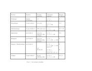
Tableau 1. Techniques biophysiques utilisées pour étudier le <strong>repliement</strong> <strong>de</strong>s <strong>protéines</strong> — Biophysical methods used in the<br />
study of the folding of proteins.<br />
Propriétés Techniques Résolution Mesures<br />
Compactage du Fluorescence < 1 ms Orientation et environnement <strong>de</strong> la chaîne latérale du<br />
cœur <strong>de</strong> la protéine intrinsèque tryptophane (Engelhard, Evans, 1996a)<br />
Absorption aux UV 1 ms Orientation et environnement <strong>de</strong> la chaîne latérale <strong>de</strong> la<br />
tyrosine (Udgaonkar, Baldwin, 1995)<br />
Fluorescence 1 ms Formation et interruption <strong>de</strong> segments hydrophobes<br />
extrinsèque (ANS) (Engelhard, Evans, 1996b)<br />
Extinction <strong>de</strong> 1 ms Localisation <strong>de</strong> la chaîne latérale du tryptophane<br />
fluorescence (Engelhard, Evans, 1996a)<br />
“Quenching Cysteinyl” 10 s Protection <strong>de</strong>s chaînes latérales <strong>de</strong>s cystéines<br />
(Ballery et al., 1993)<br />
Dimensions Anisotropie <strong>de</strong> 1 ms Mobilité <strong>de</strong> la chaîne du tryptophane et dimensions<br />
moléculaires fluorescence moléculaires globales (Jones et al., 1995)<br />
Transfert d’énergie 1 ms Distance scalaire entre un tryptophane et un fluorofore<br />
<strong>de</strong> fluorescence attachés covalemment (Rischel, Poulsen, 1995)<br />
Diffusion <strong>de</strong>s rayons X < 100 ms Rayon <strong>de</strong> giration moyen (Eliezer et al., 1995)<br />
aux petits angles<br />
Diffusion <strong>de</strong> la lumière 1 s Rayon <strong>de</strong> giration moyen (Feng, Widom, 1994)<br />
quasi élastique<br />
Structures secondaires Dichroïsme circulaire 1 ms Conformation du squelette, moyennée sur la séquence et<br />
et liaisons hydrogènes dans l’UV lointain la population étudiée (Evans, Radford, 1994)<br />
persistantes RMN en marquage 5–10 ms Formation <strong>de</strong> liaisons hydrogènes stables du squelette<br />
pulsé peptidique (Baldwin, 1993)<br />
Spectroscopie <strong>de</strong> masse 5–10 ms Formation <strong>de</strong> liaisons hydrogènes persistantes pour les<br />
en marquage pulsé intermédiaires du <strong>repliement</strong> (Miranker et al., 1996)<br />
Contacts tertiaires Activité biologique 1 ms–1 s Formation <strong>de</strong> la structure tertiaire native et du site actif<br />
et structure native (Evans, Radford, 1994)<br />
Repliement 10 ms Vitesse <strong>de</strong> dépliement <strong>de</strong>s intermédiaires discrets<br />
interrompu (Schreiber, Fersht, 1993)<br />
Dichroïsme circulaire 1 ms Formation <strong>de</strong> contacts tertiaires stables (entre<br />
en proche UV aromatiques, pont disulfures) (Evans, Radford, 1994)<br />
RMN en temps réel 1 s Formation <strong>de</strong> contacts tertiaires entre chaînes latérales<br />
spécifiques (Balbach et al., 1995)<br />
Ingénierie <strong>de</strong>s <strong>protéines</strong> Contribution énergétique <strong>de</strong>s chaînes latérales aux<br />
intermédiaires (Fersht, 1995c)<br />
1996)]. Ces populations, observées transitoirement,<br />
mènent la protéine monomérique native à l’étape lente<br />
du <strong>repliement</strong>.<br />
Il semble qu’au vu <strong>de</strong> ces nombreuses observations,<br />
on peut suggérer que l’étape précoce passe par<br />
la formation rapi<strong>de</strong> d’intermédiaires (transitoires) qui<br />
possè<strong>de</strong>nt un pourcentage élevé <strong>de</strong> structures secondaires<br />
et un faible taux <strong>de</strong> structures tertiaires (Dobson,<br />
1991). Le ou les chemins <strong>de</strong> <strong>repliement</strong> empruntés<br />
semblent dépendre <strong>de</strong>s <strong>protéines</strong> étudiées et <strong>de</strong>s<br />
conditions expérimentales. Néanmoins, il semble que<br />
le premier évènement du <strong>repliement</strong> soit un collapsus<br />
hydrophobe ayant lieu avant la formation <strong>de</strong>s structures<br />
secondaires (Dill, 1985). Le <strong>repliement</strong> serait initié par<br />
la con<strong>de</strong>nsation <strong>de</strong> la chaîne polypeptidique grâce aux<br />
interactions hydrophobes, puis cette étape serait suivie<br />
d’un réarrangement d’un petit nombre d’états con<strong>de</strong>nsés<br />
(Dill et al., 1993). De nombreuses données expérimentales<br />
montrent que le collapsus hydrophobe serait<br />
le mécanisme précurseur du <strong>repliement</strong> (Shortle,<br />
Abeygunawarda, 1993). Ensuite, les chemins <strong>de</strong><br />
<strong>repliement</strong> suivis pourraient être ceux du J i g s a w<br />
Puzzle, <strong>de</strong> la diffusion-collision, <strong>de</strong> la nucléation, ou<br />
<strong>de</strong> la nucléation-con<strong>de</strong>nsation, etc.
76 Biotechnol. Agron. Soc. Environ. 2000 4 (2), 71–81 N. Benhabilès, A. Thomas, R. Brasseur<br />
<strong>Les</strong> évènements tardifs du <strong>repliement</strong>. Ils se situent<br />
après la formation du globule fondu et avant la<br />
formation <strong>de</strong> la protéine native et fonctionnelle. Dans<br />
cette phase, on voit apparaître les sites <strong>de</strong> liaisons au<br />
substrat ou au ligand (sites <strong>de</strong> liaisons au Ca2+ <strong>de</strong> l’αlactalbumine<br />
(Kuwajima et al., 1989) par exemple),<br />
ou <strong>de</strong>s épitopes réactifs dans la sous-unité β <strong>de</strong> la<br />
tryptophane synthase (Blond-Elguindi, Goldberg ,<br />
1990). C’est dans cette étape limitante qu’apparaissent<br />
également l’activité enzymatique [structures II es et I I I<br />
bien agencées (Matousek e t a l ., 1990), compaction<br />
hydrophobe correcte (Lecomte, Matthews, 1993)],<br />
l’assemblage <strong>de</strong>s sous-unités dans les <strong>protéines</strong><br />
oligomériques (Ptitsyn, 1991) ou <strong>de</strong>s domaines dans<br />
les <strong>protéines</strong> multidomaines (Jaenicke, 1987), et le<br />
remaniement <strong>de</strong>s ponts disulfures (Levitt, Chothia,<br />
1976). Il faut noter que les ponts disulfures ne semblent<br />
jouer qu’un rôle stabilisateur <strong>de</strong> la structure 3-D, le<br />
<strong>repliement</strong> étant dirigé majoritairement par <strong>de</strong>s interactions<br />
non covalentes.<br />
La proline jouerait <strong>de</strong>s rôles particuliers au cours<br />
<strong>de</strong>s évènements tardifs du <strong>repliement</strong> (Brandts et al.,<br />
1975). Par exemple, certaines mutations <strong>de</strong> la proline<br />
ne semblent pas modifier le <strong>repliement</strong>, d’autres<br />
d i m i n u e r a i ent la vitesse, enfin, certaines pourraient le<br />
bloquer (Levitt, 1981).<br />
Que se passe-t-il pourles <strong>protéines</strong> multimériques ?<br />
<strong>Les</strong> domaines protéiques sont généralement considérés<br />
comme <strong>de</strong>s unités <strong>de</strong> <strong>repliement</strong> autonomes, <strong>de</strong> même<br />
que les sous-unités <strong>de</strong>s <strong>protéines</strong> oligomériques. Pour<br />
les <strong>protéines</strong> oligomériques, l’association entre sousunités<br />
a lieu lors <strong>de</strong> l’étape finale du <strong>repliement</strong>,<br />
induisant <strong>de</strong>s réajustements conformationnels pour<br />
générer les propriétés fonctionnelles <strong>de</strong> la protéine.<br />
Mais <strong>de</strong>s scénarios différents sont décrits selon les<br />
<strong>protéines</strong> (Baldwin, 1996b ; Creighton, 1994). Par<br />
exemple, les fragments C-terminaux <strong>de</strong> la thermolysine<br />
semblent se replier <strong>de</strong> façon autonome et coopérative<br />
pour un nombre d’hélices supérieur ou égal à trois<br />
(Vita et al., 1989), alors que les expériences menées<br />
sur les fragments du BPTI montrent que les sousdomaines<br />
se replient simultanément puis s’associent<br />
(Oas, Kim, 1988). En général les domaines isolés se<br />
replient plus vite que lorsqu’ils sont intégrés dans<br />
l’ensemble <strong>de</strong> la protéine (Missiakas et al., 1992).<br />
<strong>Les</strong> résultats théoriques et expérimentaux permettent<br />
<strong>de</strong> poser quelques principes <strong>de</strong> base sur le <strong>repliement</strong><br />
(Figure 2) qui peuvent être schématisés comme suit.<br />
1. Le <strong>repliement</strong> d’une chaîne polypeptidique paraît<br />
être largement déterminé par sa séquence, cela est en<br />
accord avec le postulat d’Anfinsen. Pour la plupart <strong>de</strong>s<br />
<strong>protéines</strong>, la structure native est sous contrôle thermodynamique<br />
(la structure native serait une structure<br />
dont l’énergie appartiendrait au bassin d’énerg i e<br />
e s<br />
Cinétiques<br />
RAPIDE<br />
LENTE<br />
Intermédiaire(s) ?<br />
Intermédiaire(s) ?<br />
CHAPERONS<br />
État dénaturé<br />
Collapse<br />
hydrophobe<br />
Globule fondu<br />
État natif<br />
F i g u re 2 . Scénario probable pour le <strong>repliement</strong> <strong>de</strong>s<br />
<strong>protéines</strong> — Probable scenario of the folding of proteins.<br />
minimum). L’existence <strong>de</strong> chemins <strong>de</strong> <strong>repliement</strong><br />
paraît être compatible avec le fait que les intermédiaires<br />
partiellement repliés sont sous contrôle cinétique. La<br />
chaîne polypeptidique semble explorer ses routes <strong>de</strong><br />
<strong>repliement</strong> vers la structure native, en passant par <strong>de</strong>s<br />
intermédiaires formant <strong>de</strong>s populations hétérogènes<br />
d’espèces partiellement repliées dont le nombre<br />
diminue au fur et à mesure que la protéine évolue vers<br />
la conformation présentant le minimum énergétique<br />
(Figure 1). Certaines espèces pourraient être piégées<br />
<strong>de</strong> façon transitoire dans <strong>de</strong>s minima locaux, ceci<br />
ralentissant le processus <strong>de</strong> <strong>repliement</strong>.<br />
2. Initialement, le collapsus hydrophobe semble générer<br />
une population hétérogène avec <strong>de</strong>s noyaux <strong>de</strong> structures<br />
natives et non natives en équilibre fluctuant, avec par<br />
exemple, <strong>de</strong>s réarrangements internes comme le proposent<br />
les modèles du Jigsaw Puzzle (Harrison, Durbin,<br />
1985) ou <strong>de</strong> diffusion-collision (Karplus, Weaver,<br />
1976). Puis, les noyaux repliés correctement peuvent<br />
ensuite s’associer pour générer <strong>de</strong>s microdomaines qui<br />
diffusent et s’associent. Un globule fondu peut se<br />
former.<br />
Ces trois évènements précoces ont lieu très rapi<strong>de</strong>ment.<br />
À partir du globule fondu démarre l’étape <strong>de</strong><br />
cinétique lente. Le processus <strong>de</strong> <strong>repliement</strong> suit <strong>de</strong>s voies<br />
plus contraintes : il y a formation <strong>de</strong>s structures tertiaires,<br />
appariement <strong>de</strong>s domaines, et les <strong>de</strong>rniers arrangements<br />
conformationnels se mettent en place. Remarquons que<br />
lors du <strong>repliement</strong>, il y a compétition cinétique (Yon,<br />
1996) entre un <strong>repliement</strong> correct et une agrégation.
<strong>Les</strong> <strong>mécanismes</strong> <strong>de</strong> <strong>repliement</strong> <strong>de</strong>s <strong>protéines</strong> <strong>solubles</strong> 77<br />
De telles situations peuvent avoir lieu aussi bien in<br />
vivo qu’in vitro (Mitraki et al., 1991). Par exemple,<br />
<strong>de</strong>s <strong>protéines</strong> étrangères surexprimées dans les cellules<br />
bactériennes peuvent former <strong>de</strong>s corps d’inclusions<br />
in<strong>solubles</strong>. Ou bien, le <strong>repliement</strong> anormal <strong>de</strong> certaines<br />
<strong>protéines</strong> peut donner <strong>de</strong>s plaques amyloï<strong>de</strong>s, responsables<br />
<strong>de</strong> maladies sévères (Perutz, 1996 ; Perutz,<br />
1997 ; Kelly, 1998).<br />
2.3. Chaperons et <strong>repliement</strong> <strong>de</strong>s <strong>protéines</strong> in vivo<br />
Certaines <strong>protéines</strong>, même celles dont les ponts<br />
disulfures ont été rompus par <strong>de</strong>s agents dénaturants, sont<br />
capables <strong>de</strong> se replier spontanément. Il a longtemps été<br />
postulé que le <strong>repliement</strong> d’une chaîne se faisait in<br />
vivo par les mêmes <strong>mécanismes</strong> que ceux démontrés<br />
in vitro. Cette vue a été modifiée par la découverte <strong>de</strong>s<br />
chaperons moléculaires (Ellis, 1992 ; Jaenicke, 1993 ;<br />
Buchner, 1996 ; Clarke, 1996 ; Ellis, 1996).<br />
<strong>Les</strong> chaperons, par leur association transitoire avec<br />
<strong>de</strong>s <strong>protéines</strong> naissantes ou déstabilisées par un s t r e s s ,<br />
sont capables d’empêcher un mauvais <strong>repliement</strong> ou<br />
une agrégation (association au chaperon par <strong>de</strong>s<br />
interactions hydrophobes). Par contre, elles ne semblent<br />
pas pouvoir interagir avec les <strong>protéines</strong> natives ou se<br />
lier à une chaîne totalement dépliée. Elles régulent la<br />
vitesse <strong>de</strong> <strong>repliement</strong> en agissant comme <strong>de</strong>s catalyseurs<br />
mais ne paraissent pas remettre en question le postulat<br />
d’Anfinsen selon lequel l’information nécessaire au<br />
<strong>repliement</strong> est contenue dans la séquence primaire. Il<br />
semble qu’in vivo peu <strong>de</strong> <strong>protéines</strong> nécessitent la<br />
présence <strong>de</strong> chaperons pour se replier correctement<br />
(Yon, 1997).<br />
D’autres molécules accessoires peuvent jouer un<br />
rôle dans le <strong>repliement</strong>, comme la protéine disulfi<strong>de</strong><br />
isomérase, la peptidyl proline cis-trans isomérase. Ces<br />
enzymes accélèrent ledit processus (Schmid, 1990 ;<br />
Lorimer, 1992) (qui peut aussi avoir lieu en leur<br />
absence i n v i t ro sous <strong>de</strong>s conditions précises). D’autres<br />
modifications, comme les glycosylations, ne semblent<br />
pas changer le chemin <strong>de</strong> <strong>repliement</strong>, ni in vivo, ni in<br />
vitro, par contre elles augmentent la stabilité <strong>de</strong> la<br />
protéine (DeKoster, Robertson, 1997) et modifient son<br />
adressage.<br />
Enfin, notons que l’étu<strong>de</strong> du comportement particulier<br />
<strong>de</strong>s <strong>protéines</strong> thermophiles pourrait apporter<br />
beaucoup à la compréhension <strong>de</strong>s <strong>mécanismes</strong> <strong>de</strong><br />
<strong>repliement</strong> et <strong>de</strong> stabilisation <strong>de</strong>s <strong>protéines</strong> (Petukhov<br />
et al., 1997; Vogt, Argos, 1997).<br />
<strong>Les</strong> informations tant expérimentales que théoriques<br />
sur le mécanisme <strong>de</strong> <strong>repliement</strong> <strong>de</strong>s <strong>protéines</strong> sont<br />
abondantes dans la littérature. Néanmoins aucun<br />
schéma consensus et prédictif n’existe en cette fin du<br />
2 0 e siècle. La compréhension <strong>de</strong> ce mécanisme<br />
fondamental sera certainement le centre d’évolution<br />
<strong>de</strong> la biotechnologie <strong>de</strong>s <strong>protéines</strong> au 21 e siècle.<br />
Remerciements<br />
Robert Brasseur est directeur <strong>de</strong> recherche au FNRS. Ce<br />
travail a été supporté par le “Interuniversity Poles of<br />
Attraction Programme – Belgian State, Prime Minister’s<br />
O ffice – Fe<strong>de</strong>ral Office for Scientific, Technical and<br />
Cultural Affairs” PAI contract n° P4/03. Le travail <strong>de</strong> Nora<br />
Benhabilès a été supporté par un grant FNRS Télévie.<br />
Bibliography<br />
A l e x a n d r o v NN., Go N. (1994). Biological meaning,<br />
statistical significance, and classification of local spatial<br />
similarities in nonhomologous proteins. Protein Sci. 3,<br />
p. 866–875.<br />
Anfinsen CB. (1973). Principles that govern the folding of<br />
protein chains. Science 181, p. 223–230.<br />
A n f i n s e n CB., Scheraga HA. (1975). Experimental and<br />
theoretical aspects of protein folding. Adv. Prot. Chem.<br />
29, p. 205–300.<br />
Anfinsen CB., Haber E., Sela M., White FH. (1961). The<br />
kinetics of formation of native ribonuclease during<br />
oxidation of the reduced polypepti<strong>de</strong> chain. Proc. Natl.<br />
Acad. Sci. USA. 47, p. 1309–1314.<br />
B a i Y., Sosnick TR., Mayne L., Englan<strong>de</strong>r S W. (1995).<br />
Protein folding intermediates: native-state hydrogen<br />
exchange. Science 259, p. 192–197.<br />
B a l b a c h J., Forg e V., Va n N u l a n d NAJ., Wi n d e r S L . ,<br />
H o r e PJ., Dobson CM. (1995). Following proteinfolding<br />
in real time using NMR-spectroscopy. Nat.<br />
Struct. Biol. 2, p. 865–870.<br />
B a l d w i n RL. (1975). Intermediates in protein folding<br />
reactions and the mechanism of protein folding. Annu.<br />
Rev. Biochem. 44, p. 453–475.<br />
B a l d w i n RL. (1993). Pulsed H/D exchange studies<br />
intermediates. Curr. Opin. Struct. Biol. 3, p. 84–91.<br />
Baldwin RL. (1996a). Why is protein folding so fast ? Proc.<br />
Natl. Acad. Sci. USA 93, p. 2627–2628.<br />
B a l d w i n RL. (1996b). On-pathway versus off - p a t h w a y<br />
folding intermediates. Folding Des. 1 (1), R1–8.<br />
B a l l e r y N., Desmadril M., Minard P., Yo n JM. (1993).<br />
Characterisation of an intermediate in the folding<br />
pathway of phosphoglycerate kinase: chemical reactivity<br />
of genetically introduced cysteinyl residues during the<br />
folding process. Biochemistry 33, p. 708–714.<br />
Ballew RM., Sabelko J., Gruebele M. (1996a). Observation<br />
of distinct nanosecond and microsecond protein folding<br />
events. Nat. Struct. Biol. 3, p. 923–926.<br />
B a l l e w RM., Sabelko J., Guebele M. (1996b). Direct<br />
observation of fast protein folding. Proc. Natl Acad. Sci.<br />
USA 93, p. 5759–5764.<br />
B l o n d - E l g u i n d i S., Goldberg ME. (1990). Kinetic<br />
characterization of early immunoreactive intermediates<br />
during the refolding of guanidine-unfol<strong>de</strong>d Escherichia<br />
coli tryptophan synthase β 2 subunits. Biochemistry 29,<br />
p. 2409–2417.
78 Biotechnol. Agron. Soc. Environ. 2000 4 (2), 71–81 N. Benhabilès, A. Thomas, R. Brasseur<br />
Blun<strong>de</strong>ll TL., Johnson MS. (1993). Catching a common<br />
fold. Protein Sci. 2, p. 877–883.<br />
B r a n d t s J F., Halvorson HR., Brennan M. (1975).<br />
Consi<strong>de</strong>ration of the possibility that the slow step in<br />
protein folding is due to cis-trans isomerism of proline<br />
residue. Biochemistry 14, p. 4953–4963.<br />
Bryngelson JD., Wolynes PG. (1987). Spin-glass and the<br />
statistical mechanics of protein folding. Proc. Natl.<br />
Acad. Sci. USA 84, p. 7524–7528.<br />
B r y n g e l s o n JD., Onuchic JN., Socci ND., Wo l y n e s P G .<br />
(1995). Funnels, pathways and the energy landscape of<br />
protein folding: a synthesis. Proteins: Struct. Funct.<br />
Genet. 21, p. 167–195.<br />
B u c h n e r J. (1996). Supervising the fold: functional<br />
principles of molecular chaperones. FASEB J. 1 0,<br />
p. 10–19.<br />
C a m a c h o CJ., T h i r u m a l a i D. (1993). Kinetics and<br />
thermodynamics of folding in mo<strong>de</strong>l proteins. Proc.<br />
Natl. Acad. Sci. USA 90, p. 6369–6372.<br />
C h a ff o t t e A F., Cadieux C., Guillou Y., Goldberg M E .<br />
(1992). A possible folding initial intermediate: the Cterminal<br />
proteolytic domain of tryptophan synthase β<br />
chain folds in less than 4 milliseconds into a con<strong>de</strong>nsed<br />
state with non-native-like secondary structure.<br />
Biochemistry 31, p. 4303–4308.<br />
C h a m b e r l a i n AK., Marqusee S. (1997). Touring the<br />
landscapes: Partially fol<strong>de</strong>d proteins examined by<br />
hydrogen exchange. Structures 5, p. 859–863.<br />
Chan HS., Dill KA. (1998). Protein folding in the landscape<br />
perspective: chevron plots and non-Arrhenius kinetics.<br />
Proteins: Struct. Funct. Genet. 30, p. 2–33.<br />
Chothia C. (1984). Principles that <strong>de</strong>termine the structure of<br />
proteins. Annu. Rev. Biochem. 53, p. 537–572.<br />
Chothia C. (1992). One thousand families for the molecular<br />
biologist. Nature 357, p. 543–544.<br />
C l a r k e AR. (1996). Molecular chaperones in protein folding<br />
and translocation. Curr. Opin. Struct. Biol. 6, p. 43–50.<br />
C l a r k e J., Itzhaki LS., Fersht AR. (1997). Hydrogen<br />
exchange at equilibrium: a short cut for analysing<br />
protein-folding pathways ? Trends Biochem. Sci. 22,<br />
p. 284–287.<br />
Creighton TE. (1984). Bisulfi<strong>de</strong> bond formation in proteins.<br />
Methods Enzymol. 107, p. 305–329.<br />
Creighton TE. (1994). The energetic ups and downs of<br />
protein folding. Nat. Struct. Biol. 1, p. 135–138.<br />
Dagget V., Levitt M. (1993). A mo<strong>de</strong>l of the molten globule<br />
state from molecular dynamics simulations. J. Mol.<br />
Biol. 232, p. 600–619.<br />
D a g g e t t V., Li AJ., Itzhaki LS., Otzen DE., Fersht A R .<br />
(1996). Structure of the transition-state for folding of a<br />
protein-<strong>de</strong>rived from experiment and simulation. J. Mol.<br />
Biol. 257, p. 430–440.<br />
DeKoster GT., Robertson AD. (1997). Thermodynamics of<br />
unfolding for Kazal-type serine protease inhibitors:<br />
entropic stabilization of ovomucoid first domain by<br />
glycosylation. Biochemistry 36, p. 2323–2331.<br />
D e l e u M., Wa t h e l e t B., Brasseur R., Paquot M. (1998).<br />
Aperçu <strong>de</strong>s techniques d’analyse conformationnelle <strong>de</strong>s<br />
macromolécules biologiques. Biotechnol. Agron. Soc.<br />
Environ. 2 (4), p. 234–247.<br />
Dill KA. (1985). Theory for the folding and stability of<br />
globular proteins. Biochemistry 24, p. 1501–1509.<br />
D i l l KA. (1990). Dominant forces in protein folding.<br />
Biochemistry 29, p. 7113–7155.<br />
Dill KA., Fiebig KM., Chan HS. (1993). Cooperativity of<br />
protein folding kinetics. Proc. Natl. Acad. Sci. USA 90,<br />
p. 1942–1946.<br />
D i l l KA., Bromberg S., Yu e K., Fiebig KM., Ye e D P. ,<br />
Thomas PD., Chan HS. (1995). Principles of protein<br />
folding: a perspective from simple extract mo<strong>de</strong>ls.<br />
Protein Sci. 4, p. 561–602.<br />
Dobson CM. (1991). Characterization of protein folding<br />
intermediates. Curr. Opin. Struct. Biol. 1, p. 22–27.<br />
E l i e z e r D., Jennings PA., Wr i g h t PE., Doniach S . ,<br />
Hodgson SK., Tsuruta H. (1995). The radius of gyration<br />
of an apomyoglobin folding intermediaite. Science 270,<br />
p. 487–488.<br />
E l l i s J. (1992). Protein folding. Cytosolic chaperonin<br />
confirmed. Nature 358, p. 191.<br />
Ellis RJ. (1996). The “bio” in biochemistry: protein folding<br />
insi<strong>de</strong> and outsi<strong>de</strong> the cell. Science 272, p. 1448–1449.<br />
E n g e l h a r dM., Evans PA. (1996a). Experimental investigation<br />
of si<strong>de</strong> chain interactions in early folding intermediates.<br />
Folding Des. 1, p. 31–37.<br />
Engelhard M., Evans PA. (1996b). Kinetics of interaction<br />
of partially fol<strong>de</strong>d proteins with a hydrophobic dye:<br />
evi<strong>de</strong>nce that molten globule character is maximal in early<br />
folding intermediates. Protein Sci. 4, p. 1553–1562.<br />
Evans PA., Radford SE. (1994). Probing the structure of<br />
folding intermediates. C u rr. Opin. Struct. Biol. 4,<br />
p. 100–106.<br />
Feng HP., Widom J. (1994). Kinetics of compaction during<br />
lysozyme refolding studied by continuous-flow<br />
quasielastic light scattering. B i o c h e m i s t ry 3 3,<br />
p. 13382–13390.<br />
F e r s h t AR. (1995a). Optimization of rates of protein<br />
folding: the nucleation-con<strong>de</strong>nsation mechanism and its<br />
implications. P roc. Natl. Acad. Sci. USA 9 2,<br />
p. 10869–10873.<br />
F e r s h t AR. (1995b). Characterizing transition states in<br />
protein folding: an essential step in the puzzle. Curr.<br />
Opin. Struct. Biol. 5, p. 79–84.<br />
F e r s h tAR. (1995c). Mapping the structure of transition states<br />
and intermediates in folding: <strong>de</strong>lineation of pathways at<br />
high resolution. Philos. Trans. R. Soc. London 438,<br />
p. 11–15.<br />
F e r s h t AR. (1997). Nucleation mechanisms in protein<br />
folding. Curr. Opin. Struct. Biol. 7, p. 3–9.<br />
Fink AL. (1995). Compact intermediate states in protein<br />
folding. Annu. Rev. Biophys. Biomol. Stru c t . 2 4,<br />
p. 495–522.<br />
F i n k e l s t e i n AV., Ptitsyn OB. (1987). Why do globular
<strong>Les</strong> <strong>mécanismes</strong> <strong>de</strong> <strong>repliement</strong> <strong>de</strong>s <strong>protéines</strong> <strong>solubles</strong> 79<br />
proteins fit the limited set of folding patterns ? Progr.<br />
Biophys. Mol. Biol. 50, p. 171–190.<br />
F r e e d m a n RB. (1992). Protein folding in the cell. I n<br />
T. Creyhton (ed). P rotein folding New York: W H<br />
Freeman p. 455–538.<br />
Godzik A., Skolnick J., Kolinski A. (1992). Simulations of<br />
the folding pathway of triose phosphate isomerase-type<br />
α/β barrel proteins. Proc. Natl. Acad. Sci. USA 89,<br />
p. 2629–2633.<br />
G o v i n d a r a j a n S., Goldstein RA. (1995). Optimal local<br />
propensities for mo<strong>de</strong>l proteins. Proteins Struct. Funct.<br />
Genet. 22, p. 413–418.<br />
Guo ZY., Thirumalai D. (1995). Kinetics of protein folding:<br />
nucleation mechanism, time scale, and pathways.<br />
Biopolymers 36, p. 83–102.<br />
G u o Z., T h i r u m a l a iD. (1996). Kinetics and thermodynamics<br />
of folding of a <strong>de</strong>-novo <strong>de</strong>signed 4-helix bundle protein.<br />
J. Mol. Biol. 263, p. 323–343.<br />
G u o Z., T h i r u m a l a i D. (1997). The nucleation-collapse<br />
mechanism in protein folding: evi<strong>de</strong>nce for the<br />
uniqueness of the folding nucleus. Folding Des. 2,<br />
p. 377–391.<br />
Harrison S., Durbin R. (1985). Is there a single pathway for<br />
the folding of a polypepti<strong>de</strong> chain ? Proc. Natl. Acad.<br />
Sci. USA 82, p. 4028–4030.<br />
Jaenicke R. (1987). Folding and association of proteins.<br />
Progr. Biophys. Mol. Biol. 49, p. 117–237.<br />
Jaenicke R. (1993). Role of accessory proteins in protein<br />
folding. Curr. Opin. Struct. Biol. 3, p. 104–112.<br />
J a g a n n a d h a m M V., Balasubramanian D. (1985). The molten<br />
globule intermediate form in the folding pathwayof<br />
human carbonic anhydrase. FEBS Lett. 188, p. 326–330.<br />
J e n g M F., Englan<strong>de</strong>r S W. (1991). Stable submolecular<br />
folding units in a non compact form of cytochrome c. J.<br />
Mol. Biol. 221, p. 215–228.<br />
Jones E., Beechem JM., Matthews CR. (1995). Local and<br />
global dynamics during the folding of E. c o l i<br />
dihydrofolate reductase by time-resolved fluorescence<br />
spectroscopy. Biochemistry 34, p. 1867–1877.<br />
Karplus M., Sali A. (1995). Theoretical studies of protein<br />
folding and unfolding. Curr. Opin. Struct. Biol. 5,<br />
p. 58–73.<br />
Karplus M., Weaver DL. (1976). Protein folding dynamics.<br />
Nature 260, p. 404–406.<br />
Karplus M., Weaver DL. (1994). Protein folding dynamics:<br />
the collision–diffusion mo<strong>de</strong>l and experimental data.<br />
Protein Sci. 3, p. 650–668.<br />
Karplus M., Sali A., Shakhnovich EI. (1995). Kinetics of<br />
protein folding. Nature 373, p. 664–665.<br />
K a t a o k aM., Goto Y. (1996). X-ray solution sacttering studies<br />
of protein folding. Folding Des. 1, p. R107–R114.<br />
Kauzmann W. (1959). Some factors in the interpretation of<br />
protein <strong>de</strong>naturation. Adv. Prot. Chem. 14, p. 1–64.<br />
Kim PS., Baldwin RL. (1982). Specific intermediates in the<br />
folding reactions of small proteins and the mechanism<br />
of protein folding. Annu. Rev. Biochem. 51, p. 459-489.<br />
K e l l y J W. (1998). The alternative conformations of<br />
amyloidogenic proteins and their multi-step assembly<br />
pathways. Curr. opin. Struct. Biol. 8, p. 101–106.<br />
K o l i n s k i A., Skolnick J. (1994). Monte Carlo simulations of<br />
protein folding, I – Lattice mo<strong>de</strong>l and interaction scheme.<br />
Proteins: Struct. Funct. Genet. 18, p. 338–352.<br />
Kuwajima K. (1989). The molten globule state as a clue for<br />
un<strong>de</strong>rstanding the folding and cooperativity of globular<br />
protein structure. Proteins: Struct. Funct. Genet. 6,<br />
p. 87–103.<br />
Kuwajima K. (1996). The molten state of α-lactalbumin.<br />
FASEB J. 10, p. 102–109.<br />
Kuwajima K., Mitani M., Sugai S. (1989). Characterization<br />
of the critical state in protein folding. Effects of<br />
guanidine hydrochlori<strong>de</strong> and specific calcium binding<br />
on the folding kinetics of α-lactalbumine. J. Mol. Biol.<br />
206, p. 547–561.<br />
L e c o m t e J T, Matthews CR. (1993). Unravelling the<br />
mechanism of protein folding: new tricks for an old<br />
problem. Prot. Eng. 6, p. 1–10.<br />
L e v i n t h a lC. (1968). Are there pathways for protein folding ?<br />
J. Chem. Phys. 65, p. 44–45.<br />
Levitt M. (1981). Effect of proline residues in protein<br />
folding. J. Mol. Biol. 145, p. 251–263.<br />
Levitt M., Chothia C. (1976). Structural patterns in globular<br />
proteins. Nature 261, p. 552–558.<br />
L e v i t t M., Wa r s h e l A. (1975). Computer simulation of<br />
protein folding. Nature 253, p. 693–698.<br />
L i f s o nS., RoigA. (1961). On the theory of helix-coil transition<br />
in polypepti<strong>de</strong>s. J. Chem. Phys. 34, p. 1963–1974.<br />
Lorimer GH. (1992). Role of accessory proteins in protein<br />
folding. Curr. Opin. Struct. Biol. 2, p. 26–34.<br />
Matagne A., Dobson CM. (1998). The folding process of<br />
hen lysozyme: a perspective from the “new view”. Cell.<br />
Mol. Life Sci. 54, p. 363–371.<br />
M a t h e s o n RR., Scheraga HA. (1978). A method for<br />
predicting nucleation sites for protein folding based on<br />
hydrophobic contacts. Macromolecules 11, p. 814–829.<br />
M a t o u s e k A., Kellis J T., Serrano L., Bycroft M., Fersht A R .<br />
(1990). Transient folding intermediates characterized by<br />
proteins engineering. Nature 346, p. 440–445.<br />
M i r a n k e r A., Robinson C V., Radford SE., Dobson C M .<br />
(1996). Investigation of protein folding by mass<br />
spectroscopy. FASEB J. 10, p. 93–101.<br />
Missiakas D., Betton JM., Chaffotte A., Yon JM. (1992).<br />
Kinetic studies of the refolding of yeast phosphoglycerate<br />
kinase: comparison with the isolated engineered<br />
domains. Protein Sci. 1, p. 1485–1493.<br />
M i t r a k i A., Fane B., Haase-Pettinger C., Sturtevant J . ,<br />
King J. (1991). Global suppression of protein folding<br />
<strong>de</strong>fects and inclusion body formation. Science 253,<br />
p. 54–58.<br />
Moore PB. (1997). Ribosomes: Protein synthesis in slow<br />
motion. Curr. Opin. Struct. Biol. 7, p. R179–R181.<br />
Nötling B., Golbik R., Fersht AR. (1995). Submillisecond<br />
events in protein folding. Proc. Natl. Acad. Sci. USA 92,
80 Biotechnol. Agron. Soc. Environ. 2000 4 (2), 71–81 N. Benhabilès, A. Thomas, R. Brasseur<br />
p. 10668–10672.<br />
Oas TG., Kim PS. (1988). A pepti<strong>de</strong> mo<strong>de</strong>l of a protein<br />
intermediate. Nature 336, p. 42–48.<br />
Ohgushi M., Wada AA. (1983). Molten globule state: a<br />
compact form of protein with mobile si<strong>de</strong> chains. FEBS<br />
Lett. 164, p. 21–24.<br />
O r e n g o CA., Jones D T., T h o r n t o n JM. (1994). Protein<br />
superfamilies and domain superfolds. N a t u re 3 7 2,<br />
p. 631–634.<br />
Pan<strong>de</strong> VS., Grosberg AY., Tanaka T., Rokhsar DS. (1998).<br />
Pathways for protein folding: is a new view nee<strong>de</strong>d ?<br />
Curr. Opin. Struct. Biol. 8, p. 68–79.<br />
P e c o r a r i F., Minard P., Desmadril M., Yo n JM. (1993).<br />
Structure and functional complementation of engineered<br />
fragments from yeast phosphoglycerate kinase. Protein<br />
Eng. 6, p. 313–325.<br />
P e c o r a r i F., Minard P., Desmadril M., Yo n JM. (1996).<br />
Occurrence of transient multimeric species during the<br />
refolding of a monomeric protein. J. Biol. Chem. 271,<br />
p. 5270–5276.<br />
Perutz MF. (1996). Glutamine repeats and inherited neuro<strong>de</strong>generative<br />
diseases: molecular aspects. Curr. Opin.<br />
Struct. Biol. 6, p. 848–858.<br />
P e r u t z M F. (1997). Amyloid fibrils. Mutations make enzyme<br />
polymerize. Nature 385, p. 773.<br />
P e t u k h o vM., KilY., Kuramitzu S., Lanzov V. (1997). Insight<br />
into thermal resistance of proteins from the intrinsic<br />
stability of their α-helices. P roteins Struct. Funct.<br />
Genet. 29, p. 309–320.<br />
P l a x c o K W., Dobson CM. (1996). Ti m e - r e l a x e d<br />
biophysical methods in the study of protein folding.<br />
Curr. Opin. Struct. Biol. 6, p. 630–636.<br />
Ptitsyn OB. (1991). How does protein synthesis give rise to<br />
the 3-D structure ? FEBS Lett. 285, p. 176–181.<br />
Ptitsyn OB. (1995). Structures of folding intermediates.<br />
Curr. Opin. Struct. Biol. 5, p. 74–78.<br />
Ptitsyn OB. (1996). How molten is the molten globule ?<br />
Nat. Struct. Biol. 3, p. 488–490.<br />
Ptitsyn OB., Finkelstein AV. (1980). Similarities of protein<br />
topologies: evolutionary divergence, functional convergence<br />
or principles of folding ? Q. Rev. Biophys. 13,<br />
p. 339–386.<br />
Ptitsyn OB., Rashin AA. (1973). Stagewise mechanism of<br />
protein folding. Dokl. Akad. Nauk. SSSR 2 1 3,<br />
p. 473–475.<br />
P t i t s y n OB., Pain RH., Semisotnov G V., Zerovnik E . ,<br />
Razgulyaev OI. (1990). Evi<strong>de</strong>nce for a molten globule<br />
state as a general intermediate in protein folding. FEBS<br />
Lett. 26, p. 20–24.<br />
Rischel C., Poulsen FM. (1995). Modification of a specific<br />
tyrosine enables tracing of the end -to-end distance<br />
during apomyoglobin folding. FEBS Lett. 3 7 4,<br />
p. 105–109<br />
Ro<strong>de</strong>r H. (1995). Watching protein folding unfold. Nat.<br />
Struct. Biol. 2, p. 817–820.<br />
Sali A., Shakhnovich EI., Karplus M. (1994a). Kinetics of<br />
protein folding: A lattice mo<strong>de</strong>l study of the<br />
requirements for folding to the native state. J. Mol. Biol.<br />
235, p. 1614–1636.<br />
Sali A., Shakhnovich EI., Karplus M. (1994b). How does a<br />
protein fold ? Nature 369, p. 248–251.<br />
Sancho J., Ferscht AR. (1992). Dissection of an enzyme by<br />
protein engineering. J. Mol. Biol. 224, p. 741–747.<br />
S c h i n d l e r TS., Herrier M., Marahiel MA., Schmid F X .<br />
(1995). Extremely rapid folding in the absence of<br />
intermediates. Nat. Struct. Biol. 2, p. 663–673.<br />
Schmid FX. (1990). Catalysis and assistance in protein<br />
folding. Curr. Opin. Struct. Biol. 1, p. 36–41<br />
Schreiber G., Fersht AR. (1993). The refolding of cis- and<br />
trans- peptidylprolyl isomers of bastar. Biochemistry 32,<br />
p. 11195–11203.<br />
Schulz GE. (1977). Structural rules for globular proteins.<br />
Angew. Chem. 16, p. 23–32.<br />
Shortle DR. (1996). Structural analysis of non-native states<br />
of proteins by NMR methods. Curr. Opin. Struct. Biol.<br />
6, p. 24–30.<br />
Shortle D., Abeygunawarda C. (1993). NMR analysis of the<br />
residual structure in the <strong>de</strong>natured state of an unusual<br />
mutant of staphylococcal nuclease. Curr. Opin. Struct.<br />
Biol. 1, p. 121–134.<br />
Skolnick J., Kolinski A. (1990). Simulations of the folding<br />
of a globular protein. Science 250, p. 1121–1125.<br />
S o c c i ND., Onuchic JN., Wo l y n e s PG. (1998). Protein<br />
folding mechanisms and the multidimensional folding<br />
funnel. Proteins Struct. Funct. Genet. 32, p. 136–158.<br />
S o s n i c kTR., Mayne L., Hiller R., Englan<strong>de</strong>r S W. (1994). T h e<br />
barriers in protein folding. Nat. Struct. Biol. 1, p. 1 4 9 – 1 5 6 .<br />
Taketomi H., Kano F., Gô N. (1988). The effect of amino<br />
acid substitutions on protein folding and unfolding<br />
transition studied by computer simulations. Biopolymer<br />
27, p. 527–560.<br />
Tanford C. (1970). Theoretical mo<strong>de</strong>ls for the mechanism<br />
of <strong>de</strong>naturation. Adv. Prot. Chem. 24, p. 1–95.<br />
Udgaonkar JB., Baldwin RL. (1995). Nature of the early<br />
intermediaites of ribonuclease A. B i o c h e m i s t ry 3 4,<br />
p. 4088–4096.<br />
Uversky VN., Ptitsyn O. (1994). Partly fol<strong>de</strong>d state, a new<br />
equilibrium state of protein molecules: four- s t a t e<br />
guanidium chlori<strong>de</strong>-induced unfolding of Β-lactamase<br />
at low temperature. Biochemistry 33, p. 2782–2791.<br />
Uversky VN., Ptitsyn O. (1996). Further evi<strong>de</strong>nce on the<br />
equilibrium “pre-molten globule state”: four state<br />
guanidium chlori<strong>de</strong>-induced unfolding of carbonic<br />
anhydrase B at low temperature. J. Mol. Biol. 255,<br />
p. 215–228.<br />
Vi d u g i r i s GJ., Royer CA. (1998). Determination of the<br />
volume changes for pressure-induced transitions of<br />
apomyoglobin between the native, molten globule, and<br />
unfol<strong>de</strong>d states. Biophys. J. 75, p. 463–470.<br />
Vidugiris GJA., Markley JL., Royer CA. (1995). Evi<strong>de</strong>nce<br />
for a molten globule-like transition state in protein<br />
folding from <strong>de</strong>termination of activation volumes.
<strong>Les</strong> <strong>mécanismes</strong> <strong>de</strong> <strong>repliement</strong> <strong>de</strong>s <strong>protéines</strong> <strong>solubles</strong> 81<br />
Biochemistry 34, p. 4909–4912.<br />
Viguera AR., Serrano L., Wilmanns M. (1996). Different<br />
folding transition-states may result in the same native<br />
structure. Nat. Struct. Biol. 3, p. 874-880.<br />
Vi t a C., Fontana A., Jaenicke R. (1989). Folding of<br />
thermolysin fragments: hydrodynamic properties of<br />
isolated domains and subdomains. Eur. J. Biochem. 183,<br />
p. 513–518.<br />
Vo g t G., A rg o s P. (1997). Protein thermal stability: hydrogen<br />
bonds or internal packing. Folding Des. 2, p. S40–S46.<br />
Wang Z. (1996). How many fold types of protein are in<br />
nature ? Proteins Struct. Func. Genet. 26, p. 186–191.<br />
We i s s m a n JA., Kim PS. (1992). Reexamination of the<br />
folding of BPTI: predominance of native intermediates.<br />
Science 253, p. 1386–1393.<br />
Wetlaufer D. (1973). Nucleation, rapid folding and globular<br />
intrachain regions in proteins. Proc. Natl. Acad. Sci.<br />
USA 70, p. 697–701.<br />
Wetlaufer DB. (1981). Folding of protein fragments. Adv.<br />
Protein Chem. 34, p. 61–92.<br />
Wetlaufer DB., Ristow S. (1973). Acquisition of the 3-D<br />
structure of proteins. Annu. Rev. Biochem. 4 2,<br />
p. 135–158.<br />
Wi l l i a m s S., Causgrove T P., Gilmanshin R., Fang K S . ,<br />
Callen<strong>de</strong>r RH., Woodruff WH., Dyer RB. (1996). Fast<br />
events in protein folding: helix melting and formation in<br />
a small pepti<strong>de</strong>. Biochemistry 35, p. 691–697.<br />
Wo l y n e sPG., Onuchic JN., T h i r u m a l a iD. (1995). Navigating<br />
the folding routes. Science 267, p. 1619–1620.<br />
Yon JM. (1996). Protein aggregation. In Meyers RA. (ed).<br />
Encyclopedia of molecular biology and molecular<br />
medicine V Weinheim VCH., Germany, p.73–93.<br />
Yon JM. (1997). Protein folding: concepts and perspectives.<br />
Cell. Mol. Life Sci. 53, p. 557–567.<br />
Z i m m BH., Bragg JK. (1959). Theory of the phase<br />
transition between helix and random coil polypepti<strong>de</strong><br />
chains. J. Chem. Phys. 31, p. 526–535.<br />
(140 réf.)