Université Louis Pasteur. Maîtrise de Biochimie BMS. Session de ...
Université Louis Pasteur. Maîtrise de Biochimie BMS. Session de ...
Université Louis Pasteur. Maîtrise de Biochimie BMS. Session de ...

Create successful ePaper yourself
Turn your PDF publications into a flip-book with our unique Google optimized e-Paper software.
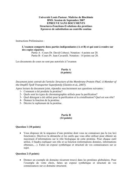
Instructions Préliminaires :<br />
<strong>Université</strong> <strong>Louis</strong> <strong>Pasteur</strong>. <strong>Maîtrise</strong> <strong>de</strong> <strong>Biochimie</strong><br />
<strong>BMS</strong>. <strong>Session</strong> <strong>de</strong> Septembre 2007<br />
EPREUVE SANS DOCUMENTS<br />
Structures-Fonctions-Evolutions <strong>de</strong>s protéines<br />
Epreuves <strong>de</strong> substitution au contrôle continu<br />
L’examen comporte <strong>de</strong>ux parties indépendantes (A et B) et qui sont à rendre sur<br />
<strong>de</strong>s copies séparées.<br />
Partie A : Cours Dr. David Cobessi. Notation : 6 points sur 20.<br />
Partie B : Cours Pr. Jean Cavarelli. Notation : 14 points sur 20.<br />
Les documents <strong>de</strong> cours ne sont pas autorisés à l’examen<br />
Partie A<br />
(6 points)<br />
Document joint: extrait <strong>de</strong> l’article: Structure of the Membrane Protein FhaC: A Member of<br />
the Omp85TpsB Transporter Superfamily (Clantin et al., 2007).<br />
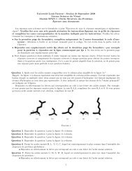
Apres lecture du document joint, répon<strong>de</strong>z succinctement aux questions suivantes :<br />
1- Comment a été produite la protéine?<br />
2- Quels sont les types <strong>de</strong> chromatographie utilisés pour la purification?<br />
3- Quel détergent a été utilisé pour la purification et la cristallisation? Quel est son rôle?<br />
4- Donnez la fonction <strong>de</strong> la protéine.<br />
5- Décrire le repliement <strong>de</strong> la protéine.<br />
Question 1 (10 points)<br />
Partie B<br />
(14 points)<br />
• Vous disposez <strong>de</strong> la séquence d’une protéine dont vous ne connaissez pas la (ou les)<br />
fonction(s). Décrivez la démarche et les outils que vous allez utiliser pour obtenir un<br />
maximum d’informations sur le rôle biologique <strong>de</strong> cette protéine. Pour chaque outil<br />
utilisé, il faudra expliquer son rôle et sa fonction (informations données, informations<br />
obtenues,…). Faites un exposé synthétique et structuré <strong>de</strong> vos connaissances sur ce<br />
sujet.<br />
Question 2 (5 points)<br />
• Donnez un exemple <strong>de</strong> domaine récurrent trouvé dans les protéines globulaires. Pour<br />
l’exemple <strong>de</strong> votre choix, faites un exposé synthétique et structuré <strong>de</strong> vos<br />
connaissances sur ce domaine structural.
Science 17 August 2007:<br />
Vol. 317. no. 5840, pp. 957 - 961<br />
DOI: 10.1126/science.1143860<br />
Reports<br />
Structure of the Membrane Protein FhaC: A Member of the Omp85TpsB<br />
Transporter Superfamily<br />
Bernard Clantin, 1,2,3 Anne-Sophie Delattre, 2,3,4 Prakash Rucktooa, 1,2,3 Nathalie Saint, 5,6<br />
Albano C. Méli, 5,6 Camille Locht, 2,3,4 Françoise Jacob-Dubuisson, 2,3,4* Vincent<br />
Villeret 1,2,3*<br />
In Gram-negative bacteria and eukaryotic organelles, ß-barrel proteins of the outer membrane<br />
protein 85–two-partner secretion B (Omp85-TpsB) superfamily are essential components of<br />
protein transport machineries. The TpsB transporter FhaC mediates the secretion of Bor<strong>de</strong>tella<br />
pertussis filamentous hemagglutinin (FHA). We report the 3.15 Å crystal structure of FhaC.<br />
The transporter comprises a 16-stran<strong>de</strong>d ß barrel that is occlu<strong>de</strong>d by an N-terminal helix and<br />
an extracellular loop and a periplasmic module composed of two aligned polypepti<strong>de</strong>transport–associated<br />
(POTRA) domains. Functional data reveal that FHA binds to the POTRA<br />
1 domain via its N-terminal domain and likely translocates the adhesin-repeated motifs in an<br />
exten<strong>de</strong>d hairpin conformation, with folding occurring at the cell surface. General features of<br />
the mechanism obtained here are likely to apply throughout the superfamily.<br />
1<br />
UMR8161 CNRS, Institut <strong>de</strong> Biologie <strong>de</strong> Lille, <strong>Université</strong> <strong>de</strong> Lille 1, <strong>Université</strong> <strong>de</strong> Lille 2, 1 rue du Prof.<br />
Calmette, F-59021 Lille ce<strong>de</strong>x, France.<br />
2<br />
Institut <strong>Pasteur</strong> <strong>de</strong> Lille, Lille, 1 rue du Prof. Calmette, F-59019 Lille ce<strong>de</strong>x, France.<br />
3<br />
IFR142, 59019 Lille, France.<br />
4<br />
INSERM, U629, 59019 Lille, France.<br />
5<br />
INSERM, U554, 34090 Montpellier, France.<br />
6<br />
UMR5048 CNRS, <strong>Université</strong> <strong>de</strong> Montpellier 1, <strong>Université</strong> <strong>de</strong> Montpellier 2, Montpellier, France.<br />
*<br />
To whom correspon<strong>de</strong>nce should be addressed. E-mail: francoise.jacob@ibl.fr (F.J.-D.);<br />
vincent.villeret@ibl.fr (V.V.)<br />
Targeting of proteins to their <strong>de</strong>dicated subcellular compartments is essential for cell function<br />
and organelle biogenesis. Translocation of proteins across or insertion into membranes is<br />
mediated by protein machineries, some of which have been conserved throughout evolution,<br />
such as the transporters of the Omp85-TpsB superfamily. TpsB transporters are components<br />
of two-partner secretion (TPS) systems in Gram-negative bacteria. They secrete large, mostly<br />
ß-helical proteins called TpsA proteins that generally serve as virulence factors (1, 2). TpsB<br />
transporters function without accessory factors. The superfamily also inclu<strong>de</strong>s the Toc75,<br />
Sam50-Tob55, and Omp85-YaeT homologs, which are the cores of large hetero-oligomeric<br />
complexes involved in protein transport across, and insertion of ß-barrel proteins into, the<br />
outer membranes of chloroplasts, mitochondria, and Gram-negative bacteria, respectively (3–<br />
9).<br />
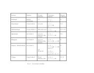
Omp85-TpsB transporters have been predicted to comprise a conserved C-terminal<br />
transmembrane ß barrel and a soluble N-terminal region harboring one to five putative<br />
polypepti<strong>de</strong>-transport-associated (POTRA) domains, which are hypothesized to mediate<br />
protein-protein interactions (10–12). The transporters also harbor conserved C-proximal<br />
signature motifs of unknown function in their pore-forming regions (13). In spite of their
implication in critical physiological processes such as membrane biogenesis and secretion of<br />
virulence proteins, the molecular mechanisms of protein translocation or insertion into<br />
membranes by those transporters remain poorly un<strong>de</strong>rstood. To address these issues, we<br />
<strong>de</strong>termined the crystal structure of the TpsB prototype FhaC that mediates the translocation to<br />
the bacterial surface of filamentous hemagglutinin (FHA), the major adhesin of the whooping<br />
cough agent Bor<strong>de</strong>tella pertussis.<br />
FhaC was crystallized in space group C2221, and the crystals contained one molecule in the<br />
asymmetric unit. The structure was solved by the single-wavelength anomalous diffusion<br />
(SAD) method (14) and is reported to a resolution of 3.15 Å (table S1 and fig. S1). The<br />
protein is a monomer and comprises a 35 Å high ß barrel composed of 16 antiparallel ß<br />
strands (B1 to B16) (Fig. 1A and fig. S2) with a shear number of 20. The ß barrel corresponds<br />
to the C-terminal moiety of the protein and encompasses residues 209 to 554. The periplasmic<br />
and extracellular si<strong>de</strong>s of the barrel are characterized by short turns and longer loops (L1 to<br />
L8), respectively, in general agreement with a prior topology mo<strong>de</strong>l (15). The N terminus of<br />
the protein is located in the extracellular milieu and folds into a 20-residue-long helix (H1)<br />
that goes right through the transmembrane ß barrel (Fig. 1, A and B). The C terminus of helix<br />
H1 emerges into the periplasm and is connected to a periplasmic module via a 30–amino acids<br />
linker that has no well-<strong>de</strong>fined electron <strong>de</strong>nsity in the crystal structure. This periplasmic<br />
module of 150 residues prece<strong>de</strong>s the ß barrel, a feature that had not been predicted earlier (15).<br />
Fig. 1. Crystal structure of FhaC. (A) Ribbon<br />
representation of FhaC viewed from the membrane<br />
plane. Putative position of the membrane (M) boundary<br />
is indicated with horizontal lines, with the extracellular<br />
si<strong>de</strong> (E) at the top and the periplasm (P) at the bottom.<br />
The helix H1 is colored red, POTRA 1 light blue,<br />
POTRA 2 purple, motif 3 green, and motif 4 blue. (B)<br />
Cutaway view of FhaC from the membrane plane,<br />
rotated about 90° relative to A. The helix H1, POTRA<br />
helices H2 and H4, and the loop L6 are indicated. The<br />
images were created with PyMOL (30). [View Larger<br />
Version of this Image (29K GIF file)]<br />
Production and purification of FhaC.<br />
The recombinant bacteria were grown at 37 °C in liquid LB broth to an absorbance of 1 (A600)<br />
and treated with 1 mM isopropyl-1-thio-β-D-galactosi<strong>de</strong> (final concentration) for 2 h. Cells<br />
were collected, washed in 20 mM sodium phosphate (pH 7), and resuspen<strong>de</strong>d in the same<br />
buffer containing 0.01 mg/ml DNase and a mixture of protease inhibitors (Roche Molecular<br />
Biochemicals, Meylan, France). Cells were broken by passages through a French pressure<br />
cell. After harvesting the membrane fractions by ultracentrifugation (100,000 x g for 1 h), two<br />
steps of extraction were performed successively with 0.8 and 1.5 % β-octyl glucosi<strong>de</strong>. The<br />
second extract was subjected to chromatography onto a cation-exchange column Poros HS20<br />
(Perkin-Elmer) equilibrated in 20 mM sodium phosphate (pH 7.0) with 1% β-octyl glucosi<strong>de</strong>.<br />
FhaC was eluted with a linear 0-1M gradient of NaCl. The FhaC-containing fractions were<br />
pooled and applied onto a 1-ml HiTrap chelating column (Amersham Biosciences)<br />
equilibrated in 20 mM sodium phosphate (pH 7.0), 1% β-octyl glucosi<strong>de</strong>. FhaC was eluted by<br />
a pulse of 400 mM imidazole (pH 6.5) in the equilibration buffer. For crystallization, FhaC
was concentrated to 40 mg/ml by using Vivaspin centrifugal <strong>de</strong>vices with a 50 kDa cut-off<br />
(Vivascience). Selenomethionine labeled FhaC (Se-Met FhaC) proteins were produced<br />
following the procedure <strong>de</strong>scribed (S5) and purified following the same protocol as for the<br />
native protein. For the secretion experiments, pAS-FcΔPot1, pAS-FcΔPot2 and pAS-FcΔL6<br />
were introduced into E. coli UT5600 together with compatible pFJD12, which enco<strong>de</strong>s the<br />
FHA <strong>de</strong>rivative Fha44, and secretion was assessed as <strong>de</strong>scribed (S3).<br />
Crystallization<br />
Native and Se-Met FhaC crystals were obtained at 20°C using the hanging drop vapor<br />
diffusion method. The protein and precipitant solutions were mixed in a 1:1 ratio. Crystals<br />
were grown at a<br />
protein concentration of 40 mg/ml for the native protein and 32 mg/ml for the<br />
<strong>de</strong>rivative in 32% PEG 1000, 1% β-octyl-glucosi<strong>de</strong>