Electrophysiologie du myocarde et du tissu nodal
Electrophysiologie du myocarde et du tissu nodal
Electrophysiologie du myocarde et du tissu nodal

You also want an ePaper? Increase the reach of your titles
YUMPU automatically turns print PDFs into web optimized ePapers that Google loves.
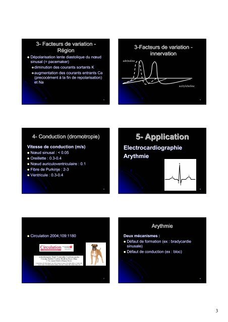
3- Facteurs de variation -<br />
Région<br />
Dépolarisation lente diastolique <strong>du</strong> nœud<br />
sinusal (= pacemaker)<br />
diminution des courants sortants K<br />
augmentation des courants entrants Ca<br />
(precocément<br />
precocément à la fin de repolarisation)<br />
repolarisation)<br />
<strong>et</strong> Na<br />
4- Con<strong>du</strong>ction (dromotropie<br />
( dromotropie)<br />
Vitesse de con<strong>du</strong>ction (m/s)<br />
Nœud sinusal : < 0.05<br />
Oreill<strong>et</strong>te : 0.3-0.4 0.3 0.4<br />
Nœud auriculoventriculaire : 0.1<br />
Fibre de Purkinje : 2-3 2<br />
Ventricule : 0.3-0.4 0.3 0.4<br />
Circulation 2004;109:1180<br />
13<br />
15<br />
17<br />
adrénaline<br />
3-Facteurs Facteurs de variation -<br />
innervation<br />
acétylcholine<br />
5- Application<br />
Electrocardiographie<br />
Arythmie<br />
Arythmie<br />
Deux mécanismes :<br />
Défaut de formation (ex : bradycardie<br />
sinusale)<br />
Défaut de con<strong>du</strong>ction (ex : bloc)<br />
14<br />
16<br />
18<br />
3