A. Ruol - Società Triveneta di Chirurgia
A. Ruol - Società Triveneta di Chirurgia
A. Ruol - Società Triveneta di Chirurgia

You also want an ePaper? Increase the reach of your titles
YUMPU automatically turns print PDFs into web optimized ePapers that Google loves.
IL CANCRO DEL CARDIAS<br />
Intervento <strong>di</strong> Ivor Lewis:<br />
come, quando e perche’<br />
Dr. A. <strong>Ruol</strong><br />
Responsabile UOS <strong>di</strong> <strong>Chirurgia</strong> delle prime vie <strong>di</strong>gestive<br />
Clinica Chirurgica 1°<br />
Azienda Ospedaliera – Universita’ <strong>di</strong> Padova<br />
Centro <strong>di</strong> Alta Specializzazione per le Malattie dell’Esofago<br />
Direttore: Prof. E. Ancona
Esophageal<br />
and<br />
Esophagogastric<br />
Junction<br />
Cancers<br />
v. 2.2011<br />
Esophagogastric Junction cancers<br />
TNM - 7 th e<strong>di</strong>tion, 2010<br />
• A tumour the epicentre of which<br />
is within 5 cm of the<br />
esophagogastric junction and<br />
also extends into the oesophagus<br />
is classified and staged<br />
accor<strong>di</strong>ng to the esophageal<br />
cancer scheme<br />
• All other tumours with an<br />
epicentre in the stomach greater<br />
than 5 cm from the EG-J or<br />
those within 5 cm of the EG-J<br />
without extension into the<br />
oesophagus are staged using the<br />
gastric carcinoma scheme
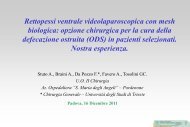
Classification for adenocarcinoma at the esophago-gastric junction<br />
Siewert 1996, 2000<br />
Type I. Adenocarcinoma of the <strong>di</strong>stal<br />
esophagus which may infiltrate the E-G<br />
junction from above & mostly develops<br />
in Barrett’s esophagus<br />
Type II. True carcinoma of the<br />
car<strong>di</strong>a, arising at the E-G<br />
junction<br />
Type III. Subcar<strong>di</strong>al gastric<br />
carcinoma which infiltrates the E-G<br />
junction from below
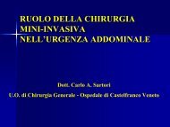
Adenocarcinoma of the esophagus &<br />
esophago-gastric junction<br />
• Type I<br />
• Type II<br />
?<br />
• Type III<br />
transthoracic esophago-gastric resection<br />
& gastric pull-up (above azygos vein)<br />
transthoracic esophago-gastric resection<br />
& gastric pull-up (above azygos vein)<br />
total gastrectomy (D2) + res. lower esophagus<br />
& Roux-en-Y esophago-jejunostomy<br />
limited resection for early cancer :<br />
short esophageal resection + proximal gastrectomy<br />
& Meren<strong>di</strong>no jejunal interposition<br />
total gastrectomy (D2)<br />
& Roux-en-Y esophago-jejunostomy
Is there a “standard of care” operation<br />
for esophageal cancer ? Kaiser, Ann Surg 2001<br />
There is no single right or ideal operation<br />
for every patient with esophageal cancer<br />
It is important to recognize that the results achieved by<br />
surgeons who ... have a large clinical volume may not<br />
be achievable by the occasional esophageal surgeon<br />
We need to continue to work toward<br />
reducing the rate of perioperative complications<br />
associated with esophagectomy and<br />
defining the variables that relate to long-term survival
The impact of complications on outcomes after resection for esophageal<br />
and gastroesophageal junction carcinoma Rizk, J Am Coll Surg 2004<br />
510 consecutive patients operated between 1996 and 2001<br />
at the Memorial Sloan-Kettering Cancer Center<br />
pts. Postop. Mortality 3-year Survival<br />
Surgical<br />
Complications<br />
138 12,3 % 31 %<br />
NO Surgical<br />
Complications 372 3,8 % 48 %<br />
Technical complications have a large negative impact on survival after<br />
esophagectomy for cancer (HR 1,41 p = 0,008)<br />
Strategies to optimize surgical technique & minimize complications<br />
improve outcomes
AdenoCa of the esophagus and E-G junction<br />
Achieving R0 complete resection should be the goal<br />
of surgery (it is the most significant independent prognostic factor)<br />
• resection margins free of tumor<br />
• adequate lymph node <strong>di</strong>ssection<br />
Strong in<strong>di</strong>vidual preference & some degree of surgical<br />
mystique often govern selection of operation for GE junction<br />
adenocarcinoma ( Rusch 2004 )<br />
The optimal surgical strategy remains controversial
AdenoCa della giunzione esofago-gastrica<br />
problemi ancora controversi relativi all’intervento chirurgico<br />
• via <strong>di</strong> accesso<br />
• volume <strong>di</strong> resezione esofagea<br />
• volume <strong>di</strong> resezione gastrica<br />
• estensione della linfoadenectomia<br />
The type of operation selected may affect :<br />
• the ability to achieve a complete (R0) resection,<br />
• the quality of lymph node clearance and staging,<br />
• the long-term chance of local control and survival,<br />
• patient’s quality of life
AdenoCa of the esophagus & esophago-gastric junction<br />
problemi ancora controversi relativi all’ intervento chirurgico:<br />
• volume <strong>di</strong> resezione esofagea :<br />
– almeno 8-10 cm <strong>di</strong> esofago indenne a monte del tumore,<br />
per via toracotomica dx (oppure sx)<br />
– tutta la mucosa con metaplasia intestinale (Barrett)<br />
– ? esofagectomia totale a torace chiuso<br />
– ? resezione esofagea <strong>di</strong>stale per via transiatale ( sec. Pinotti )<br />
– ??? resezione esofago per via addominale esclusiva
Adenocarcinoma of the E-G junction<br />
Microscopic evidence of cancer (R1)<br />
at a resection margin (on prefixed fresh specimen)<br />
cm proximal margin length <strong>di</strong>stal margin length<br />
< 2 14/30 (47%) 3/ 8 (37.5%)<br />
2 -3.9 1/ 9 (11%) 2/17 (12%)<br />
4 - 5.9 2/ 8 (25%) 0/37 (0%)<br />
> 6 0/24 (0%)<br />
Total 23% 6%<br />
Ito 2004
Centro <strong>di</strong> Alta Specializzazione<br />
per le Malattie dell’ Esofago<br />
University of Padova, Italy<br />
AdenoCa giunzione esofago-gastrica Tipo II<br />
1980-2003: 267 resecati<br />
trancia <strong>di</strong> sezione prossimale positiva per tumore<br />
Livello anastomosi<br />
Trancia sup. positiva<br />
esofago cervicale 1/34 2.9%<br />
apice torace 1/28 3.6%<br />
sopra arco v. azigos 0/56 0%<br />
arco v. azigos 1/50 2%<br />
sotto arco v. azigos 4/35 11%<br />
vena polmonare inf. 2/35 5.7%<br />
sotto vena polmonare inf. 1/22 4.5%<br />
3/168 = 1.8%<br />
10 / 260<br />
= 3.8 %<br />
7/92 = 7.6%
Adenocarcinoma of the E-G junction<br />
Residual cancer at the resection margin<br />
2.5 - 35% microscopic evidence of cancer (i.e. R1)<br />
Papachristou 1980, Mandard 1981, Sons 1986, Husemann 1989, Peracchia 1991, Stipa 1992, Bozzetti 1982 e 2000, Fekete 1997,<br />
Kodera 1999, Guillem 1999, Mattioli 2001, Mariette 2003, Ito 2004<br />
– palpation & gross inspection: unreliable<br />
– intraoperative frozen section: 9-21% false negative rates<br />
proximal resection margin length: at least 6-8 cm (on prefixed fresh<br />
resected specimen *) Papachristou 1980, Suzuki 1990, Cor<strong>di</strong>ano 1996, Mattioli 2001, Mariette 2003, Ito 2004<br />
<strong>di</strong>stal resection margin length:<br />
at least 4 cm (on prefixed fresh<br />
resected specimen *) Siewert 2000, Mattioli 2001, Ito 2004<br />
* prefixed fresh specimen measures 44-55% of the in situ length before resection
Centro <strong>di</strong> Alta Specializzazione<br />
per le Malattie dell’ Esofago<br />
University of Padova, Italy<br />
Paziente con adenoCa del car<strong>di</strong>as <strong>di</strong> tipo Siewert II,<br />
al quale era stato proposto in altra sede<br />
l’intervento per sola via addominale<br />
Tumore car<strong>di</strong>ale<br />
Seconda<br />
localizzazione<br />
(da <strong>di</strong>ffusione linfatica<br />
intraparietale ?)
Centro <strong>di</strong> Alta Specializzazione<br />
per le Malattie dell’ Esofago<br />
University of Padova, Italy<br />
ESOPHAGEAL<br />
SECTION MARGIN<br />
with microscopic tumor nests in the submucosa<br />
ANASTOMOTIC<br />
RECURRENCE<br />
the safety resection margin proximal to the tumor (as measured<br />
in vivo) should be at least 6 cm long, but preferably 8 cm long
Centro <strong>di</strong> Alta Specializzazione<br />
per le Malattie dell’ Esofago<br />
University of Padova, Italy<br />
Take down & Colon interposition
ISDE Consensus Conference 1995<br />
Carcinoma of the thoracic esophagus and EG-junction<br />
the role of lymph node <strong>di</strong>ssection<br />
• improves the accuracy of pathologic staging<br />
• reduces local-regional recurrences<br />
• the need for & extent of lymphadenectomy is debated:<br />
possible improvement of long-term prognosis, but<br />
no randomized study has shown a survival advantage related to a<br />
more extensive lymph node <strong>di</strong>ssection<br />
number of lymph nodes to be examined<br />
in the operative specimen = at least 15
AdenoCa della giunzione esofago-gastrica<br />
problema ancora controverso<br />
• linfoadenectomia: almeno 15 linfono<strong>di</strong><br />
– LN paracar<strong>di</strong>ali, piccola curva gastrica, tripode celiaco, origine<br />
art. epatica e splenica, periesofagei me<strong>di</strong> e inferiori, sottocarenali<br />
= linfoadenectomia me<strong>di</strong>astino inferiore + addominale D2<br />
– linfoadenectomia D0 linfoadenectomia D1 = solo LN peritumorali<br />
– linfoadenectomia D3 o D4 = D2 + LN paraortici, interaortocavali,<br />
retropancreatici + LN me<strong>di</strong>astinici superiori, recurrenziali, sovraclaveari
Centro <strong>di</strong> Alta Specializzazione<br />
per le Malattie dell’ Esofago<br />
University of Padova, Italy<br />
v. porta art. epatica moncone art. gastrica sx<br />
art. splenica
Centro <strong>di</strong> Alta Specializzazione<br />
per le Malattie dell’ Esofago<br />
University of Padova, Italy<br />
arco v. azigos bronco sx bronco dx pericar<strong>di</strong>o esofago
Differential pathologic variables and outcomes across the<br />
spectrum of adenocarcinoma of the esophagogastric junction<br />
Reynolds, World J Surg 2010;34:2821–2829<br />
AEG type I = 180, type II = 182, type III = 158<br />
Factor<br />
Multivariate analysis for survival<br />
Hazard<br />
ratio<br />
95% CI p Value<br />
AEG type I vs. II vs. III 0.976 0.911, 1.008 0.367<br />
Node status 3.578 1.678, 3.987 < 0.0001<br />
No. involved nodes 2.367 1.076, 2.987 < 0.0001<br />
pT 1.099 1.001, 1.189 0.079
Prevalence of LN metastases in Distal Esophagus (DE)<br />
and Gastro-Esophageal Junction (GEJ) cancers<br />
Leers, DeMeester et al. J Thor Car<strong>di</strong>ovasc Surg 2009;138:594-<br />
Lymph node status DE tumors (n = 301) GEJ tumors (n = 208) P<br />
• N classification<br />
N + 150 ( 49.8% ) 118 ( 56.7% ) .18<br />
• No. of involved nodes<br />
1-4 66 (21.9%) 50 (22.1%) .74<br />
5-8 24 (8.0%) 22 (11.1%)<br />
> 8 61 (20.3%) 42 (21.1%)<br />
*Depth of invasion & N+ DE tumors (n = 245) GEJ tumors (n = 172) P<br />
Intramucosal 2/71 ( 2.8%) 1/36 ( 2.8%) 1.0<br />
Submucosal 11/38 (28.9%) 6/29 (20.7%) .59<br />
Intramural 15/30 (50.0%) 8/16 (50.0%) 1.0<br />
Transmural 96/106 (90.6%) 79/91 (86.8%) .92<br />
( * patients with neoadjuvant therapy excluded )
Prevalence and location of node metastases in patients who<br />
had en-bloc resection (patients not having en-bloc resection excluded)<br />
Leers, DeMeester et al. J Thor Car<strong>di</strong>ovasc Surg 2009;138:594-<br />
DE tumors (n = 150) GEJ tumors (n = 100) P<br />
•Prevalence of N1 <strong>di</strong>sease 83 (55%) 61 (61%)<br />
.43<br />
•Location of positive LN<br />
Me<strong>di</strong>astinal: 39 ( 26% ) 25 ( 25% ) .88<br />
Paratracheal 3 (2%) 0<br />
Subcarinal 12 (8%) 3 (3%)<br />
Paraesophageal 38 (25%) 25 (25%)<br />
Abdominal: 70 (47%) 52 (52%) .44<br />
Parahiatal 33 (22%) 20 (20%)<br />
Perigastric 52 (35%) 45 (45%)<br />
Celiac 9 (6%) 3 (3%)<br />
Other 13 (9%) 8 (8%)
Prevalence and location of node metastases in patients who<br />
had en-bloc resection (patients not having en-bloc resection excluded)<br />
Leers, DeMeester et al. J Thor Car<strong>di</strong>ovasc Surg 2009;138:594-<br />
Lymph node status DE tumors (n = 150) GEJ tumors (n = 100) P<br />
Prevalence of N1 <strong>di</strong>sease 83 (55%) 61 (61%) .43<br />
Location of positive LN:<br />
Me<strong>di</strong>astinal 39 ( 26% ) 25 ( 25%) .88<br />
Abdominal 70 (47%) 52 (52%) .44<br />
• Positive me<strong>di</strong>astinal LN<br />
in pts with N+<br />
• Positive me<strong>di</strong>astinal LN<br />
as the only site of N+<br />
47% 41% n.s.<br />
9% 8% n.s.
The pattern of lymph node involvement in patients<br />
with N1 <strong>di</strong>sease after en bloc esophagectomy (n = 144)<br />
with <strong>di</strong>stal esophageal (DE) and gastroesophageal junction (GEJ) tumors<br />
Leers, DeMeester et al. J Thor Car<strong>di</strong>ovasc Surg 2009;138:594-<br />
DE<br />
GEJ<br />
In 9% of pts.<br />
with DE tumors,<br />
a positive<br />
me<strong>di</strong>astinal LN<br />
was the only<br />
site of LN<br />
involvement<br />
In 8% of pts.<br />
with GEJ<br />
tumors,<br />
a positive<br />
me<strong>di</strong>astinal LN<br />
was the only<br />
site of LN<br />
involvement
Lymph node metastasis in adenocarcinoma of the<br />
gastroesophageal junction (GEJ) and <strong>di</strong>stal esophagus (DE)<br />
Leers, DeMeester et al. J Thor Car<strong>di</strong>ovasc Surg 2009;138:594-<br />
a lower me<strong>di</strong>astinal node <strong>di</strong>ssection needs to be<br />
included in the surgical therapy of both tumors<br />
• the danger with recommen<strong>di</strong>ng total gastrectomy for GEJ cancer<br />
is the tendency (especially among low-volume centers) to minimize<br />
the me<strong>di</strong>astinal <strong>di</strong>ssection<br />
inadequate me<strong>di</strong>astinal lymph node <strong>di</strong>ssection<br />
increased frequency of a positive proximal resection margin
Location of nodal metastases accor<strong>di</strong>ng to Siewert<br />
classification for the 111 patients with pN+ <strong>di</strong>sease<br />
Pedrazzani, DeManzoni 2007<br />
Site of<br />
nodal metastasis<br />
Abdomen<br />
(n = 87)<br />
Abdomen and chest<br />
(n = 23)<br />
Chest<br />
(n = 1)<br />
Type I<br />
(n = 13)<br />
Siewert classification<br />
Type II<br />
(n = 44)<br />
Type III<br />
(n = 54)<br />
7 (53.8%) 31 (70.5%) 49 (90.7%)<br />
6 (46.2%) 13 (29.5%) 4 (7.4%)<br />
— — 1 (1.9%)
Adenocarcinoma of the E-G junction<br />
Frequency of metastasis in me<strong>di</strong>astinal lymph nodes<br />
Peracchia 1987 Type II 20%<br />
Aikou 1989 Type II 12%<br />
Wang 1993 Type II 18%<br />
Clark 1994 Type I-II 28%<br />
Tachimori 1996 Type II 19%<br />
Nigro 1999 Type II 33%<br />
Mauvais 2000 Type II 19%<br />
Siewert 2000 Type II 16%<br />
Mattioli 2001 Type II 7%<br />
Altorki 2002 Type I-II 32%<br />
Monig 2002 Type II 11%<br />
Nakamura 2002 Type II 3.6%<br />
Ichikura 2003 Type II 14%<br />
Lerut 2004 Type II 22%<br />
Ancona- <strong>Ruol</strong> 2005 Type II 20.5%<br />
Leers-DeMeester 2009 Type II 25%<br />
7 - 33%
Total gastrectomy is not always necessary<br />
for advanced Type II cancer of the car<strong>di</strong>a<br />
LN # 4d : along right gastroepiploic vessels<br />
LN # 5 : suprapyloric<br />
LN # 6 : infrapyloric<br />
% metastatic LN<br />
in Type II cancer<br />
of the EG-J:<br />
LN # 4d 5%<br />
LN # 5 5%<br />
LN # 6 0%<br />
(Kobayashi, 2002<br />
Ichikura, 2003)
Type II-III adenocarcinoma of the E-G junction<br />
• Total gastrectomy + <strong>di</strong>stal esophagectomy<br />
+ D2 LN-<strong>di</strong>ssection (88 patients)<br />
- frequency of metastasis to LN # 4d , 5 , 6 : all < 6%<br />
- no long-term survivor among patients with these metastases<br />
• Proximal gastrectomy + subtotal esophagectomy<br />
+ D2 LN-<strong>di</strong>ssection, exclu<strong>di</strong>ng LN # 4d, 5, 6 (89 patients)<br />
- same survival curves for total gastrectomy and proximal<br />
gastrectomy ( p = 0.3 for pT1; p = 0.7 for pT2-4 )<br />
Kodera, 1999
Cancer of the esophagus and EG-junction<br />
Sentinel node mapping ( 99m TC-ra<strong>di</strong>oguided or blue dye technique)<br />
• Sentinel node mapping theoretically could allow the extent of<br />
lymphadenectomy to be tailored to the in<strong>di</strong>vidual patient<br />
• High false-negative rate, especially after neoadjuvant treatments<br />
• Skip metastases can lead to positive <strong>di</strong>stant LN, despite of negative SLNs<br />
• High rate of positive sentinel nodes in >1 nodal stations<br />
• High rate of metastases also in non-sentinel nodes<br />
still experimental and requires validation,<br />
inclu<strong>di</strong>ng the need of a method for intraoperative<br />
examination with rapid immunohistochemisty
Adenoca della giunzione esofago-gastrica <strong>di</strong> Tipo II<br />
Conclusioni<br />
l’intervento <strong>di</strong> riferimento e’<br />
• la resezione esofago-gastrica<br />
• con linfoadenectomia<br />
del me<strong>di</strong>astino me<strong>di</strong>o-inferiore e addominale D2<br />
• e’ consigliabile che l’intervento venga eseguito<br />
in Centri ad alto volume da chirurghi esperti in<br />
questo tipo <strong>di</strong> chirurgia
Centro <strong>di</strong> Alta Specializzazione<br />
per le Malattie dell’ Esofago<br />
University of Padova, Italy<br />
• Neoadjuvant therapy decreases the frequency of regional lymph<br />
node metastases, and also changes their location<br />
• The map of the <strong>di</strong>stribution of nodal metastasis after neoadjuvant<br />
therapy might be useful to plan the operative technique and<br />
adequate lymphadenectomy
Centro <strong>di</strong> Alta Specializzazione<br />
per le Malattie dell’ Esofago<br />
University of Padova, Italy<br />
Adenocarcinoma of the esophagus & EG-junction:<br />
Main metastatic lymph node sites<br />
The area of the circles is proportional to the frequency of nodal metastasis<br />
Adenocarcinoma<br />
29.6%<br />
p=0.49<br />
21.9%<br />
37.1%<br />
p=0.002<br />
12.5%<br />
35.6%<br />
p=0.20<br />
18.8%<br />
Surgery alone<br />
Neoadjuvant Ther. + Surgery
Centro <strong>di</strong> Alta Specializzazione<br />
per le Malattie dell’ Esofago<br />
University of Padova, Italy<br />
pTNM (Adenocarcinoma n = 181)<br />
Pathological stage<br />
T0-Tis N0<br />
T0 N+<br />
T1 N0<br />
T1 N+<br />
T2 N0<br />
T2 N+<br />
T3 N0<br />
T3 N+<br />
T4 N0<br />
T4 N+<br />
Surgery alone<br />
n= 132 (%)<br />
0<br />
0<br />
2 ( 1.5)<br />
2 ( 1.5)<br />
14 (10.6)<br />
10 ( 7.6)<br />
27 (20.5)<br />
73 (55.3)<br />
1 ( 0.8)<br />
3 ( 2.3)<br />
Neoad. Ther. + surgery<br />
n= 49 (%)<br />
6 (12.2)<br />
5 (10.2)<br />
1 ( 2.0)<br />
1 ( 2.0)<br />
8 (16.3)<br />
6 (12.2)<br />
5 (10.2)<br />
15 (30.6)<br />
1 ( 2.0)<br />
1 ( 2.0)<br />
p=0.002<br />
Me<strong>di</strong>an number of examined<br />
nodes:<br />
19.5 (15-27)<br />
20 (15-25)<br />
p=n.s.<br />
Me<strong>di</strong>an number of<br />
metastatic nodes:<br />
2 (0-5.5)<br />
1 (0-2)<br />
p=0.03
Centro <strong>di</strong> Alta Specializzazione<br />
per le Malattie dell’ Esofago<br />
University of Padova, Italy<br />
Adenocarcinoma of the EG-junction<br />
Survival after Surgery alone and Neoad.Ther.+Surgery<br />
100<br />
80<br />
60<br />
40<br />
20<br />
0<br />
100<br />
80<br />
60<br />
40<br />
20<br />
0<br />
0 1 2 3 4 5<br />
0 1 2 3 4 5<br />
N0<br />
N1<br />
N2<br />
N3<br />
N0<br />
N1<br />
N2<br />
N3<br />
Surgery alone<br />
(n=132)<br />
(p
AdenoCa localmente avanzato dell’esofago inferiore e car<strong>di</strong>as<br />
Chemioterapia versus Chemio-Ra<strong>di</strong>oterapia neoa<strong>di</strong>uvante<br />
Centro <strong>di</strong> Alta Specializzazione<br />
per le Malattie dell’ Esofago<br />
University of Padova, Italy<br />
Analisi retrospettiva, eseguita su dati raccolti<br />
prospetticamente, me<strong>di</strong>ante un database de<strong>di</strong>cato<br />
Gennaio 1992 – Dicembre 2007<br />
Adenocarcinoma dell’esofago e<br />
della giunzione esofago-gastrica<br />
Sta<strong>di</strong>o clinico localmente avanzato ( T1N+, T2N+, T3-T4 ogni N )<br />
Assenza <strong>di</strong> metastasi a <strong>di</strong>stanza
Centro <strong>di</strong> Alta Specializzazione<br />
per le Malattie dell’ Esofago<br />
University of Padova, Italy<br />
RISULTATI<br />
1752 pazienti<br />
con cancro dell’esofago e del car<strong>di</strong>as, osservati tra 1992 e 2007<br />
- 1238 pazienti<br />
Esclusi per istologia <strong>di</strong>versa da<br />
Adenocarcinoma<br />
- 192 pazienti<br />
Esclusi per sta<strong>di</strong>azione clinica<br />
T1-2 N0 o M+<br />
- 45 pazienti<br />
Esclusi per trattamento<br />
palliativo<br />
277 pazienti<br />
Trattati con intento curativo<br />
179 pazienti<br />
Trattati con INTERVENTO<br />
CHIRURGICO DI PRIMA ISTANZA<br />
39 pazienti<br />
Trattati con<br />
CHEMIOTERAPIA<br />
NEOADIUVANTE<br />
98 pazienti<br />
Trattati con TERAPIA<br />
NEOADIUVANTE<br />
59 pazienti<br />
Trattati con<br />
CHEMIO-RADIOTERAPIA<br />
NEOADIUVANTE
Centro <strong>di</strong> Alta Specializzazione<br />
per le Malattie dell’ Esofago<br />
University of Padova, Italy<br />
RISULTATI<br />
39 pazienti<br />
Trattati con<br />
CHEMIOTERAPIA<br />
NEOADIUVANTE<br />
59 pazienti<br />
Trattati con<br />
CHEMIO-RADIOTERAPIA<br />
NEOADIUVANTE<br />
179 pazienti<br />
Trattati con INTERVENTO<br />
CHIRURGICO DI PRIMA ISTANZA
Centro <strong>di</strong> Alta Specializzazione<br />
per le Malattie dell’ Esofago<br />
University of Padova, Italy<br />
RISULTATI<br />
Pazienti sottoposti a sola<br />
chemioterapia neoa<strong>di</strong>uvante (39)<br />
Pazienti sottoposti a chemiora<strong>di</strong>oterapia<br />
neoa<strong>di</strong>uvante (59)<br />
• 98% Siewert I-II<br />
• 65.8% dei pazienti era in<br />
classe ASA 1-2<br />
p: n.s<br />
• 98% Siewert I-II<br />
• 66.1% dei pazienti era in<br />
classe ASA 1-2<br />
• 95% sta<strong>di</strong>o clinico 3-4<br />
• 94.9% cN +<br />
• 95% sta<strong>di</strong>o clinico 3-4<br />
• 89.8% cN +
Centro <strong>di</strong> Alta Specializzazione<br />
per le Malattie dell’ Esofago<br />
University of Padova, Italy<br />
RISULTATI<br />
Pazienti sottoposti a sola<br />
chemioterapia neoa<strong>di</strong>uvante (39)<br />
78,4% chemioterapia con<br />
derivati del platino e 5-FU<br />
Pazienti sottoposti a chemiora<strong>di</strong>oterapia<br />
neoa<strong>di</strong>uvante (59)<br />
77,6% chemioterapia con<br />
derivati del platino e 5-FU<br />
90% >40 Gy <strong>di</strong> RT<br />
p: n.s<br />
Nessun decesso per tossicità<br />
Nessun decesso per tossicità<br />
Tossicità <strong>di</strong> grado WHO 3-4<br />
nel 12,8%<br />
Tossicità <strong>di</strong> grado WHO 3-4<br />
nel 18,6%
Centro <strong>di</strong> Alta Specializzazione<br />
per le Malattie dell’ Esofago<br />
University of Padova, Italy<br />
RISULTATI<br />
Pazienti sottoposti a sola<br />
chemioterapia neoa<strong>di</strong>uvante (39)<br />
27 pazienti (69,2%) sottoposti a<br />
intervento resettivo<br />
96,3% R0<br />
Pazienti sottoposti a chemiora<strong>di</strong>oterapia<br />
neoa<strong>di</strong>uvante (59)<br />
44 pazienti (74,6%) sottoposti a<br />
intervento resettivo<br />
93.2% R0<br />
Nessun decesso perioperatorio<br />
Nessun decesso perioperatorio<br />
Morbilità postop. 29,6% Morbilità postop. 20,5%<br />
p: n.s<br />
Complicanze postop. Chirurgiche: 7,4%<br />
Complicanze postop. Me<strong>di</strong>che: 18,5%<br />
Complicanze postop. Chir.+Med: 3,7%<br />
Complicanze postop. Chirurgiche: 11,4%<br />
Complicanze postop. Me<strong>di</strong>che: 9,1%
Centro <strong>di</strong> Alta Specializzazione<br />
per le Malattie dell’ Esofago<br />
University of Padova, Italy<br />
RISULTATI<br />
Valutazione istopatologica del Tumore primitivo (pT)<br />
e del Downstaging linfonodale (pN)<br />
RISPOSTA AL TRATTAMENTO<br />
pT 0<br />
pT 1-2<br />
pT 3-4<br />
CHEMIOTERAPIA<br />
NEOADIUVANTE<br />
27 pazienti operati<br />
0<br />
6 (22.2%)<br />
21 (77.8%)<br />
CHEMIO-RT<br />
NEOADIUVANTE<br />
44 pazienti operati<br />
11 (25.0%)<br />
13 (29.6%)<br />
20 (45.4%)<br />
Downstaging da cN+ a pN0 5/26 (19.2%) 20/38 (52.6%)<br />
p 0,009
Centro <strong>di</strong> Alta Specializzazione<br />
per le Malattie dell’ Esofago<br />
University of Padova, Italy<br />
RISULTATI<br />
Risposta patologica pTNM<br />
RISPOSTA AL TRATTAMENTO<br />
CHEMIOTERAPIA<br />
NEOADIUVANTE<br />
27 pazienti operati<br />
CHEMIO-RT<br />
NEOADIUVANTE<br />
44 pazienti operati<br />
Risposta patologica Completa<br />
(p T0 N0 M0)<br />
0 6 (13.6%)<br />
Risposta Parziale 12 (44.4%) 28 (63.6%)<br />
Nessuna risposta 15 (55.6%) 10 (22.7%)<br />
p 0,006
%<br />
Centro <strong>di</strong> Alta Specializzazione<br />
per le Malattie dell’ Esofago<br />
University of Padova, Italy<br />
RISULTATI<br />
Sopravvivenza globale dei pazienti sottoposti a trattamento<br />
neoa<strong>di</strong>uvante e successivo intervento resettivo R0<br />
100<br />
p 0,05<br />
80<br />
60<br />
40<br />
20<br />
Chemiora<strong>di</strong>oterapia<br />
neoa<strong>di</strong>uvante<br />
Chemioterapia<br />
neoa<strong>di</strong>uvante<br />
58%<br />
29%<br />
0<br />
0 1 2 3 4 5<br />
Anni