Associação de Arco Aórtico Cervical.pdf
Associação de Arco Aórtico Cervical.pdf
Associação de Arco Aórtico Cervical.pdf

You also want an ePaper? Increase the reach of your titles
YUMPU automatically turns print PDFs into web optimized ePapers that Google loves.
1242<br />
As anomalias do arco aórtico, associadas ou<br />
não a malformações cardíacas, são mais frequentes<br />
em indivíduos com a <strong>de</strong>lecção do cromossoma<br />
22q11 (2) , fazendo parte do espectro<br />
<strong>de</strong> anomalias cardiovasculares associadas a<br />
esta <strong>de</strong>lecção.<br />
Até recentemente o diagnóstico baseava-se<br />
no Rx tórax, Rx esofágico contrastado, ecocardiografia<br />
ou aortografia. A ressonância magnética<br />
nuclear (RMN) tem vindo a ser progressivamente<br />
mais utilizada para <strong>de</strong>finir com<br />
precisão a anatomia vascular (nomeadamente<br />
da aorta) e as suas relações com a traqueia e<br />
esófago (1) .<br />
Os casos que a seguir se apresentam referem-se<br />
a duas situações raras <strong>de</strong> arco aórtico<br />
cervical e comunicação interventricular (CIV)<br />
associadas ao síndrome <strong>de</strong> DiGeorge, ilustrando<br />
comparativamente o papel da angiografia<br />
convencional e da angioressonância no seu<br />
diagnóstico.<br />
CASO CLÍNICO I<br />
Criança do sexo masculino, fruto <strong>de</strong> gestação<br />
normal (IV gesta II para), parto <strong>de</strong> termo<br />
por cesariana com um peso ao nascimento a<strong>de</strong>quado<br />
à ida<strong>de</strong> gestacional (AIG) e índice <strong>de</strong><br />
APGAR 9/10 ao primeiro e quinto minutos,<br />
respectivamente. Sem antece<strong>de</strong>ntes familiares<br />
relevantes, salientando-se apenas dois abortos<br />
espontâneos prévios.<br />
Observado pela primeira vez aos oito dias<br />
<strong>de</strong> vida, apresentava no exame clínico facies<br />
dismórfica (boca pequena, orelhas pequenas e<br />
<strong>de</strong> implantação baixa, muito orladas); sopro<br />
ru<strong>de</strong>, pansistólico, <strong>de</strong> grau III/VI, sem cianose,<br />
pulsos femurais presentes, sem diferenças tensionais<br />
entre os membros superiores e inferiores;<br />
hérnia inguinal direita e hérnia supraumbilical.<br />
Dos exames complementares efectuados regista-se:<br />
Rx tórax evi<strong>de</strong>nciando índice cardiotorácico<br />
(ICT) <strong>de</strong> 61 % e aumento da vascularização<br />
pulmonar; electrocardiograma (ECG)<br />
mostrando hipertrofia biventricular e <strong>de</strong>svio<br />
direito do eixo; ecocardiograma bidimensional/Doppler<br />
(ECO 2D/Doppler) <strong>de</strong>notando CIV<br />
subaórtica, hipertensão pulmonar, muito difícil<br />
visualização do arco aórtico e ausência <strong>de</strong><br />
timo. O cateterismo cardíaco confirmou a CIV<br />
<strong>de</strong> tamanho mo<strong>de</strong>rado (Qp/Qs = 2:1) e <strong>de</strong>monstrou<br />
um arco aórtico anómalo (cervical direito),<br />
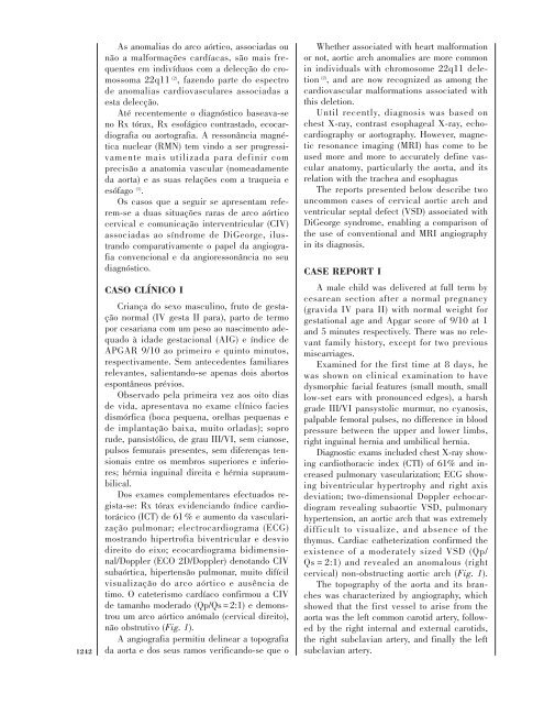
não obstrutivo (Fig. 1).<br />
A angiografia permitiu <strong>de</strong>linear a topografia<br />
da aorta e dos seus ramos verificando-se que o<br />
Whether associated with heart malformation<br />
or not, aortic arch anomalies are more common<br />
in individuals with chromosome 22q11 <strong>de</strong>letion<br />
(2) , and are now recognized as among the<br />
cardiovascular malformations associated with<br />
this <strong>de</strong>letion.<br />
Until recently, diagnosis was based on<br />
chest X-ray, contrast esophageal X-ray, echocardiography<br />
or aortography. However, magnetic<br />
resonance imaging (MRI) has come to be<br />
used more and more to accurately <strong>de</strong>fine vascular<br />
anatomy, particularly the aorta, and its<br />
relation with the trachea and esophagus<br />
The reports presented below <strong>de</strong>scribe two<br />
uncommon cases of cervical aortic arch and<br />
ventricular septal <strong>de</strong>fect (VSD) associated with<br />
DiGeorge syndrome, enabling a comparison of<br />
the use of conventional and MRI angiography<br />
in its diagnosis.<br />
CASE REPORT I<br />
A male child was <strong>de</strong>livered at full term by<br />
cesarean section after a normal pregnancy<br />
(gravida IV para II) with normal weight for<br />
gestational age and Apgar score of 9/10 at 1<br />
and 5 minutes respectively. There was no relevant<br />
family history, except for two previous<br />
miscarriages.<br />
Examined for the first time at 8 days, he<br />
was shown on clinical examination to have<br />
dysmorphic facial features (small mouth, small<br />
low-set ears with pronounced edges), a harsh<br />
gra<strong>de</strong> III/VI pansystolic murmur, no cyanosis,<br />
palpable femoral pulses, no difference in blood<br />
pressure between the upper and lower limbs,<br />
right inguinal hernia and umbilical hernia.<br />
Diagnostic exams inclu<strong>de</strong>d chest X-ray showing<br />
cardiothoracic in<strong>de</strong>x (CTI) of 61% and increased<br />
pulmonary vascularization; ECG showing<br />
biventricular hypertrophy and right axis<br />
<strong>de</strong>viation; two-dimensional Doppler echocardiogram<br />
revealing subaortic VSD, pulmonary<br />
hypertension, an aortic arch that was extremely<br />
difficult to visualize, and absence of the<br />
thymus. Cardiac catheterization confirmed the<br />
existence of a mo<strong>de</strong>rately sized VSD (Qp/<br />
Qs = 2:1) and revealed an anomalous (right<br />
cervical) non-obstructing aortic arch (Fig. 1).<br />
The topography of the aorta and its branches<br />
was characterized by angiography, which<br />
showed that the first vessel to arise from the<br />
aorta was the left common carotid artery, followed<br />
by the right internal and external carotids,<br />
the right subclavian artery, and finally the left<br />
subclavian artery.