Associação de Arco Aórtico Cervical.pdf
Associação de Arco Aórtico Cervical.pdf
Associação de Arco Aórtico Cervical.pdf

Create successful ePaper yourself
Turn your PDF publications into a flip-book with our unique Google optimized e-Paper software.
CASO CLÍNICO<br />
<strong>Associação</strong> <strong>de</strong> <strong>Arco</strong> <strong>Aórtico</strong> <strong>Cervical</strong><br />
a Delecção 22q11 – Papel da RMN<br />
no Diagnóstico [98]<br />
RUI ALMEIDA, SÍLVIA ÁLVARES, ANA FORTUNA, JORGE MOREIRA, ALBERTO VIEIRA<br />
RESUMO<br />
As anomalias do arco aórtico são<br />
relativamente comuns, ocorrendo em 0,5-3 %<br />
da população, tendo recentemente sido<br />
reconhecidas como fazendo parte do espectro<br />
<strong>de</strong> anomalias cardiovasculares associadas à<br />
<strong>de</strong>lecção do cromossoma 22q11.<br />
Actualmente a RMN surge como mais um<br />
método disponível para o seu diagnóstico,<br />
pois permite <strong>de</strong>finir com precisão a anatomia<br />
vascular (nomeadamente da aorta) e as suas<br />
relações com a traqueia e esófago, obviando<br />
as limitações da ecocardiografia<br />
convencional <strong>de</strong> superfície e evitando<br />
algumas das <strong>de</strong>svantagens da angiografia<br />
convencional, nomeadamente o uso <strong>de</strong><br />
radiação ionizante e <strong>de</strong> contraste iodado.<br />
Apresentam-se dois casos <strong>de</strong> arco aórtico<br />
cervical e CIV associados ao síndrome <strong>de</strong><br />
DiGeorge (CATCH22 +), em cujos<br />
diagnósticos foram utilizadas a angiografia<br />
convencional e a angioressonância,<br />
respectivamente.<br />
INTRODUÇÃO<br />
As anomalias do arco aórtico são relativamente<br />
comuns, ocorrendo em 0,5-3 % da<br />
população (1) . Entre estas contam-se o arco aórtico<br />
direito e, mais raramente, o arco aórtico<br />
cervical, bem como origens anómalas das artérias<br />
<strong>de</strong>le emergentes. Po<strong>de</strong>m estar associadas a<br />
doença cardíaca congénita, formar um anel<br />
vascular ou po<strong>de</strong>m ser achados isolados em indivíduos<br />
assintomáticos.<br />
Serviço <strong>de</strong> Cardiologia Pediática do Hospital Maria Pia, Porto<br />
Rev Port Cardiol 2003;22 (10):1241-1248<br />
Palavras-Chave<br />
<strong>Arco</strong> aórtico cervical; Delecção 22q11;<br />
Síndrome CATCH 22; RMN<br />
ABSTRACT<br />
Aortic arch anomalies are relatively common,<br />
occurring in 0.5-3 % of the population. In<br />
recent years, they have been recognized as<br />
being among the cardiovascular<br />
malformations found in chromosome 22q11<br />
<strong>de</strong>letion.<br />
MRI is now an alternative method of<br />
diagnosing aortic arch anomalies since it<br />
accurately <strong>de</strong>fines aortic anatomy and its<br />
relation with the trachea and esophagus, with<br />
some advantages in comparison with<br />
echocardiography and conventional<br />
angiography.<br />
The authors present two cases of cervical<br />
aortic arch and VSD associated with<br />
DiGeorge syndrome (CATCH22 +), diagnosed<br />
by conventional angiography and magnetic<br />
resonance imaging, respectively.<br />
Key words<br />
<strong>Cervical</strong> aortic arch; 22q11 <strong>de</strong>letion;<br />
CATCH 22 syndrome; MRI<br />
INTRODUCTION<br />
Aortic arch anomalies are relatively common,<br />
occurring in 0.5-3 % of the population<br />
(1) . They inclu<strong>de</strong> right aortic arch and, less<br />
commonly, cervical aortic arch, as well as abnormal<br />
origins of the arteries that emerge from<br />
it. They may be associated with congenital<br />
heart disease or may form a vascular ring, or<br />
be isolated findings in asymptomatic individuals.<br />
Recebido para publicação: Agosto <strong>de</strong> 2003 Aceite para publicação: Outubro <strong>de</strong> 2003<br />
Received for publication: August 2003 Accepted for publication: October 2003<br />
1241
1242<br />
As anomalias do arco aórtico, associadas ou<br />
não a malformações cardíacas, são mais frequentes<br />
em indivíduos com a <strong>de</strong>lecção do cromossoma<br />
22q11 (2) , fazendo parte do espectro<br />
<strong>de</strong> anomalias cardiovasculares associadas a<br />
esta <strong>de</strong>lecção.<br />
Até recentemente o diagnóstico baseava-se<br />
no Rx tórax, Rx esofágico contrastado, ecocardiografia<br />
ou aortografia. A ressonância magnética<br />
nuclear (RMN) tem vindo a ser progressivamente<br />
mais utilizada para <strong>de</strong>finir com<br />
precisão a anatomia vascular (nomeadamente<br />
da aorta) e as suas relações com a traqueia e<br />
esófago (1) .<br />
Os casos que a seguir se apresentam referem-se<br />
a duas situações raras <strong>de</strong> arco aórtico<br />
cervical e comunicação interventricular (CIV)<br />
associadas ao síndrome <strong>de</strong> DiGeorge, ilustrando<br />
comparativamente o papel da angiografia<br />
convencional e da angioressonância no seu<br />
diagnóstico.<br />
CASO CLÍNICO I<br />
Criança do sexo masculino, fruto <strong>de</strong> gestação<br />
normal (IV gesta II para), parto <strong>de</strong> termo<br />
por cesariana com um peso ao nascimento a<strong>de</strong>quado<br />
à ida<strong>de</strong> gestacional (AIG) e índice <strong>de</strong><br />
APGAR 9/10 ao primeiro e quinto minutos,<br />
respectivamente. Sem antece<strong>de</strong>ntes familiares<br />
relevantes, salientando-se apenas dois abortos<br />
espontâneos prévios.<br />
Observado pela primeira vez aos oito dias<br />
<strong>de</strong> vida, apresentava no exame clínico facies<br />
dismórfica (boca pequena, orelhas pequenas e<br />
<strong>de</strong> implantação baixa, muito orladas); sopro<br />
ru<strong>de</strong>, pansistólico, <strong>de</strong> grau III/VI, sem cianose,<br />
pulsos femurais presentes, sem diferenças tensionais<br />
entre os membros superiores e inferiores;<br />
hérnia inguinal direita e hérnia supraumbilical.<br />
Dos exames complementares efectuados regista-se:<br />
Rx tórax evi<strong>de</strong>nciando índice cardiotorácico<br />
(ICT) <strong>de</strong> 61 % e aumento da vascularização<br />
pulmonar; electrocardiograma (ECG)<br />
mostrando hipertrofia biventricular e <strong>de</strong>svio<br />
direito do eixo; ecocardiograma bidimensional/Doppler<br />
(ECO 2D/Doppler) <strong>de</strong>notando CIV<br />
subaórtica, hipertensão pulmonar, muito difícil<br />
visualização do arco aórtico e ausência <strong>de</strong><br />
timo. O cateterismo cardíaco confirmou a CIV<br />
<strong>de</strong> tamanho mo<strong>de</strong>rado (Qp/Qs = 2:1) e <strong>de</strong>monstrou<br />
um arco aórtico anómalo (cervical direito),<br />
não obstrutivo (Fig. 1).<br />
A angiografia permitiu <strong>de</strong>linear a topografia<br />
da aorta e dos seus ramos verificando-se que o<br />
Whether associated with heart malformation<br />
or not, aortic arch anomalies are more common<br />
in individuals with chromosome 22q11 <strong>de</strong>letion<br />
(2) , and are now recognized as among the<br />
cardiovascular malformations associated with<br />
this <strong>de</strong>letion.<br />
Until recently, diagnosis was based on<br />
chest X-ray, contrast esophageal X-ray, echocardiography<br />
or aortography. However, magnetic<br />
resonance imaging (MRI) has come to be<br />
used more and more to accurately <strong>de</strong>fine vascular<br />
anatomy, particularly the aorta, and its<br />
relation with the trachea and esophagus<br />
The reports presented below <strong>de</strong>scribe two<br />
uncommon cases of cervical aortic arch and<br />
ventricular septal <strong>de</strong>fect (VSD) associated with<br />
DiGeorge syndrome, enabling a comparison of<br />
the use of conventional and MRI angiography<br />
in its diagnosis.<br />
CASE REPORT I<br />
A male child was <strong>de</strong>livered at full term by<br />
cesarean section after a normal pregnancy<br />
(gravida IV para II) with normal weight for<br />
gestational age and Apgar score of 9/10 at 1<br />
and 5 minutes respectively. There was no relevant<br />
family history, except for two previous<br />
miscarriages.<br />
Examined for the first time at 8 days, he<br />
was shown on clinical examination to have<br />
dysmorphic facial features (small mouth, small<br />
low-set ears with pronounced edges), a harsh<br />
gra<strong>de</strong> III/VI pansystolic murmur, no cyanosis,<br />
palpable femoral pulses, no difference in blood<br />
pressure between the upper and lower limbs,<br />
right inguinal hernia and umbilical hernia.<br />
Diagnostic exams inclu<strong>de</strong>d chest X-ray showing<br />
cardiothoracic in<strong>de</strong>x (CTI) of 61% and increased<br />
pulmonary vascularization; ECG showing<br />
biventricular hypertrophy and right axis<br />
<strong>de</strong>viation; two-dimensional Doppler echocardiogram<br />
revealing subaortic VSD, pulmonary<br />
hypertension, an aortic arch that was extremely<br />
difficult to visualize, and absence of the<br />
thymus. Cardiac catheterization confirmed the<br />
existence of a mo<strong>de</strong>rately sized VSD (Qp/<br />
Qs = 2:1) and revealed an anomalous (right<br />
cervical) non-obstructing aortic arch (Fig. 1).<br />
The topography of the aorta and its branches<br />
was characterized by angiography, which<br />
showed that the first vessel to arise from the<br />
aorta was the left common carotid artery, followed<br />
by the right internal and external carotids,<br />
the right subclavian artery, and finally the left<br />
subclavian artery.
primeiro vaso a originar-se da aorta era a artéria<br />
carótida comum esquerda, seguindo-se as<br />
artérias carótidas interna e externa direitas, a<br />
artéria subclávia direita e, por fim, a artéria<br />
subclávia esquerda.<br />
Os outros exames efectuados <strong>de</strong>monstraram:<br />
cálcio, fósforo e paratormona normais, imunofenotipagem<br />
linfocitária em sangue periférico<br />
mostrando diminuição dos linfócitos T (com<br />
número normal <strong>de</strong> linfócitos B e NK) e pesquisa<br />
<strong>de</strong> <strong>de</strong>leção 22q11 pela técnica <strong>de</strong> FISH<br />
(fluorescent in situ hybridization) positiva.<br />
Quanto à evolução verificou-se uma má<br />
progressão estaturo-pon<strong>de</strong>ral, associada a infecções<br />
pulmonares <strong>de</strong> repetição e insuficiência<br />
cardíaca difícil <strong>de</strong> controlar, motivo pelo<br />
qual foi realizado encerramento cirúrgico da<br />
CIV aos cinco meses <strong>de</strong> vida.<br />
CASO CLÍNICO II<br />
Criança do sexo masculino; fruto <strong>de</strong> gestação<br />
<strong>de</strong> risco (ameaça <strong>de</strong> abortamento no primeiro<br />
trimestre), I gesta, I para. Parto às 38<br />
semanas, eutócico, com peso AIG e índice <strong>de</strong><br />
APGAR <strong>de</strong>sconhecido. Os antece<strong>de</strong>ntes familiares<br />
eram irrelevantes.<br />
Actualmente com 17 anos <strong>de</strong> ida<strong>de</strong>, sempre<br />
assintomático do ponto <strong>de</strong> vista cardíaco, apresenta<br />
cardiopatia congénita (diagnosticada aos<br />
10 anos), sendo <strong>de</strong> referir na história pregressa<br />
os seguintes antece<strong>de</strong>ntes: membrana laríngea<br />
anterior congénita operada aos 14 anos, infecções<br />
respiratórias altas frequentes; gastrite crónica<br />
(H. pylori +) e dificulda<strong>de</strong>s na aprendiza-<br />
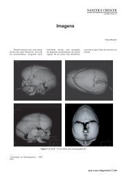
Fig. 1 Aortograma anterógrado em<br />
posição oblíqua anterior esquerda,<br />
evi<strong>de</strong>nciando a origem anómala dos<br />
ramos arteriais emergentes da aorta<br />
torácica. AoD, aorta <strong>de</strong>scen<strong>de</strong>nte;<br />
ASD - artéria subclávia direita;<br />
ASE - artéria subclávia esquerda;<br />
CCE - carótida comum esquerda;<br />
CE - carótida externa direita; CI -<br />
carótida interna direita.<br />
Fig. 1 Antegra<strong>de</strong> aortogram in<br />
oblique left anterior view, showing<br />
the anomalous origin of the arterial<br />
branches of the thoracic aorta. AoD<br />
- <strong>de</strong>scending aorta; ASD - right<br />
subclavian artery; ASE - left subclavian<br />
artery; CCE - left common<br />
carotid; CE - right external carotid;<br />
CI - right internal carotid.<br />
Other exams showed normal calcium, phosphorus<br />
and parathyroid hormone; lymphocyte<br />
immunophenotyping in peripheral blood revealed<br />
reduced T lymphocytes, with normal B and<br />
NK lymphocytes; and positive test for 22q11<br />
<strong>de</strong>letion using the FISH technique (fluorescent<br />
in situ hybridization).<br />
The patient’s evolution inclu<strong>de</strong>d poor height<br />
and weight progression, repeated pulmonary<br />
infections and heart failure that was resistant<br />
to treatment, and so the VSD was closed surgically<br />
at 5 months of life.<br />
CASE REPORT II<br />
After an at-risk pregnancy (threat of miscarriage<br />
in the first trimester), a male child<br />
was born (gravida I, para I) at 38 weeks, full<br />
term, with normal weight for gestational age<br />
and unknown Apgar score; there was no relevant<br />
family history.<br />
Now 17 years old, and without cardiac<br />
symptoms since birth, he has congenital heart<br />
disease diagnosed at age 10, and among other<br />
history, congenital anterior laryngeal web operated<br />
at age 14, frequent upper respiratory<br />
tract infections, chronic gastritis (H. pylori positive),<br />
and learning difficulties.<br />
On clinical examination he had good height<br />
(P25-50) and weight (P10-25) <strong>de</strong>velopment,<br />
dysmorphic facial features (round nose with<br />
hypoplasia of the nostrils, hypoplasia of the<br />
central face with high narrow palate and small<br />
ears), hoarse and nasal speech, and a gra<strong>de</strong><br />
II/VI pansystolic murmur, lou<strong>de</strong>st at the lower<br />
left sternal bor<strong>de</strong>r.<br />
gem. 1243
1244<br />
Ao exame objectivo apresenta boa evolução<br />
estatural (P25-50) e pon<strong>de</strong>ral (P10-25; fácies<br />
dismórfica (nariz globoso com hipoplasia das<br />
asas do nariz, hipoplasia da face média com palato<br />
alto e estreito e orelhas pequenas), voz<br />
rouca e hipernasalada; sopro pansistólico, II/VI,<br />
máximo no bordo esternal esquerdo baixo.<br />
Nos exames complementares salienta-se:<br />
Rx tórax com ICT <strong>de</strong> 50 %, vascularização pulmonar<br />
sem alterações evi<strong>de</strong>ntes e anomalia<br />
congénita da primeira costela do hemitórax esquerdo;<br />
ECG normal; ECO 2D/Doppler mostrando<br />
CIV subaórtica restritiva, <strong>de</strong>tectando-se<br />
um gradiente interventricular instantâneo máximo<br />
<strong>de</strong> 80 mmHg, boa função biventricular e<br />
provável arco aórtico cervical. A angioressonância<br />
corroborou os achados ecocardiográficos<br />
evi<strong>de</strong>nciando arco aórtico direito em topografia<br />
mais alta do que o habitual (cervical)<br />
associado a CIV subaórtica (com opacificação<br />
precoce da artéria pulmonar e do ventrículo<br />
direito) (Fig. 2).<br />
Fig. 2 Corte sagital obtido por angioresonância<br />
(spin <strong>de</strong> eco pon<strong>de</strong>rado<br />
em T1), mostrando uma angulação<br />
mais pronunciada e posição<br />
mais elevada do arco aórtico.<br />
Fig. 2 Sagittal section obtained by<br />
MRI angiography (T1-weighted spin<br />
echo), showing the aortic arch’s<br />
more pronounced angulation and<br />
higher position.<br />
The main findings of the diagnostic exams<br />
were as follows: chest X-ray with CTI of 50 %,<br />
no obvious changes in pulmonary vascularization,<br />
and congenital abnormality of the first rib<br />
in the left hemithorax; normal ECG; 2D/Doppler<br />
echo showing restrictive subaortic VSD<br />
with a maximum instantaneous intraventricular<br />
gradient of 80 mmHg, good biventricular function<br />
and probable cervical aortic arch. MRI<br />
confirmed the echocardiographic findings, showing<br />
right aortic arch, higher than usual (cervical),<br />
together with subaortic VSD, with early<br />
opacification of the pulmonary artery and the<br />
right ventricle (Fig. 2).<br />
The MRI images show the topography of<br />
the aorta and its branches, with a similar anatomy<br />
to the first case, particularly in the emergence<br />
of the carotid and subclavian arteries<br />
(Fig. 3).<br />
Other exams showed normal calcium and<br />
phosphorus and lower than normal parathyroid
Nas imagens da angiorressonância po<strong>de</strong>mos<br />
observar a topografia da aorta e dos seus ramos<br />
com uma anatomia sobreponível à do primeiro<br />
caso (nomeadamente na emergência das artérias<br />
carótidas e subclávias) (Fig. 3).<br />
Os outros exames efectuados <strong>de</strong>monstraram:<br />
cálcio e fósforo normais e paratormona inferior<br />
ao normal; diminuição dos linfócitos T (número<br />
normal <strong>de</strong> linfócitos B, número normal <strong>de</strong> linfócitos<br />
NK) e pesquisa <strong>de</strong> <strong>de</strong>lecção 22q11 por<br />
FISH positiva.<br />
Quanto à evolução, esta criança mantém-se<br />
activa e assintomática (tendo efectuado Holter-<br />
-24 horas e prova <strong>de</strong> esforço que foram normais).<br />
Foi pesquisada a <strong>de</strong>lecção 22q11 por<br />
FISH em ambos os pais, que foi negativa.<br />
DISCUSSÃO<br />
O arco aórtico cervical, <strong>de</strong>scrito pela primeira<br />
vez em 1914 por Reid, constitui uma<br />
hormone; reduction in T lymphocytes with normal<br />
B and NK lymphocytes; and positive FISH<br />
test for 22q11 <strong>de</strong>letion.<br />
With regard to evolution, this child has remained<br />
active and asymptomatic (24-hour Holter<br />
monitoring and exercise testing proved normal).<br />
Testing by FISH for 22q11 <strong>de</strong>letion was<br />
negative in both parents.<br />
DISCUSSION<br />
<strong>Cervical</strong> aortic arch, <strong>de</strong>scribed for the first<br />
time by Reid in 1914, is a rare malformation,<br />
<strong>de</strong>fined as an aortic arch located above the<br />
clavicle, and may be right (more frequent) or<br />
left. Its cause appears to be abnormal persistence<br />
of the third aortic arch and regression of<br />
the normal fourth arch, which pushes the aortic<br />
arch towards the cervical region (3) .<br />
Most cases remain asymptomatic, but there<br />
may be associated respiratory problems or<br />
Fig. 3 Corte oblíquo obtido por angioressonância<br />
(spin <strong>de</strong> eco pon<strong>de</strong>rado<br />
em T1) com contraste – gadolíneo,<br />
<strong>de</strong>monstrando uma anatomia<br />
arterial anómala sobreponível à do<br />
1.º caso. AoD, aorta <strong>de</strong>scen<strong>de</strong>nte;<br />
ASD, artéria subclávia direita;<br />
ASE, artéria subclávia esquerda;<br />
CCE, carótida comum esquerda;<br />
CE, carótida externa direita; CI, carótida<br />
interna direita.<br />
Fig. 3 Oblique section obtained by<br />
MRI angiography (T1-weighted spin<br />
echo) with gadolinium contrast, revealing<br />
anomalous arterial anatomy<br />
similar to the first case. AoD, <strong>de</strong>scending<br />
aorta; ASD, right subclavian<br />
artery; ASE, left subclavian artery;<br />
CCE, left common carotid; CE,<br />
right external carotid; CI, right internal<br />
carotid.<br />
1245
1246<br />
malformação rara <strong>de</strong>finida por um arco que se<br />
coloca acima do nível da clavícula, po<strong>de</strong>ndo<br />
ser direito (mais frequente) ou esquerdo. Na<br />
sua origem parece estar uma persistência anormal<br />
do terceiro arco aórtico (e regressão do<br />
quarto arco normal) que redirecciona o arco<br />
aórtico para a região cervical (3) .<br />
Na maioria dos casos é assintomático, mas<br />
po<strong>de</strong> estar associado a problemas respiratórios<br />
ou disfagia por compressão da traqueia ou esófago,<br />
respectivamente, provocados por um anel<br />
vascular. Outros achados incluem uma massa<br />
pulsátil supraclavicular, diferenças tensionais<br />
entre os membros superiores ou ausência <strong>de</strong><br />
pulso femural ou do membro superior contralateral<br />
após compressão da massa cervical. Num<br />
número significativo <strong>de</strong> casos po<strong>de</strong>m ser encontradas<br />
anomalias estruturais do arco como<br />
obstrução, aneurismas e tortuosida<strong>de</strong>s que justificam<br />
uma correcção cirúrgica (4) . Em 30 %<br />
dos casos associa-se a outras malformações<br />
cardíacas como CIV, tetralogia <strong>de</strong> Fallot ou<br />
ventrículo direito com dupla câmara <strong>de</strong> saída (5) .<br />
O diagnóstico através da ecocardiografia, é<br />
feito por abordagem suprasternal, colocando a<br />
sonda ecocardiográfica numa posição mais alta<br />
do que a habitual, para permitir o <strong>de</strong>sdobramento<br />
do arco que se encontra numa localização<br />
mais cefálica (6) . No entanto, as imagens<br />
ecocardiográficas convencionais <strong>de</strong> superfície<br />
não são muitas vezes diagnósticas por inexistência<br />
<strong>de</strong> uma janela sonográfica a<strong>de</strong>quada.<br />
A angiografia convencional constituiu até<br />
recentemente o gold standard para o diagnóstico<br />
<strong>de</strong>sta patologia, apresentando como principais<br />
inconvenientes ser um método invasivo,<br />
utilizar radiação ionizante e contraste iodado.<br />
A RMN é uma técnica inócua, semi-invasiva<br />
e particularmente eficaz na avaliação da<br />
aorta torácica sendo, actualmente, a maioria<br />
dos exames cardiovasculares realizados para o<br />
seu estudo. A utilização do gadolíneo, como<br />
meio <strong>de</strong> contraste (não nefrotóxico), veio aumentar<br />
a precisão diagnóstica <strong>de</strong>ste exame (1, 7) .<br />
Nestes dois casos, referentes à mesma anomalia<br />
(arco aórtico cervical), foi utilizada uma<br />
metodologia diferente: o recurso à angioressonância<br />
no segundo caso, permitiu <strong>de</strong> igual<br />
modo fazer o diagnóstico com as vantagens já<br />
enumeradas em relação à angiografia convencional.<br />
O síndrome <strong>de</strong> DiGeorge juntamente com o<br />
síndrome velocardiofacial e o síndrome face-<br />
-anomalia conotruncal incluem-se no espectro<br />
dysphagia due to compression respectively of<br />
the trachea or esophagus, caused by a vascular<br />
ring. Other findings inclu<strong>de</strong> a pulsatile supraclavicular<br />
mass, differences in blood pressure<br />
between the upper limbs, and absence of femoral<br />
or contralateral upper limb pulse after compression<br />
of the cervical mass. In a significant<br />
number of cases there are structural anomalies<br />
of the arch such as obstruction, aneurysms and<br />
twisting, which justify surgical correction (4) . In<br />
30 % of cases there are associated cardiac<br />
malformations such as VSD, tetralogy of Fallot<br />
or double outlet right ventricle (5) .<br />
Diagnosis using echocardiography is by a<br />
suprasternal approach, with the probe placed<br />
in a higher position than usual to allow the<br />
complete visualization of the arch, which is in<br />
a more cephalic position than normal (6) . However,<br />
conventional echocardiographic images<br />
are not often diagnostic, due to the lack of an<br />
a<strong>de</strong>quate echocardiographic window.<br />
Until recently, conventional angiography<br />
has been the gold standard for diagnosing this<br />
pathology, its main disadvantages being its invasive<br />
nature and the ionizing radiation used.<br />
MRI angiography is harmless, semi-invasive<br />
and particularly effective in assessment of<br />
the thoracic aorta, and most cardiovascular<br />
exams for aortic arch anomalies now use this<br />
technique. The use of gadolinium as a contrast<br />
agent, which has the advantage of not being<br />
nephrotoxic, has increased the diagnostic accuracy<br />
of this exam (1, 7) .<br />
In the two cases presented, which both <strong>de</strong>scribe<br />
the same anomaly (cervical aortic arch),<br />
different methods were used; in the second,<br />
MRI enabled the diagnosis to be ma<strong>de</strong> with<br />
the above-mentioned advantages over conventional<br />
angiography.<br />
DiGeorge syndrome, together with velocardiofacial<br />
syndrome and conotruncal face anomaly<br />
syndrome, are among the range of clinical<br />
conditions associated with 22q11 <strong>de</strong>letion,<br />
which are generally known as the CATCH-22<br />
syndrome (for Cardiac <strong>de</strong>fects, Abnormal facies,<br />
Thymic hypoplasia, Cleft palate, and<br />
Hypocalcemia). It is characterized by hypoplasia<br />
or aplasia of the thymus and the parathyroid<br />
glands due to dysmorphogenesis of the<br />
third and fourth pharyngeal pouches during<br />
early embryogenesis, and results in greater<br />
susceptibility to infection and neonatal hypocalcemia,<br />
at times severe. In ol<strong>de</strong>r children it<br />
has characteristics in common with velocardiofacial<br />
syndrome, including velopharyngeal in-
clínico das condições com <strong>de</strong>lecção 22q11 (genericamente<br />
conhecido como síndrome CATCH<br />
22 – cujas iniciais significam Cardiac <strong>de</strong>fects,<br />
Abnormal facies, Thymic hypoplasia, Cleft palate,<br />
Hypocalcemia). Caracteriza-se por uma<br />
hipoplasia/aplasia tímica e das glândulas paratirói<strong>de</strong>s<br />
por dismorfogénese da terceira e<br />
quarta bolsas faríngeas durante a embriogénese<br />
precoce, <strong>de</strong> que resultam maior susceptibilida<strong>de</strong><br />
às infecções e hipocalcemia neonatal,<br />
por vezes grave. Em crianças mais velhas partilha<br />
características com o síndrome velocardiofacial<br />
(como incompetência velofaríngea e voz<br />
hipernasalada) po<strong>de</strong>ndo tornarem-se ainda evi<strong>de</strong>ntes<br />
uma baixa estatura, dificulda<strong>de</strong>s <strong>de</strong><br />
aprendizagem e doenças psiquiátricas.<br />
Este síndrome está frequentemente associado<br />
a anomalias dos gran<strong>de</strong>s vasos (arco aórtico<br />
direito) e malformações cardíacas (<strong>de</strong>feitos<br />
septais auriculares e ventriculares).<br />
A associação <strong>de</strong> membrana laríngea anterior<br />
a <strong>de</strong>lecção 22q11, presente no caso II, tem<br />
sido também <strong>de</strong>scrita (8) . As membranas laríngeas<br />
anteriores são pouco comuns (cerca <strong>de</strong><br />
5 % das malformações laríngeas), e ocorrem<br />
frequentemente associadas a outras anomalias,<br />
tais como cardiopatia congénita, úvula bífida,<br />
fenda palatina, atresia esofágica, sinus preauricular<br />
e anomalias urogenitais. Stoler preconiza<br />
a pesquisa da <strong>de</strong>lecção 22q11 em todos os<br />
doentes com membrana laríngea, cardiopatia e<br />
anomalias do palato (8) .<br />
competence and hypernasal speech, and may<br />
also be linked with short stature, learning difficulties<br />
and psychiatric disor<strong>de</strong>rs.<br />
This syndrome is often associated with abnormalities<br />
of the great vessels (right aortic<br />
arch) and heart malformations (atrial and ventricular<br />
septal <strong>de</strong>fects).<br />
An association of anterior laryngeal web<br />
with 22q11 <strong>de</strong>letion, as found in case II, has<br />
also been <strong>de</strong>scribed (8) . Anterior laryngeal webs<br />
are uncommon (accounting for around 5 % of<br />
laryngeal malformations), and are frequently<br />
found in association with other anomalies,<br />
such as congenital heart disease, bifid uvula<br />
and cleft palate, esophageal atresia, preauricular<br />
sinus and urogenital abnormalities. Stoler<br />
recommends testing for 22q11 <strong>de</strong>letion in all<br />
patients with laryngeal webs, heart disease and<br />
palatal <strong>de</strong>fects (8) .<br />
CONCLUSÕES<br />
Este trabalho exemplica o contributo da angioressonância<br />
para o diagnóstico <strong>de</strong> malformações<br />
do arco aórtico, constituindo uma alternativa<br />
extremamente válida no estudo<br />
cardiovascular.<br />
Salienta-se a importância da pesquisa sistemática<br />
da <strong>de</strong>lecção 22q11 nas anomalias do<br />
arco aórtico isoladas ou associadas a <strong>de</strong>feitos<br />
cardíacos (conotruncais), sobretudo se reforçados<br />
por outros dados clínicos sugestivos (2, 9, 10) CONCLUSIONS<br />
This work highlights the contribution of<br />
MRI angiography in the diagnosis of aortic<br />
arch malformations, for which it is a valuable<br />
alternative method of cardiovascular study.<br />
It is important to test systematically for<br />
22q11 <strong>de</strong>letion in cases of aortic arch anomalies,<br />
either in isolation or with associated heart<br />
<strong>de</strong>fects (conotruncal), especially in the presence<br />
of other suggestive clinical findings<br />
.<br />
O diagnóstico precoce <strong>de</strong>sta alteração cromossómica<br />
torna-se importante pelas suas diversas<br />
complicações médicas, dificulda<strong>de</strong>s <strong>de</strong> aprendizagem<br />
associadas e possíveis implicações<br />
hereditárias em familiares próximos.<br />
Por fim, é <strong>de</strong> referir a associação entre arco<br />
aórtico cervical e síndrome <strong>de</strong> DiGeorge em<br />
ambos os casos, o que constitui uma rarida<strong>de</strong>,<br />
dado o reduzido número <strong>de</strong> casos idênticos pu- 1247<br />
(2, 9, 10) .<br />
Early diagnosis of this chromosome alteration<br />
is important, given its various medical complications,<br />
associated learning difficulties, and<br />
possible implications for family members who<br />
may also inherit the <strong>de</strong>letion.<br />
Finally, we should stress the association<br />
between cervical aortic arch and DiGeorge<br />
syndrome in both these cases, which is unusual<br />
if the small number of similar cases in<br />
the literature is anything to go by (11) . This finding<br />
suggests that cervical aortic arch may also<br />
be one of the cardiac abnormalities associated<br />
with this syndrome.
1248<br />
blicados na literatura (11) . Este achado sugere<br />
que o arco aórtico cervical seja também uma<br />
das anomalias cardíacas associadas a este síndrome.<br />
Pedido <strong>de</strong> separatas para:<br />
Address for reprints:<br />
RUI ALMEIDA<br />
Alameda Dr. Fernando Azeredo Antas 47, 7.ºB<br />
4150-314 PORTO<br />
Tel.: 914 023 536<br />
e-mail: rui.mc.almeida@netcabo.pt<br />
1. Berlin SC. Magnetic resonance imaging of the cardiovascular<br />
system and airway. Pediatr Clin North Am 1997;44:<br />
659-79.<br />
2. Momma K, Matsuoka R, Takao A. Aortic arch anomalies<br />
associated with chromosome 22q11 <strong>de</strong>letion (CATCH 22).<br />
Pediatr Cardiol 1999;20:97-102.<br />
3. Weinberg PM. Aortic arch anomalies. In: Moss AJ, Adams<br />
F, eds. Heart Disease in Infants, Children and Adolescents.<br />
Baltimore: Williams & Wilkins 1995;810-37.<br />
4. McElhinney DB, Thompson LD, Weinberg PM, Jue KL,<br />
Hanley FL. Surgical approach to complicated cervical aortic<br />
arch: anatomic, <strong>de</strong>velopmental, and surgical consi<strong>de</strong>rations.<br />
Cardiol Young 2000;10:212-9.<br />
5. Kazuma N, Murakami M, Suzuki Y, Umezu R, Murata M.<br />
<strong>Cervical</strong> aortic arch associated with 22q11.2 <strong>de</strong>letion. Pediatr<br />
Cardiol 1997;18:149-51.<br />
BIBLIOGRAFIA / REFERENCES<br />
6. Sni<strong>de</strong>r R, Silverman NH. Suprasternal notch echocardiography:<br />
a two-dimensional technique for evaluating congenital<br />
heart disease. Circulation 1981;63:165.<br />
7. Carpenter JP, Holland GA, Gol<strong>de</strong>n MA, Barker CF, Lexa<br />
FJ, Gilfeather M et al. Magnetic resonance angiography of<br />
the aortic arch. J Vasc Surg 1997;25:145-51.<br />
8. Stoler JM, Ladouylis M, Holmes LB. Anterior Laryngeal<br />
Webs and 22q11 Deletions. Am J Med Genet 1998;79:152.<br />
9. Tobias ES, Morrison N, Whiteford ML, Tolmie JL. Towards<br />
earlier diagnosis of 22q11 <strong>de</strong>letions. Arch Dis Child<br />
1999;81:513-4.<br />
10. McElhinney DB, Clark BJ, Weinberg PM, Kenton ML,<br />
McDonald-McGinn D, Driscoll DA et al. Association of chromosome<br />
22q11 <strong>de</strong>letion with isolated anomalies of aortic arch<br />
laterality and branching. J Am Coll Cardiol 2001;37:2114-9.<br />
11. Kumar A, McCombs JL, Sapire DW. Deletions in chromosome<br />
22q11 region in cervical aortic arch. Am J Cardiol<br />
1997;79:388-90.