Associação de Arco Aórtico Cervical.pdf
Associação de Arco Aórtico Cervical.pdf
Associação de Arco Aórtico Cervical.pdf

You also want an ePaper? Increase the reach of your titles
YUMPU automatically turns print PDFs into web optimized ePapers that Google loves.
1244<br />
Ao exame objectivo apresenta boa evolução<br />
estatural (P25-50) e pon<strong>de</strong>ral (P10-25; fácies<br />
dismórfica (nariz globoso com hipoplasia das<br />
asas do nariz, hipoplasia da face média com palato<br />
alto e estreito e orelhas pequenas), voz<br />
rouca e hipernasalada; sopro pansistólico, II/VI,<br />
máximo no bordo esternal esquerdo baixo.<br />
Nos exames complementares salienta-se:<br />
Rx tórax com ICT <strong>de</strong> 50 %, vascularização pulmonar<br />
sem alterações evi<strong>de</strong>ntes e anomalia<br />
congénita da primeira costela do hemitórax esquerdo;<br />
ECG normal; ECO 2D/Doppler mostrando<br />
CIV subaórtica restritiva, <strong>de</strong>tectando-se<br />
um gradiente interventricular instantâneo máximo<br />
<strong>de</strong> 80 mmHg, boa função biventricular e<br />
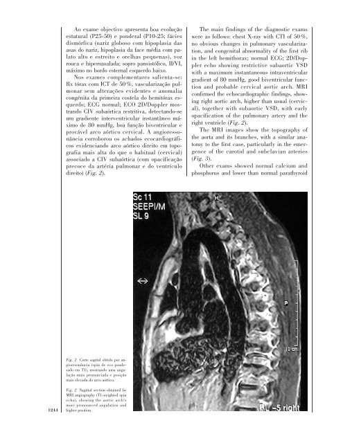
provável arco aórtico cervical. A angioressonância<br />
corroborou os achados ecocardiográficos<br />
evi<strong>de</strong>nciando arco aórtico direito em topografia<br />
mais alta do que o habitual (cervical)<br />
associado a CIV subaórtica (com opacificação<br />
precoce da artéria pulmonar e do ventrículo<br />
direito) (Fig. 2).<br />
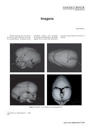
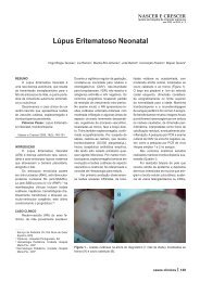
Fig. 2 Corte sagital obtido por angioresonância<br />
(spin <strong>de</strong> eco pon<strong>de</strong>rado<br />
em T1), mostrando uma angulação<br />
mais pronunciada e posição<br />
mais elevada do arco aórtico.<br />
Fig. 2 Sagittal section obtained by<br />
MRI angiography (T1-weighted spin<br />
echo), showing the aortic arch’s<br />
more pronounced angulation and<br />
higher position.<br />
The main findings of the diagnostic exams<br />
were as follows: chest X-ray with CTI of 50 %,<br />
no obvious changes in pulmonary vascularization,<br />
and congenital abnormality of the first rib<br />
in the left hemithorax; normal ECG; 2D/Doppler<br />
echo showing restrictive subaortic VSD<br />
with a maximum instantaneous intraventricular<br />
gradient of 80 mmHg, good biventricular function<br />
and probable cervical aortic arch. MRI<br />
confirmed the echocardiographic findings, showing<br />
right aortic arch, higher than usual (cervical),<br />
together with subaortic VSD, with early<br />
opacification of the pulmonary artery and the<br />
right ventricle (Fig. 2).<br />
The MRI images show the topography of<br />
the aorta and its branches, with a similar anatomy<br />
to the first case, particularly in the emergence<br />
of the carotid and subclavian arteries<br />
(Fig. 3).<br />
Other exams showed normal calcium and<br />
phosphorus and lower than normal parathyroid