

a rquivos b rasileiros - Conselho Brasileiro de Oftalmologia
a rquivos b rasileiros - Conselho Brasileiro de Oftalmologia
a rquivos b rasileiros - Conselho Brasileiro de Oftalmologia

Create successful ePaper yourself
Turn your PDF publications into a flip-book with our unique Google optimized e-Paper software.
ZENHA F, ET AL.<br />
Ages ranged from 35 to 90 years (median: 65 years). According<br />
to skin color, 18 patients were characterized as Caucasians, 6 as afro<strong>de</strong>scen<strong>de</strong>nts<br />
and 1 as Oriental.<br />
HLA class I antigens were typed using a microlymphocytotoxity<br />
assay (8) .<br />
Regarding the HLA profile, <strong>de</strong>termined in the time of inclusion<br />
in the study, 6 patients were found to have one of the haplotypes<br />
<strong>de</strong>scribed by Torres et al. (9) , a fact that led to the formation of two<br />
groups. Group A consisted of 6 patients presenting one of the A2-<br />
B40 and A1-B8 HLA haplotypes and Group B consisted of the remaining<br />
19 patients who did not present either haplotype.<br />
The control population consisted of a group of 257 normal individuals<br />
who had donated kidneys at the São Paulo Interior Transplant<br />
(SPIT) Center.<br />
At initial examination the visual acuity in the Group A subjects<br />
ranged from absence of light perception (ALP, 0 for statistical purposes)<br />
to 1 (median: 0.7). In the current exam, visual acuity ranged from<br />
ALP (0 for statistical purposes) to 0.8 (median: 0.3). For Group B, visual<br />
acuity ranged from ALP to 1 (median: 0.85) at initial examination and<br />
from ALP to 1 (median: 0.5) at current examination.<br />
Patients were classified as having POAG according gonioscopy<br />
findings, intraocular pressure of 21 mmHg or more, accompanied<br />
by changes suggestive of glaucoma in the head of the optic nerve<br />
and, or in the visual field.<br />
Visual fields were classified according to the criteria of the Ocular<br />
Hypertension Treatment Study (OHTS) (10) . In addition, perimetric<br />
losses were classified according to the following criteria:<br />
1- Stable (no change in the visual field);<br />
2- Mild (changes present also in another quadrant and/or increased<br />
lesions in the same quadrant);<br />
3- Mo<strong>de</strong>rate (changes present in two more quadrants and/or<br />
marked increase of lesions in the same quadrant(s);<br />
4- Severe (changes progressing to all quadrants or worsening<br />
of tubular loss).<br />
Data from the patients’ medical records were used to fill out cards<br />
which contained, in addition to data regarding the ophthalmologic<br />
exam, patient i<strong>de</strong>ntification data and records of campimetric changes.<br />
All the patients who returned for re-evaluation were submitted<br />
to anamnesis, measurement of visual acuity, tonometry, campimetry<br />
(at least two examinations), fundoscopy with retinography, and<br />
optical coherence tomography (OCT - Topcon, São Paulo, Brazil).<br />
A classification was elaborated to assess the loss of nerve fibers<br />
by OCT using as reference the percentiles of normal distribution of<br />
the apparatus itself:<br />
1 = p < 1% - High involvement of nerve fibers<br />
2 = 1% < p < 5% - Bor<strong>de</strong>rline involvement<br />
3 = p > 5% - Low involvement or normal condition<br />
Data were analyzed statistically by the Fisher two-tailed exact<br />
test, with the level of significance set at p < 0.05 (95% confi<strong>de</strong>nce<br />
interval). When significant differences in frequency were <strong>de</strong>tected<br />
between groups, the odds ratio (OR) was calculated, using statistical<br />
program EpiInfo 6.0.<br />
RESULTS<br />
HLA class I haplotypes associated with POAG in Group A patients<br />
were HLA A1-B8 (4 patients) and HLA A2-B40 (2 patients).<br />
On the basis of the HLA typing, it should be pointed out that<br />
other participants presented at least one of the class I specificities<br />
composing these haplotypes (HLA-A1-B8, -A2-B40 and -A9-B12), as<br />
shown in table 1.<br />
At initial examination, the cup-to-disc ratio ranged from 0.2 to<br />
0.9, with a median of 0.4, for Group A and from 0.4 to 1.0, with a<br />
median of 0.85, for Group B. At the current examination, the values<br />
ranged from 0.5 to 0.7, with a median of 0.8, for Group A and from<br />
0.7 to 1.0, with a median of 0.85, for Group B.<br />
No significant differences in cup-to-disc ratio were <strong>de</strong>tected<br />
between the two groups at either time of evaluation (Group A x Group<br />
B in the first examination: p=0.8500; Group A x Group B last examination:<br />
p=0.2994). However, when the variation in the cup-to-disc<br />
ratio of group A patients was compared to that of Group B patients,<br />
significant differences were <strong>de</strong>tected both when Group A was compared<br />
to Group B patients as a whole (p=0.012) and when comparisons<br />
were ma<strong>de</strong> between different age ranges, as shown in table 2.<br />
When visual fields were compared between groups and between<br />
times, the variation was 1 to 3 (median: 2) for Group A and also<br />
for Group B (median: 1), with the difference being nonsignificant<br />
(p=0.61879). In other words, there was a significant anatomical loss,<br />
but not a significant physiological loss.<br />
Regarding optical coherence tomography, the cup ranged from<br />
0.6 to 0.98 (median: 0.85) in Group A and from 0.48 to 1.0 (median:<br />
0.85) in Group B, with the difference being nonsignificant (p=0.55825).<br />
Nerve fiber layer losses ranged from 1 to 3 (median:1) in Group<br />
A and also ranged from 1 to 3 (median: 2) in Group B, with the difference<br />
being nonsignificant (p=0.26948).<br />
DISCUSSION<br />
The present study is the continuation of previous investigations<br />
which corroborated the involvement of the major histocompatibility<br />
complex in the <strong>de</strong>velopment of different types of glaucoma,<br />
important cause of unavoidable blindness in Brazil (11-14) .<br />
Torres et al., only studied Caucasian patients, whereas in the<br />
present study, were inclu<strong>de</strong>d patients with different ethnicities (9) .<br />
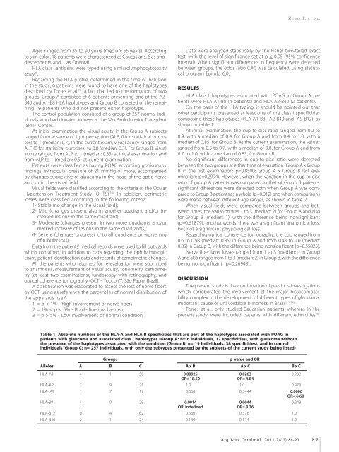
Table 1. Absolute numbers of the HLA-A and HLA-B specificities that are part of the haplotypes associated with POAG in<br />
patients with glaucoma and associated class I haplotypes (Group A: n= 6 individuals, 12 specificities), with glaucoma without<br />
the presence of the haplotypes associated with the condition (Group B: n= 19 individuals, 38 specificities), and in control<br />
individuals (Group C: n= 257 individuals, with only the subtypes presented by the subjects of the current study being listed)<br />
Groups<br />
p value and OR<br />
Alleles A B C A x B A x C B x C<br />
HLA-A1 4 1 050 0.00925 0.0263 0.239<br />
OR= 18.50 OR= 4.84<br />
HLA-A2 3 9 128 1.000 1.0000 0.978<br />
HLA- A9 1 7 017 0.660 0.3444 0.0006<br />
OR= 6.60<br />
HLA-B8 4 0 029 0.0014 0.0044 0.249<br />
OR in<strong>de</strong>fined OR= 8.36<br />
HLA-B12 0 4 062 0.560 0.3760 1.000<br />
HLA-B40 2 1 024 0.139 0.1140 1.000<br />
Arq Bras Oftalmol. 2011;74(2):88-90<br />
89


