a rquivos b rasileiros - Conselho Brasileiro de Oftalmologia
a rquivos b rasileiros - Conselho Brasileiro de Oftalmologia
a rquivos b rasileiros - Conselho Brasileiro de Oftalmologia
You also want an ePaper? Increase the reach of your titles
YUMPU automatically turns print PDFs into web optimized ePapers that Google loves.
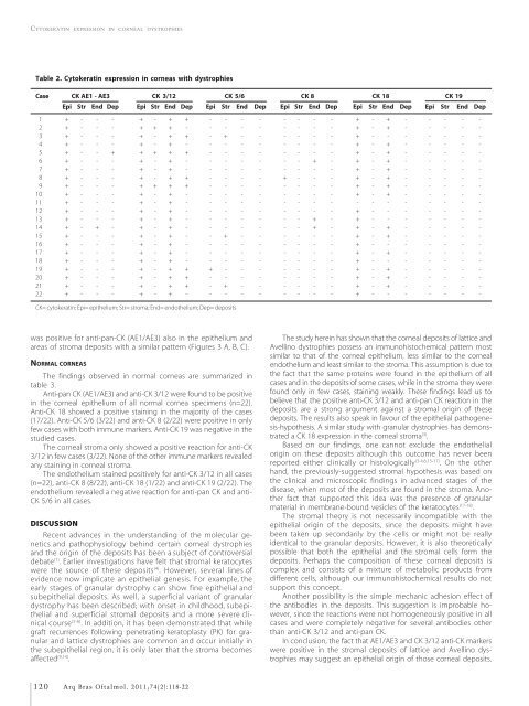
CYTOKERATIN EXPRESSION IN CORNEAL DYSTROPHIES<br />
Table 2. Cytokeratin expression in corneas with dystrophies<br />
Case CK AE1 - AE3 CK 3/12 CK 5/6 CK 8 CK 18 CK 19<br />
Epi Str End Dep Epi Str End Dep Epi Str End Dep Epi Str End Dep Epi Str End Dep Epi Str End Dep<br />
01 + - - - + - + + - - - - - - - - + - + - - - - -<br />
02 + - - - + + + - - - - - - - - - + - + - - - - -<br />
03 + - - - + - + + - + - - - - - - + - - - - - - -<br />
04 + - - - + - + - - - - - - - - - + - + - - - - -<br />
05 + - - + + + + + - - - - - - - - + - + - - - - -<br />
06 + - - - + - + - - - - - - - + - + - + - - - - -<br />
07 + - - - + - + - - - - - - - - - + - + - - - - -<br />
08 + - - - + - + + - - - - + - - - + - + - - - - -<br />
09 + - - - + + + + - - - - - - - - + - + - - - - -<br />
10 + - - - + - + - - - - - - - - - + - + - - - - -<br />
11 + - - - + - + - - - - - - - - - - - - - - - - -<br />
12 + - - - + - + - - - - - - - - - + - - - - - - -<br />
13 + - - - + - + - - - - - - - + - + - - - - - - -<br />
14 + - + - + - + - - - - - - - + - + - + - - - - -<br />
15 + - - - + - + - - + - - - - - - + - + - - - - -<br />
16 + - - - + - + - - - - - - - - - + - - - - - - -<br />
17 + - - - + - + - - - - - - - - - + - + - - - - -<br />
18 + - - - + - + - - - - - - - - - + - - - - - - -<br />
19 + - - - + - + + + - - - - - - - + - + - - - - -<br />
20 + - - - + - + + - - - - - - - - + - + - - - - -<br />
21 + - - - + - + + - + - - - - - - + - + - - - - -<br />
22 + - - - + - + - - - - - - - - - + - - - - - - -<br />
CK= cytokeratin; Epi= epithelium; Str= stroma; End= endothelium; Dep= <strong>de</strong>posits<br />
was positive for anti-pan-CK (AE1/AE3) also in the epithelium and<br />
areas of stroma <strong>de</strong>posits with a similar pattern (Figures 3 A, B, C).<br />
NORMAL CORNEAS<br />
The findings observed in normal corneas are summarized in<br />
table 3.<br />
Anti-pan CK (AE1/AE3) and anti-CK 3/12 were found to be positive<br />
in the corneal epithelium of all normal cornea specimens (n=22).<br />
Anti-CK 18 showed a positive staining in the majority of the cases<br />
(17/22). Anti-CK 5/6 (3/22) and anti-CK 8 (2/22) were positive in only<br />
few cases with both immune markers. Anti-CK 19 was negative in the<br />
studied cases.<br />
The corneal stroma only showed a positive reaction for anti-CK<br />
3/12 in few cases (3/22). None of the other immune markers revealed<br />
any staining in corneal stroma.<br />
The endothelium stained positively for anti-CK 3/12 in all cases<br />
(n=22), anti-CK 8 (8/22), anti-CK 18 (1/22) and anti-CK 19 (2/22). The<br />
endothelium revealed a negative reaction for anti-pan CK and anti-<br />
CK 5/6 in all cases.<br />
DISCUSSION<br />
Recent advances in the un<strong>de</strong>rstanding of the molecular genetics<br />
and pathophysiology behind certain corneal dystrophies<br />
and the origin of the <strong>de</strong>posits has been a subject of controversial<br />
<strong>de</strong>bate (1) . Earlier investigations have felt that stromal keratocytes<br />
were the source of these <strong>de</strong>posits (4) . However, several lines of<br />
evi<strong>de</strong>nce now implicate an epithelial genesis. For example, the<br />
early stages of granular dystrophy can show fine epithelial and<br />
subepithelial <strong>de</strong>posits. As well, a superficial variant of granular<br />
dystrophy has been <strong>de</strong>scribed; with onset in childhood, subepithelial<br />
and superficial stromal <strong>de</strong>posits and a more severe clinical<br />
course (5-6) . In addition, it has been <strong>de</strong>monstrated that while<br />
graft recurrences following penetrating keratoplasty (PK) for granular<br />
and lattice dystrophies are common and occur initially in<br />
the subepithelial region, it is only later that the stroma becomes<br />
affected (9,14) .<br />
The study herein has shown that the corneal <strong>de</strong>posits of lattice and<br />
Avellino dystrophies possess an immunohistochemical pattern most<br />
similar to that of the corneal epithelium, less similar to the corneal<br />
endothelium and least similar to the stroma. This assumption is due to<br />
the fact that the same proteins were found in the epithelium of all<br />
cases and in the <strong>de</strong>posits of some cases, while in the stroma they were<br />
found only in few cases, staining weakly. These findings lead us to<br />
believe that the positive anti-CK 3/12 and anti-pan CK reaction in the<br />
<strong>de</strong>posits are a strong argument against a stromal origin of these<br />
<strong>de</strong>posits. The results also speak in favour of the epithelial pathogenesis-hypothesis.<br />
A similar study with granular dystrophies has <strong>de</strong>monstrated<br />
a CK 18 expression in the corneal stroma (3) .<br />
Based on our findings, one cannot exclu<strong>de</strong> the endothelial<br />
origin on these <strong>de</strong>posits although this outcome has never been<br />
reported either clinically or histologically (3-4,6,15-17) . On the other<br />
hand, the previously-suggested stromal hypothesis was based on<br />
the clinical and microscopic findings in advanced stages of the<br />
disease, when most of the <strong>de</strong>posits are found in the stroma. Another<br />
fact that supported this i<strong>de</strong>a was the presence of granular<br />
material in membrane-bound vesicles of the keratocytes (17-18) .<br />
The stromal theory is not necessarily incompatible with the<br />
epithelial origin of the <strong>de</strong>posits, since the <strong>de</strong>posits might have<br />
been taken up secondarily by the cells or might not be really<br />
i<strong>de</strong>ntical to the granular <strong>de</strong>posits. However, it is also theoretically<br />
possible that both the epithelial and the stromal cells form the<br />
<strong>de</strong>posits. Perhaps the composition of these corneal <strong>de</strong>posits is<br />
complex and consists of a mixture of metabolic products from<br />
different cells, although our immunohistochemical results do not<br />
support this concept.<br />
Another possibility is the simple mechanic adhesion effect of<br />
the antibodies in the <strong>de</strong>posits. This suggestion is improbable however,<br />
since the reactions were not homogeneously positive in all<br />
cases and were completely negative for several antibodies other<br />
than anti-CK 3/12 and anti-pan CK.<br />
In conclusion, the fact that AE1/AE3 and CK 3/12 anti-CK markers<br />
were positive in the stromal <strong>de</strong>posits of lattice and Avellino dystrophies<br />
may suggest an epithelial origin of those corneal <strong>de</strong>posits.<br />
120 Arq Bras Oftalmol. 2011;74(2):118-22


