

Access to cataract surgery in public and private health systems ...
Access to cataract surgery in public and private health systems ...
Access to cataract surgery in public and private health systems ...

You also want an ePaper? Increase the reach of your titles
YUMPU automatically turns print PDFs into web optimized ePapers that Google loves.
TORRICELLI AAM, ET AL.<br />
elapsed from the bl<strong>in</strong>k<strong>in</strong>g until the tear film breaks up (26,29) . Values<br />
below 10 seconds are considered abnormal (30) . More recently, values of<br />
less than five seconds have been considered abnormal by some<br />
authors who suggest lower cut po<strong>in</strong>ts when a smaller volume of<br />
fluoresce<strong>in</strong> is <strong>in</strong>stilled (5 micro-litres of fluoresce<strong>in</strong> at 2%) (31) . Choos<strong>in</strong>g<br />
lower values decreases the test sensibility <strong>and</strong> <strong>in</strong>creases its specificity.<br />
Others tests <strong>to</strong> evaluate the tear film stability are: the <strong>in</strong>terferometry,<br />
that measures the central precorneal tear film thickness non<strong>in</strong>vasively<br />
us<strong>in</strong>g an <strong>in</strong>terference th<strong>in</strong>-film thickness measurement device (32-34) <strong>and</strong><br />
the meibomiometry, that uses a new laser meibometer <strong>to</strong> measure<br />
meibomian lipid levels on the lid marg<strong>in</strong> by blott<strong>in</strong>g meibomian<br />
gl<strong>and</strong> oil <strong>in</strong><strong>to</strong> a piece of plastic <strong>and</strong> moni<strong>to</strong>r<strong>in</strong>g its transparency <strong>in</strong><br />
order <strong>to</strong> calculate the amount of oil <strong>and</strong> its rate of delivery (35) . These<br />
<strong>to</strong>ols are not commonly used <strong>in</strong> daily rout<strong>in</strong>e.<br />
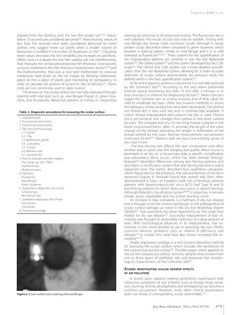
The lesions on the ocular surface are normally evaluated through<br />
sta<strong>in</strong><strong>in</strong>g with vital dyes such as: rose Bengal (Figure 2), green lissam<strong>in</strong>e,<br />
<strong>and</strong> fluoresce<strong>in</strong>. Abnormal patterns of cornea or conjunctiva<br />
Table 2. Diagnostic procedures for assess<strong>in</strong>g the ocular surface<br />
1. Questionnaires<br />
2. Visual acuity assessment<br />
3. External eye exam<strong>in</strong>ation<br />
2. Slip lamp biomicroscopy<br />
3.1. Eyelids<br />
3.2. Cilia<br />
3.3. Meibomian gl<strong>and</strong>s<br />
3.4. Conjuntiva<br />
3.5. Cornea<br />
3.6. Menisco tear<br />
3.7. Lacrimal Film<br />
3 Tests <strong>to</strong> evaluate tear film stability<br />
Tear break-up test (TBUT)<br />
Interferometry<br />
Meibomiometry<br />
4. Vital Dyes<br />
Fluoresce<strong>in</strong><br />
Rose Bengal<br />
Green lissam<strong>in</strong>e<br />
5. Quantitative diagnostic test of tear<br />
Schirmer test<br />
Red Phenol Test<br />
7. Qualitative diagnostic test of tear<br />
Fern<strong>in</strong>g test<br />
Osmolarity<br />
8. Impression cy<strong>to</strong>logy<br />
Figure 2. Ocular surface lesion sta<strong>in</strong><strong>in</strong>g with rose Bengal.<br />
sta<strong>in</strong><strong>in</strong>g are observed at slit lamp exam<strong>in</strong>ation. The fluoresce<strong>in</strong> dye is<br />
well <strong>to</strong>lerated. The results of such test may be variable. Test<strong>in</strong>g with<br />
rose Bengal dye reveals more consistent results although it causes<br />
greater ocular discomfort when compared <strong>to</strong> green lissam<strong>in</strong>e, which<br />
presents a sta<strong>in</strong><strong>in</strong>g pattern similar <strong>to</strong> rose Bengal <strong>and</strong> it is as well<br />
<strong>to</strong>lerated as fluoresce<strong>in</strong> (26,36,37) . Three <strong>systems</strong> for the quantification of<br />
the impregnation patterns are currently <strong>in</strong> use: the Van Bijsterveld<br />
system (38) , the Oxford system (39) <strong>and</strong> the system developed by the CLEK<br />
study (40) . The Oxford <strong>and</strong> CLEK studies use a more detailed quantification<br />
than the van Bijsterveld system, allow<strong>in</strong>g for a more accurate<br />
detection of ocular surface abnormalities. No previous study has<br />
def<strong>in</strong>ed which is the best quantification system (26) .<br />
As far as the aqueous portion is concerned, it is normally assessed<br />
by the Schirmer’s test (26) . Accord<strong>in</strong>g <strong>to</strong> this test, when performed<br />
without <strong>to</strong>pical anesthesia, less than 10 mm after 5 m<strong>in</strong>utes <strong>in</strong> at<br />
least one eye is a criterion for diagnos<strong>in</strong>g dry eye (41) . Many cl<strong>in</strong>icians<br />
regard the Schirmer test as unduly <strong>in</strong>vasive <strong>and</strong> of little value for<br />
mild <strong>to</strong> moderate dry eyes. Other less <strong>in</strong>vasive methods <strong>to</strong> assess<br />
the adequacy of tear production have been developed. The phenol<br />
red thread test is one such test <strong>and</strong> is commercially available. A<br />
cot<strong>to</strong>n thread impregnated with phenol red dye is used. Phenol<br />
red is pH sensitive <strong>and</strong> changes from yellow <strong>to</strong> red when wetted<br />
by tears. The crimped end of a 70 mm long thread is placed <strong>in</strong> the<br />
lower conjunctival fornix. After 15 seconds, the length of the color<br />
change on the thread, <strong>in</strong>dicat<strong>in</strong>g the length <strong>in</strong> millimeters of the<br />
thread wetted by the tears. Normal measurements are between<br />
9 mm <strong>and</strong> 20 mm (42) . Patients with dry eyes have wett<strong>in</strong>g values of<br />
less than 9 mm.<br />
The tear fern<strong>in</strong>g test reflects the tear composition <strong>and</strong> offers<br />
another way <strong>to</strong> assess tear film <strong>in</strong>tegrity <strong>and</strong> quality. When mucus is<br />
permitted <strong>to</strong> air dry on a microscope slide, a specific crystallization<br />
<strong>and</strong> arborisation (fern) occurs, which has been termed “fern<strong>in</strong>g”.<br />
Rol<strong>and</strong>o (43) identified differences among tear fern<strong>in</strong>g patterns <strong>and</strong><br />
described a classification system that tear fern<strong>in</strong>g became a useful<br />
diagnostic <strong>to</strong>ol. The author described four qualitative categories,<br />
which depended on the presence, the size <strong>and</strong> density of the ferns<br />
observed (Figure 3). Rol<strong>and</strong>o found that normal tear films often<br />
demonstrated a Type I or II pattern (with lots of fern<strong>in</strong>g), whereas<br />
patients with kera<strong>to</strong>conjunctivitis sicca (KCS) had Type III <strong>and</strong> IV<br />
tear fern<strong>in</strong>g patterns (<strong>in</strong> which there was scarce or absent fern<strong>in</strong>g).<br />
Although Rol<strong>and</strong>o’s classification system (43,44) is subjective, it rema<strong>in</strong>s<br />
simple, quick, repeatable <strong>and</strong> has a short learn<strong>in</strong>g curve.<br />
An <strong>in</strong>crease <strong>in</strong> tear osmolarity is a hallmark of dry eye disease<br />
<strong>and</strong> is thought <strong>to</strong> be the central mechanism <strong>in</strong> the pathogenesis of<br />
ocular surface damage, as noted <strong>in</strong> the Dry Eye WorkShop Report<br />
(DEWS) (25) . Tear osmolarity has been reported <strong>to</strong> be the s<strong>in</strong>gle best<br />
marker for dry eye disease (25) . Successful measurement of tear osmolarity<br />
was thought <strong>to</strong> necessitate collection of a large amount of<br />
tears. With technological advances <strong>in</strong> its measurement, tear osmolarity<br />
is now more feasible <strong>to</strong> use <strong>in</strong> assess<strong>in</strong>g dry eye. Others<br />
common extr<strong>in</strong>sic problems such as vitam<strong>in</strong> A deficiency <strong>and</strong><br />
allergies (25) or contact lens wear have also shown <strong>in</strong>creased tear osmolarity<br />
(45-47) .<br />
F<strong>in</strong>ally, impression cy<strong>to</strong>logy is a non-<strong>in</strong>vasive labora<strong>to</strong>ry method<br />
for assess<strong>in</strong>g the ocular surface, which <strong>in</strong>cludes the epithelium of<br />
the conjunctiva <strong>and</strong> the cornea (48) . The filter paper, when applied on<br />
the corneo-conjunctiva surface, removes samples that conta<strong>in</strong> from<br />
one <strong>to</strong> three layers of epithelial cells <strong>and</strong> preserves the morphological<br />
characteristics of the collected cells (49) .<br />
STUDIES INVESTIGATING OCULAR ADVERSE EFFECTS<br />
OF AIR POLLUTION<br />
In recent years, patients seek<strong>in</strong>g ophthalmic exam<strong>in</strong>ation with<br />
subjective symp<strong>to</strong>ms of eye irritation such as foreign body sensation,<br />
burn<strong>in</strong>g, itch<strong>in</strong>g, pho<strong>to</strong>phobia <strong>and</strong> whimper<strong>in</strong>g has become a<br />
common occurrence. However, most often cl<strong>in</strong>ical exam<strong>in</strong>ation<br />
does not reveal a correspond<strong>in</strong>g ocular abnormality (10) .<br />
Arq Bras Oftalmol. 2011;74(5):377-82<br />
379


